XiuZhen Han

Find an error

Name: 韩秀珍; XiuZhen Han
Organization: Shandong University
Department: Department of Pharmacology
Title: Associate Professor
Co-reporter:Hong Yao;Zhimei Shang;Penghong Wang;Shuixian Li
Cardiovascular Toxicology 2016 Volume 16( Issue 2) pp:101-110
Publication Date(Web):2016 April
DOI:10.1007/s12012-015-9317-z
Luteolin-7-O-glucoside (LUTG) was isolated from the plants of Dracocephalum tanguticum Maxim. Previous research has showed that LUTG pretreatment had a significant protective effect against doxorubicin (DOX)-induced cardiotoxicity by reducing intracellular calcium overload and leakage of creatine kinase and lactate dehydrogenase. But the underlying mechanisms have not been completely elucidated. In the present study, we investigated the effects of LUTG on H9c2 cell morphology, viability, apoptosis, reactive oxygen species generation, and the mitochondrial transmembrane potentials. The expression of p-PTEN, p-Akt, p-ERK, p-mTOR, and p-GSK-3β were detected by Western blotting. Compared with DOX alone treatment group, the morphological injury and apoptosis of the cells in groups treated by DOX plus LUTG were alleviated, cell viability was increased, ROS generation was lowered remarkably, and mitochondrial depolarization was mitigated. In DOX group, the expression of p-PTEN was lower than normal group and the expression of p-Akt and p-ERK was higher than normal group. In the groups treated with LUTG (20 μM), the expression of p-PTEN was upregulated and the expression of p-Akt, p-ERK, p-mTOR, and p-GSK-3β was downregulated. These results indicated that the protective effects of LUTG against DOX-induced cardiotoxicity may be related to anti-apoptosis through PTEN/Akt and ERK pathway.
Co-reporter:Yong-Li Jiang;Shui-Xian Li;Yu-Jing Liu;Lian-Ping Ge;Xiu-Zhen Han;Zhao-Peng Liu
Chemical Biology & Drug Design 2015 Volume 86( Issue 5) pp:1017-1029
Publication Date(Web):
DOI:10.1111/cbdd.12569

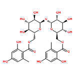
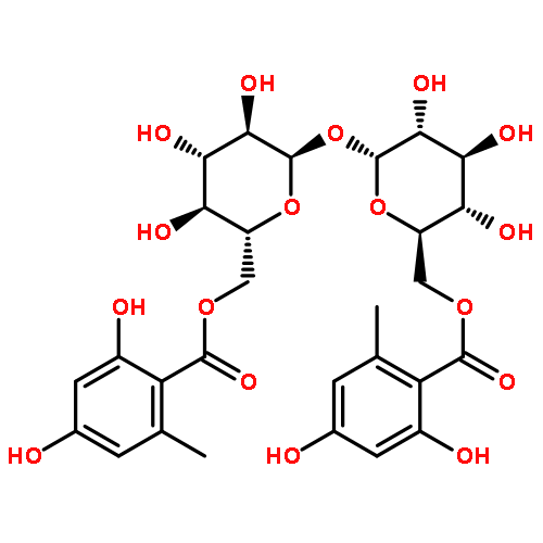
As a continuous research for the discovery of trehalose-based anti-invasive agents, we developed a convenient synthetic approach for the preparation of 6,6′-dideoxy-6,6′-bis(acylamino)-α,α-D-trehaloses. A series of trehalose-based amides were prepared through the trityl protection of the two primary hydroxyls of α,α-D-trehalose, benzoylation, the removal of the trityl protective group, mesylation, azidation, catalytic hydrogenation in the presence of hydrochloride, coupling reaction with a variety of acids, and subsequent debenzoylation and deacetylation in some cases. Compound 8b, 6,6′-dideoxy-6,6′-bis(2-hydroxybenzamide)-α,α-D-trehalose, was just as potent as the natural brartemicin against the invasion of murine colon 26-L5 cells. It exhibited no cytotoxicity on human breast adenocarcinoma MDA-MB-231 and murine colon 26-L5 cells. It can significantly inhibit the migration and invasion of the MDA-MB-231 cells. The anti-invasive effect of 8b was possibly related to its inhibitory activity on MMP-9, its suppression on the expression of MMP-9 and VEGF, and its deactivation of Akt.

Co-reporter:Hong Yao;Xiangyang Han
American Journal of Cardiovascular Drugs 2014 Volume 14( Issue 6) pp:433-442
Publication Date(Web):2014 December
DOI:10.1007/s40256-014-0089-9
Apoptosis occurs frequently in myocardial infarction, oxidative stress injury, and ischemia/reperfusion injury, and plays a pivotal role in the development of heart diseases. Inhibition of apoptosis alone does not necessarily lead to meaningful rescue in terms of either cardiomyocyte survival or function. Activation of the PI3K/Akt signaling pathway induced by insulin not only inhibits cardiomyocyte apoptosis but also substantially preserves and even improves regional and overall cardiac function. Insulin can protect cardiomyocytes from apoptosis by regulating a number of signaling molecules, such as eNOS, FOXOs, Bad, GSK-3β, mTOR, NDRG2, and Nrf2, through activating PI3K and Akt. This review focuses on the protective mechanisms and targets of insulin identified in the prevention and treatment of myocardial injury.
brartemicin
Gelatinase B
Mitogen-activated protein kinase
Phosphatidylinositol 3-kinase
Luteolin-7-glucoside
Adriamycin