Co-reporter:Changrong Wang, Lili Du, Junhui Zhou, Lingwei Meng, Qiang Cheng, Chun Wang, Xiaoxia Wang, Deyao Zhao, Yuanyu Huang, Shuquan Zheng, Huiqing Cao, Jianhua Zhang, Liandong Deng, Zicai Liang, and Anjie Dong
ACS Applied Materials & Interfaces September 27, 2017 Volume 9(Issue 38) pp:32463-32463
Publication Date(Web):September 1, 2017
DOI:10.1021/acsami.7b07337
Hydrophobization of cationic polymers, as an efficient strategy, had been widely developed in the structure of cationic polymer micelles to improve the delivery efficiency of nucleic acids. However, the distribution of hydrophobic segments in the polymer chains is rarely considered. Here, we have elaborated three types of hydrophobized polyethylene glycol (PEG)-blocked cationic polymers with different distributions of the hydrophobic segments in the polymer chains PEG–PAM–PDP (E–A–D), PEG–PDP–PAM (E–D–A), and PEG–P(AM/DP) (E–(A/D)), which were synthesized by reversible addition–fragmentation chain transfer polymerization of methoxy PEG, cationic monomer aminoethyl methacrylate, and pH-sensitive hydrophobic monomer 2-diisopropylaminoethyl methacrylate, respectively. In aqueous solution, all of the three copolymers, E–A–D, E–D–A, and E–(A/D), were able to spontaneously form nanosized micelles (100–150 nm) (ME–A–D, ME–D–A, and ME–(A/D)) and well-incorporated small interfering RNA (siRNA) into complex micelles (CMs). The effect of distributions of the hydrophobic segments on siRNA delivery had been evaluated in vitro and in vivo. Compared with ME–D–A and ME–(A/D), ME–A–D showed the best siRNA binding capacity to form stable ME–A–D/siRNA CMs less than 100 nm, mediated the best gene-silencing efficiency and inhibition effect of tumor cell growth in vitro, and showed better liver gene-silencing effect in vivo. In the case of ME–(A/D) with a random distribution of cationic and hydrophobic segments, a gene-silencing efficiency higher than Lipo2000 but lesser than ME–A–D and ME–D–A was obtained. As the mole ratio of positive and negative charges increased, ME–D–A/siRNA and ME–A–D/siRNA showed similar performances in size, zeta potential, cell uptake, and gene silencing, but ME–(A/D)/siRNA showed reversed performances. In addition, ME–A–D as the best siRNA carrier was evaluated in the tumor tissue in the xenograft murine model and showed good anticancer capacity. Obviously, the distribution of the hydrophobic segments in the amphiphilic cationic polymer chains should be seriously considered in the design of siRNA vectors.Keywords: amphiphilic cationic polymers; delivery efficiency; distribution of the hydrophobic segments; gene silence; siRNA delivery;
Co-reporter:Hongzhang Deng, Anjie Dong
Journal of Controlled Release 2017 Volume 259(Volume 259) pp:
Publication Date(Web):10 August 2017
DOI:10.1016/j.jconrel.2017.03.116
Co-reporter:Gan Chen, Hongzhang Deng, Xiang Song, Mingzi Lu, Lian Zhao, Sha Xia, Guoxing You, Jingxiang Zhao, Yulong Zhang, Anjie Dong, Hong Zhou
Biomaterials 2017 Volume 144(Volume 144) pp:
Publication Date(Web):1 November 2017
DOI:10.1016/j.biomaterials.2017.08.008
Sepsis-associated acute liver injury contributes to the pathogenesis of multiple organ dysfunction syndrome and is associated with increased mortality. Currently, no specific therapeutics for sepsis-associated liver injury are available. With excess levels of reactive oxygen species (ROS) being implicated as key players in sepsis-induced liver injury, we hypothesize that ROS-responsive nanoparticles (NPs) formed via the self-assembly of diblock copolymers of poly(ethylene glycol) (PEG) and poly(propylene sulfide) (PPS) may function as an effective drug delivery system for alleviating sepsis-induced liver injury by preferentially releasing drug molecules at the disease site. However, there are no reports available on the biocompatibility and effect of PEG-b-PPS-NPs in vivo. Herein, this platform was tested for delivering the promising antioxidant therapeutic molecule melatonin (Mel), which currently has limited therapeutic efficacy because of its poor pharmacokinetic properties. The mPEG-b-PPS-NPs efficiently encapsulated Mel using the oil-in-water emulsion technique and provided sustained, on-demand release that was modulated in vitro by the hydrogen peroxide concentration. Animal studies using a mouse model of sepsis-induced acute liver injury revealed that Mel-loaded mPEG-b-PPS-NPs are biocompatible and much more efficacious than an equivalent amount of free drug in attenuating oxidative stress, the inflammatory response, and subsequent liver injury. Accordingly, this work indicates that mPEG-b-PPS-NPs show potential as an ROS-mediated on-demand drug delivery system for improving Mel bioavailability and treating oxidative stress-associated diseases such as sepsis-induced acute liver injury.
Co-reporter:Hongzhang Deng, Xuefei Zhao, Liandong Deng, Jianfeng Liu, Anjie Dong
Journal of Controlled Release 2017 Volume 255(Volume 255) pp:
Publication Date(Web):10 June 2017
DOI:10.1016/j.jconrel.2017.04.002
The fact that the sensitivities of different tumor cells and different individuals to the actions of drug delivery system varied greatly, restricted the anticarcinogen to a desired therapeutic concentration. How to determine the destiny of drug delivery system in space and time is the main challenge to realize the precise anticancer therapy. In this paper, we reported a preparation of degradable nanoparticles (designated Pros-PDC) loaded DOX and IR780 with three functional domains: the charge-conversional feature with long circulation time and enhanced internalization, light-triggered reactive oxygen species (ROS) generation and subsequently ROS responsive anticancer drug release with a spatially and temporally precise fashion. The spatiotemporal drug release from the ROS activated Pros-PDC nanoparticles could be controlled by when and how long to perform laser irradiation. In the present work, multifunctions of DOX and IR780 loaded Pros-PDC nanoparticles, as a flexible, easily controllable drug release platform, had been certificated in vitro and in vivo.Download high-res image (198KB)Download full-size image
Co-reporter:Junhui Zhou, Yidi Wu, Changrong Wang, Qiang Cheng, Shangcong Han, Xiaoxia Wang, Jianhua Zhang, Liandong Deng, Deyao Zhao, Lili Du, Huiqing Cao, Zicai Liang, Yuanyu Huang, and Anjie Dong
Nano Letters 2016 Volume 16(Issue 11) pp:6916-6923
Publication Date(Web):October 13, 2016
DOI:10.1021/acs.nanolett.6b02915
The extremely low efficient cytosolic release of the internalized siRNA has emerged recently as a central issue for siRNA delivery, while there is a lack of guidelines to facilitate the cytosolic release of internalized siRNA. To address these concerns, we studied the contribution of the pH-sensitive inner core on handling the cytosolic release of siRNA delivered by a series of PG-P(DPAx-co-DMAEMAy)-PCB amphiphilic polycation nanomicelles (GDDC-Ms) with extremely low internalization (<1/4 of lipofactamine 2000 (Lipo2000)). Significantly, just by varying the mole ratio of DPA and DMAEMA to adjust the initial disassembly pH (pHdis) of the core near to 6.8, GDDC4-Ms/siRNA could get nearly 98.8% silencing efficiency at w/w = 12 with 50 nM siRNA and ∼78% silencing efficiency at w/w = 30 with a very low dose of 5 nM siRNA in HepG-2 cell lines, while Lipo2000 only got 65.7% with 50 nM siRNA. Furthermore, ∼98.4% silencing efficiency was also realized in the hard-to-transfect human acute monoblastic leukemia cell line U937 by GDDC4-Ms/siRNA (at w/w = 15, 50 nM siRNA), in the inefficient case for Lipo2000. Additionally, the high silencing efficiency (∼80%) in skin tissue in vivo was discovered. Undoubtedly, the robust potential of GDDC4-Ms in handling the cytosolic release paves a simple but efficient new way for the design of the nonviral siRNA vector.Keywords: Cytosolic release; high efficiency; low doses; low internalization; monoblastic cell; siRNA delivery;
Co-reporter:Hongzhang Deng, Xuefei Zhao, Jinjian Liu, Jianhua Zhang, Liandong Deng, Jianfeng Liu and Anjie Dong
Nanoscale 2016 vol. 8(Issue 3) pp:1437-1450
Publication Date(Web):03 Dec 2015
DOI:10.1039/C5NR06745F
The tuning of the structure of nanocarriers with fast acidic-degradation rate and high stability in physiological conditions or during storage is under intensive study. In this context, a kind of dual-pH responsive micelles with well-balanced stability, that is, fast hydrolysis in acidic environment and stability towards blood drug release at 7.4 were developed. This is achieved by the self-assembly of micelles of poly(ethylene glycol)-b-(poly ε-caprolactone-g-poly(2,2-dimethyl-1,3-dioxolane-4-yl)methylacrylate-co-2(dimethylamino)ethyl methacrylate) (mPEG-b-(PCL-g-P(DA-co-DMAEMA))) copolymers with two inert pH responsive moieties of DA and DMAEMA. The fast synergistic acid-triggered disassembly and high stability at physiological condition of the mPEG-b-(PCL-g-P(DA-co-DMAEMA)) micelles was verified by 1H NMR, particle size and optical stability measurements, which was induced and mediated by the synergistic pH responses of the hydrolysis of the ketal in DA moieties and the switch in solubility of tertiary amino moieties (DMAEMA) under mild acid conditions. It was observed that the hydrolysis rate of the ketal could be promoted by increasing the content of DMAEMA moieties. The fast intracellular disassembly of the micelles depending on the contents of DMAEMA moieties was also traced by fluorescence resonance energy transfer (FRET). The in vitro release studies showed that the release of DOX from mPEG-b-(PCL-g-P(DA-co-DMAEMA)) micelles at mild acid condition was significantly accelerated by increasing the content of DMAEMA moieties, while greatly impeding drug release in physiological conditions. The antitumor activity of DOX-loaded micelles was studied in MCF-7 and 4T1 cells in vitro and in 4T1 tumor-bearing Balb-c mice in vivo. The results indicated the DOX-loaded micelles with higher content of DMAEMA moieties exhibited enhanced anticancer activity. Collectively, the synergistic dual-pH responsive design of mPEG-b-(PCL-g-P(DA-co-DMAEMA)) micelles provided a new route for improving anticancer drug delivery efficiency.
Co-reporter:Hongzhang Deng, Xuefei Zhao, Dongxuan He, Weisheng Guo, Keni Yang, Anjie Dong and Xing-Jie Liang
Chemical Communications 2016 vol. 52(Issue 46) pp:7406-7408
Publication Date(Web):26 Apr 2016
DOI:10.1039/C6CC01996J
A new type of membrane fusogenic lipid was prepared to deliver DNA or siRNA into the cytoplasm directly in a fusion-dependent manner in order to bypass the cellular endocytosis to avoid the inefficient escape from the endosome and low transfection efficacy.
Co-reporter:Shuxin Xu;Liong Deng;Jianhua Zhang;Li Yin
Journal of Biomedical Materials Research Part B: Applied Biomaterials 2016 Volume 104( Issue 3) pp:640-656
Publication Date(Web):
DOI:10.1002/jbm.b.33420
Abstract
With increasingly rigorous requirements for biomaterials, the design and fabrication of novel materials with smart functions are urgently needed. The fabrication of composite materials that can surmount individual shortcomings as well as bring synergistic benefits represents an efficient route to improve the performances and expand application scopes of biomaterials. Due to their unique structures and properties, electrospun-fibers and hydrogels have been widely applied in many biological and biomedical fields. Based on this, more and more attentions have been paid on the composites of electrospun-fibers and hydrogels as biomaterials, aiming to bring their individual superiority into full play as well as remedy their intrinsic defects. This review summarizes approaches used to integrate electrospun-fibers and hydrogels into various structures and the development of their composites as a potential solution to some current challenges in drug delivery, tissue engineering and some other bio-related aspects. The individual roles and mutual synergy of electrospun-fibers and hydrogels in the composites will be emphasized. © 2015 Wiley Periodicals, Inc. J Biomed Mater Res Part B: Appl Biomater, 104B: 640–656, 2016.
Co-reporter:Hongzhang Deng, Xuefei Zhao, Jinjian Liu, Liandong Deng, Jianhua Zhang, Jianfeng Liu and Anjie Dong
Journal of Materials Chemistry A 2015 vol. 3(Issue 48) pp:9397-9408
Publication Date(Web):16 Nov 2015
DOI:10.1039/C5TB01939G
Nanocarriers have been extensively explored for cancer drug delivery with their ability to respond to cancer heterogeneity which is recently recognized as a critical doorway to a high therapeutic index. We proposed to develop a polycaprolactone bearing acid-labile β-carboxylic amide segments with charge reversal properties, which was coupled to mPEG with thioether as a linker. The linker could respond to overproduced reactive oxygen species (ROS) of cancer cells (ROS, e.g., perhaps more than one order of magnitude higher than healthy cells). This tailor-made surface charged nanoparticles (NPs) exhibited a capacity of reversing its surface charge from negative to positive at a tumor extracellular environment (pH ∼ 6.8) for enhancing cell internalization and an ability of response to the tumor ROS heterogeneity at the tumor intracellular environment to accelerate the release of drugs from NPs. The in vitro release studies showed that DOX release was greatly accelerated under the intracellular prevailing ROS (hydrogen peroxide (H2O2) simulating the oxidative stress). Cell uptake showed that the NPs could be more effectively internalized at pH 6.8 (simulating tumor extracellular conditions) than at pH 7.4. The MTT assay demonstrated that the DOX loaded NPs showed significant cytotoxicity to HepG2 cancer cells while no influence on the L02 normal cells. These ROS sensitive and surface charged NPs with superior cell internalization ability and rapid intracellular drug release provided a novel platform for tumor-targeting drug delivery.
Co-reporter:Pingsheng Huang, Yumin Zhang, Weiwei Wang, Junhui Zhou, Yu Sun, Jinjian Liu, Deling Kong, Jianfeng Liu, Anjie Dong
Journal of Controlled Release 2015 Volume 220(Part A) pp:456-464
Publication Date(Web):28 December 2015
DOI:10.1016/j.jconrel.2015.11.007
Combined chemoradiotherapy is potent to defeat malignant tumor. Concurrent delivery of radioisotope with chemotherapeutic drugs, which also act as the radiosensitizer, to tumor tissues by a single vehicle is essential to achieve this objective. To this end, a macroscale injectable and thermosensitive micellar-hydrogel (MHg) depot was constructed by thermo-induced self-aggregation of poly(ε-caprolactone-co-1,4,8-trioxa[4.6]spiro-9-undecanone)-poly(ethyleneglycol)-poly(ε-caprolactone-co-1,4,8-trioxa[4.6]spiro-9-undecanone) (PECT) triblock copolymer micelles (Ms), which could not only serve as a micellar drug reservoir to locally deliver concentrated nano chemotherapeutic drugs, but also immobilize radioisotopes at the internal irradiation hot focus. Doxorubicin (DOX) and iodine-131 labeled hyaluronic acid (131I-HA) were used as the model therapeutic agents. The aqueous mixture of drug-loaded PECT micelles and 131I-HA exhibited sol-to-gel transition around body temperature. In vitro drug release study indicated that PECT/DOX Ms were sustainedly shed from the native PECT/DOX MHg formulation, which could be internalized by tumor cells with rapid intracellular DOX release. This hydrogel formulation demonstrated considerable in vitro antitumor effect as well as remarkable radiosensitization. In vivo subcutaneous injection of PECT MHg demonstrated that 131I isotope was immobilized stably at the injection location and no obvious indication of damage to major organs were observed as indicated by the histopathological analysis. Furthermore, the peritumoral injection of chemo-radiation therapeutic agents-encapsulated MHg formulation on tumor-bearing nude mice resulted in the desired combined treatment effect, which significantly improved the tumor growth inhibition efficiency with minimized drug-associated side effects to major organs. Consequently, such a thermosensitive MHg formulation, which enabled the precise control over the dosage and ratio of combination therapeutic agents to obtain the desired therapeutic effect with a single drug administration and reduced side effects, holds great potential for spatiotemporally delivery of multiple bioactive agents for sustained combination therapy.The schematic diagram of design concept: formation of PECT/DOX MHg nanodrug depot as well as radionuclide reservoir for in situ chemoradiotherapy.
Co-reporter:Shangcong Han, Qiang Cheng, Yidi Wu, Junhui Zhou, Xingwen Long, Tuo Wei, Yuanyu Huang, Shuquan Zheng, Jianhua Zhang, Liandong Deng, Xiaoxia Wang, Xing-Jie Liang, Huiqing Cao, Zicai Liang, Anjie Dong
Biomaterials 2015 48() pp: 45-55
Publication Date(Web):
DOI:10.1016/j.biomaterials.2015.01.026
Co-reporter:Weiwei Wang, Jinjian Liu, Chen Li, Ju Zhang, Jianfeng Liu, Anjie Dong and Deling Kong
Journal of Materials Chemistry A 2014 vol. 2(Issue 26) pp:4185-4192
Publication Date(Web):10 Apr 2014
DOI:10.1039/C4TB00275J
The real-time monitoring of materials degradation is crucial to determine the in vivo retention time and the design or screening of degradable biomaterials. However, in vivo performance cannot always be predicted through the traditional determination of in vitro erosion and current standard methods sacrifice samples or animals, preventing the sequential measurement of the same specimen. Herein, a non-invasive fluorescence imaging method was developed to sequentially follow in vivo loss of fluorescence signal to simultaneously characterize the hydrolytic and enzymatic degradation of PEGlyated polyester hydrogel. Rhodamine B was conjugated to thermosensitive amphiphilic triblock copolymer based on cyclic ether modified PCL and PEG (abbreviated as PECT) and no obvious influence on gelation time or gel strength was observed with the conjugation content under 0.121% (w/w). Both in vitro and in vivo degradation profiles followed linear fittings while in vivo and in vitro hydrogel degradation rates correlated in an exponential mathematical model, enabling the general prediction of in vivo erosion trends of new biomaterial formulations from in vitro data. This methodology possibly enabled rational design and rapid in vitro screening of degradable materials, and might be potentially extended to simultaneously determine the material erosion and speculate the drug release from a drug-incorporated scaffold, or the cell growth profile in tissue-engineering formulations.
Co-reporter:Pingsheng Huang, Cuihong Yang, Jinjian Liu, Weiwei Wang, Shutao Guo, Jiao Li, Yu Sun, Hongxu Dong, Liandong Deng, Jianhua Zhang, Jianfeng Liu and Anjie Dong
Journal of Materials Chemistry A 2014 vol. 2(Issue 25) pp:4021-4033
Publication Date(Web):17 Apr 2014
DOI:10.1039/C4TB00273C
Sequentially overcoming the obstacles mainly from the low water solubility of lipophilic anticancer drugs, gastrointestinal microenvironment and systemic circulation is the major concern for designing oral anticancer drug carriers. Herein, we prepared the multifunctional polyelectrolyte complex nanoparticles (CNPs), engineered by hyaluronic acid (HA) grafted polycaprolactone (PCL) nanoparticles (HA-g-PCL NPs) coated with chitosan (CS) electrostatically, as a platform to improve the oral delivery efficiency of lipophilic anticancer drugs. Paclitaxel (PTX) and doxorubicin (DOX) were used as the model medicine and fluorescence probe, respectively. The size, zeta potential, morphology and pH-sensitivity of the NPs were studied systematically. The results indicated that the core–shell structure of CS/HA-g-PCL CNPs was formed at pH 5.0, which remained intact in the pH ranging from 3.0 to 6.8, while the CS layer detached gradually with the increase of pH to 7.4 and the HA-g-PCL NPs were released. In vitro drug release studies showed that accelerated drug release was triggered by hyaluronidase-1 (Hyal-1), which was a major HA degradation enzyme abundant within tumor cells. Cell uptake studies showed that HA-g-PCL NPs were internalized into cancer cells (EC109) via receptor-mediated endocytosis, but were rarely taken up by normal fibroblasts (NIH3T3). Furthermore, intracellular drug release indicated that HA-g-PCL NPs could provide an effective approach for transport of loaded cargoes into the cytoplasm. Therefore, higher cytotoxicity for PTX loaded HA-g-PCL NPs (HA-g-PCL/PTX NPs) against cancer cells EC109 but lower cytotoxicity against normal cells NIH3T3 was observed. In vivo studies showed that CS/HA-g-PCL CNPs via oral administration were able to preferentially deliver drugs into tumor tissue with commendable antitumor efficiency and few side effects. Overall, CS/HA-g-PCL CNPs showed great potential for improving oral delivery efficiency of lipophilic anticancer drugs.
Co-reporter:Pingsheng Huang, Jinjian Liu, Weiwei Wang, Chen Li, Junhui Zhou, Xue Wang, Liandong Deng, Deling Kong, Jianfeng Liu, and Anjie Dong
ACS Applied Materials & Interfaces 2014 Volume 6(Issue 16) pp:14631
Publication Date(Web):August 6, 2014
DOI:10.1021/am503974y
Enabling nanocarriers to complete the sophisticated journey from the initial injection site to the targeted tumor cells and achieve “spatiotemporally pinpointed” drug release intracellularly is a challenging task in anticancer drug delivery. Herein, versatile shell-cross-linked nanoparticles (SCNPs) were prepared by one-step assembly of triblock zwitterionic copolymers, polycarboxybetaine methacrylate-block-poly(N-(2-(2-pyridyl disulde) ethyl methacrylamide-block-poly(2-(diisopropylamino) ethyl methacrylate) (PCB-b-PDS-b-PDPA, termed as PCSSD), which was well-defined via reversible additive fragment transfer (RAFT) polymerization, followed by functionalization with Arg-Gly-Asp (RGD). Thus, the RGD-PCSSD SCNPs cooperatively combine the ultra pH-sensitive PDPA core for efficient drug loading and pH-responsive drug release, the disulfide-cross-linked PDS shell that prevents premature drug release, the zwitterionic PCB corona to stabilize the SCNPs and prolong its systemic circulation, the RGD ligand for active tumor targeting and receptor-mediated endocytosis. Doxorubicin (DOX) was loaded as a model medicine (termed as RGD-PCSSD/DOX SCNPs). The dual-sensitivity studies showed that the pH-sensitivity of PDPA core could be adjusted by the shell-cross-linking density, accompanied by better control over premature drug release. Furthermore, results obtained by flow cytometry and fluorescence microscopy analysis demonstrated that once the RGD-PCSS10D/DOX SCNPs were internalized into tumor cells via receptor-mediated endocytosis, boost drug release was observed with considerable cytotoxicity in vitro. The results of ex vivo imaging studies further confirmed the successful drug delivery from the injection site to the tumor tissue. In summary, the well-constructed RGD-PCSS10D/DOX SCNPs with cooperative multifunctionality showed great potential as novel nanocarriers for tumor targeted anticancer drug delivery.Keywords: doxorubicin; pH-sensitive; shell-cross-linked; tumor targeting; zwitterionic copolymer
Co-reporter:Shangcong Han, Haiying Wan, Daoshu Lin, Shutao Guo, Hongxu Dong, Jianhua Zhang, Liandong Deng, Ruming Liu, Hua Tang, Anjie Dong
Acta Biomaterialia 2014 Volume 10(Issue 2) pp:670-679
Publication Date(Web):February 2014
DOI:10.1016/j.actbio.2013.09.035
Abstract
Nanoparticles (NPs) assembled from amphiphilic polycations have been certified as potential carriers for gene delivery. Structural modification of polycation moieties may be an efficient route to further enhance gene delivery efficiency. In this study two electroneutral monomers with different hydrophobicities, 2-hydroxyethyl methacrylate (HEMA) and 2-hydroxyethyl acrylate (HEA), were incorporated into the cationic poly(dimethylamino ethyl methacrylate) (PDMAEMA) side-chains of amphiphilic poly(ε-caprolactone)-graft-poly(dimethylamino ethylmethacrylate) (PCD) by random co-polymerization, to obtain poly(ε-caprolactone)-graft-poly(dimethylamino ethyl methacrylate-co-2-hydroxyethyl methacrylate) (PCD-HEMA) and poly(ε-caprolactone)-graft-poly(dimethylamino ethyl methacrylate-co-2-hydroxyethyl acrylate) (PCD-HEA). Minimal HEA or HEMA moieties in PDMAEMA do not lead to statistically significant changes in particle size, zeta potential, DNA condensation properties and buffering capacity of the naked NPs. However, the incorporation of HEMA and HEA lead to reductions and increases, respectively, in the surface hydrophilicity of the naked NPs and NPs/DNA complexes, which was confirmed by water contact angle assay. These simple modifications of PDMAEMA with HEA and HEMA moieties significantly affect the gene transfection efficiency on HeLa cells in vitro: PCD-HEMA NP/DNA complexes show a much higher transfection efficiency than PCD NPs/DNA complexes, while PCD-HEA NPs/DNA complexes show a lower transfection efficiency than PCD NP/DNA complexes. Fluorescence activated cell sorter and confocal laser scanning microscope results indicate that the incorporation of hydrophobic HEMA moieties facilitates an enhancement in both cellular uptake and endosomal/lysosomal escape, leading to a higher transfection efficiency. Moreover, the process of endosomal/lysosomal escape confirmed in our research that PCD and its derivatives do not just rely on the proton sponge mechanism, but also on membrane damage due to the polycation chains, especially hydrophobic modified ones. Hence, it is proved that hydrophobic modification of cationic side-chains is a crucial route to improve gene transfection mediated by polycation NPs.
Co-reporter:Weiwei Wang;Liong Deng;Pingsheng Huang;Shuxin Xu;Xu Li;Nan Lv;Lei Wang;Renjie Hu;Jianhua Zhang
Journal of Biomedical Materials Research Part A 2014 Volume 102( Issue 1) pp:17-29
Publication Date(Web):
DOI:10.1002/jbm.a.34694
Injectable thermosensitive hydrogels provide local non-invasive platforms for sustained drug release,tissue engineering and cellular immunity. As a long-term implant, the toxicity and in vivo biological effect should be concerned.Previously we developed a novel type of injectable nanoparticular self-supported hydrogel (PECT NPsGel)of PEG and pendent cycle ethers modified poly(ε-caprolactone) triblock copolymer (PECT), which could sustainedly release PECT or drug-loaded PECT nanoparticles with the hydrogel disassembly and provided efficient antitumor activity and significant decrease of side effects. Herein, the aim of this work was to reveal the toxicity and in vivo biological effect of PECT nanoparticles and PECT NPsGel. In vitro cytotoxicity indicated no cell cytotoxicity was observed when the concentration of PECT nanoparticle was up to 500 µg/mL, and also nomutagenic effect and no genotoxicity were observed.In vivo intravenous injection of PECT nanoparticles demonstrated that the LD50 was approximate high to 2.564 g/kg, and compared with the control mice, the mice treated with daily administration of PECT nanoparticles showed no difference in the physical or behavioral alterations, body weight changes, biochemical and hematological parameters as well as organ coefficients. The in vivo chronic effect of PECT NPsGelconfirmed no toxic lesions to animals in a whole period of three months even the dosage was high to 20 g/kg. These findings indicated PECT nanoparticles and PECT NPsGel were of well biocompatibility and did not provoke any side effect to body, which represented a new class of injectable and non-invasive systemic or site-specific delivery carrier. © 2013 Wiley Periodicals, Inc. J Biomed Mater Res Part A: 102A: 17–29, 2014.
Co-reporter:Zhen Wei, Hui Zhao, Jianhua Zhang, Liandong Deng, Siyu Wu, Junyu He and Anjie Dong
RSC Advances 2014 vol. 4(Issue 93) pp:51381-51388
Publication Date(Web):26 Sep 2014
DOI:10.1039/C4RA07505F
Poly(vinyl alcohol) electrospun nanofibrous (PVANF) membranes that could sensitively detect and adsorb metal ions were modified with spirolactam–rhodamineine derivatives (PVANF–SRD) membranes and sulfo-spirolactam–rhodamine derivatives (PVANF–SSRD) membranes. Surface chemistry and morphology during functionalization of PVANF membranes were monitored using Fourier transform-infrared spectroscopy (FT-IR), X-ray photoelectron spectroscopy (XPS) and scanning electron microscopy (SEM). These two membranes could display real-time sensing by the naked eye based on ring-opening reaction of spirolactam–rhodamine derivatives induced by corresponding metals. PVANF–SRD and PVANF–SSRD membranes exhibited high selectivity and sensitivity toward Fe3+/Cr3+ and Hg2+, respectively. In terms of PVANF–SSRD membranes, adsorption capacity for Hg2+ in contaminated water was studied. Freundlich isotherm could better describe the interactions than Langmuir: Kf = 7.0175 mg g−1 (r2 = 0.9996) for Hg2+. The regenerability of these two membranes was investigated via Na4EDTA solution treatment, and results demonstrated good sustainability in detection and adsorption efficiency.
Co-reporter:Pingsheng Huang, Huijuan Song, Weiwei Wang, Yu Sun, Junhui Zhou, Xue Wang, Jinjian Liu, Jianfeng Liu, Deling Kong, and Anjie Dong
Biomacromolecules 2014 Volume 15(Issue 8) pp:
Publication Date(Web):July 23, 2014
DOI:10.1021/bm500764p
Reasonably structural design of nanoparticles (NPs) to combine functions of prolonged systemic circulation, enhanced tumor targeting and specific intracellular drug release is crucial for antitumor drug delivery. Combining advantages of Arg-Gly-Asp (RGD) for active tumor targeting, zwitterionic polycarboxybetaine methacrylate (PCB) for prolonged systemic circulation, poly(2-(diisopropylamino) ethyl methacrylate) (PDPA) for acid-triggered intracellular release, novel RGD-PCB-b-PDPA (RGD-PCD) block copolymers were prepared via reversible addition–fragmentation chain transfer (RAFT) polymerization and followed by functionalization with RGD. Doxorubicine (DOX) was encapsulated within the RGD-PCD NPs as model medicine (RGD-PCD/DOX NPs). With ultra pH-sensitivity of PDPA, the drug release was restrained at pH 7.4 for only 24% within 36 h, which was increased to 60% at pH 6.0 within 24 h, and released more rapidly at pH 5.0 for 100% within 5 h, indicating that the RGD-PCD/DOX NPs were able to turn drug release “off” at neutral pH (e.g., systemic circulation) whereas “on” under acidic conditions (e.g., inside endo/lysosomes). Furthermore, the results of fluorescence microscopy and flow cytometry analysis demonstrated improved internalization of RGD-PCD/DOX NPs in HepG2 cells via integrin-mediated endocytosis with rapid DOX release intracellularly. Consequently, the RGD-PCD/DOX NPs showed considerable cytotoxicity against HepG2 and HeLa cells in comparison with free DOX. Importantly, the RGD-PCD/DOX NPs exhibited little protein adsorption property with excellent serum stability, which led to prolonged systemic circulation and enhanced tumor accumulation in tumor-bearing nude mice. Therefore, this multifunctional RGD-PCD NPs, which represented the flexible design approach, showed great potential for the development of novel nanocarriers in tumor-targeted drug delivery.
Co-reporter:Weiwei Wang, Ju Zhang, Chen Li, Pingsheng Huang, Shan Gao, Shangcong Han, Anjie Dong, Deling Kong
Colloids and Surfaces B: Biointerfaces 2014 Volume 115() pp:302-309
Publication Date(Web):1 March 2014
DOI:10.1016/j.colsurfb.2013.12.026
•Novel poly(ɛ-caprolactone)-based terpolymers with an A-block-B-graft-C structure are proposed.•Two consecutive controlled polymerization, ring-opening polymerization and atom transfer radical polymerization are employed to prepare the terpolymers.•Multicompartment micelles (MCMs) with well-segregated Janus-cores are generated from the aqueous assembly of obtained terpolymers.•The compartment balance in the core is adjustable by altering the B and C composition.•The MCMs show excellent cytocompatibility in mouse and human cells.The architecture of hydrophobic segments can determine the specific morphology of multicompartment micelles (MCMs) that are generated from aqueous assembly of amphiphilic terpolymers. In this study, we aimed to design and generate poly(ɛ-caprolactone)-based multicompartment micelles with adjustable Janus-cores. Well-defined terpolymers with a novel A-block-B-graft-C architecture composed of biologically compatible polymers, methoxy poly(ethylene glycol) (PEG), poly(ɛ-caprolactone) (PCL) and poly(2-(perfluorobutyl)ethyl methacrylate) (PPFEMA), were prepared by the stepwise use of ring-opening polymerization and atom transfer radical polymerization. Characterization of the obtained terpolymers was carried out by 1H NMR and gel permeation chromatography. Results from differential scanning calorimetry and X-ray diffraction studies indicated that within the terpolymer structure, the PCL segments are in the crystalline state, while fluorocarbon segments belong to the amorphous domains. Due to the thermodynamic incompatibility of PCL and PPFEMA, MCMs could be obtained upon aqueous self-assembly of the terpolymer. The well-segregated Janus-cores with adjustable compartment balance were revealed by transmission electron microscopy. In vitro cell viability assays further demonstrated an excellent cytocompatibility of the MCMs both in mouse embryonic fibroblasts (3T3) and human acute monocytic leukemia (THP-1) cells.
Co-reporter:Jianhua Zhang, Xiaona Lin, Jinjian Liu, Junqiang Zhao, Hongxu Dong, Liandong Deng, Jianfeng Liu and Anjie Dong
Journal of Materials Chemistry A 2013 vol. 1(Issue 36) pp:4667-4677
Publication Date(Web):11 Jul 2013
DOI:10.1039/C3TB20597E
A novel strategy was purposed to sustainedly deliver drug-loaded nanoparticles (NPs) to tumor sites and further enhance intracellular drug release. NPs with the ability of sequential self-gelation and self-release in response to a tumor-specific microenvironment were developed, which involved the conjugation of doxorubicin (DOX) on a thermosensitive amphiphilic copolymer (poly(ε-caprolactone)-b-poly(ethylene glycol)-b-poly(ε-caprolactone), PCEC) via acid-cleavable hydrazone linkages. The conjugate (PCEC-co-DOX) can self-assemble into micelle-like NPs in water. Moreover, the freeze-dried PCEC-co-DOX NP powder with good dispersibility in water can easily be constructed into an injectable NP aqueous dispersion at ambient temperature, making it convenient for storage and clinical applications. After injection, the dispersion can in situ thermosensitively self-gelate, anchoring large amounts of NPs at the tumor site. Subsequently, the formed NP self-supported gel can sustainedly release NPs themselves in an acidic tumor microenvironment. The released DOX-co-PCEC NPs were taken up by tumor cells and finally realized in intracellular drug release by the acid-triggered cleavage of the hydrazone bond. Compared with the repeated injection of free DOX, a single peritumoral injection of DOX-co-PCEC NP aqueous dispersion achieved a similar tumor inhibition effect but exhibited lower systemic toxicity. In vivo biodistribution studies indicated that DOX delivered by the DOX-co-PCEC NP hydrogel accumulated mainly in tumor tissue rather than in healthy tissue in mice treated with a single peritumoral injection. These results suggest that the design presented here provides a promising nanomedicine platform for cancer therapy.
Co-reporter:Weiwei Wang, Liandong Deng, Shuxin Xu, Xiumei Zhao, Nan Lv, Guixian Zhang, Na Gu, Renjie Hu, Jianhua Zhang, Jinjian Liu and Anjie Dong
Journal of Materials Chemistry A 2013 vol. 1(Issue 4) pp:552-563
Publication Date(Web):13 Nov 2012
DOI:10.1039/C2TB00068G
Combination delivery systems composed of injectable hydrogels and drug-incorporated micelles or nanoparticles with tunable and convenient properties for clinical operation and storage are urgently demanded in regional cancer chemotherapy to prolong and control drug release, enhance antitumor efficiency and decrease side effects. Previously, we developed a novel thermosensitive amphiphilic triblock copolymer, poly(ε-caprolactone-co-1,4,8-trioxa[4.6]spiro-9-undecanone)–poly(ethylene glycol)–poly(ε-caprolactone-co-1,4,8-trioxa[4.6]spiro-9-undecanone) (PECT), and fabricated a reconstituted “two into one” combination system of thermosensitive injectable hydrogel PTX/PECTGel, assembled from paclitaxel (PTX)-loaded PECT nanoparticles (NPs). PTX/PECTGel could be stored as freeze-dried powders of paclitaxel-loaded PECT NPs, which could be reconstituted into aqueous fluid dispersions at ambient temperature just by mixing with water after gentle stirring for several minutes, and form a hydrogel at the injected site in vivo. Herein, the drug release, in vivo morphology, antitumor efficiency and pharmacokinetic properties of PTX/PECTGel were evaluated. The PTX/PECTGel combination system could continuously release PTX in a near linear manner over 42 days in vitro, and simultaneously, PTX/PECT NPs containing 75% of the total released PTX could dissociate from the PTX/PECTGel. PTX/PECTGel exhibited remarkable in vitro anti-proliferative activities against Ehrlich ascites carcinoma (EAC) cancer cells. The peritumorally or intratumorally injected PECT gel could cover the entire surface or fill up the interior space of the tumor, respectively. A single peritumoral injection of the PTX/PECTGel formulation at a low dosage of 10 mg kg−1 could completely inhibit the growth of an EAC tumor inoculated in Balb/c mice after the first week, and the inhibition could be sustained for more than 21 days. The plasma pharmacokinetic study demonstrated that PTX/PECTGel could greatly decrease the systemic exposure of PTX, as confirmed by the rather low plasma concentration. On the other hand, the PTX concentration in normal tissues with the intratumoral injection of PTX/PECTGel was approximately 2 μg g−1, which was 3–10 times lower than that with the intraperitoneal or intratumoral injection of Taxol®, implying fewer off-target side effects. These data confirmed that the PTX/PECTGel combination local delivery system could vastly prolong the in vitro and in vivo paclitaxel release, enhance the local tumor inhibition effect and lower the systemic exposure and tissue distribution of paclitaxel. Hence, the “two into one” PTX/PECTGel system holds underlying value for regional cancer chemotherapy.
Co-reporter:Junqiang Zhao, Jinjian Liu, Shuxin Xu, Junhui Zhou, Shangcong Han, Liandong Deng, Jianhua Zhang, Jianfeng Liu, Aimin Meng, and Anjie Dong
ACS Applied Materials & Interfaces 2013 Volume 5(Issue 24) pp:13216
Publication Date(Web):December 6, 2013
DOI:10.1021/am404213w
Nanoparticle (NP)-assisted drug delivery systems with disassemblable behaviors in response to intracellular microenvironment are urgently demanded in systemic cancer chemotherapy for enhanced intracellular drug release. Curcumin (CUR), an effective and safe anticancer agent, was limited by its water insolubility and poor bioavailability. Herein, pH and reduction dual-induced disassemblable NPs for high loading efficiency and improved intracellular release of CUR were developed based on an acid degradable cyclic benzylidene acetal groups (CBAs)-functionalized poly(2,4,6-trimethoxybenzylidene-1,1,1-tris(hydroxymethyl)ethane methacrylate)-g-SS-poly(ethylene glycol) (PTTMA-g-SS-PEG) graft copolymer, which was readily prepared via RAFT copolymerization and coupling reaction. The NPs self-assembled from PTTMA-g-SS-PEG copolymers were stable at physiological pH, and quickly disassembled in mildly acidic and reductive environments because of the hydrolysis of CBAs in hydrophobic PTTMA core and the cleavage of disulfide-linked detachable PEG shell. PTTMA-g-SS-PEG NPs exhibited excellent CUR loading capacity with drug loading content up to 19.2% and entrapment efficiency of 96.0%. Within 20 h in vitro, less than 15.0% of CUR was released from the CUR-loaded NPs in normal physiological conditions, whereas 94.3% was released in the presence of reductive agent and mildly acidic conditions analogous to the microenvironment in endosome/lysosome and cytoplasm. Confocal fluorescence microscopies revealed that the CUR-loaded PTTMA-g-SS-PEG NPs exhibited more efficiently intracellular CUR release for EC-109 cells than that of CUR-loaded reduction-unresponsive PTTMA-g-PEG NPs and free CUR. In vitro cytotoxicity studies displayed blank PTTMA-g-SS-PEG NPs showed low toxicity at concentrations up to 1.0 mg/mL, whereas CUR-loaded PTTMA-g-SS-PEG NPs demonstrated more efficient growth inhibition toward EC-109 and HepG-2 cells than reduction-unresponsive controls and free CUR. Therefore, the above results indicated that pH and reduction dual-induced disassemblable PTTMA-g-SS-PEG NPs may have emerged as superior nanocarriers for active loading and promoted intracellular drug delivery in systemic cancer chemotherapy.Keywords: curcumin; cyclic benzylidene acetal; disassembly; graft copolymer nanoparticle; pH and reduction dual-responsive;
Co-reporter:Daoshu Lin, Qian Jiang, Qiang Cheng, Yuanyu Huang, Pingsheng Huang, Shangcong Han, Shutao Guo, Zicai Liang, Anjie Dong
Acta Biomaterialia 2013 Volume 9(Issue 8) pp:7746-7757
Publication Date(Web):August 2013
DOI:10.1016/j.actbio.2013.04.031
Abstract
Long circulation, cell internalization, endosomal escape and small interfering RNA (siRNA) release to the cytoplasm are the prerequisite considerations for siRNA delivery vectors. Herein, a kind of sheddable nanoparticles (NPs) with micelle architecture for siRNA delivery were fabricated by using an intracellular-activated polycation-detachable copolymer (PECssD), which was prepared by introducing highly reducing environment-responsive disulfide linkages between PEGylated polycaprolactone (PCL) and the grafted polycation, poly(2-dimethylaminoethyl methacrylate) (PDMAEMA). The architecture of PECssD self-assembled NPs includes a biodegradable hydrophobic PCL core, a PEG shield and a detachable comb-like polycation surface. The stable nanosized complexes of PECssD NPs with siRNA, termed PECssD/siRNA micelleplexes, were formed, which could prolong circulation, improve accumulation and retention in tumor tissue, and be favorable for internalization. In particular, the cleavage of the disulfide linkages in the intracellular microenvironment and the subsequent dissociation of the PDMAEMA/siRNA polyplexes from the PEGylated PCL cores of PECssD/siRNA micelleplexes were also confirmed, which facilitated the endosomal escape and the efficient release of siRNA. As a result, the distribution of siRNA in cytoplasm was enhanced and subsequently promoted the efficiency of siRNA in gene silencing. Furthermore, systemic administration of the NPs carrying siPlk1 (polo-like kinase 1 specific siRNA) induced a tumor-suppressing effect in the HeLa-Luc xenograft murine model. Therefore, the devised strategy of the polycation-detachable copolymer PECssD NPs could address the requirements of the multistep systemic delivery process of siRNA. The hydrophobic core of the PECssD/siRNA micelleplexes is expected to entrap antitumor drugs or other therapeutic agents for combined therapies.
Co-reporter:Wendi Zhang, Qiang Cheng, Shutao Guo, Daoshu Lin, Pingsheng Huang, Juan Liu, Tuo Wei, Liandong Deng, Zicai Liang, Xing-Jie Liang, Anjie Dong
Biomaterials 2013 34(27) pp: 6495-6503
Publication Date(Web):
DOI:10.1016/j.biomaterials.2013.04.030
Co-reporter:Weiwei Wang, Liandong Deng, Shasha Liu, Xu Li, Xiumei Zhao, Renjie Hu, Jianhua Zhang, Haijie Han, Anjie Dong
Acta Biomaterialia 2012 Volume 8(Issue 11) pp:3963-3973
Publication Date(Web):November 2012
DOI:10.1016/j.actbio.2012.07.021
Abstract
The convenient and precise fabrication of drug–hydrogel formulations with satisfactory degradability and a well-controlled drug release profile are crucial factors for injectable hydrogel formulations in clinical applications. Here a new injectable thermosensitive hydrogel formed from poly(ε-caprolactone) (PCL)–poly(ethylene glycol)–poly(ε-caprolactone) amphiphilicco-polymers with 1,4,8-trioxa[4.6]spiro-9-undecanone (TOSUO) moieties incorporated in the poly(ε-caprolactone) (PCL)block (PECT) was constructed to provide a route to tailor the degradation and drug release behavior. The effect of hydrophilic cyclic ether moieties on the degradation of and drug release by PECT hydrogels were evaluated in vitro and in vivo. The results indicated that a freeze-dried powder of paclitaxel-loaded PECT nanoparticles rapidly dissolved in water at ambient temperature with slightly shaking and formed a stable injectable in situ drug–hydrogel formulation at body temperature, which is convenient for clinical operations because it avoids the need for pre-quenching or long-term incubation. The paclitaxel distribution was also more quantitative and homogeneous on entrapping paclitaxel in PECT nanoparticles. Further, the small number of pendant cyclic ether groups in PCL could decrease the cystallinity and hydrophobicity and, as a result, the in vitro and in vivo retention time of PECT hydrogels and the release of entrapped paclitaxel could be tuned from a few weeks to months by varying the amount of PTOSUO in the hydrophobic block. Significantly, paclitaxel-loaded PECT nanoparticles and free paclitaxel could be simultaneously released during the in vitro paclitaxel release from PECT hydrogels. A histopathological evaluation indicated that in vivo injected PECT hydrogels produced only a modest inflammatory response. Thus pendant cyclic ether modification of PCL could be an effective way to achieve the desired degradation and drug release profiles of amphiphilicco-polymer thermosensitive hydrogels and PECT hydrogels may be suitable for local drug delivery.
Co-reporter:Xiaona Lin, Liandong Deng, Yongshen Xu and Anjie Dong
Soft Matter 2012 vol. 8(Issue 12) pp:3470-3477
Publication Date(Web):16 Feb 2012
DOI:10.1039/C2SM07172J
This paper aimed at engineering a multifunctional drug delivery system to provide efficient drug loading and sustained and site-specific drug release for prolonged formulation. Paclitaxel conjugated poly(ε-caprolactone)-poly(ethylene glycol)-poly(ε-caprolactone)(PCEC/PTX) was synthesized by conjugating paclitxel(PTX) to carboxylic acid-terminated poly(ε-caprolactone)-b-poly(ethylene glycol)-b-poly(ε-caprolactone)(PCEC). PCEC/PTX could self-assemble into nanoparticles (NPs) with high PTX content (20%, w/w) in the cores. The PCEC/PTX NPs performed pH sensitive drug release and presented a similar anticancer efficacy as PTX. Significantly, the aqueous solution of PCEC/PTX NPs freeze-dried powder presented a thermosensitive reversible sol-to-gel transition property around body temperature, which provided a possibility of injectable site-specific administration. Not only free PTX but also PTX-loaded NPs could be released from PCEC/PTX hydrogel. The in vitrocell uptake results of Rhodamine B conjugated PCEC NPs indicated the PCEC/PTX NPs could be endocytosized by cancer cells efficiently. The in vivo gel degradation studies showed that PCEC/PTX hydrogel could sustain at least 28 days after subcutaneous injection. These results suggested PCEC/PTX hydrogel could be employed as an injectable anti-cancer drug for site-specific administration to achieve the sustained release of PTX.
Co-reporter:Ning Li;Meihua Yu;Liandong Deng;Jun Yang
Journal of Materials Science: Materials in Medicine 2012 Volume 23( Issue 8) pp:1913-1919
Publication Date(Web):2012 August
DOI:10.1007/s10856-012-4664-9
Hydrogels with the advantages of prolonging drug release and administration convenience are necessary for intravaginal drug delivery to prevent sexual transmission of human immunodeficiency virus and other vaginal infections. In this study, the thermosensitive hydrogel of methylcellulose modified by stearic acid (MCS) were evaluated in the presence of NaCl and phosphates, which exhibited sol-to-gel transition performance at body temperature or even lower. The in vitro cytotoxicity and in vivo mucosal irritation were investigated and the results showed that MCS hydrogel possessed good biocompatibility similar with hydroxyethyl cellulose (HEC) gel. Significantly, the release studies revealed that MCS hydrogel could control tenofovir sustained release for 10 h without burst release, longer than that from HEC gel or poloxamer 407 hydrogel. Therefore, MCS thermosensitive hydrogel would be a promising carrier for intravaginal delivery of antiviral drugs for long time controlled release.
Co-reporter:Yuanyu Huang, Daoshu Lin, Qian Jiang, Wendi Zhang, Shutao Guo, Ping Xiao, Shuquan Zheng, Xiaoxia Wang, Hongbo Chen, Hong-Yan Zhang, Liandong Deng, Jinfeng Xing, Quan Du, Anjie Dong, Zicai Liang
Biomaterials 2012 33(18) pp: 4653-4664
Publication Date(Web):
DOI:10.1016/j.biomaterials.2012.02.052
Co-reporter:Yanqin Liang, Liandong Deng, Chen Chen, Juan Zhang, Ruimei Zhou, Xu Li, Renjie Hu, Anjie Dong
Carbohydrate Polymers 2011 Volume 83(Issue 4) pp:1828-1833
Publication Date(Web):1 February 2011
DOI:10.1016/j.carbpol.2010.10.048
A thermoreversible nanoparticle hydrogels based on carboxylated methoxy poly(ethylene glycol)-grafted chitosan (N-CS-g-mPEG) were investigated in this paper. The aqueous dispersion of N-CS-g-mPEG nanoparticle freeze-dried powder (NP-FDP) can undergo reversible gel–sol transition as the temperature changes. The structures of N-CS-g-mPEG NPs and hydrogels were characterized by TEM, DLS and ESEM. Using 5-Fluorouracil (5-FU), bovine serum albumin (BSA) and dexamethasone (DXM) as model drugs, in vitro release behaviors of drug-loaded hydrogels were also investigated. The release results show that there is a sustained release of BSA and DXM from the drug-loaded hydrogel. In vitro cytotoxicity tests investigated by MTT assay indicate that N-CS-g-mPEG is non-toxic to normal mouse embryonic fibroblast cells (3T3). Therefore, the thermoreversible N-CS-g-mPEG NP hydrogels could be potentially applied in a local minimally invasive drug delivery system.
Co-reporter:Daoshu Lin, Yuanyu Huang, Qian Jiang, Wendi Zhang, Xinye Yue, Shutao Guo, Ping Xiao, Quan Du, Jinfeng Xing, Liandong Deng, Zicai Liang, Anjie Dong
Biomaterials 2011 32(33) pp: 8730-8742
Publication Date(Web):
DOI:10.1016/j.biomaterials.2011.07.089
Co-reporter:Shutao Guo, Yuanyu Huang, Tuo Wei, Wendi Zhang, Weiwei Wang, Daoshu Lin, Xu Zhang, Anil Kumar, Quan Du, Jinfeng Xing, Liandong Deng, Zicai Liang, Paul C. Wang, Anjie Dong, Xing-Jie Liang
Biomaterials 2011 32(3) pp: 879-889
Publication Date(Web):
DOI:10.1016/j.biomaterials.2010.09.052
Co-reporter:Shutao Guo, Yuanyu Huang, Wendi Zhang, Weiwei Wang, Tuo Wei, Daoshu Lin, Jinfeng Xing, Liandong Deng, Quan Du, Zicai Liang, Xing-Jie Liang, Anjie Dong
Biomaterials 2011 32(18) pp: 4283-4292
Publication Date(Web):
DOI:10.1016/j.biomaterials.2011.02.034
Co-reporter:Shutao Guo, Yong Qiao, Weiwei Wang, Haiyong He, Liandong Deng, Jinfeng Xing, Jianqing Xu, Xing-Jie Liang and Anjie Dong
Journal of Materials Chemistry A 2010 vol. 20(Issue 33) pp:6935-6941
Publication Date(Web):07 Jul 2010
DOI:10.1039/C0JM00506A
pH-dependent temperature- sensitive poly(ε-caprolactone)-graft-poly(2-(dimethylamino) ethyl methacrylate) (PCL-g-PDMAEMA), a kind of degradable, amphiphilic, cationic copolymer, was synthesized. PCL-g-PDMAEMA was self-assembled into core-shell nanoparticles with an ultralow critical association concentration at about 8.1 × 10⁻⁴ g L−1. It was found that PCL-g-PDMAEMA nanoparticles were able to simultaneously entrap hydrophobic paclitaxel and load DNA. Hydrophobic drug paclitaxel, loaded by PCL-g-PDMAEMA NPs, could be released faster in an acidic environment than in a neutral environment, and PCL-g-PDMAEMA NPs showed a comparable in vitro gene transfection efficiency to Lipofectamine 2000. In addition, the gene transfection efficiency was enhanced by the addition of 5% serum. Besides, confocal microscopic measurements indicated that PCL-g-PDMAEMA nanoparticles/DNA polyplexes could escape from the endosome and release the payloads effectively in cytoplasm. These results suggest PCL-g-PDMAEMA has great potential for achieving the synergistic effect of drug and gene therapies in vivo.
Co-reporter:Jinfeng Xing;Liong Deng
Journal of Applied Polymer Science 2010 Volume 117( Issue 4) pp:2354-2359
Publication Date(Web):
DOI:10.1002/app.32083
Abstract
In this study, stable 5-fluorouracil (5-FU)-loaded chitosan (CS)/alginate (Alg) nanoparticles (NPs) were prepared with poloxamer as a surfactant. The effects of the Alg concentration, CS/Alg weight ratio, and poloxamer concentration on the properties of the 5-FU-loaded CS/Alg NPs were studied. The results of dynamic light scattering and transmission electron microscopy indicated that stable 5-FU-loaded CS/Alg NPs of around 200 nm with low-size polydispersities were achieved. Furthermore, the in vitro release of the 5-FU-loaded CS/Alg NPs was investigated in phosphate buffer solution at pH 7.4. The results show that the encapsulation efficiency of 5-FU depended on the drug feeding amount (DFA), poloxamer concentration, Alg concentration, and CS concentration. However, the in vitro release rate of the 5-FU-loaded CS/Alg NPs was only related to the DFA, Alg concentration, and CS concentration and was independent of the poloxamer concentration. The time of 5-FU release from the CS/Alg NPs could becontrolled to be sustained for more than 12 h. According to this study, CS/Alg NPs stabilized by poloxamer could serve as a suitable candidate for the controlled release of 5-FU. © 2010 Wiley Periodicals, Inc. J Appl Polym Sci, 2010
Co-reporter:Yong Qiao, Yang Huang, Chao Qiu, Xinye Yue, Liandong Deng, Yanmin Wan, Jinfeng Xing, Congyou Zhang, Songhua Yuan, Anjie Dong, Jianqing Xu
Biomaterials 2010 Volume 31(Issue 1) pp:115-123
Publication Date(Web):January 2010
DOI:10.1016/j.biomaterials.2009.09.032
To minimize the cytotoxicity of poly (2-(dimethylamino) ethyl methacrylate) (PDMAEMA) as a gene delivery vector, we synthesized PEGylated PDMAEMA by atom transfer radical polymerization (ATRP). Here we report its effects on transfection efficiency in vitro delivered with a GFP expression plasmid and immunogenicity in vivo after complexed with a HIV gag gene DNA vaccine. mPEG113-b-PDMAEMA94 was efficient in condensing DNA and formed polyplexes with an average diameter of about 150 nm. The in vitro transfection experiments demonstrated that PEGylation dramatically decreased the cytotoxicity at the N/P ratios above 30, although the transfection efficiency in vitro was reduced. Interestingly, mice in vivo vaccination study clearly showed that PEGylated PDMAEMA used as DNA delivery vector significantly improved the prime effect of DNA vaccine through intranasal administration. Importantly, PEGylated PDMAEMA was further proved its ability to induce cytokines production by murine macrophages. Overall, mPEG-b-PDMAEMA can be used as an efficient DNA vaccine vector which enhances adaptive immune responses by activating innate immunity.
Co-reporter:Liandong Deng;Yinglei Zhai;Shutao Guo;Fengmin Jin
Journal of Nanoparticle Research 2009 Volume 11( Issue 2) pp:365-374
Publication Date(Web):2009 February
DOI:10.1007/s11051-008-9391-2
P((MAA-co-DMAEMA)-g-EG) polyampholyte nanogels (PANGs) were prepared by distillation-dispersion copolymerization of poly(ethylene glycol) methyl ether methacrylate (MPEGMA), N,N-dimethylaminoethyl methacrylate (DMAEMA), and methacrylic acid (MAA) using acetonitrile (AN) as dispersion medium. The results of FTIR spectra indicate that the composition of P((MAA-co-DMAEMA)-g-EG) PANGs is consistent with the designed structure. The results of TEM and laser particle size analyzer (LPSA) show that P((MAA-co-DMAEMA)-g-EG) PANGs present spherical morphology and a bimodal size distribution after and before swelling. P((MAA-co-DMAEMA)-g-EG) PANGs have typically amphoteric characters responding to pH, whose isoelectric point (IEP) increases with decreasing the ratio of MAA/DMAEMA and equilibrium swelling degree (ESD) is greater than that at IEP when the pH value is distant from IEP. P((MAA-co-DMAEMA)-g-EG) PANGs also represent ionic strength sensitivity. Using the water-soluble chitosan (CS, Mn = 5 kDa) as model drug, in vitro release indicates that CS can be effectively incorporated into PANGs and the release rate of CS at pH 1.89 is an order of magnitude greater than that at pH 8.36. P((MAA-co-DMAEMA)-g-EG) PANGs may be useful in biomedicine, especially in oral drug delivery of biomacromolecule.
Co-reporter:YingLei Zhai;LianDong Deng;XiaoNa Lin;Li Xiao;FengMin Jin
Science Bulletin 2009 Volume 54( Issue 17) pp:2918-2924
Publication Date(Web):2009 September
DOI:10.1007/s11434-009-0246-8
Methoxy poly(ethylene oxide)-b-poly(ethyl cyanoacrylate) (mPEG-b-PECA), amphiphilic block copolymer, was synthesized via oxyanion-initiated polymerization with a sodium alcoholate-terminated monomethoxy poly(ethylene glycol) as the macroinitiator. mPEG-b-PECA was characterized by GPC, 1H-NMR and FTIR. The results indicate that the structure of mPEG-b-PECA is well controlled with narrow molecular weight distribution. The dexamethasone (DXM)-loaded mPEG-b-PECA nanoparticles (NPs) were prepared by the nanoprecipitation technique and characterized by LPSA, 1H-NMR and TEM. The DXM-loaded mPEG-b-PECA NPs are of spherical shape with the size of less than 100 nm. The drug-loaded amount (DL) and encapsulation efficiency (EE) of DXM-loaded NPs were investigated by HPLC. The results show that DXM can be effectively incorporated into mPEG-b-PECA NPs, which provides a potential delivery system for DXM and other hydrophobic drugs.
Co-reporter:Jianhua Zhang;Zhipeng Liu;Hai Du;Yong Zeng;Liandong Deng
Pharmaceutical Research 2009 Volume 26( Issue 6) pp:1398-1406
Publication Date(Web):2009 June
DOI:10.1007/s11095-009-9850-1
In transdermal drug delivery system (TDDS), chemical enhancers and crystallization inhibitors added into the adhesive matrixes to improve drug permeation and formulation stability often result in some negative effect on adhesive properties and dressing performance. The aim of this paper is to develop a hydrophilic pressure sensitive adhesive (PSA) for TDDS without using additional chemical enhancers and crystallization inhibitors.A quaternary blend (PDGW) composed of polyvinyl pyrrolidone, D,L-lactic acid oligomers, glycerol and water was prepared. The adhesive strength, drug loading capacity, drug state and stability of PDGW were characterized by using ibuprofen (IBU) and salicylic acid (SA) as model drugs. Moreover, In vitro and in vivo drug permeation through rat skin from PDGW patch in comparison to acrylate adhesive (ACA) and nature rubber adhesive (NRA) was investigated.PDGW performs excellent drug loading and crystallization inhibition capacity. Furthermore, the accumulative amount for 24 h in vitro from PDGW patch is far higher than that from ACA and NRA patch. And the plasma concentration of drugs in vivo from PDGW patch is bigger than that from ACA patch.PDGW possesses excellent PSA properties and self-enhancement for drug percutaneous permeation, which can be used to develop new formulation of TDDS.
Co-reporter:Jun Li;Yinglei Zhai;Bin Zhang;Liong Deng;Yongshen Xu
Polymer International 2008 Volume 57( Issue 2) pp:
Publication Date(Web):
DOI:10.1002/pi.2339
Abstract
This work evaluates the transdermal drug delivery properties of amphiphilic copolymer self-assembled nanoparticles by skin penetration experiments in vitro. Paclitaxel-loaded methoxy poly(ethylene glycol)-block-poly(D,L-lactic acid) diblock copolymer nanoparticles (PNPs) were prepared by a solid dispersion technique and were applied to the surface of excised full-thickness rat skin in Franz diffusion cells. HPLC, transmission electron microscopy, Fourier transform infrared spectroscopy and 1H NMR were used to assay the receptor fluid. The results show that the amphiphilic copolymer nanoparticles with the entrapped paclitaxel are able to penetrate rat skin. Ethanol can improve the delivery of PNPs and increase the cumulative amount of paclitaxel in the receptor fluid by 3 times. Fluorescence microscopy measurements indicate that the PNPs can penetrate the skin not only via appendage routes including sweat ducts and hair follicles but also via epidermal routes. Copyright © 2007 Society of Chemical Industry
Co-reporter:Liong Deng;Chunmei Yao;Aigui Li
Polymer International 2005 Volume 54(Issue 7) pp:
Publication Date(Web):11 MAR 2005
DOI:10.1002/pi.1797
Poly{[α-maleic anhydride-ω-methoxy-poly(ethylene glycol)]-co-(ethyl cyanoacrylate)} (PEGECA) copolymers were prepared by radical polymerization of macromolecular poly(ethylene glycol) monomers (PEGylated) and ethyl 2-cyanoacrylate in solvent. The structures of the copolymer were characterized by Fourier-transform infrared (FTIR) and proton nuclear magnetic resonance (1H-NMR). The morphology and size of the PEGECA nanoparticles prepared by nanoprecipitation techniques were investigated by transmission electron microscopy (TEM) and photon correlation spectroscopy (PCS) methods. The results show that the PEGECA can self-assemble into highly stable nanoparticles in aqueous media, and inner core and outer shell morphology. The size of the nanoparticles was strongly influenced by the solvent character and the copolymer concentration in the organic solvents. A hydrophobic drug, ibuprofen, was effectively incorporated into the nanoparticles, which provides a delivery system for ibuprofen and other hydrophobic compounds. Copyright © 2005 Society of Chemical Industry
Co-reporter:Liong Deng;Aigui Li;Chunmei Yao;Duoxian Sun
Journal of Applied Polymer Science 2005 Volume 98(Issue 5) pp:2116-2122
Publication Date(Web):23 SEP 2005
DOI:10.1002/app.22367
Methoxy poly(ethylene glycol)-b-poly(L-lactic acid) (MPELLA) was prepared by the melt polycondensation of methoxy poly(ethylene glycol) and L-lactic acid. The structure and properties of MPELLA were characterized by IR, 1H-NMR, differential scanning calorimetry, and wide-angle X-ray diffraction. To estimate its feasibility as a vehicle for paclitaxel, MPELLA nanoparticles were prepared by a self-emulsification/solvent evaporation method. The paclitaxel-loaded nanoparticles (PMTs) showed a spherical morphology with an inner core and an outer shell. The size, size distribution, and loading capacity of PMTs were also measured. The release kinetics of paclitaxel from PMTs in vitro was studied. The results show that paclitaxel can be effectively incorporated into MPELLA nanoparticles, which provide a delivery system for paclitaxel and other hydrophobic or toxic compounds. © 2005 Wiley Periodicals, Inc. J Appl Polym Sci 98: 2116–2122, 2005
Co-reporter:Anjie Dong;Guoling Hou;Menghuang Feng;Aigui Li
Journal of Polymer Science Part B: Polymer Physics 2002 Volume 40(Issue 21) pp:2440-2448
Publication Date(Web):12 SEP 2002
DOI:10.1002/polb.10303
The isoelectric points of amphoteric polyurethane (APU) with hydrophobic soft segments and pendent COOH and CH2N(CH3)2 groups and the effect of inorganic salts on the isoelectric points were investigated by the conductivity titration method. The pH values at isoelectric points agreed with the theoretical isoelectric point equations of Patrickios and Merle with pKa = 6.1 and pKb = 8.3, even though, as an ionomer, APU has a low density of ionizable groups on the macromolecular chains and takes a micellar state in aqueous media. The precipitation of APU waterborne dispersions also was observed around the isoelectric points and around lower or higher pH values with the ultraviolet spectrophotometric method. © 2002 Wiley Periodicals, Inc. J Polym Sci Part B: Polym Phys 40: 2440–2448, 2002
Co-reporter:Dongxuan He, Wei Zhang, Hongzhang Deng, Shuaidong Huo, Yi-Feng Wang, Ningqiang Gong, Liandong Deng, Xing-Jie Liang and Anjie Dong
Chemical Communications 2016 - vol. 52(Issue 98) pp:NaN14148-14148
Publication Date(Web):2016/11/11
DOI:10.1039/C6CC07595A
A novel amphiphilic camptothecin prodrug, CPT-ss-Ir, consisting of CPT, Ir and a disulfide bond linker, was synthesized, and it could self-assemble into nanowires in aqueous solution. Upon intracellular triggering, active CPT and Ir species were released to exert a considerable anticancer effect.
Co-reporter:Shutao Guo, Yong Qiao, Weiwei Wang, Haiyong He, Liandong Deng, Jinfeng Xing, Jianqing Xu, Xing-Jie Liang and Anjie Dong
Journal of Materials Chemistry A 2010 - vol. 20(Issue 33) pp:NaN6941-6941
Publication Date(Web):2010/07/07
DOI:10.1039/C0JM00506A
pH-dependent temperature- sensitive poly(ε-caprolactone)-graft-poly(2-(dimethylamino) ethyl methacrylate) (PCL-g-PDMAEMA), a kind of degradable, amphiphilic, cationic copolymer, was synthesized. PCL-g-PDMAEMA was self-assembled into core-shell nanoparticles with an ultralow critical association concentration at about 8.1 × 10⁻⁴ g L−1. It was found that PCL-g-PDMAEMA nanoparticles were able to simultaneously entrap hydrophobic paclitaxel and load DNA. Hydrophobic drug paclitaxel, loaded by PCL-g-PDMAEMA NPs, could be released faster in an acidic environment than in a neutral environment, and PCL-g-PDMAEMA NPs showed a comparable in vitro gene transfection efficiency to Lipofectamine 2000. In addition, the gene transfection efficiency was enhanced by the addition of 5% serum. Besides, confocal microscopic measurements indicated that PCL-g-PDMAEMA nanoparticles/DNA polyplexes could escape from the endosome and release the payloads effectively in cytoplasm. These results suggest PCL-g-PDMAEMA has great potential for achieving the synergistic effect of drug and gene therapies in vivo.
Co-reporter:Weiwei Wang, Jinjian Liu, Chen Li, Ju Zhang, Jianfeng Liu, Anjie Dong and Deling Kong
Journal of Materials Chemistry A 2014 - vol. 2(Issue 26) pp:NaN4192-4192
Publication Date(Web):2014/04/10
DOI:10.1039/C4TB00275J
The real-time monitoring of materials degradation is crucial to determine the in vivo retention time and the design or screening of degradable biomaterials. However, in vivo performance cannot always be predicted through the traditional determination of in vitro erosion and current standard methods sacrifice samples or animals, preventing the sequential measurement of the same specimen. Herein, a non-invasive fluorescence imaging method was developed to sequentially follow in vivo loss of fluorescence signal to simultaneously characterize the hydrolytic and enzymatic degradation of PEGlyated polyester hydrogel. Rhodamine B was conjugated to thermosensitive amphiphilic triblock copolymer based on cyclic ether modified PCL and PEG (abbreviated as PECT) and no obvious influence on gelation time or gel strength was observed with the conjugation content under 0.121% (w/w). Both in vitro and in vivo degradation profiles followed linear fittings while in vivo and in vitro hydrogel degradation rates correlated in an exponential mathematical model, enabling the general prediction of in vivo erosion trends of new biomaterial formulations from in vitro data. This methodology possibly enabled rational design and rapid in vitro screening of degradable materials, and might be potentially extended to simultaneously determine the material erosion and speculate the drug release from a drug-incorporated scaffold, or the cell growth profile in tissue-engineering formulations.
Co-reporter:Pingsheng Huang, Cuihong Yang, Jinjian Liu, Weiwei Wang, Shutao Guo, Jiao Li, Yu Sun, Hongxu Dong, Liandong Deng, Jianhua Zhang, Jianfeng Liu and Anjie Dong
Journal of Materials Chemistry A 2014 - vol. 2(Issue 25) pp:NaN4033-4033
Publication Date(Web):2014/04/17
DOI:10.1039/C4TB00273C
Sequentially overcoming the obstacles mainly from the low water solubility of lipophilic anticancer drugs, gastrointestinal microenvironment and systemic circulation is the major concern for designing oral anticancer drug carriers. Herein, we prepared the multifunctional polyelectrolyte complex nanoparticles (CNPs), engineered by hyaluronic acid (HA) grafted polycaprolactone (PCL) nanoparticles (HA-g-PCL NPs) coated with chitosan (CS) electrostatically, as a platform to improve the oral delivery efficiency of lipophilic anticancer drugs. Paclitaxel (PTX) and doxorubicin (DOX) were used as the model medicine and fluorescence probe, respectively. The size, zeta potential, morphology and pH-sensitivity of the NPs were studied systematically. The results indicated that the core–shell structure of CS/HA-g-PCL CNPs was formed at pH 5.0, which remained intact in the pH ranging from 3.0 to 6.8, while the CS layer detached gradually with the increase of pH to 7.4 and the HA-g-PCL NPs were released. In vitro drug release studies showed that accelerated drug release was triggered by hyaluronidase-1 (Hyal-1), which was a major HA degradation enzyme abundant within tumor cells. Cell uptake studies showed that HA-g-PCL NPs were internalized into cancer cells (EC109) via receptor-mediated endocytosis, but were rarely taken up by normal fibroblasts (NIH3T3). Furthermore, intracellular drug release indicated that HA-g-PCL NPs could provide an effective approach for transport of loaded cargoes into the cytoplasm. Therefore, higher cytotoxicity for PTX loaded HA-g-PCL NPs (HA-g-PCL/PTX NPs) against cancer cells EC109 but lower cytotoxicity against normal cells NIH3T3 was observed. In vivo studies showed that CS/HA-g-PCL CNPs via oral administration were able to preferentially deliver drugs into tumor tissue with commendable antitumor efficiency and few side effects. Overall, CS/HA-g-PCL CNPs showed great potential for improving oral delivery efficiency of lipophilic anticancer drugs.
Co-reporter:Hongzhang Deng, Xuefei Zhao, Jinjian Liu, Liandong Deng, Jianhua Zhang, Jianfeng Liu and Anjie Dong
Journal of Materials Chemistry A 2015 - vol. 3(Issue 48) pp:NaN9408-9408
Publication Date(Web):2015/11/16
DOI:10.1039/C5TB01939G
Nanocarriers have been extensively explored for cancer drug delivery with their ability to respond to cancer heterogeneity which is recently recognized as a critical doorway to a high therapeutic index. We proposed to develop a polycaprolactone bearing acid-labile β-carboxylic amide segments with charge reversal properties, which was coupled to mPEG with thioether as a linker. The linker could respond to overproduced reactive oxygen species (ROS) of cancer cells (ROS, e.g., perhaps more than one order of magnitude higher than healthy cells). This tailor-made surface charged nanoparticles (NPs) exhibited a capacity of reversing its surface charge from negative to positive at a tumor extracellular environment (pH ∼ 6.8) for enhancing cell internalization and an ability of response to the tumor ROS heterogeneity at the tumor intracellular environment to accelerate the release of drugs from NPs. The in vitro release studies showed that DOX release was greatly accelerated under the intracellular prevailing ROS (hydrogen peroxide (H2O2) simulating the oxidative stress). Cell uptake showed that the NPs could be more effectively internalized at pH 6.8 (simulating tumor extracellular conditions) than at pH 7.4. The MTT assay demonstrated that the DOX loaded NPs showed significant cytotoxicity to HepG2 cancer cells while no influence on the L02 normal cells. These ROS sensitive and surface charged NPs with superior cell internalization ability and rapid intracellular drug release provided a novel platform for tumor-targeting drug delivery.
Co-reporter:Jianhua Zhang, Xiaona Lin, Jinjian Liu, Junqiang Zhao, Hongxu Dong, Liandong Deng, Jianfeng Liu and Anjie Dong
Journal of Materials Chemistry A 2013 - vol. 1(Issue 36) pp:NaN4677-4677
Publication Date(Web):2013/07/11
DOI:10.1039/C3TB20597E
A novel strategy was purposed to sustainedly deliver drug-loaded nanoparticles (NPs) to tumor sites and further enhance intracellular drug release. NPs with the ability of sequential self-gelation and self-release in response to a tumor-specific microenvironment were developed, which involved the conjugation of doxorubicin (DOX) on a thermosensitive amphiphilic copolymer (poly(ε-caprolactone)-b-poly(ethylene glycol)-b-poly(ε-caprolactone), PCEC) via acid-cleavable hydrazone linkages. The conjugate (PCEC-co-DOX) can self-assemble into micelle-like NPs in water. Moreover, the freeze-dried PCEC-co-DOX NP powder with good dispersibility in water can easily be constructed into an injectable NP aqueous dispersion at ambient temperature, making it convenient for storage and clinical applications. After injection, the dispersion can in situ thermosensitively self-gelate, anchoring large amounts of NPs at the tumor site. Subsequently, the formed NP self-supported gel can sustainedly release NPs themselves in an acidic tumor microenvironment. The released DOX-co-PCEC NPs were taken up by tumor cells and finally realized in intracellular drug release by the acid-triggered cleavage of the hydrazone bond. Compared with the repeated injection of free DOX, a single peritumoral injection of DOX-co-PCEC NP aqueous dispersion achieved a similar tumor inhibition effect but exhibited lower systemic toxicity. In vivo biodistribution studies indicated that DOX delivered by the DOX-co-PCEC NP hydrogel accumulated mainly in tumor tissue rather than in healthy tissue in mice treated with a single peritumoral injection. These results suggest that the design presented here provides a promising nanomedicine platform for cancer therapy.
Co-reporter:Weiwei Wang, Liandong Deng, Shuxin Xu, Xiumei Zhao, Nan Lv, Guixian Zhang, Na Gu, Renjie Hu, Jianhua Zhang, Jinjian Liu and Anjie Dong
Journal of Materials Chemistry A 2013 - vol. 1(Issue 4) pp:NaN563-563
Publication Date(Web):2012/11/13
DOI:10.1039/C2TB00068G
Combination delivery systems composed of injectable hydrogels and drug-incorporated micelles or nanoparticles with tunable and convenient properties for clinical operation and storage are urgently demanded in regional cancer chemotherapy to prolong and control drug release, enhance antitumor efficiency and decrease side effects. Previously, we developed a novel thermosensitive amphiphilic triblock copolymer, poly(ε-caprolactone-co-1,4,8-trioxa[4.6]spiro-9-undecanone)–poly(ethylene glycol)–poly(ε-caprolactone-co-1,4,8-trioxa[4.6]spiro-9-undecanone) (PECT), and fabricated a reconstituted “two into one” combination system of thermosensitive injectable hydrogel PTX/PECTGel, assembled from paclitaxel (PTX)-loaded PECT nanoparticles (NPs). PTX/PECTGel could be stored as freeze-dried powders of paclitaxel-loaded PECT NPs, which could be reconstituted into aqueous fluid dispersions at ambient temperature just by mixing with water after gentle stirring for several minutes, and form a hydrogel at the injected site in vivo. Herein, the drug release, in vivo morphology, antitumor efficiency and pharmacokinetic properties of PTX/PECTGel were evaluated. The PTX/PECTGel combination system could continuously release PTX in a near linear manner over 42 days in vitro, and simultaneously, PTX/PECT NPs containing 75% of the total released PTX could dissociate from the PTX/PECTGel. PTX/PECTGel exhibited remarkable in vitro anti-proliferative activities against Ehrlich ascites carcinoma (EAC) cancer cells. The peritumorally or intratumorally injected PECT gel could cover the entire surface or fill up the interior space of the tumor, respectively. A single peritumoral injection of the PTX/PECTGel formulation at a low dosage of 10 mg kg−1 could completely inhibit the growth of an EAC tumor inoculated in Balb/c mice after the first week, and the inhibition could be sustained for more than 21 days. The plasma pharmacokinetic study demonstrated that PTX/PECTGel could greatly decrease the systemic exposure of PTX, as confirmed by the rather low plasma concentration. On the other hand, the PTX concentration in normal tissues with the intratumoral injection of PTX/PECTGel was approximately 2 μg g−1, which was 3–10 times lower than that with the intraperitoneal or intratumoral injection of Taxol®, implying fewer off-target side effects. These data confirmed that the PTX/PECTGel combination local delivery system could vastly prolong the in vitro and in vivo paclitaxel release, enhance the local tumor inhibition effect and lower the systemic exposure and tissue distribution of paclitaxel. Hence, the “two into one” PTX/PECTGel system holds underlying value for regional cancer chemotherapy.
Co-reporter:Hongzhang Deng, Xuefei Zhao, Dongxuan He, Weisheng Guo, Keni Yang, Anjie Dong and Xing-Jie Liang
Chemical Communications 2016 - vol. 52(Issue 46) pp:NaN7408-7408
Publication Date(Web):2016/04/26
DOI:10.1039/C6CC01996J
A new type of membrane fusogenic lipid was prepared to deliver DNA or siRNA into the cytoplasm directly in a fusion-dependent manner in order to bypass the cellular endocytosis to avoid the inefficient escape from the endosome and low transfection efficacy.

![5,12-Naphthacenedione,7,8,9,10-tetrahydro-6,8,11-trihydroxy-8-(hydroxyacetyl)-1-methoxy-10-[[2,3,6-trideoxy-3-[[(2Z)-3,4-dicarboxy-1-oxo-2-butenyl]amino]-a-L-lyxo-hexopyranosyl]oxy]-,(8S,10S)- (9CI)](/data/chemimg/136700/114390-31-7.png)
![5,12-Naphthacenedione,7,8,9,10-tetrahydro-6,8,11-trihydroxy-8-(hydroxyacetyl)-1-methoxy-10-[[2,3,6-trideoxy-3-[[(2Z)-3,4-dicarboxy-1-oxo-2-butenyl]amino]-a-L-lyxo-hexopyranosyl]oxy]-,(8S,10S)- (9CI)](/data/chemimg/136700/114390-31-7_b.png)


![2-Propenamide, 2-methyl-N-[2-(2-pyridinyldithio)ethyl]-](http://img.cochemist.com/ccimg/217100/217093-78-2.png)
![2-Propenamide, 2-methyl-N-[2-(2-pyridinyldithio)ethyl]-](http://img.cochemist.com/ccimg/217100/217093-78-2_b.png)
![1,4,8-Trioxaspiro[4.6]undecan-9-one](http://img.cochemist.com/ccimg/110700/110674-74-3.png)
![1,4,8-Trioxaspiro[4.6]undecan-9-one](http://img.cochemist.com/ccimg/110700/110674-74-3_b.png)

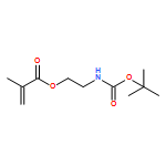
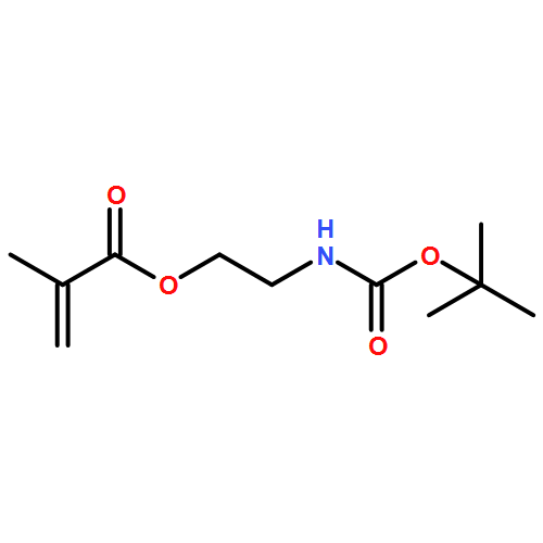
![2-Propenoic acid, 2-methyl-, 2-[[(1,1-dimethylethoxy)carbonyl]amino]ethyl ester](/data/chemimg/1566300/89743-52-2.png)
![2-Propenoic acid, 2-methyl-, 2-[[(1,1-dimethylethoxy)carbonyl]amino]ethyl ester](/data/chemimg/1566300/89743-52-2_b.png)
![2-PROPENAMIDE, 2-METHYL-N-[4-[(2-PYRIMIDINYLAMINO)SULFONYL]PHENYL]-](http://img.cochemist.com/ccimg/52300/52205-02-4.png)
![2-PROPENAMIDE, 2-METHYL-N-[4-[(2-PYRIMIDINYLAMINO)SULFONYL]PHENYL]-](http://img.cochemist.com/ccimg/52300/52205-02-4_b.png)


![3,5,9-Trioxa-4-phosphaheptacos-18-en-1-aminium,4-hydroxy-N,N,N-trimethyl-10-oxo-7-[[(9Z)-1-oxo-9-octadecen-1-yl]oxy]-, innersalt, 4-oxide, (7R,18Z)-](http://img.cochemist.com/ccimg/4300/4235-95-4.png)
![3,5,9-Trioxa-4-phosphaheptacos-18-en-1-aminium,4-hydroxy-N,N,N-trimethyl-10-oxo-7-[[(9Z)-1-oxo-9-octadecen-1-yl]oxy]-, innersalt, 4-oxide, (7R,18Z)-](http://img.cochemist.com/ccimg/4300/4235-95-4_b.png)