Co-reporter:Hua Jin;Jiang Pi;Yue Zhao;Jinhuan Jiang;Ting Li;Xueyi Zeng;Peihui Yang;Colin E. Evans
Nanoscale (2009-Present) 2017 vol. 9(Issue 42) pp:16365-16374
Publication Date(Web):2017/11/02
DOI:10.1039/C7NR06898K
Poor bioavailability and non-specificity of chemotherapeutic agents are major challenges in breast cancer treatment. Antibodies and small molecules that block cell signaling pathways have shown promise in the clinic, but their application is also limited by the high costs and treatment dosages required. Novel therapies that aim to rapidly and specifically target malignant cells with long-lasting impact in the tumor microenvironment may ultimately improve clinical outcome in cancer patients. Here, we demonstrate that epidermal growth factor receptor (EGFR)-targeting GE11 peptides conjugated with PEGylated polylactic-co-glycolic acid (PLGA) nanoparticles can be used to effectively deliver an anti-cancer agent, curcumin, into EGFR-expressing MCF-7 cells in vitro and in vivo. Treatment of breast cancer cells and tumor-bearing mice with these curcumin-loaded nanoparticles gave rise to reduced phosphoinositide 3-kinase signaling, decreased cancer cell viability, attenuated drug clearance from the circulation, and suppressed tumor burden compared with free curcumin or non-EGFR targeting nanoparticles. The targeted nanoscale drug delivery system we describe here may provide a new strategy for the design of targeted cancer therapy vectors. Our study provides evidence that the efficacy of pharmacologic anti-cancer agents can be enhanced through their delivery in the form of modified nanoparticles that effectively target specific malignant cell types.
Co-reporter:Jiang Pi, Hua Jin, Jinhuan Jiang, Fen Yang, Huaihong Cai, Peihui Yang, Jiye Cai, Zheng W. Chen
Pharmacological Research 2017 Volume 119(Volume 119) pp:
Publication Date(Web):1 May 2017
DOI:10.1016/j.phrs.2016.11.036
As the active anticancer component of Rabdosia Rubescens, oridonin has been proved to show strong anticancer activity in cancer cells, which is also found to be closely related to its specific inhibition effects on the EGFR tyrosine kinase activity. In this study, atomic force microscopy based single molecule force spectroscopy (AFM-SMFS) was used for real-time and in-situ detection of EGF-EGFR interactions in living esophageal cancer KYSE-150 cells to evaluate the anticancer activity of oridonin for the first time. Oridonin was found to induce apoptosis and also reduce EGFR expression in KYSE-150 cells. AFM-SMFS results demonstrated that oridonin could inhibit the binding between EGF and EGFR in KYSE-150 cells by decreasing the unbinding force and binding probability for EGF-EGFR complexes, which was further proved to be closely associated with the intracellular ROS level. More precise mechanism studies based on AFM-SMFS demonstrated that oridonin treatment could decrease the energy barrier width, increase the dissociation off rate constant and decrease the activation energy of EGF-EGFR complexes in ROS dependent way, suggesting oridonin as a strong anticancer agent targeting EGF-EGFR interactions in cancer cells through ROS dependent mechanism. Our results not only suggested oridonin as a strong anticancer agent targeting EGF-EGFR interactions in ROS dependent mechanism, but also highlighted AFM-SMFS as a powerful technique for pharmacodynamic studies by detecting ligand-receptor interactions, which was also expected to be developed into a promising tool for the screening and mechanism studies of drugs.Download high-res image (181KB)Download full-size image
Co-reporter:Hua Jin;Jiang Pi;Fen Yang;Chaomin Wu
Applied Microbiology and Biotechnology 2016 Volume 100( Issue 15) pp:6643-6652
Publication Date(Web):2016 August
DOI:10.1007/s00253-016-7360-8
Angiogenesis provides necessary nutrients and oxygen for tumor growth and metastasis; thus, every stage of angiogenesis process is the potential target for cancer therapies. Ursolic acid (UA) is reported to decrease tumor burden through anti-angiogenesis pathway, but its poor water solubility greatly limits its efficiency and clinical application. Here, a simple method for preparing UA-loaded chitosan nanoparticles (CH-UA-NPs) with anti-angiogenesis and anti-tumor activity was demonstrated. In vitro, CH-UA-NPs could significantly inhibit the proliferation, migration, and tube formation of human umbilical vascular endothelial cells (HUVECs). After uptake by HUVECs, CH-UA-NPs were mainly localized in lysosomes and mitochondria, but not nuclei. CH-UA-NPs induced the destruction of lysosome membrane integrity, collapse of mitochondrial membrane potential, and reorganization of cell cytoskeleton. All these changes led to the apoptosis or necrosis in HUVECs. In vivo, CH-UA-NPs could inhibit the angiogenesis in chicken chorioallantoic membrane (CAM) model and H22 xenograft model. Notably, comparing with free UA, such synthesized CH-UA-NPs could save about tenfold of UA doses, implying that this could significantly decrease the side effects induced by high doses of UA in biological organism. Our data showed that CH-UA-NPs and this nanoparticle-based drug delivery system could be as a potential drug candidate for anti-angiogenesis treatment.
Co-reporter:Jiang Pi, Hua Jin, Fen Yang, Zheng W. Chen and Jiye Cai
Nanoscale 2014 vol. 6(Issue 21) pp:12229-12249
Publication Date(Web):26 Aug 2014
DOI:10.1039/C4NR04195J
The cell membrane, which consists of a viscous phospholipid bilayer, different kinds of proteins and various nano/micrometer-sized domains, plays a very important role in ensuring the stability of the intracellular environment and the order of cellular signal transductions. Exploring the precise cell membrane structure and detailed functions of the biomolecules in a cell membrane would be helpful to understand the underlying mechanisms involved in cell membrane signal transductions, which could further benefit research into cell biology, immunology and medicine. The detection of membrane biomolecules at the single molecule level can provide some subtle information about the molecular structure and the functions of the cell membrane. In particular, information obtained about the molecular mechanisms and other information at the single molecule level are significantly different from that detected from a large amount of biomolecules at the large-scale through traditional techniques, and can thus provide a novel perspective for the study of cell membrane structures and functions. However, the precise investigations of membrane biomolecules prompts researchers to explore cell membranes at the single molecule level by the use of in situ imaging methods, as the exact conformation and functions of biomolecules are highly controlled by the native cellular environment. Recently, the in situ single molecule imaging of cell membranes has attracted increasing attention from cell biologists and immunologists. The size of biomolecules and their clusters on the cell surface are set at the nanoscale, which makes it mandatory to use high- and super-resolution imaging techniques to realize the in situ single molecule imaging of cell membranes. In the past few decades, some amazing imaging techniques and instruments with super resolution have been widely developed for molecule imaging, which can also be further employed for the in situ single molecule imaging of cell membranes. In this review, we attempt to summarize the characteristics of these advanced techniques for use in the in situ single molecule imaging of cell membranes. We believe that this work will help to promote the technological and methodological developments of super-resolution techniques for the single molecule imaging of cell membranes and help researchers better understand which technique is most suitable for their future exploring of membrane biomolecules; ultimately promoting further developments in cell biology, immunology and medicine.
Co-reporter:Jiang Pi, Ting Li, Jianxin Liu, Xiaohui Su, Rui Wang, Fen Yang, Haihua Bai, Hua Jin, Jiye Cai
Micron 2014 Volume 65() pp:1-9
Publication Date(Web):October 2014
DOI:10.1016/j.micron.2014.03.012
•We firstly reported that RAW264.7 macrophages would undergo morphological, ultrastructural and bio-mechanical changes after LPS stimulation.•LPS activated RAW264.7 macrophages became much bigger than control cells.•The surface ultrastructure of LPS activated RAW264.7 macrophages was altered as the surface particle sizes and roughness increased.•The adhesive capacity and stiffness of RAW264.7 macrophages increased after LPS stimulation.•The F-actin in RAW264.7 macrophages tended to assemble into the nuclei area after LPS stimulation.In recent years, LPS activated RAW264.7 cells are widely used as an in vitro inflammatory model for the screen of effective anti-inflammation drugs and the investigation of exact anti-inflammation mechanism of these drugs. But up to now, there are few data about the effect of LPS on the morphology, especially on the membrane ultrastructure and bio-mechanical properties of RAW264.7 macrophages. In this work, the topographical and biophysical changes of RAW264.7 macrophages upon LPS stimulation are detected by high resolution atomic force microscopy (AFM). The AFM results suggested that LPS activated RAW264.7 macrophages changed to be much bigger than control cells with some holes emerged on cell surface. The size of membrane protein clusters and the roughness of membrane significantly increased after LPS exposure. In addition, the AFM force measurement results demonstrated that LPS stimulation increased the adhesion force of RAW264.7 macrophages, and also increased the stiffness of RAW264.7 macrophages, which were attributed to the re-distribution of intracellular F-actin structures induced by LPS. These findings suggested that LPS stimulation could also induce the pathophysiological changes of RAW264.7 macrophages, which would benefit our understanding of the inflammatory processes in macrophages upon pathogen stimulation at nano-scale.
Co-reporter:Dong-zhi Yin 银东智;Ji-ye Cai 蔡继业
Journal of Huazhong University of Science and Technology [Medical Sciences] 2014 Volume 34( Issue 1) pp:1-9
Publication Date(Web):2014 February
DOI:10.1007/s11596-014-1223-2
Oval cells have a potential to differentiate into a variety of cell lineages including hepatocytes and biliary epithelia. Several models have been established to activate the oval cells by incorporating a variety of toxins and carcinogens, alone or combined with surgical treatment. Those models are obviously not suitable for the study on human hepatic oval cells. It is necessary to establish a new and efficient model to study the human hepatic oval cells. In this study, the hepatocyte growth factor (HGF) and epidermal growth factor (EGF) were used to induce differentiation of mouse embryonic stem (ES) cells into hepatic oval cells. We first confirmed that hepatic oval cells derived from ES cells, which are bipotential, do exist during the course of mouse ES cells’ differentiation into hepatic parenchymal cells. RT-PCR and transmission electron microscopy were applied in this study. The ratio of Sca-1+/CD34+ cells sorted by FACS in the induction group was increased from day 4 and reached the maximum on the day 8, whereas that in the control group remained at a low level. The differentiation ratio of Sca-1+/CD34+ cells in the induction group was significantly higher than that in the control group. About 92.48% of the sorted Sca-1+/CD34+ cells on the day 8 were A6 positive. Highly purified A6+/Sca-1+/CD34+ hepatic oval cells derived from ES cells could be obtained by FACS. The differentiation ratio of hepatic oval cells in the induction group (up to 4.46%) was significantly higher than that in the control group. The number of hepatic oval cells could be increased significantly by HGF and EGF. The study also examined the ultrastructures of ES-derived hepatic oval cells’ membrane surface by atomic force microscopy. The ES-derived hepatic oval cells cultured and sorted by our protocols may be available for the future clinical application.
Co-reporter:Qiping Shi;Simin Luo;Haiying Jia;Lie Feng;Xiaohua Lu;Lixin Zhou
Journal of Cellular Biochemistry 2013 Volume 114( Issue 10) pp:2221-2230
Publication Date(Web):
DOI:10.1002/jcb.24555
ABSTRACT
Transplantation of functional insulin-producing cells (IPCs) provides a novel mode for insulin replacement, but is often accompanied by many undesirable side effects. Our previous studies suggested that IPCs could not mimic the physiological regulation of insulin secretion performed by pancreatic beta cells. To obtain a better method through which to acquire more similar IPCs, we compared the difference between IPCs of the GLP-1 group and IPCs of the non-GLP-1 group in the morphological features in cellular level and physiological function. The levels of insulin secretion were measured by ELISA. The insulin and glucagon-like peptide-1 (GLP-1) mRNA gene expression was determined by real-time quantitative PCR. The morphological features were detected by atomic force microscopy (AFM) and laser confocal scanning microscopy (LCSM). Intracellular Ca2+ levels and Glucagon-like peptide-1 receptor (GLP-1R) levels were determined by flow cytometer (FCM). We found that IPCs of the GLP-1 group had bigger membrane particle size and average roughness (Ra) than IPCs of the non-GLP-1 group but still smaller than normal human pancreatic beta cells. The physiology function of IPCs of the GLP-1 group were much closer to normal human pancreatic beta cells than IPCs of the non-GLP-1 group. GLP-1 could improve the similarity of IPCs from human adipose tissue-derived mesenchymal stem cells and pancreatic beta cells in cellular ultrastructure and function. J. Cell. Biochem. 114: 2221–2230, 2013. © 2013 Wiley Periodicals, Inc.
Co-reporter:Jiang Pi, Fen Yang, Hua Jin, Xun Huang, Ruiying Liu, Peihui Yang, Jiye Cai
Bioorganic & Medicinal Chemistry Letters 2013 Volume 23(Issue 23) pp:6296-6303
Publication Date(Web):1 December 2013
DOI:10.1016/j.bmcl.2013.09.078
Selenium nanoparticles (Se NPs) have been served as promising materials for biomedical applications, especially for cancer treatment. The anti-cancer effects of Se NPs against cancer cells have been widely studied in recent years, but whether Se NPs can induce the changes of cell membrane bio-mechanical properties in cancer cells still remain unexplored. In this Letter, we prepared Se NPs for investigating the intracellular localization of Se NPs in MCF-7 cells and determined the effects of Se NPs on apoptosis and necrosis in MCF-7 cells. Especially, we reported for the first time about the effects of Se NPs on the bio-mechanical properties of cancer cells and found that Se NPs could remarkably decrease the adhesion force and Young’s modulus of MCF-7 cells. To further understand the potential mechanisms about how Se NPs affect the bio-mechanical properties of MCF-7 cells, we also investigated the expression of CD44 molecules, the structure and the amounts of F-actin. The results indicated that the decreased adhesion force between AFM tip and cell membrane was partially due to the changes of membrane molecules induced by Se NPs, such as the down-regulation of trans-membrane CD44 molecules. Additionally, the decrease of Young’s modulus of MCF-7 cells was due to the dis-organization and down-regulation of F-actin induced by Se NPs. These results collectively suggested that cell membrane was of vital importance in Se NPs induced toxicity in cancer cells, which could be served as a potential target for cancer treatment by Se NPs.
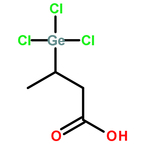
Co-reporter:Jiang Pi, Jing Zeng, Jian-Jun Luo, Pei-Hui Yang, Ji-Ye Cai
Bioorganic & Medicinal Chemistry Letters 2013 Volume 23(Issue 10) pp:2902-2908
Publication Date(Web):15 May 2013
DOI:10.1016/j.bmcl.2013.03.061
Germanium (Ge) is considered to play a key role in the pharmacological effects of some medicinal plants. Here, two new Ge(IV)–polyphenol complexes were synthesized and measured for their potential biological activities. The results indicated that these Ge(IV)–polyphenol complexes possessed great anti-oxidative activities, both showing stronger hydroxyl scavenging effects than their corresponding ligands. We also demonstrated the strong intercalating abilities of Ge(IV)–polyphenol complexes into calf thymus-DNA molecules. In addition, these two Ge(IV)–polyphenol complexes showed strong proliferative inhibition effect on HepG2 cancer cells. Moreover, the morphological changes in HepG2 cells induced by Ge(IV)–polyphenol complexes were detected by atomic force microscopy. All these results collectively suggested that Ge(IV)–polyphenol complexes could be served as promising pharmacologically active substances against cancer treatment.
Co-reporter:Yanjuan Tang, Guimin Sun, Jiye Cai and Peihui Yang
Analytical Methods 2013 vol. 5(Issue 18) pp:4602-4607
Publication Date(Web):05 Jun 2013
DOI:10.1039/C3AY40249E
A versatile and facile strategy for detecting the inhibitory action of curcumin on cancer cells has been developed by using the specific recognition between folic acid conjugated-gold nanorods (F-AuNRs) and folate receptors on the cell surface, based on the localized surface plasmon resonance (LSPR) and fluorescence spectra of gold nanorods (AuNRs). The F-AuNR probe was used to detect cancer cells with a detection limit down to 10 cells per mL, and it could replace traditional organic dyes as it has a better detection limit. Combined MTT, flow cytometry, fluorescence images and high-resolution AFM images further confirmed the feasibility of the spectroscopy methods. This approach can be used as a simple, rapid and sensitive tool to study the inhibitory action of drugs on cancer cells.
Co-reporter:Jiang Pi;Hua Jin;RuiYing Liu;Bing Song
Applied Microbiology and Biotechnology 2013 Volume 97( Issue 3) pp:1051-1062
Publication Date(Web):2013 February
DOI:10.1007/s00253-012-4359-7
Selenium nanoparticles (Se NPs) have been recognized as promising materials for biomedical applications. To prepare Se NPs which contained cancer targeting methods and to clarify the cellular localization and cytotoxicity mechanisms of these Se NPs against cancer cells, folic acid protected/modified selenium nanoparticles (FA–Se NPs) were first prepared by a one-step method. Some morphologic and spectroscopic methods were obtained to prove the successfully formation of FA–Se NPs while free folate competitive inhibition assay, microscope, and several biological methods were used to determine the in vitro uptake, subcellular localization, and cytotoxicity mechanism of FA–Se NPs in MCF-7 cells. The results indicated that the 70-nm FA–Se NPs were internalized by MCF-7 cells through folate receptor-mediated endocytosis and targeted to mitochondria located regions through endocytic vesicles transporting. Then, the FA–Se NPs entered into mitochondria; triggered the mitochondria-dependent apoptosis of MCF-7 cells which involved oxidative stress, Ca2+ stress changes, and mitochondrial dysfunction; and finally caused the damage of mitochondria. FA–Se NPs released from broken mitochondria were transported into nucleus and further into nucleolus which then induced MCF-7 cell cycle arrest. In addition, FA–Se NPs could induce cytoskeleton disorganization and induce MCF-7 cell membrane morphology alterations. These results collectively suggested that FA–Se NPs could be served as potential therapeutic agents and organelle-targeted drug carriers in cancer therapy.
Co-reporter:Li Liu;Hua Jin;JinLai Ou;JinHuan Jiang;Jiang Pi
Science Bulletin 2013 Volume 58( Issue 21) pp:2584-2593
Publication Date(Web):2013 July
DOI:10.1007/s11434-013-5739-9
When used in combination with certain chemotherapies, curcumin has been shown to increase apoptosis in several cancer cell lines. Here, we report the combined effects of curcumin and cinobufacini on human cervical carcinoma cells. The aim of this study was to examine whether curcumin could enhance apoptosis induced by cinobufacini. 3-(4,5-Dimethylthiazol-2-y1)-2,5-diphenytetrazolium bromide (MTT) assays revealed that the growth and proliferation of HeLa cells could be inhibited by 75% after a combined treatment of 25 μg/mL cinobufacini and 8 μg/mL curcumin. The combined treatment is 3 times more effective than treatment with 25 μg/mL cinobufacini alone. Annexin V-FITC/PI staining, morphological changes and immunofluorescence verified a significant enhancement in cinobufacini-induced apoptosis when cells were also exposed to curcumin. The data showed that the proportion of early apoptotic cells significantly increased from 15.43% in cells treated only with 25 μg/mL cinobufacini to 49.2% in cells treated with 25 μg/mL cinobufacini and 8 μg/mL curcumin. Moreover, compared with treatment of only 25 μg/mL cinobufacini, ROS production increased 1.7-fold, the intracellular free Ca2+ concentration increased 1.5-fold, and the mitochondrial membrane potential decreased by 20% in the combined treatment. Simultaneously, the atomic force microscopy (AFM) results suggest that cells treated with a combination of cinobufacini and curcumin varied significantly in shape and ultrastructure. Collapsed cells with leaking cytoplasm, blebbing pores and emerging apoptotic bodies were prevalent. The nanoparticle size increased from 70 nm when the cells were treated with 25 μg/mL cinobufacini to 190 nm when the cells were treated with 25 μg/mL cinobufacini and 8 μg/mL curcumin. The size increase resulted in the cell membrane becoming considerably rough. These results can improve our understanding of combination treatments. Specifically, the combination of cinobufacini and curcumin may potentially find use as a novel cervical carcinoma treatment. Additionally, AFM is a powerful tool that can be used to explore cellular morphologies and ultrastructures.
Co-reporter:Lina Ma, Bing Song, Hua Jin, Jiang Pi, Li Liu, Jinhuan Jiang, Jiye Cai
Bioorganic & Medicinal Chemistry Letters 2012 Volume 22(Issue 3) pp:1459-1463
Publication Date(Web):1 February 2012
DOI:10.1016/j.bmcl.2011.11.095
Cinobufacini is a traditional Chinese anti-tumor drug and widely used in clinic experiences. But little is known about its effect on the cells. In this study, the effects of cinobufacini on breast cancer MDA-MB-231 cell were evaluated by CCK-8 assay, and the data showed cinobufacini could inhibit the MDA-MB-231 cells growth effectively in dose-dependent and time-dependent manners. Cell apoptosis and cell cycle were detected by flow cytometry analysis. After the cells being treated with 50 μg/mL cinobufacini for 48 h, the early apoptosis percentage (20.45 ± 1.46%) is much higher than the normal group (7.73 ± 1.21%). The cell cycle data indicated that cinobufacini caused a cell cycle arrest at S phase. What’s more, cinobufacini can affect the disruption of cytoskeleton, and these alterations changed the cell-surface ultrastructure and the cell morphology which were detected by atomic force microscopy (AFM) at nanoscale level. It indicated that the cell membrane structure and cytoskeleton networks were destroyed and the cell tails were narrowed after the cell being treated with cinobufacini. The present study is to provide valuable new insights to understand the mechanism of the drug in anti-tumor process. Furthermore, the knowledge concerning the signaling of cell cycle is potentially important to clinical utility.Cinobufacini could induce MDA-MB-231 cell apoptosis, AFM images showed that cinobufacini could changes the cell surface significantly (Fig. 2). LCSM images showed the cells before being treated with cinobufacini, the cells were spindle, a large number of F-actin around the nucleus regularly in the cells and the fibers in the cytoplasm arranged as regular rays. The cells after being treated with cinobufacini (50 μg/mL) for 48 h, F-actin dispersed in the cells and stress fibers around the nucleus arranged in disorder and the numbers were decreased, the nuclei deformed, there were some holes in the middle of the nuclei (Fig. 4) and cinobufacini caused a cell cycle arrest at S phase.
Co-reporter:Qiping Shi;Simin Luo;Hua Jin
Applied Microbiology and Biotechnology 2012 Volume 94( Issue 2) pp:479-486
Publication Date(Web):2012 April
DOI:10.1007/s00253-012-3904-8
We successfully differentiated human adipose tissue-derived mesenchymal stem cells (haMSCs) into insulin-producing cells (IPCs) in vitro and did not use any insulin which might be absorbed by cells during in vitro culture. Expression of insulin gene was massively increased by 28,000-fold at day 12 compared with haMSCs (P < 0.05). IPCs could secrete insulin after glucose was stimulated. The higher the concentration of glucose, the more production of insulin was noted. We reported AFM images of IPCs for the first time. AFM images showed that the sizes of cells were similar to each other, and all IPC surface had a porous structure in the cytoplasm area. In sugar-free group, the size of holes was similar (diameter, 1,086.98 ± 156.70 nm; depth, 185.22 ± 52.14 nm). In higher sugar-stimulated group, there were more holes with bigger diameter and smaller depth. (diameter, 3,183.65 ± 2,229.18 nm; depth 109.42 ± 56.26 nm, P < 0.05). We found that the hole diameter and depth could change with the concentration of glucose in media. Concurrently, laser scanning confocal microscopy images indicated that cortical actin network beneath plasma membrane in IPCs was dense and continuous. After glucose stimulation, we found the actin web depolymerized and became discontinuous in IPCs. We speculated that diameter augmentation of holes located in the cytoplasm area in IPCs was one manifestation of excytosis increase.
Co-reporter:Mu Wang, Yuxia Ruan, Xiaobo Xing, Qian Chen, Yuan Peng, Jiye Cai
Analytica Chimica Acta 2011 Volume 697(1–2) pp:83-89
Publication Date(Web):4 July 2011
DOI:10.1016/j.aca.2011.04.028
The cell surface glycoprotein CD44 was implicated in the progression, metastasis and apoptosis of certain human tumors. In this study, we used atomic force microscope (AFM) to monitor the effect of curcumin on human hepatocellular carcinoma (HepG2) cell surface nanoscale structure. High-resolution imaging revealed that cell morphology and ultrastructure changed a lot after being treated with curcumin. The membrane average roughness increased (10.88 ± 4.62 nm to 129.70 ± 43.72 nm) and the expression of CD44 decreased (99.79 ± 0.16% to 75.14 ± 8.37%). Laser scanning confocal microscope (LSCM) imaging showed that CD44 molecules were located on the cell membrane. The florescence intensity in control group was weaker than that in curcumin treated cells. Most of the binding forces between CD44 antibodies and untreated HepG2 cell membrane were around 120–220 pN. After being incubated with curcumin, the major forces focused on 70–150 pN (10 μM curcumin-treated) and 50–120 pN (20 μM curcumin-treated). These results suggested that, as result of nanoscale molecular redistribution, changes of the cell surface were in response to external treatment of curcumin. The combination of AFM and LSCM could be a powerful method to detect the distribution of cell surface molecules and interactions between molecules and their ligands.Graphical abstractHighlights► In this study, we investigate the changes of CD44 expression and distribution on HepG2 cells after curcumin treatment. ► We find curcumin is able to change the morphology and ultrastructure of HepG2 cells. ► Curcumin can reduce the expression of CD44 molecules and induce the nanoscale molecular redistribution on cell surface. ► The binding force between CD44-modified AFM tip and the HepG2 cell surface decreases after curcumin-treatment.
Co-reporter:Xiaofang Cai, Pengtao You, Jiye Cai, Xiaoxi Yang, Qian Chen, Feicheng Huang
Micron 2011 Volume 42(Issue 1) pp:17-28
Publication Date(Web):January 2011
DOI:10.1016/j.micron.2010.08.009
Integrity of the cell membrane is a basic requirement for maintaining the biological characteristics of a cell. In this study, cell membrane as the target of drug action was investigated. CCK-8 assay suggested that Artesunate (ART) could significantly suppress the proliferation of Jurkat cells in a dose-dependent manner. Changes in the morphology and mechanics of Jurkat cells were studied by atomic force microscopy (AFM). These changes included decrease of Young's modulus (from 3.18 ± 0.54 to 1.72 ± 0.54 kPa), increase in the fluctuation of surface components, increase in shrinkage, or even the appearance of pores. The Young's modulus change was according to the F-actin protein, not the Tubulin-β or integrin β1 protein. Meanwhile, the activities of plasma membrane Ca2+–Mg2+-ATPase and Na+–K+-ATPase were also repressed following ART exposure as well as membrane potential. Western blot was used to detect Caspase 3 and Cyclin D1 protein level. The Cyclin D1 was downregulated and Caspase 3 was activated. Hence, cellular membrane represented a plausible target for ART-induced injury.
Co-reporter:Xiaobo Xing, Hua Jin, Yuhong Lu, Qiulan Wang, Yunlong Pan, Jiye Cai, Haiyan Wang
Micron 2011 Volume 42(Issue 1) pp:42-46
Publication Date(Web):January 2011
DOI:10.1016/j.micron.2010.08.004
The pathological changes of erythrocytes were detected at the nanometer scale, which was important for revealing the onset of diseases, early diagnosis, and effective therapies. Diseases may disturb the morphology and function of erythrocytes at molecular scale. There were dramatic surface deformations in topography of erythrocytes from a patient with elliptocytosis complicating idiopathic thrombocytopenic purpura (ITP). The overall shape and surface membrane of the healthy, pre- and post-therapeutic erythrocytes have been studied by high-resolution atomic force microscopy imaging. The results showed that we can detect healthy and pathological erythrocytes by the morphologic parameters of the length, width, ratio of length to width, peak, valley, valley-to-peak, surface fluctuation, and standard deviations of the erythrocytes. Therefore, the morphologic information of erythrocytes is very important indictor for diagnosing the healthy and disease, as well as evaluating therapeutic effect.
Co-reporter:Jianan Chen, Yin Pei, Zhengwei Chen, Jiye Cai
Micron 2010 Volume 41(Issue 3) pp:198-202
Publication Date(Web):April 2010
DOI:10.1016/j.micron.2009.11.002
The lateral organization of membrane proteins and lipids domains has a direct impact on many cellular processes, but generally these domains are too small to be resolved by diffraction-limited resolution of fluorescence microscopy. Here, we use quantum dot (QD) labeling based on near-field optical imaging, to study the nanoscale organization of hyaluronan receptor CD44 molecules of fixed mesenchymal stem cells (MSCs) in air, with a optical resolution down to 50 nm. The photostability and high luminance of QD evidently improve the signal-to-noise ratio and reproducibility of near-field optical data. Importantly, the blinking-intensity analysis was proposed to identify single QD, providing a calibration to relate intensity to numbers of antibody for the first time. Additionally, the fluorescence-topographic imaging enables us to investigate the topographic location pattern. Our results demonstrate that CD44 molecules on MSCs are enriched into nanosized domain and they predominantly locate on the peak of the membrane protrusions, which may contribute to clarify the underlying mechanism of functions ascribed to these molecules.
Co-reporter:Xiaofang Cai, Xiaobo Xing, Jiye Cai, Qian Chen, Shixian Wu, Feicheng Huang
Micron 2010 Volume 41(Issue 3) pp:257-262
Publication Date(Web):April 2010
DOI:10.1016/j.micron.2009.08.011
The mechanical properties of cells are important for many cellular processes. Here, atomic force microscopy (AFM) and laser scanning confocal microscopy (LSCM) were carried out to characterize lymphocyte and Jurkat cells. The average elastic modulus of lymphocyte is 1.24 ± 0.09 kPa, which is almost twofold higher than that of Jurkat cell (0.51 ± 0.06 kPa). LSCM images of sub-membrane cytoskeleton showed a significant difference in the organization of their F-actin structures. Lymphocyte cells had more and thicker actin bundles than that of Jurkat cells. Lymphocyte and Jurkat cells after adding the F-actin destabilizing agent Cytochalasin-B (Cyt-B) were also investigated by AFM. A decrease in the elastic modulus of lymphocyte from a value of 1.24 ± 0.09 kPa down to 0.34 ± 0.04 kPa for 24 h was observed, and that of Jurkat cell decreased from 0.51 ± 0.06 kPa to 0.23 ± 0.04 kPa. We really believe that this technology will be used for cancer detection and opens a door to study the biophysical properties of signaling domains extending from the cell surface to deeper parts of the cell.
Co-reporter:Xiaofang Cai, Xiaoxi Yang, Jiye Cai, Shixian Wu and Qian Chen
The Journal of Physical Chemistry B 2010 Volume 114(Issue 11) pp:3833-3839
Publication Date(Web):March 2, 2010
DOI:10.1021/jp910682q
Mitomycin C (MMC) has been shown to have a therapeutic effect against human pterygium fibroblasts (HPFs) by inducing apoptosis. However, there is little data about the effect of it on plasma membrane. In the present study, the cytotoxicity of MMC to HPFs including inhibiting cell growth, inducing apoptosis and bringing about membrane toxicity was investigated. It was found that MMC could significantly suppress the proliferation of HPFs in a dose-dependent manner by CCK-8 assay. Flow cytometric analysis also revealed that treatment with MMC resulted in increased percentages of apoptotic cells in a dose-dependent manner. Membrane lipid peroxidation level, lactate dehydrogenase (LDH) leakage, membrane surface topography, and membrane rigidity alterations were investigated to assess the membrane toxicity induced by MMC. Treatment with MMC at different concentrations accelerated membrane lipid peroxidation and potentiated LDH leakage, which was consistent with disturbance of membrane surface and decrease of membrane elasticity detected by atomic force microscopy. All the above changes led to the disturbed intracellular Ca2+ homeostasis, which was an important signal triggering apoptosis. Hence, the membrane toxicity induced by MMC might play an important role in the process of apoptotic induction and the calcium channel may be one of the apoptosis mechanisms.
Co-reporter:Hua Jin;Xun Huang;Yong Chen;Hongxia Zhao
Applied Microbiology and Biotechnology 2010 Volume 88( Issue 3) pp:761-770
Publication Date(Web):2010 October
DOI:10.1007/s00253-010-2747-4
The photodynamic antimicrobial chemotherapy as a promising approach for efficiently killing pathogenic microbes is attracting increasing interest. In this study, the cytotoxic and phototoxic effects of hematoporphyrin monomethyl ether (HMME) on the Gram-positive and Gram-negative bacteria were investigated. The cell viability was assessed by colony-forming unit method, and the results indicated that there was no significant cytotoxicity but high phototoxicity in the examined concentrations. Notably, the Gram-positive bacteria were more sensitive to HMME in phototoxicity. Simultaneously, an atomic force microscope (AFM) was used to detect the changes in morphological and nanomechanical properties of bacteria before and after HMME treatment. AFM images indicate that upon photoinactivation, the bacterial surface changed from a smooth, homogeneous architecture to a heterogenous, crackled morphology. The force spectroscopy measurements reveal that the cell wall became less rigid and the Young’s modulus decreased about 50%, whereas the tip-cell-surface adhesion forces increased significantly compared to those of native cells. It was speculated that the photodynamic effects of HMME induced the changes in the chemical composition of the outer membrane and exposure of some proteins inside the envelope. AFM can be utilized as a powerful and sensitive method for studying the interaction between bacteria and drugs.
Co-reporter:Huai-Hong Cai, Xing Zhong, Pei-Hui Yang, Wei Wei, Jianan Chen, Jiye Cai
Colloids and Surfaces A: Physicochemical and Engineering Aspects 2010 Volume 372(1–3) pp:35-40
Publication Date(Web):3 December 2010
DOI:10.1016/j.colsurfa.2010.09.017
With the objective of probing site-selective binding mechanisms of rhodamine dye with serum albumin, the interactions between rhodamine B (RB) and bovine serum (BSA) have been investigated by the complementary methods of absorption spectroscopy, steady-state and time-resolved fluorescence spectroscopy, circular dichroism (CD) spectroscopy, and cyclic voltammetry, respectively. The results from fluorescence analysis indicated that the site-selective binding of RB with BSA originated from the complex formation with high association constant. The conformational changes of BSA after binding with RB were characterized by decreased absorbance and CD signal of BSA, and significant weakened electrochemical signal of RB. The site-selective binding of RB with BSA was confirmed by analysis of time-resolved fluorescence spectra, which exhibited biexponential decay with significantly enhanced lifetime when compared to the lifetime of RB alone. Employing ligand displacement cum fluorescence measurements using known site-specific binding ligands, such as phenylbutazone and ibuprofen, further confirmed that RB selectively bound with BSA at site I and II, which induced the unfolding of BSA. In addition, a reliable mechanism was proposed to explain the interactions of RB with BSA, which were mediated by concentrations-dependent site-selective binding of dye molecules with protein in residues of different subdomains. These results provide further information for better understanding the interaction mechanisms of rhodamine dyes with serum albumin.Graphical abstractResearch highlights▶ With the objective of probing binding mechanisms of rhodamine dye with protein, the interaction of rhodamine B and bovine serum albumin was investigated by the complementary spectroscopic methods. ▶ Results showed that the binding of rhodamine B to bovine serum albumin was concentrations-dependently site-selective binding. ▶ Rhodamine molecules preferentially bound to site I of protein when dye concentrations were low, whereas dye molecules selectively bound to site II of protein at their higher concentration.
Co-reporter:Hongxia Zhao, Hua Jin, Jiye Cai, Shan Ding
Ultramicroscopy 2010 Volume 110(Issue 10) pp:1306-1311
Publication Date(Web):September 2010
DOI:10.1016/j.ultramic.2010.06.002
Investigation and simulation of naturally occurring mineralization can offer some new ideas in the design and fabrication of new functional materials for bone analogues. In this paper, a model dual membrane diffusion system (DMDS) was used to study the mineralization behaviour of collagen. The process of mineralization was observed by atomic force microscope (AFM). The results showed that the surface roughness and hardness of mineralized collagen fibers increased with time during the process of mineralization. The adhesion force of mineralized collagen fibers decreased with mineralization time. The micromechanical properties and microstructure changes of mineralized collagen fibers suggested that the mineralization was a step-by-step assembling process.
Co-reporter:Huai-Hong Cai, Pei-Hui Yang, Jianan Chen, Zhi-Hong Liang, Qiongyu Chen, Jiye Cai
Electrochimica Acta 2009 Volume 54(Issue 13) pp:3651-3656
Publication Date(Web):1 May 2009
DOI:10.1016/j.electacta.2009.01.042
DNA conformational change and breakage induced by artemisinin, a traditional Chinese herbal medicine, have been visually characterized and quantitatively measured by the multiple tools of electrochemistry, UV–vis absorption spectroscopy, atomic force microscopy (AFM), and DNA electrophoresis. Electrochemical and spectroscopic results confirm that artemisinin can intercalate into DNA double helix, which causes DNA conformational changes. AFM imaging vividly demonstrates uneven DNA strand breaking induced by QHS interaction. To assess these DNA breakages, quantitative analysis of the extent of DNA breakage has been performed by analyzing AFM images. Basing on the statistical analysis, the occurrence of DNA breaks is found to depend on the concentration of artemisinin. DNA electrophoresis further validates that the intact DNA molecules are unwound due to the breakages occur at the single strands. A reliable scheme is proposed to explain the process of artemisinin-induced DNA cleavage. These results can provide further information for better understanding the anticancer activity of artemisinin.
Co-reporter:Yangzhe Wu, Yi Hu, Jiye Cai, Shuyuan Ma, Xiaoping Wang, Yong Chen, Yunlong Pan
Micron 2009 Volume 40(Issue 3) pp:359-364
Publication Date(Web):April 2009
DOI:10.1016/j.micron.2008.10.003
Atomic force microscopy (AFM) is a rapidly developing tool recently introduced into the evaluation of the age of bloodstains, potentially providing legal medical experts useful information for forensic investigation. In this study, the time-dependent, morphological changes of red blood cells (RBC) under three different conditions (including controlled, room-temperature condition, uncontrolled, outdoor-environmental condition, and controlled, low-temperature condition) were observed by AFM, as well as the cellular viscoelasticity via force-vs-distance curve measurements. Firstly, the data indicate that substrate types have different effects on cellular morphology of RBC. RBC presented the typical biconcave shape on mica, whereas either the biconcave shape or flattened shape was evident on glass. The mean volume of RBCs on mica was significantly larger than that of cells on glass. Surprisingly, the adhesive property of RBC membrane surfaces was substrate type-independent (the adhesive forces were statistically similar on glass and mica). With time lapse, the changes in cell volume and adhesive force of RBC under the controlled room-temperature condition were similar to those under the uncontrolled outdoor-environmental condition. Under the controlled low-temperature condition, however, the changes in cell volume occurred mainly due to the collapse of RBCs, and the curves of adhesive force showed the dramatic alternations in viscoelasticity of RBC. Taken together, the AFM detections on the time-dependent, substrate type-dependent, environment (temperature/humidity)-dependent changes in morphology and surface viscoelasticity of RBC imply a potential application of AFM in forensic medicine or investigations, e.g., estimating age of bloodstain or death time.
Co-reporter:Mingqian Hu;Jianan Chen;Jiongkun Wang;Xiaoping Wang;Shuyuan Ma;Crystal Y. Chen;Zheng W. Chen
Journal of Molecular Recognition 2009 Volume 22( Issue 6) pp:516-520
Publication Date(Web):
DOI:10.1002/jmr.976
Abstract
Although CD69 is well known as an early T cell-activation marker, the possibility that CD69 are distributed as nano-structures on membrane for immune regulation during T cell activation has not been tested. In this study, nanoscale features of CD69 expression on activated T cells were determined using the atomic force microscopy (AFM) topographic and force-binding nanotechnology as well as near-field scanning optical microscopy (NSOM)-/fluorescence quantum dot (QD)-based nanosacle imaging. Unstimulated CD4+ T cells showed neglectable numbers of membrane CD69 spots binding to the CD69 Ab-functinalized AFM tip, and no detectable QD-bound CD69 as examined by NSOM/QD-based imaging. In contrast, Phytohemagglutinin (PHA)-activated CD4+ T cells expressed CD69, and displayed many force-binding spots binding to the CD69 Ab-functionalized AFM tip on about 45% of cell membrane, with mean binding-rupture forces 276 ± 71 pN. Most CD69 molecules appeared to be expressed as 100–200 nm nanoclusters on the membrane of PHA-activated CD4+ T cells. Meanwhile, NSOM/QD-based nanoscale imaging showed that CD69 were non-uniformly distributed as 80–200 nm nanoclusters on cell-membrane of PHA-activated CD4+ T cells. This study represents the first demonstration of the nano-biology of CD69 expression during T cell activation. Copyright © 2009 John Wiley & Sons, Ltd.
Co-reporter:Yangzhe Wu;Hongsong Lu;Xianhui He;Yi Hu
Nanoscale Research Letters 2009 Volume 4( Issue 8) pp:
Publication Date(Web):2009 August
DOI:10.1007/s11671-009-9340-8
The activation of T lymphocytes plays a very important role in T-cell-mediated immune response. Though there are many related literatures, the changes of membrane surface nanostructures and adhesion property of T lymphocytes at different activation stages have not been reported yet. However, these investigations will help us further understand the biophysical and immunologic function of T lymphocytes in the context of activation. In the present study, the membrane architectures of peripheral blood T lymphocytes were obtained by AFM, and adhesion force of the cell membrane were measured by acquiring force–distance curves. The results indicated that the cell volume increased with the increases of activation time, whereas membrane surface adhesion force decreased, even though the local stiffness for resting and activated cells is similar. The results provided complementary and important data to further understand the variation of biophysical properties of T lymphocytes in the context of in vitro activation.
Co-reporter:Yangzhe Wu;Yi Hu;Shuyuan Ma
Journal of Materials Science: Materials in Medicine 2008 Volume 19( Issue 12) pp:3621-3629
Publication Date(Web):2008 December
DOI:10.1007/s10856-008-3477-3
Biomacromolecule has been widely used as biomedical material. Because different biomacromolecules possess different properties, how to exhibit the respective advantages of different components on one type of biomaterial becomes the hot spot in the field of biomaterial studying. This work reported a type of complex film that consisted of hyaluronic acid (HA), type I collagen (Col-I), and chitosan (CS) (HA–Col-I/CS, HCC). Then, a series of experiments were performed, such as inverted microscopic observation, atomic force microscopic (AFM) imaging, flow cytometry (FCM) measurement, MTT assay, and MIC assay. In the present work, we observed the growing condition of 3T3 fibroblasts on the surface of the HCC complex film, visualized the morphological changes of platelets during the coagulation process, and discovered microparticles on the platelet membrane. Moreover, we confirmed the microparticles are the platelet-derived microparticles (PMPs) using the FCM. In addition, the minimal inhibitory concentration (MIC) of HCC against Escherichia coli (E. coli) 8099 was 0.025 mg/ml, against Staphylococcus aureus (S. aureus) ATCC 6538 was 0.1 mg/ml. The results together indicated that the HCC film possessed promising coagulation property, cell compatibility and anti-bacteria property, and the potential in future clinical application such as wound healing and bandage.
Co-reporter:Yun-Qi Wang;Ji-Ye Cai
Applied Biochemistry and Biotechnology 2007 Volume 141( Issue 2-3) pp:203-213
Publication Date(Web):2007 June
DOI:10.1007/BF02729062
A novel strategy for constructing multiple joined genes of acidic partner-mediated antimicrobial peptide is described. This strategy allows the expression of antimicrobial peptide byEscherichia coli in a stable form and with high yield. Cecropin A (1–8)-melittin (1–10) (CAME) hybrid peptide was selected as a model of antimicrobial peptide. An acidic fragment from magainin intervening sequence was fused to the antimicrobial peptide as a partner to neutralize the lethal effects on the host cells. Multiple copies of the fusion peptide gene were tandemly linked and cloned into the expression vector pET21a. Multimers were expressed at high levels, reaching up to 36% of total cell proteins, and expression levels were proportional to the degree of multimerization. The fusion proteins were mainly expressed as inclusion bodies, probably owing to cysteine residues in the multimers. The target CAME peptide was obtained by cleaving the multimers with cyanogen bromide and purified by cation-exchange chromatography. Recombinant CAME peptide showed strong antimicrobial activities against both Gram-negative and -positive bacteria. These results might provide an efficient solution for high-level expression of various kinds of antimicrobial peptides that are toxic to the host.
Co-reporter:Yangzhe Wu, Jiye Cai, Longqiu Cheng, Yanfang Xu, Zhiyan Lin, Chenxi Wang, Yong Chen
Micron 2006 Volume 37(Issue 2) pp:139-145
Publication Date(Web):February 2006
DOI:10.1016/j.micron.2005.08.007
CHO cells possess easily identifiable karyotypes, and CHO cell chromosomes are large and few in number, making these cells ideal for mutational and drug toxicity studies and suitable for investigations of animal chromosome structure. Here, we used atomic force microscopy (AFM) in the tapping mode for detailed visualizations of Chinese hamster ovary (CHO) cell chromosomes during various mitotic phases, including typical prophase, prometaphase, metaphase, anaphase and telophase. Based on our detailed observations, we were able to divide metaphase and anaphase into sub-phases: metaphase I, II and III, and anaphase I and II. Furthermore, we used the AFM error-signal mode to visualize chromosomal ultrastructures and cytokinesis. While these visualizations were all successful, we found that the image quality was affected by cellular debris, contamination. Collectively, our results show that the AFM technique has great potential for the detailed study of chromosomes and chromosomal ultrastructures during all phases of the cell cycle, but that careful standards of sample preparation must be maintained.
Co-reporter:Yong Chen, Jiye Cai
Micron 2006 Volume 37(Issue 4) pp:339-346
Publication Date(Web):June 2006
DOI:10.1016/j.micron.2005.11.011
Estimation of the time of death is one of the most important problems for forensic medicine and law. Physical and chemical postmortem changes are evaluated together while estimating the time of death. The pattern analysis of antemortem and postmortem bloodstains is one of the important parameters for forensic science, and cellular changes of blood cells can be useful for the quantitative assessment of the time of death. In this study, by successively investigating erythrocytes exposed in air on mica for 5 days using tapping mode atomic force microscopy (TM-AFM), we observed deformation of whole cell and membrane surface of unfixed erythrocytes with time lapse. We found that the time-dependent cellular changes occurred after exposure of erythrocytes in air for several days. At 0.5 days of exposure, fissures and cell shrinkage were observed. At 2.5 days of exposure, the emergence of nanometer-scale protuberances were observed and these protuberances increased in number with increasing time. The changes of cell shape and cell membrane surface ultrastructure can be used to estimate the time of death. Futhermore, smear-induced abnormal erythrocytes and immunostained erythrocytes were observed here. The need for more precise research is indicated, such as the correlation of membrane changes to intervals of less than 0.5 day of air exposure, and use of various substrates in addition to mica, including glass, metals, fabrics, among others, on which the bloodstains appear in crime scenes. The results of this research demonstrate the efficacy of AFM as a potentially powerful analytical tool in forensic science.
Co-reporter:Yong Chen, Jiye Cai, Tao Zhao, Chenxi Wang, Shuo Dong, Shuqian Luo, Zheng W. Chen
Ultramicroscopy 2005 Volume 103(Issue 3) pp:173-182
Publication Date(Web):June 2005
DOI:10.1016/j.ultramic.2004.11.019
The thin sectioning has been widely applied in electron microscopy (EM), and successfully used for an in situ observation of inner ultrastructure of cells. This powerful technique has recently been extended to the research field of atomic force microscopy (AFM). However, there have been no reports describing AFM imaging of serial thin sections and three-dimensional (3-D) reconstruction of cells and their inner structures. In the present study, we used AFM to scan serial thin sections approximately 60 nm thick of a mouse embryonic stem (ES) cell, and to observe the in situ inner ultrastructure including cell membrane, cytoplasm, mitochondria, nucleus membrane, and linear chromatin. The high-magnification AFM imaging of single mitochondria clearly demonstrated the outer membrane, inner boundary membrane and cristal membrane of mitochondria in the cellular compartment. Importantly, AFM imaging on six serial thin sections of a single mouse ES cell showed that mitochondria underwent sequential changes in the number, morphology and distribution. These nanoscale images allowed us to perform 3-D surface reconstruction of interested interior structures in cells. Based on the serial in situ images, 3-D models of morphological characteristics, numbers and distributions of interior structures of the single ES cells were validated and reconstructed. Our results suggest that the combined AFM and serial-thin-section technique is useful for the nanoscale imaging and 3-D reconstruction of single cells and their inner structures. This technique may facilitate studies of proliferating and differentiating stages of stem cells or somatic cells at a nanoscale.
Co-reporter:Qian Feng, Gucheng Zeng, Peihui Yang, Chenxi Wang, Jiye Cai
Colloids and Surfaces A: Physicochemical and Engineering Aspects 2005 Volumes 257–258() pp:85-88
Publication Date(Web):5 May 2005
DOI:10.1016/j.colsurfa.2004.10.099
Due to the theoretical and practical importance of hyaluronic acid (HA) and chitosan in biomedical engineering, self-assembly polyelectrolyte complex films of HA/chitosan were fabricated with two different strategies and characterized by atomic force microscopy (AFM). With the first strategy, while the mica was rich in negative charges, chitosan was fabricated first and the HA were fabricated secondly. The films exhibited cluster features on the substrate. With the second strategy, while the mica was modified to be in positive charges, HA was fabricated first and chitosan was fabricated secondly. The films exhibited a more homogeneous feature and a nice miscibility at nanometer scale and had a lower roughness than pure chitosan films. The self-assembly polyelectrolyte complex films by strategy two were smoother and more uniform than the ones by strategy one.
Co-reporter:Aiqun Li, Huimei Liu, Pei Ouyang, Pei-Hui Yang, Huai-Hong Cai, Jiye Cai
Sensors and Actuators B: Chemical (July 2017) Volume 246() pp:190-196
Publication Date(Web):July 2017
DOI:10.1016/j.snb.2017.01.125
Co-reporter:Huai-Hong Cai, Xiang Yu, Hai Dong, Jiye Cai, Pei-Hui Yang
Journal of Food Engineering (December 2014) Volume 142() pp:163-169
Publication Date(Web):1 December 2014
DOI:10.1016/j.jfoodeng.2014.04.018
•Nanoparticle aggregates induced by 3-mercaptopriopionic acid and gold nanoparticles can be used for detection of melamine.•This method allows a detection limit of melamine was 0.4 μg/ml by UV–vis absorption spectroscopy.•Color change can be observed with naked eye at 30 μg/ml melamine without the aid of any advanced instruments.•This method has been successfully applied to detect melamine in infant formula with good reproducibility.Nanoparticle aggregates induced by synergistic effect of electrostatic interaction and hydrogen-bonding recognition between melamine and 3-mercaptopriopionic acid (MA), which conjugated on the surface of gold nanoparticles (AuNPs) are used for detection of melamine. MA molecules are conjugated on AuNP surfaces to form MA-modified AuNPs (MA-AuNPs), acting as nanoprobes in the detection of melamine. Since such nanoparticle aggregates-mediated signal amplification can be measured by absorption spectroscopy, the method enables sensitive and real-time detection of melamine at the detection limit as low as 0.4 μg/ml, and the linear detection ranging from 0.6 μg/ml to 42 μg/ml. The color change can be readily seen by the naked eye at 30 μg/ml melamine without the aid of any advanced instruments. This method has been successfully applied to detect melamine in infant formula with good reproducibility, and it has the potential of being used in food safety detection.
Co-reporter:Mingqian Hu, Jiongkun Wang, Hongxia Zhao, Shisong Dong, Jiye Cai
Journal of Biomechanics (22 July 2009) Volume 42(Issue 10) pp:1513-1519
Publication Date(Web):22 July 2009
DOI:10.1016/j.jbiomech.2009.03.051
The ultrastructural and mechanical properties of single resting, activated and apoptosis lymphocyte have been investigated by atomic force microscopy (AFM). Using topographic imaging, we showed that the surface of the resting lymphocyte is smooth, while lymphocyte activation and apoptosis are often accompanied by changes in cell morphology. The apoptosis lymphocyte is rougher than those of the two other morphotypes, and coated with many big particles. Using spatially resolved force–distance curves, we found that the valve of the activated lymphocyte is about two to three times stiffer (Young's modulus of ∼20 kPa) than those of the two other morphotypes (5–11 kPa). These results can improve our understanding of the mechanical properties of cells during growth and differentiation.
Co-reporter:Shengpu Li, Ruyi Shi, Qiulan Wang, Jiye Cai, Shouquan Zhang
Gene (10 March 2012) Volume 495(Issue 2) pp:189-193
Publication Date(Web):10 March 2012
DOI:10.1016/j.gene.2011.12.044
Spermatogonial stem cells (SSCs) provide the foundation for spermatogenesis and male fertility. However, spermatogenesis has direct links with some adhesion molecules on SSCs membrane. Β1-integrin (CD29) is such a kind of adhesion molecule and a biomarker of pig's SSCs. Therefore, quantitative characteristics of β1-integrin expression level in a single cell could help us to capture the signal switch and understand the mechanism of spermatogenesis. In this study, atomic force microscopy (AFM) was used to obtain the morphology and ultrastructure of SSCs at nanometer level, and the CD29 Ab-functionalized AFM tip was used to examine β1-integrin distribution on the cell membrane. There were many force-binding spots on about 50% of cell membrane binding to the CD29 Ab-functionalized AFM tip, and the mean bind rupture force was 283.63 ± 12.56PN which was much larger than the non-specific average force 70.75 ± 10.95PN. Meanwhile, β1-integrin on SSCs membrane was distributed non-uniformly, and there were some β1-integrins appeared to be expressed as 150–350 nm nanoclusters on the membrane. Our results discovered the structure of SSCs at nanometer level by AFM. The force between β1-integrin antigen–antibody interactions and the distribution of β1-integrin protein on SSCs membrane were also firstly demonstrated.Highlights► AFM was used to obtain the morphology and ultrastructure of SSCs at nanometer level. ► The functionalized tip was used to explore the force and β1-integrin distribution. ► The force is 283.63 ± 12.56PN, which is four times higher than the non-specific force. ► There was about 50% of cell membrane binding to the CD29 Ab-functionalized AFM tip.
Co-reporter:
Analytical Methods (2009-Present) 2013 - vol. 5(Issue 18) pp:
Publication Date(Web):
DOI:10.1039/C3AY40249E
A versatile and facile strategy for detecting the inhibitory action of curcumin on cancer cells has been developed by using the specific recognition between folic acid conjugated-gold nanorods (F-AuNRs) and folate receptors on the cell surface, based on the localized surface plasmon resonance (LSPR) and fluorescence spectra of gold nanorods (AuNRs). The F-AuNR probe was used to detect cancer cells with a detection limit down to 10 cells per mL, and it could replace traditional organic dyes as it has a better detection limit. Combined MTT, flow cytometry, fluorescence images and high-resolution AFM images further confirmed the feasibility of the spectroscopy methods. This approach can be used as a simple, rapid and sensitive tool to study the inhibitory action of drugs on cancer cells.


![10,16-Docosadienoicacid,11,19,21-trihydroxy-4,6,8,12,14,18,20-heptamethyl-22-[(2S,2'R,5S,5'S)-octahydro-5'-[(1R)-1-hydroxyethyl]-2,5'-dimethyl[2,2'-bifuran]-5-yl]-9-oxo-,(4R,6S,8S,10Z,12R,14R,16E,18R,19R,20S,21S)-](http://img.cochemist.com/ccimg/56100/56092-81-0.png)
![10,16-Docosadienoicacid,11,19,21-trihydroxy-4,6,8,12,14,18,20-heptamethyl-22-[(2S,2'R,5S,5'S)-octahydro-5'-[(1R)-1-hydroxyethyl]-2,5'-dimethyl[2,2'-bifuran]-5-yl]-9-oxo-,(4R,6S,8S,10Z,12R,14R,16E,18R,19R,20S,21S)-](http://img.cochemist.com/ccimg/56100/56092-81-0_b.png)
![PHORBOL 12,13-DIBUTYRATE;(1AR,1BS,4AR,7AS,7BS,8R,9R,9AS)-1A,1B,4,4A,5,7A,7B,8,9,9A-DECAHYDRO-4A,7B-DIHYDROXY-3-(HYDROXYMETHYL)-1,1,6,8-TETRAMETHYL-5-OXO-1H-CYCLOPROPA[3,4]BENZ[1,2-E]AZULEN-9,9A-DIYLBUTANOICACIDESTER](http://img.cochemist.com/ccimg/37600/37558-16-0.png)
![PHORBOL 12,13-DIBUTYRATE;(1AR,1BS,4AR,7AS,7BS,8R,9R,9AS)-1A,1B,4,4A,5,7A,7B,8,9,9A-DECAHYDRO-4A,7B-DIHYDROXY-3-(HYDROXYMETHYL)-1,1,6,8-TETRAMETHYL-5-OXO-1H-CYCLOPROPA[3,4]BENZ[1,2-E]AZULEN-9,9A-DIYLBUTANOICACIDESTER](http://img.cochemist.com/ccimg/37600/37558-16-0_b.png)