Co-reporter:Qiang Jin, Lei Feng, Shui-Jun Zhang, Dan-Dan Wang, Fang-Jun Wang, Yi Zhang, Jing-Nan Cui, Wen-Zhi Guo, Guang-Bo Ge, and Ling Yang
Analytical Chemistry September 19, 2017 Volume 89(Issue 18) pp:9884-9884
Publication Date(Web):August 15, 2017
DOI:10.1021/acs.analchem.7b01975
In this study, a novel fluorescent detection system for biological sensing of human albumin (HA) was developed on the basis of the pseudoesterase activity and substrate preference of HA. The designed near-infrared (NIR) fluorescent probe (DDAP) could be effectively hydrolyzed by HA, accompanied by significant changes in both color and fluorescence spectrum. The sensing mechanism was fully investigated by fluorescence spectroscopy, NMR, and mass spectra. DDAP exhibited excellent selectivity and sensitivity toward HA over a variety of human plasma proteins, hydrolases, and abundant biomolecules found in human body. The probe has been successfully applied to measure native HA in diluted plasma samples and the secreted HA in the hepatocyte culture supernatant. DDAP has also been used for fluorescence imaging of HA reabsorption in living renal cells, and the results show that the probe exhibits good cell permeability, low cytotoxicity and high imaging resolution. Furthermore, DDAP has been successfully used for real-time tracking the uptaking and degradation of albumin in ex vivo mouse kidney models for the first time. All these results clearly demonstrated that DDAP-based assay held great promise for real-time sensing and tracking HA in complex biological systems, which would be very useful for basic researches and clinical diagnosis of HA-associated diseases.
Co-reporter:Jing-Jing Wu, Yun-Feng Cao, Liang Feng, Yu-Qi He, James Y. Hong, Tong-Yi Dou, Ping Wang, Da-Cheng Hao, Guang-Bo Ge, and Ling Yang
Journal of Medicinal Chemistry May 11, 2017 Volume 60(Issue 9) pp:3804-3804
Publication Date(Web):April 25, 2017
DOI:10.1021/acs.jmedchem.7b00001
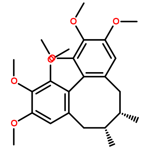
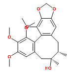
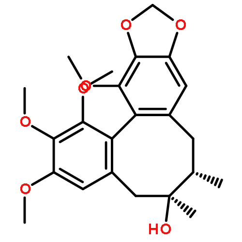
Cytochrome P450 (CYP) 3A5 characterized with polymorphic and extensive expression in multiple tissues is the most important P450 enzyme among the minor CYP3A isoforms. However, a selective and sensitive probe for CYP3A5 remains unavailable. In this study, we identified and characterized a naturally occurring lignan 12 (schisantherin E) as an isoform-specific probe for selective detection of CYP3A5 activity in complex biological samples. With thorough characterization including LC-MS and NMR, we found that 12 can be metabolized by CYP3A5 to generate a major metabolite 2-O-demethylated 12. Meanwhile, both reaction phenotyping and chemical inhibition experiments further revealed that CYP3A5 selectively catalyzed the 2-O-demethylation of 12. Specifically, the interactions between the Phe210 residue of CYP3A5 and methyl benzoate of 12 might play key roles in 12-O-demethylation, which was revealed by docking simulation and site-directed mutagenesis studies. These findings are beneficial for exploring the role of CYP3A5 in drug metabolism and pathologic process.
Co-reporter:Zi-Ru Dai;Lei Feng;Qiang Jin;Hailing Cheng;Yan Li;Jing Ning;Yang Yu;Jing-Nan Cui;Ling Yang
Chemical Science (2010-Present) 2017 vol. 8(Issue 4) pp:2795-2803
Publication Date(Web):2017/03/28
DOI:10.1039/C6SC03970G
The development of isoform-specific probe(s) for a target enzyme with multiple homologs is always challenging. Herein, a practical strategy was used to design and develop an isoform-specific probe for CYP1A1, a key cytochrome P450 isoenzyme involved in xenobiotic metabolism and bioactivation. On the basis of the subtle differences in 3D structure and substrate preference between CYP1A1 and its homolog CYP1A2, we proposed that it was possible to design a CYP1A1-specific probe via local modification of the reaction site on known CYP1A substrates. To validate this hypothesis, 4-hydroxy-1,8-naphthalimide (HN) was selected as the basic fluorophore due to its excellent optical properties, while a series of O-alkylated HN derivatives were prepared to evaluate their specificity towards CYP1A1. Our results revealed that the introduction of a chloroethyl to HN could get the best isoform selectivity towards CYP1A1 over other CYPs including CYP1A2. The newly developed probe NBCeN exhibited excellent specificity, high sensitivity, and a ratiometric fluorescence response following CYP1A1-catalyzed O-dechloroethylation. NBCeN was successfully used to real-time monitor the activity of CYP1A1 in complex biological samples and to rapidly screen CYP1A1 modulators in living systems. NBCeN could also be used for two-photon imaging of intracellular CYP1A1 in living cells and tissues with high ratiometric imaging resolution and deep tissue penetration. All these findings demonstrated that local modification of non-specific substrates was a practical strategy to develop an isoform-specific probe for a target isoenzyme, while NBCeN could serve as a specific imaging tool to explore the biological functions of CYP1A1 in complex biological systems.
Co-reporter:Li-Wei Zou, Ping Wang, Xing-Kai Qian, Lei Feng, Yang Yu, Dan-Dan Wang, Qiang Jin, Jie Hou, Zhi-Hong Liu, Guang-Bo Ge, Ling Yang
Biosensors and Bioelectronics 2017 Volume 90(Volume 90) pp:
Publication Date(Web):15 April 2017
DOI:10.1016/j.bios.2016.11.068
•A highly specific two-photon fluorescent probe GP-BAN has been developed.•The probe was successfully applied to detect DPP-IV in complex biological samples.•The probe could be used for high throughput screening of DPP-IV inhibitors.•The probe was successfully used for two-photon imaging of DPP-IV in living cells and tissues.In this study, a highly specific ratiometric two-photon fluorescent probe GP-BAN was developed and well-characterized to monitor dipeptidyl peptidase IV in plasma and living systems. GP-BAN was designed on the basis of the catalytic properties and substrate preference of DPP-IV, and it could be readily hydrolyzed upon addition of DPP-IV under physiological conditions. Both reaction phenotyping and inhibition assays demonstrated that GP-BAN displayed good reactivity and high selectivity towards DPP-IV over other human serine hydrolases including FAP, DPP-VIII, and DPP-IX. The probe was successfully used to monitor the real activities of DPP-IV in complex biological systems including diluted plasma, while it could be used for high throughput screening of DPP-IV inhibitors by using human plasma or tissue preparations as enzyme sources. As a two-photon fluorescent probe, GP-BAN was also successfully used for two-photon imaging of endogenous DPP-IV in living cells and tissues, and showed high ratiometric imaging resolution and deep-tissue penetration ability. Taken together, a ratiometric two-photon fluorescent probe GP-BAN was developed and well-characterized for highly selective and sensitive detection of DPP-IV in complex biological systems, which could serve as a promising imaging tool to explore the biological functions and physiological roles of this key enzyme in living systems.
Co-reporter:Chen Chen;Ping Wang;Liwei Zou;Ling Yang
Chemical Research in Chinese Universities 2017 Volume 33( Issue 2) pp:194-199
Publication Date(Web):2017/04/01
DOI:10.1007/s40242-017-6411-8
Co-reporter:Xuewei Cheng, Xia Lv, Hengyan Qu, Dandan Li, Mengmeng Hu, Wenzhi Guo, Guangbo Ge, Ruihua Dong
Acta Pharmaceutica Sinica B 2017 Volume 7, Issue 6(Issue 6) pp:
Publication Date(Web):1 November 2017
DOI:10.1016/j.apsb.2017.07.004
UDP-glucuronosyltransferase 1A1 (UGT1A1) plays a key role in detoxification of many potentially harmful compounds and drugs. UGT1A1 inhibition may bring risks of drug–drug interactions (DDIs), hyperbilirubinemia and drug-induced liver injury. This study aimed to investigate and compare the inhibitory effects of icotinib and erlotinib against UGT1A1, as well as to evaluate their potential DDI risks via UGT1A1 inhibition. The results demonstrated that both icotinib and erlotinib are UGT1A1 inhibitors, but the inhibitory effect of icotinib on UGT1A1 is weaker than that of erlotinib. The IC50 values of icotinib and erlotinib against UGT1A1-mediated NCHN-O-glucuronidation in human liver microsomes (HLMs) were 5.15 and 0.68 μmol/L, respectively. Inhibition kinetic analyses demonstrated that both icotinib and erlotinib were non-competitive inhibitors against UGT1A1-mediated glucuronidation of NCHN in HLMs, with the Ki values of 8.55 and 1.23 μmol/L, respectively. Furthermore, their potential DDI risks via UGT1A1 inhibition were quantitatively predicted by the ratio of the areas under the concentration–time curve (AUC) of NCHN. These findings are helpful for the medicinal chemists to design and develop next generation tyrosine kinase inhibitors with improved safety, as well as to guide reasonable applications of icotinib and erlotinib in clinic, especially for avoiding their potential DDI risks via UGT1A1 inhibition.The inhibitory potentials of icotinib and erlotinib against UDP-glucuronosyltransferase 1A1 (UGT1A1) were investigated and compared for the first time. Icotinib exhibited relatively weak inhibition against UGT1A1 in human liver microsomes compared to erlotinib. In addition, icotinib was unlikely to cause a significant drug–drug interaction (DDI) through inhibition of UGT1A1, while erlotinib exhibited much higher DDI potentials.Download high-res image (147KB)Download full-size image
Co-reporter:Hong-Ying Ma, Jia-Da Yang, Jie Hou, Li-Wei Zou, Qiang Jin, Da-Cheng Hao, Jing Ning, Guang-Bo Ge, Ling Yang
Toxicology in Vitro 2017 Volume 44(Volume 44) pp:
Publication Date(Web):1 October 2017
DOI:10.1016/j.tiv.2017.06.020
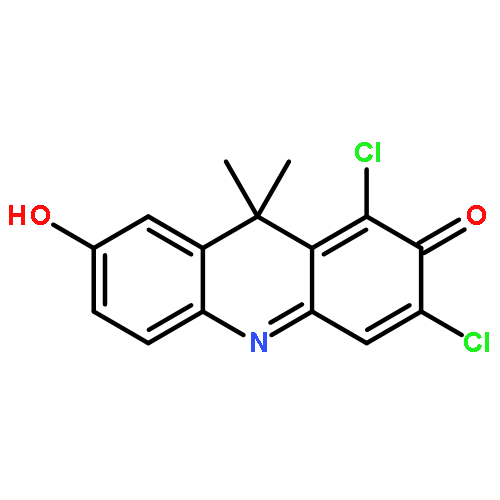
•DDAB could be hydrolyzed by liver microsomes from various animal species.•Mammalian carboxylesterases (CEs) were the major enzymes involved in DDAB hydrolysis.•Loperamide displayed different inhibitory effects on DDAB hydrolysis in different species.•DDAB displayed similar apparent substrate affinity towards mammalian CEs from different speciesDDAB (6,8-dichloro-9,9-dimethyl-7-oxo-7,9-dihydroacridin-2-yl benzoate) is a newly developed near-infrared fluorescent probe for human carboxylesterase 2 (hCE2), exhibiting high specificity and good reactivity for real-time monitoring the enzymatic activities of hCE2 in complex biological systems. In order to explore the applicability of DDAB in commonly used animal species, the interspecies difference in DDAB hydrolysis was carefully investigated by using liver microsomes from human and five experimental animals including mouse, rat, dog, minipig and monkey. Metabolite profiling demonstrated that DDAB hydrolysis could be catalyzed by all tested liver microsomes from different animals but displayed significant difference in the reaction rate. Chemical inhibition assays demonstrated that carboxylesterases (CEs) were the major enzymes involved in DDAB hydrolysis in all tested liver microsomes, indicating that DDAB was a selective substrate of CEs in a variety of mammals. However, the differential effects of loperamide (LPA, a specific inhibitor against hCE2) on DDAB hydrolysis among various species were observed. The apparent kinetic parameters and the maximum intrinsic clearances (CLmax) for DDAB hydrolysis in liver microsomes from different animals were determined, and the order of CLmax values for the formation of DDAO was CyLM > MLM ≈ PLM > RLM > HLM ≈ DLM. These findings were helpful for the rational use of DDAB as an imaging tool for CE2 in different mammals, as well as for translational researches on the function of mammalian CEs and CE2-associated drug-drug interactions.
Co-reporter:Yi-Ru Wang, Lei Feng, Liang Xu, Yan Li, Dan-Dan Wang, Jie Hou, Kun Zhou, Qiang Jin, Guang-Bo Ge, Jing-Nan Cui and Ling Yang
Chemical Communications 2016 vol. 52(Issue 36) pp:6064-6067
Publication Date(Web):01 Mar 2016
DOI:10.1039/C6CC00119J
A rapid-response fluorescent probe ACDM was developed for the selective and sensitive detection of human albumin (HA) via binding onto a non-drug binding site. ACDM was successfully used to detect trace HA in various biological samples including diluted plasma and cell culture supernatants.
Co-reporter:Dan-Dan Wang, Qiang Jin, Li-Wei Zou, Jie Hou, Xia Lv, Wei Lei, Hai-Ling Cheng, Guang-Bo Ge and Ling Yang
Chemical Communications 2016 vol. 52(Issue 15) pp:3183-3186
Publication Date(Web):05 Jan 2016
DOI:10.1039/C5CC09874B
A highly selective and sensitive bioluminescent sensor (DME) for human carboxylesterase 1 (hCE1) has been developed and well characterized. DME could be used for real-time monitoring of hCE1 activities in complex biological samples and for bio-imaging of endogenous hCE1 in living cells.
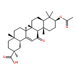
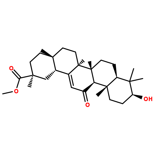
Co-reporter:Li-Wei Zou, Yao-Guang Li, Ping Wang, Kun Zhou, Jie Hou, Qiang Jin, Da-Cheng Hao, Guang-Bo Ge, Ling Yang
European Journal of Medicinal Chemistry 2016 Volume 112() pp:280-288
Publication Date(Web):13 April 2016
DOI:10.1016/j.ejmech.2016.02.020
•GA derivatives were designed, synthesized and evaluated for their inhibitory activities against hCE1 and hCE2.•Compound 15 was discovered as a novel and highly selective inhibitor against hCE2 (IC50 0.02 μM).•Molecular docking revealed the essential structural features of compound 15 as potent and selective hCE2 inhibitor.Human carboxylesterase 2 (hCE2), one of the major carboxylesterases in the human intestine and various tumour tissues, plays important roles in the oral bioavailability and treatment outcomes of ester- or amide-containing drugs or prodrugs, such as anticancer agents CPT-11 (irinotecan) and LY2334737 (gemcitabine). In this study, 18β-glycyrrhetinic acid (GA), the most abundant pentacyclic triterpenoid from natural source, was selected as a reference compound for the development of potent and specific inhibitors against hCE2. Simple semi-synthetic modulation on GA was performed to obtain a series of GA derivatives. Structure-activity relationship analysis brought novel insights into the structure modification of GA. Converting the 11-oxo-12-ene of GA to 12-diene moiety, and C-3 hydroxyl and C-30 carboxyl group to 3-O-β-carboxypropionyl and ethyl ester respectively, led to a significant enhancement of the inhibitory effect on hCE2 and the selectivity over hCE1. These exciting findings inspired us to design and synthesize the more potent compound 15 (IC50 0.02 μM) as a novel and highly selective inhibitor against hCE2, which was 3463-fold more potent than the parent compound GA and demonstrated excellent selectivity (>1000-fold over hCE1). The molecular docking study of compound 15 and the active site of hCE1 and hCE2 demonstrated that the potent and selective inhibition of compound 15 toward hCE2 could partially be attributed to its relatively stronger interactions with hCE2 than with hCE1.
Co-reporter:Xia Lv, Dan-Dan Wang, Lei Feng, Ping Wang, Li-Wei Zou, Da-Cheng Hao, Jie Hou, Jing-Nan Cui, Guang-Bo Ge and Ling Yang
RSC Advances 2016 vol. 6(Issue 6) pp:4302-4309
Publication Date(Web):21 Dec 2015
DOI:10.1039/C5RA23614B
Human carboxylesterase 1 (hCE1), plays pivotal roles in endobiotics homeostasis and xenobiotic metabolism. The plasma level of hCE1 could serve as a useful serologic biomarker for several hepatic diseases, such as hepatocellular carcinoma. However, no probe reaction has been fruitfully used to determine the activity of trace hCE1 in human plasma. This study aims to design and develop a highly selective marker reaction for measuring the enzymatic activities of hCE1 in complex biological samples including human plasma. N-(4-Methyl butyrate)-4-hydroxy-1,8-naphthalimide (NMHN), which contains a small alcohol group and a large acyl moiety, was intentionally designed based on the substrate preference of hCE1. NMHN could be easily hydrolyzed by hCE1 with very high catalytic efficacy under physiological conditions, while both reaction-phenotyping assays and chemical inhibition assays demonstrated that this reaction exhibited super selectivity towards hCE1 over other human hydrolases. Furthermore, the marker reaction possessed ideal kinetic behaviour (classic Michaelis–Menten) with high intrinsic clearance (5.8 mL min−1 mg−1). Based on this probe, a rapid hCE1 quantification method was developed and fully validated, which was successfully applied to determine the real activities of hCE1 in various biological samples including human plasma. Our findings afforded a promising tool for measuring the real activity of hCE1 and laid a solid foundation for further investigations on the biological functions of hCE1 in complex biological samples.
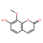
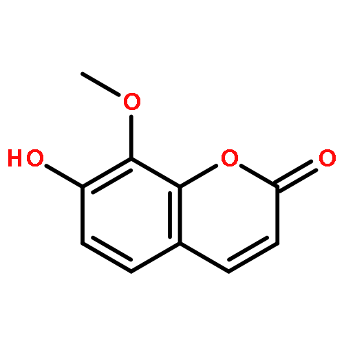
Co-reporter:Xing-Kai Qian, Ping Wang, Yang-Liu Xia, Tong-Yi Dou, Qiang Jin, Dan-Dan Wang, Da-Cheng Hao, Xiao-Lin Bi, Guang-Bo Ge, Ling Yang
Sensors and Actuators B: Chemical 2016 Volume 231() pp:615-623
Publication Date(Web):August 2016
DOI:10.1016/j.snb.2016.03.074
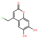
•A new fluorescent probe for the selective detection of human catechol-O-methyltransferase (COMT) was developed.•The probe exhibited excellent selectivity and sensitivity towards COMT.•The probe could be used to monitor the real activity of COMT in living cells.Catechol-O-methyltransferase (COMT), one of the most important phase II drug metabolizing enzymes, plays important roles in the metabolism of endogenous and xenobiotic catechols. In this study, a highly selective fluorescent probe for sensing activities of catechol-O-methyltransferase in complex biological samples was discovered and well characterized. Under physiological conditions, COMT selectively catalyzes the conversion of the probe (7,8-dihydroxy-4-methylcoumarin, DHMC) to 7-hydroxy-8-methoxy-4-methylcoumarin (HMMC), which brings a strong turn-on fluorescence signal at 520 nm. The probe substrate has been used for monitoring the real activities of COMT in complex biological samples, as well as for rapid screening of potential COMT inhibitors which are useful in the treatment of Parkinson's diseases Furthermore, the probe has been successfully used to monitor endogenous COMT in living cells for the first time, and the results demonstrate that DHMC is cell membrane permeable and low toxic to the cells. All these features of DHMC suggested that this probe holds great promise for COMT-related regulation and inhibition assays in drug discovery, as well as for further investigation on the biological functions of COMT in living cells.A highly selective fluorescent probe for catechol-O-methyltransferase.
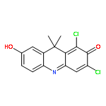
Co-reporter:Qiang Jin, Lei Feng, Dan-Dan Wang, Jing-Jing Wu, Jie Hou, Zi-Ru Dai, Shi-Guo Sun, Jia-Yue Wang, Guang-Bo Ge, Jing-Nan Cui, Ling Yang
Biosensors and Bioelectronics 2016 Volume 83() pp:193-199
Publication Date(Web):15 September 2016
DOI:10.1016/j.bios.2016.04.075
•A near-infrared fluorescent probe for carboxylesterase 2 (CE2) has been designed.•This probe exhibits good specificity, ultrahigh sensitivity, and fast reaction kinetics.•This newly probe can be used for sensing CE2 in living cells and animals.A near-infrared fluorescent probe (DDAB) for highly selective and sensitive detection of carboxylesterase 2 (CE2) has been designed, synthesized, and systematically studied both in vitro and in vivo. Upon addition of CE2, the ester bond of DDAB could be rapidly cleaved and then release a near-infrared (NIR) fluorophore DDAO, which brings a remarkable yellow-to-blue color change and strong NIR fluorescence emission in physiological solutions. The newly developed probe exhibits excellent properties including good specificity, ultrahigh sensitivity and high imaging resolution. Moreover, DDAB has been applied to measure the real activities of CE2 in complex biological samples, as well as to screen CE2 inhibitors by using tissue preparations as the enzymes sources. The probe has also been successfully used to detect endogenous CE2 in living cells and in vivo for the first time, and the results demonstrate that such detection is highly reliable. All these prominent features of DDAB make it holds great promise for further investigation on CE2-associated biological process and for exploring the physiological functions of CE2 in living systems.A colorimetric near-infrared fluorescent probe (DDAB) for highly selective and sensitive detection of carboxylesterase 2 (CE2) has been designed and well-characterized. This newly developed probe can be used for sensing CE2 in living cells and animals, and holds great promise for exploring the biological functions of CE2 in complex biological systems.
Co-reporter:Dan-Dan Wang, Qiang Jin, Jie Hou, Lei Feng, Na Li, Shi-Yang Li, Qi Zhou, Li-Wei Zou, Guang-Bo Ge, Jin-Guang Wang, Ling Yang
Journal of Chromatography B 2016 Volume 1008() pp:212-218
Publication Date(Web):1 January 2016
DOI:10.1016/j.jchromb.2015.11.046
•A LC-FD method was developed to detect the activity of human carboxylesterase 1 (hCE1).•The method was fully validated and all validation parameters fulfilled ICH guildlines.•Low detection and quantitation limits were achieved.•The method was successfully used for the rapid measuring of hCE1 activity in complex biological samples.Human carboxylesterases 1 (hCE1), one of the most important human drug metabolizing enzymes, catalyzes the hydrolysis of a large number of structurally diverse of endogenous and exogenous substrates. However, a practical, reliable and sensitive method for the precise measurement of hCE1 activities in complex biological samples has been rarely reported. In this study, a liquid chromatography-fluorescence detection (LC-FD) based method was developed for highly selective and sensitive measurement of hCE1 activities in human tissue and cell preparations. This method was based on the fluorimetric detection of HMBT, the hydrolyzed product of BMBT which was a newly developed specific probe substrate for hCE1. The developed LC-FD method was fully validated in terms of specificity, sensitivity, linearity, precision, recovery and stability. With the help of LC separation, most polar endogenous compounds in biological samples could be eluted in the column dead time, which is very beneficial for accurate determination of hCE1 activities in complex biological samples. The lower limit of quantification for HMBT (product of hCE1) of this LC-FD based method was as low as 20 nM, which was quite lower than other reported methods. The method also exhibited good precision, both intra- and inter- assay variances were both lower than 2.5%. Furthermore, the newly developed method was successfully applied to measure hCE1 activity in human liver preparations from individual donors (n = 12), as well as in homogenates from eleven different human cell lines. All these findings combined with this practical method are very helpful for the deep understanding of the expression and function of hCE1 in human biological samples.
Co-reporter:Pan Cui;Tong-Yi Dou;Shi-Yang Li;Jun-Xia Lu;Li-Wei Zou
Biotechnology Letters 2016 Volume 38( Issue 8) pp:1367-1373
Publication Date(Web):2016 August
DOI:10.1007/s10529-016-2116-1
To develop a practical method to prepare tilianin by highly selective and efficient hydrolysis of the C-7 rhamnosyl group from linarin.Naringinase was utilized to selectively catalyze the formation of tilianin using linarin as the starting material. The reaction conditions, including temperature, pH, metal ions, substrate concentration and enzyme concentration, were optimized. At 60 °C, naringinase showed enhanced α-l-rhamnosidase activity while the β-d-glucosidase activity was abrogated. The addition of Mg2+, Fe2+ and Co2+ was also beneficial for selective biotransformation of linarin to tilianin. Under the optimized conditions (pH 7.0 at 60 °C), linarin could be nearly completely transformed to tilianin with excellent selectivity (>98.9 %), while that of the by-product acacetin was less than 1.1 %. In addition, the structure of target product tilianin was fully characterized by HR-MS and 1H-NMR.A highly selective and efficient biotransformation of linarin to tilianin was developed by the proper control of incubation temperature, which enhanced the α-l-rhamnosidase activity of naringinase and blocked its β-d-glucosidase activity.
Co-reporter:Junxia Lu;Ping Wang;Jie Hou;Liwei Zou
Chemical Research in Chinese Universities 2016 Volume 32( Issue 5) pp:786-791
Publication Date(Web):2016 October
DOI:10.1007/s40242-016-6147-x
An expedient and efficient method for selective methylation of catechol coumarins by working with different alkalis under appropriate reaction conditions is reported. Esculetins were selectively methylated at position 6 and position 7 in good yield using CH3I in the presence of NaH and Na2CO3, respectively. However, daphnetins showed less regioselectivity under the same reaction conditions. Furthermore, the site preference for the methylation reaction was interpreted by the density functional theory at B3LYP/6-31+G(d) level.
Co-reporter:Qiang Jin, Lei Feng, Dan-Dan Wang, Zi-Ru Dai, Ping Wang, Li-Wei Zou, Zhi-Hong Liu, Jia-Yue Wang, Yang Yu, Guang-Bo Ge, Jing-Nan Cui, and Ling Yang
ACS Applied Materials & Interfaces 2015 Volume 7(Issue 51) pp:28474
Publication Date(Web):December 7, 2015
DOI:10.1021/acsami.5b09573
In this study, a two-photon ratiometric fluorescent probe NCEN has been designed and developed for highly selective and sensitive sensing of human carboxylesterase 2 (hCE2) based on the catalytic properties and substrate preference of hCE2. Upon addition of hCE2, the probe could be readily hydrolyzed to release 4-amino-1,8-naphthalimide (NAH), which brings remarkable red-shift in fluorescence (90 nm) spectrum. The newly developed probe exhibits good specificity, ultrahigh sensitivity, and has been successfully applied to determine the real activities of hCE2 in complex biological samples such as cell and tissue preparations. NCEN has also been used for two-photon imaging of intracellular hCE2 in living cells as well as in deep-tissues for the first time, and the results showed that the probe exhibited high ratiometric imaging resolution and deep-tissue imaging depth. All these findings suggested that this probe holds great promise for applications in bioimaging of endogenous hCE2 in living cells and in exploring the biological functions of hCE2 in complex biological systems.Keywords: bioimaging; carboxylesterase 2; ratiometric fluorescent probe; specific sensing; two-photon
Co-reporter:Ping Wang, Yang-Liu Xia, Yang Yu, Jun-Xia Lu, Li-Wei Zou, Lei Feng, Guang-Bo Ge and Ling Yang
RSC Advances 2015 vol. 5(Issue 66) pp:53477-53483
Publication Date(Web):02 Jun 2015
DOI:10.1039/C5RA06070B
Esculetin, a naturally catecholic coumarin, possess multiple pharmacological activities including anti-tumour, anti-inflammatory and anti-oxidant. However, the extensive phase II metabolism and rapid elimination from the human body significantly hinder esculetin and its derivatives as drug leads/candidates. To improve both the metabolic stability and the anti-tumour activity of esculetin via rational modification, a series of C-4 and C-8 substituted derivatives were designed and synthesized by perchloric acid catalysed von Pechmann reaction and Mannich reaction, respectively. The in vitro metabolic half-life in human liver S9 and anti-tumour activities in A549 and B16 cell lines of the newly synthesized compounds were assayed. Of these compounds, 8-(pyrrolidin-1-ylmethyl)-4-trifluoromethyl esculetin 15 was the most potent candidate compound, with almost a 20-fold increase in antiproliferative activity and a 3-fold prolonged half-life in human liver S9 compared with the parent compound 1. In addition, the potential structure–activity relationship and structure–metabolic stability relationship were discussed. Notably, the introduction of a nitrogen containing group as a hydrogen bond acceptor at the C-8 position of esculetin can improve both the metabolic stability and anti-tumour activity. All of these findings are very helpful for the structural modification of esculetin and other bioactive phenolic compounds to improve their phase II metabolic stability and bioactivity synchronously.
Co-reporter:Guiyuan He, Shixuan Zhang, Liang Xu, Yangliu Xia, Ping Wang, Shiyang Li, Liangliang Zhu, Hongxi Xu, Guangbo Ge and Ling Yang
RSC Advances 2015 vol. 5(Issue 109) pp:89818-89826
Publication Date(Web):14 Oct 2015
DOI:10.1039/C5RA20213B
Baicalein (BA), a natural flavonoid compound, possesses many desirable pharmacological activities. However, poor solubility and extensive metabolism by human UDP-glucuronosyltransferases (UGTs) strongly restrict the clinical applications of BA. We previously reported that two C-8 Mannich base derivatives of BA (BA-a and BA-j) displayed enhanced solubility and anti-cyclin dependent kinase 1 activity, yet the metabolic stabilities of these compounds remained unknown. This study aimed to evaluate the in vitro glucuronidation stability of these BA derivatives and to explore the key factors affecting the UGT-mediated biotransformation. The results showed that the glucuronidation stabilities of these BA derivatives were much higher than BA. BA-a exhibited 12-fold and BA-j exhibited 5-fold improved stability in human liver S9, while in human intestine S9, BA-a and BA-j exhibited 42-fold and 33-fold improved stability, respectively. Further investigations found that the major glucuronidation site(s) were changed from 7-OH and 6-OH in BA to 6-OH in the BA derivatives. Also, both the involved enzymes and their catalytic efficacy in 6-O-glucuronidation of BA derivatives were much lower than that of BA. The formation of an intramolecular hydrogen bond between the C-8 Mannich base substituents and C-7 phenolic groups played a predominant role in these glucuronidation changes. The calculated bond dissociation energy (BDE) of each phenolic group in BA and its derivatives agreed well with their glucuronidation activities. All these findings bring new insights into the structure–glucuronidation relationship and provide a practical strategy for the structural modification to improve the glucuronidation stability of drug candidates, especially for those phenolic compounds.
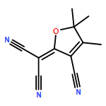
Co-reporter:Lei Feng, Zhao-Ming Liu, Jie Hou, Xia Lv, Jing Ning, Guang-Bo Ge, Jing-Nan Cui, Ling Yang
Biosensors and Bioelectronics 2015 Volume 65() pp:9-15
Publication Date(Web):15 March 2015
DOI:10.1016/j.bios.2014.10.002
•A ratiometric fluorescent ESIPT probe for selective detection of human CE2 was developed for the first time.•The probe can be selectively hydrolyzed by CE2 to release 3-hydroxylflavone which brings the remarkable changes in fluorescence spectrum.•The probe can be used to efficiently detect the real activity of CE2 in biological samples.•The probe also can monitor the real function of endogenous CE2 in living cells.A new ratiometric florescence probe derived from 3-hydroxyflavone (3-HF) has been developed for selective and sensitive detection of human carboxylesterase 2 (CE2). The probe is designed by modulating the excited state intramolecular proton transfer (ESIPT) emission of 3-HF via introducing of 4-ethylbenzoyloxy group. Under physiological conditions, probe 1 displays satisfying stability with very low background signal, but it can be selectively hydrolyzed by CE2 to release free 3-HF which brings remarkable changes in fluorescence spectrum. Both reaction phenotyping and chemical inhibition assays demonstrate that probe 1 is highly selective for CE2 over other human hydrolases including carboxylesterase 1, cholinesterases and paraoxonases. Probe 1 has been applied successfully to measure the real activities of CE2 in human biological samples, as well as to screen CE2 inhibitors by using tissue preparations as the enzymes sources. Additionally, probe 1 is cell membrane permeable and can be used for cellular imaging of endogenous CE2 in living cells. All of these features make it possible to serve as a promising tool for exploring the individual differences in biological function of CE2, as well as for rapid screening of selective and potent inhibitors of CE2 for further clinical use.
Co-reporter:Lei Feng, Zhao-Ming Liu, Liang Xu, Xia Lv, Jing Ning, Jie Hou, Guang-Bo Ge, Jing-Nan Cui and Ling Yang
Chemical Communications 2014 vol. 50(Issue 93) pp:14519-14522
Publication Date(Web):30 Sep 2014
DOI:10.1039/C4CC06642A
A highly selective long-wavelength fluorescent probe TCFB has been developed for the detection of hCE2. The probe can be used for real-time monitoring of hCE2 activity in complex biological systems.
Co-reporter:Xia Lv, Jie Hou, Yang-Liu Xia, Jing Ning, ... Ling Yang
Drug Metabolism and Pharmacokinetics (October 2015) Volume 30(Issue 5) pp:358-365
Publication Date(Web):1 October 2015
DOI:10.1016/j.dmpk.2015.07.001
Bavachinin (BCI), a major bioactive compound in Chinese herbal Psoralea corylifolia, possesses a wide range of biological activities. In this study, the glucuronidation pathway of BCI was characterized for the first time, by using pooled human liver microsomes (HLM), pooled human intestine microsomes (HIM) and recombinant human UDP-glucosyltransferases (UGTs). One mono-glucuronide was detected in HLM in the presence of uridine-diphosphate glucuronic acid (UDPGA), and it was biosynthesized and well-characterized as BCI-4′-O-glucuronide (BCIG). Reaction phenotyping assay showed that UGT1A1, UGT1A3 and UGT1A8 were involved in BCI-4′-O-glucuronidation, while UGT1A1 and UGT1A8 displayed the higher catalytic ability among all tested UGT isoforms. Kinetic analysis demonstrated that BCI-4′-O-glucuronidation in both HLM and UGT1A1 followed sigmoidal kinetic behaviors and displayed much close Km values (12.4 μM in HLM & 9.7 μM in UGT1A1). Both chemical inhibition assays and correlation analysis demonstrated that UGT1A1 displayed a predominant role in BCI-4′-O-glucuronidation in HLM. Both HIM and UGT1A8 exhibited substrate inhibition at high concentrations, and Km values of HIM and UGT1A8 were 3.6 and 2.3 μM, respectively. Similar catalytic efficiencies were observed for HIM (199.3 μL/min/mg) and UGT1A8 (216.2 μL/min/mg). These findings suggested that UGT1A1 and UGT1A8 were the primary isoforms involved in BCI-4′-O-glucuronidation in HLM, and HIM, respectively.Bavachinin (BCI), a major bioactive compound in Chinese herbal Psoralea corylifolia, possesses various biological activities. UGT1A1 and UGT1A8 were the primary isoforms involved in BCI-4′-O-glucuronidation in HLM and HIM, respectively.Download full-size image
Co-reporter:Dan-Dan Wang, Li-Wei Zou, Qiang Jin, Jie Hou, Guang-Bo Ge, Ling Yang
Fitoterapia (March 2017) Volume 117() pp:84-95
Publication Date(Web):1 March 2017
DOI:10.1016/j.fitote.2017.01.010
Mammalian carboxylesterases (CEs) are important serine hydrolases catalyzing the hydrolysis of ester- or amide-containing compounds into the corresponding alcohols and carboxylic acids. In human, two primary carboxylesterases including hCE1 and hCE2 have been identified and extensively studied in the past decade. hCE1 is known to play crucial roles in the metabolism of a wide variety of endogenous esters, clinical drugs and insecticides, while hCE2 plays a key role in the metabolic activation of anticancer agents including irinotecan and capecitabine. The key roles of hCEs in both human health and xenobiotic metabolism arouse great interest in the discovery of potent and selective hCEs inhibitors to modulate endobiotic metabolism or to improve the outcomes of patients administrated with ester drugs. This review covers the significance and recent progress in the discovery of natural inhibitors against hCEs. The tools for screening and characterization of inhibitors against human CEs, including traditional LC-based approaches and the newly developed optical substrate-based assays, are summarized and discussed for the first time. Furthermore, the structural information and inhibitory capacities of all reported hCEs inhibitors including fatty acids, flavonoids, tanshinones and triterpenoids have been systematically summarized. All information and knowledge presented in this review will be very helpful for medicinal chemists to develop more potent and highly selective inhibitors against hCEs for potential biomedical applications.Download high-res image (97KB)Download full-size image
Co-reporter:Si-Cheng Liang, Yang-Liu Xia, Jie Hou, Guang-Bo Ge, ... Ling Yang
Journal of Pharmaceutical Sciences (February 2016) Volume 105(Issue 2) pp:808-816
Publication Date(Web):1 February 2016
DOI:10.1016/j.xphs.2015.10.010
Our previous study demonstrated that daphnetin is subject to glucuronidation in vitro. However, daphnetin metabolism is still poorly documented. This study aimed to investigate daphnetin metabolism and its consequent effect on the bioactivity. Metabolic profiles obtained by human liver S9 fractions and human hepatocytes showed that daphnetin was metabolized by glucuronidation, sulfonation, and methylation to form 6 conjugates which were synthesized and identified as 7-O-glucuronide, 8-O-glucuronide, 7-O-sulfate and 8-O-sulfate, 8-O-methylate, and 7-O-suflo-8-O-methylate. Regioselective 8-O-methylation of daphnetin was investigated using in silico docking calculations, and the results suggested that a close proximity (2.03 Å) of 8-OH to the critical residue Lysine 144 might be the responsible mechanism. Compared with glucuronidation and sulfonation pathways, the methylation of daphnetin had a high clearance rate (470 μL/min/mg) in human liver S9 fractions and contributed to a large amount (37.3%) of the methyl-derived metabolites in human hepatocyte. Reaction phenotyping studies showed the major role of SULT1A1, -1A2, and -1A3 in daphnetin sulfonation, and soluble COMT in daphnetin 8-O-methylation. Of the metabolites, only 8-O-methyldaphnetin exhibited an inhibitory activity on lymphocyte proliferation comparable to that of daphnetin. In conclusion, methylation is a crucial pathway for daphnetin clearance and might be involved in pharmacologic actions of daphnetin in humans.
Co-reporter:Ya-Jing Liu, Shi-Yang Li, Jie Hou, Yan-Fang Liu, Dan-Dan Wang, Yong-Shan Jiang, Guang-Bo Ge, Xin-Miao Liang, Ling Yang
Fitoterapia (December 2016) Volume 115() pp:57-63
Publication Date(Web):1 December 2016
DOI:10.1016/j.fitote.2016.09.022
White Mulberry Root-bark (WMR) is an edible Chinese herbal used for the treatment of inflammation, nephritis and asthma. This study aimed to investigate the inhibitory effects of ethanol extract from WMR against human carboxylesterase 2 (hCE2), as well as to identity and character natural hCE2 inhibitors in this herbal. Our results demonstrated that the ethanol extract of WMR displayed potent inhibitory effects against hCE2, while three major bioactive constitutes in WMR were identified on the basis of LC fingerprinting combined with activity-based screening of LC fractions. Three bioactive compounds including SD, KG and SC were efficiently identified by comparison of LC retention times, UV and MS spectral data, with the help of authentic standards. The inhibition potentials and inhibition types of these natural compounds against hCE2 were further investigated in human liver microsomes. The results demonstrated that these bioactive compounds are potent non-competitive inhibitors against hCE2, with the Ki values ranging from 0.76 μM to 1.09 μM. All these findings suggested that three abundant natural compounds in WMR displayed potent inhibitory effects against hCE2, which could be used as lead compounds to develop more potent hCE2 inhibitors for the alleviation of hCE2-mediated severe delayed-onset diarrhea.Graphic abstractDownload high-res image (79KB)Download full-size image
Co-reporter:Pan Cui, Tong-Yi Dou, Yan-Ping Sun, Shi-Yang Li, Lei Feng, Li-Wei Zou, Ping Wang, Da-Cheng Hao, Guang-Bo Ge, Ling Yang
Journal of Molecular Catalysis B: Enzymatic (August 2016) Volume 130() pp:25-31
Publication Date(Web):1 August 2016
DOI:10.1016/j.molcatb.2016.04.013
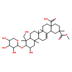
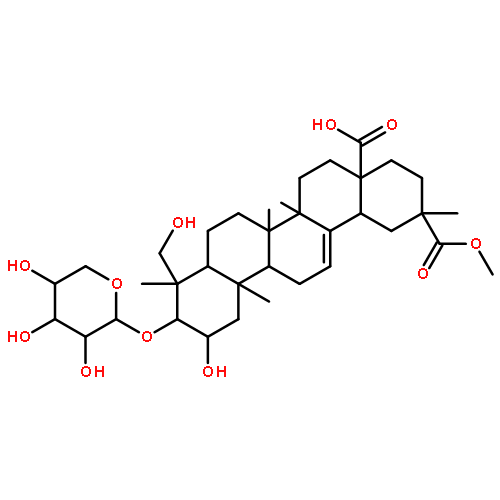
•Efficient preparation of esculentoside B was achieved by biotransformation.•β-d-glucosidase from snailase was used to perform such biotransformation.•Response surface methodology was used to optimize the reaction conditions.•Kinetic studies determined the catalytic efficiency in this preparation process.Esculentoside B (EsB, also named phytolaccagenin 3-O-β-d-xylopyranoside), a pentacyclic triterpene isolated from herbal medicine Radix phytolaccae, has been found to possess multiple pharmacological activities. Nonetheless, the low content in nature and the difficulties in the total synthesis of EsB strongly limit its extensive investigations and further development as a drug candidate. This study aims to provide a practical method for highly efficient preparation of EsB using esculentoside A (EsA, phytolaccagenin 3-O-β-d-glucopyranosyl (1 → 4)-β-d-xylopyranoside) as the starting material. β-d-glucosidase from snailase was used to catalyze the formation of EsB, and the product was then purified and fully characterized by both HRMS and NMR. To prepare EsB in a more cost-effective way, response surface methodology (RSM) was used to explore the potential effects of the reaction conditions (such as reaction temperature, pH, enzyme load, and reaction time) on the conversion rates of EsA. The highest EsB yield of 0.66 mg/ml was obtained experimentally under optimized conditions as follows: temperature 48.28 °C, pH 6.4, enzyme load 4.43%, and reaction time 2.73 h. This result agreed well with the predicted yield of 0.68 mg/ml by RSM. The enzymatic kinetics of this biotransformation was characterized at the optimum pH and temperature. The S50 value was evaluated as 167.4 μM, while the Vmax value was 345.6 nmol/min/mg. In summary, this study provided a mild and practical method for the highly efficient preparation of EsB from EsA, which held great promise for large scale production of EsB.Download full-size image
Co-reporter:Hong Xin, Xiao-Yi Qi, Jing-Jing Wu, Xin-Xin Wang, Yan Li, James Y. Hong, Wei He, Wei Xu, Guang-Bo Ge, Ling Yang
Food and Chemical Toxicology (April 2016) Volume 90() pp:112-122
Publication Date(Web):1 April 2016
DOI:10.1016/j.fct.2016.02.007
•Licochalcone A (LCA) displays broad-spectrum inhibition against human UGTs.•LCA displays strong inhibitory effects against UGT1A1, 1A3, 1A4, 1A6, 1A7, 1A9, and 2B7.•LCA displays potent inhibitory effects against UGT1A1 and 1A9 in human liver microsomes.•The potential risks of LCA via inhibition of UGT1A1 and 1A9 were predicted for the first time.Licochalcone A (LCA) is a major bioactive compound in Licorice, a widely used herbal medicine. In this study, the inhibitory effects of LCA against human UDP-glucuronosyltransferases (UGTs) and LCA associated herb-drug interactions were systematically investigated. Our results demonstrated that LCA displayed broad-spectrum inhibition against human UGTs. LCA exhibited strong inhibitory effects against UGT1A1, 1A3, 1A4, 1A6, 1A7, 1A9, and 2B7 (both IC50 and Ki values lower than 5 μM), while showing moderate inhibitory effects against UGT1A8, 1A10, 2B4, 2B15, and 2B17. The inhibitory effects of LCA against two major UGTs, including UGT1A1 and 1A9, were further investigated in human liver microsomes (HLMs), where the potential risks of LCA via inhibition of UGT1A1 and 1A9 were predicted by combining the in vitro inhibitory data and physiological data. The results from this study also showed that several LCA-containing products were able to increase the area under the curve (AUC) of the substrates that were predominantly metabolized by UGT1A1 or 1A9. These findings together demonstrate that LCA has a potent and broad-spectrum inhibitory effect against most human UGTs and thus suggest that much caution should be exercised when high-dose LCA is co-administered with UGT substrates.
Co-reporter:Yang-Liu Xia, Si-Cheng Liang, Liang-Liang Zhu, Guang-Bo Ge, ... Sheng-Li Yang
Drug Metabolism and Pharmacokinetics (2014) Volume 29(Issue 2) pp:135-140
Publication Date(Web):1 January 2014
DOI:10.2133/dmpk.DMPK-13-RG-059
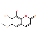
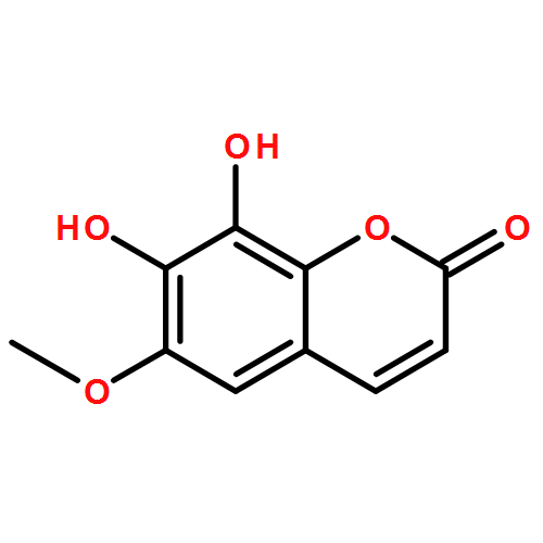
Fraxetin, a major constituent of the traditional medicine plant Fraxinus rhynchophylla Hance (Oleaceae), has been found to possess multiple bioactivities. However, the metabolic pathway(s) of fraxetin in human tissues has not been reported yet. This study aimed to characterize the glucuronidation pathway(s) of fraxetin in human tissues. Fraxetin could be metabolized to two glucuronides in human liver microsomes (HLMs). These two glucuronides were biosynthesized and characterized as 7-O-glucuronide (7-O-G) and 8-O-glucuronide (8-O-G). UGT1A1, -1A6, -1A7, -1A8, -1A9 and -1A10 participated in the formation of 7-O-G, while the formation of 8-O-G was catalyzed selectively by UGT1A6 and UGT1A9. UGT1A9 showed the highest catalytic activities in the formation of 7-O-G and 8-O-G. Both kinetic characterization and inhibition assays demonstrated that UGT1A9 played important roles in fraxetin glucuronidations in HLMs, especially in the formation of the major metabolite 8-O-G. Furthermore, the intrinsic clearance of fraxetin in both human liver microsomes and UGT1A9 was greater than that of 7,8-dihydroxylcoumarin, revealing that the addition of a C-6 methoxy group led to the higher metabolic clearance. In summary, the glucuronidation pathways of fraxetin in human liver microsomes were well-characterized, and UGT1A9 was the major isoform responsible for the glucuronidations of fraxetin.
Co-reporter:Xin-Xin Wang, Xia Lv, Shi-Yang Li, Jie Hou, Jing Ning, Jia-Yue Wang, Yun-Feng Cao, Guang-Bo Ge, Bin Guo, Ling Yang
Toxicology and Applied Pharmacology (15 November 2015) Volume 289(Issue 1) pp:70-78
Publication Date(Web):15 November 2015
DOI:10.1016/j.taap.2015.09.003
•A method for rapid identification of UGT1A1 inhibitors from herbals was developed.•Fructus psoraleae (FP) extract displayed strong inhibitory effects on UGT1A1.•Five UGT1A1 inhibitory constituents in FP were identified for the first time.•The inhibition mechanism and the inhibition constant were well-characterized.As an edible traditional Chinese herb, Fructus psoraleae (FP) has been widely used in Asia for the treatment of vitiligo, bone fracture and osteoporosis. Several cases on markedly elevated bilirubin and acute liver injury following administration of FP and its related proprietary medicine have been reported, but the mechanism in FP-associated toxicity has not been well investigated yet. This study aimed to investigate the inhibitory effects of FP extract and its major constituents against human UDP-glucuronosyltransferase 1A1 (UGT1A1), the key enzyme responsible for metabolic elimination of bilirubin. To this end, N-(3-carboxy propyl)-4-hydroxy-1,8-naphthalimide (NCHN), a newly developed specific fluorescent probe for UGT1A1, was used to evaluate the inhibitory effects of FP extract or its fractions in human liver microsomes (HLM), while LC-UV fingerprint and UGT1A1 inhibition profile were combined to identity and characterize the naturally occurring inhibitors of UGT1A1 in FP. Our results demonstrated that both the extract of FP and five major components of FP displayed evident inhibitory effects on UGT1A1 in HLM. Among these five identified naturally occurring inhibitors, bavachin and corylifol A were found to be strong inhibitors of UGT1A1 with the inhibition kinetic parameters (Ki) values lower than 1 μM, while neobavaisoflavone, isobavachalcone, and bavachinin displayed moderate inhibitory effects against UGT1A1 in HLM, with the Ki values ranging from 1.61 to 9.86 μM. These findings suggested that FP contains natural compounds with potent inhibitory effects against human UGT1A1, which may be one of the important reasons for triggering FP-associated toxicity, including elevated bilirubin levels and liver injury.LC-UV fingerprint and UGT1A1 inhibition profiles were combined to identity and characterize the natural inhibitors of UGT1A1 in F. psoraleae for the first time. Five major components in F. psoraleae were identified as strong inhibitors against UGT1A1 in human liver microsomes, with the Ki values ranging from 1.18–9.86 μM.Download high-res image (250KB)Download full-size image
Co-reporter:Wei Lei, Dan-Dan Wang, Tong-Yi Dou, Jie Hou, Liang Feng, Heng Yin, Qun Luo, Jie Sun, Guang-Bo Ge, Ling Yang
Toxicology and Applied Pharmacology (15 April 2017) Volume 321() pp:48-56
Publication Date(Web):15 April 2017
DOI:10.1016/j.taap.2017.02.018
•The inhibitory effects of six commonly used pyrethroids on human carboxylesterases were investigated.•Deltamethrin displayed strong inhibitory effects against human carboxylesterase 1 (CES1).•Deltamethrin was cell membrane permeable and could inhibit intracellular CES1 in living cells.•Both experimental and docking studies demonstrated that CES1 had at least two different ligand-binding sites.Pyrethroids are broad-spectrum insecticides that widely used in many countries, while humans may be exposed to these toxins by drinking or eating pesticide-contaminated foods. This study aimed to investigate the inhibitory effects of six commonly used pyrethroids against two major human carboxylesterases (CES) including CES1 and CES2. Three optical probe substrates for CES1 (DME, BMBT and DMCB) and a fluorescent probe substrate for CES2 (DDAB) were used to characterize the inhibitory effects of these pyrethroids. The results demonstrated that most of the tested pyrethroids showed moderate to weak inhibitory effects against both CES1 and CES2, but deltamethrin displayed strong inhibition towards CES1. The IC50 values of deltamethrin against CES1-mediated BMBT, DME, and DMCB hydrolysis were determined as 1.58 μM, 2.39 μM, and 3.3 μM, respectively. Moreover, deltamethrin was cell membrane permeable and capable of inhibition endogenous CES1 in living cells. Further investigation revealed that deltamethrin inhibited CES1-mediated BMBT hydrolysis via competitive manner but noncompetitively inhibited DME or DMCB hydrolysis. The inhibition behaviors of deltamethrin against CES1 were also studied by molecular docking simulation. The results demonstrated that CES1 had at least two different ligand-binding sites, one was the DME site and another was the BMBT site which was identical to the binding site of deltamethrin. In summary, deltamethrin was a strong reversible inhibitor against CES1 and it could tightly bind on CES1 at the same ligand-binding site as BMBT. These findings are helpful for the deep understanding of the interactions between xenobiotics and CES1.
Co-reporter:Liangliang Zhu, Ling Xiao, Yangliu Xia, Kun Zhou, Huili Wang, Minyi Huang, Guangbo Ge, Yan Wu, Ganlin Wu, Ling Yang
Toxicology and Applied Pharmacology (1 March 2015) Volume 283(Issue 2) pp:109-116
Publication Date(Web):1 March 2015
DOI:10.1016/j.taap.2015.01.003
•E2-3-O-glucuronidation in HLM is inhibited when co-incubated with DES.•E2-17-O-glucuronidation in HLM is stimulated when co-incubated with DES.•Acceleration of E2-17-O-glucuronidationin in HLM by DES is via activating the activity of UGT1A4.This in vitro study investigates the effects of diethylstilbestrol (DES), a widely used toxic synthetic estrogen, on estradiol-3- and 17-O- (E2-3/17-O) glucuronidation, via culturing human liver microsomes (HLMs) or recombinant UDP-glucuronosyltransferases (UGTs) with DES and E2. DES can potently inhibit E2-3-O-glucuronidation in HLM, a probe reaction for UGT1A1. Kinetic assays indicate that the inhibition follows a competitive inhibition mechanism, with the Ki value of 2.1 ± 0.3 μM, which is less than the possible in vivo level. In contrast to the inhibition on E2-3-O-glucuronidation, the acceleration is observed on E2-17-O-glucuronidation in HLM, in which cholestatic E2-17-O-glucuronide is generated. In the presence of DES (0–6.25 μM), Km values for E2-17-O-glucuronidation are located in the range of 7.2–7.4 μM, while Vmax values range from 0.38 to 1.54 nmol/min/mg. The mechanism behind the activation in HLM is further demonstrated by the fact that DES can efficiently elevate the activity of UGT1A4 in catalyzing E2-17-O-glucuronidation. The presence of DES (2 μM) can elevate Vmax from 0.016 to 0.81 nmol/min/mg, while lifting Km in a much lesser extent from 4.4 to 11 μM. Activation of E2-17-O-glucuronidation is well described by a two binding site model, with KA, α, and β values of 0.077 ± 0.18 μM, 3.3 ± 1.1 and 104 ± 56, respectively. However, diverse effects of DES towards E2-3/17-O-glucuronidation are not observed in liver microsomes from several common experimental animals. In summary, this study issues new potential toxic mechanisms for DES: potently inhibiting the activity of UGT1A1 and powerfully accelerating the formation of cholestatic E2-17-O-glucuronide by UGT1A4.Download high-res image (201KB)Download full-size image
Co-reporter:Yi-Ru Wang, Lei Feng, Liang Xu, Yan Li, Dan-Dan Wang, Jie Hou, Kun Zhou, Qiang Jin, Guang-Bo Ge, Jing-Nan Cui and Ling Yang
Chemical Communications 2016 - vol. 52(Issue 36) pp:NaN6067-6067
Publication Date(Web):2016/03/01
DOI:10.1039/C6CC00119J
A rapid-response fluorescent probe ACDM was developed for the selective and sensitive detection of human albumin (HA) via binding onto a non-drug binding site. ACDM was successfully used to detect trace HA in various biological samples including diluted plasma and cell culture supernatants.
Co-reporter:Zi-Ru Dai, Lei Feng, Qiang Jin, Hailing Cheng, Yan Li, Jing Ning, Yang Yu, Guang-Bo Ge, Jing-Nan Cui and Ling Yang
Chemical Science (2010-Present) 2017 - vol. 8(Issue 4) pp:
Publication Date(Web):
DOI:10.1039/C6SC03970G
Co-reporter:Lei Feng, Zhao-Ming Liu, Liang Xu, Xia Lv, Jing Ning, Jie Hou, Guang-Bo Ge, Jing-Nan Cui and Ling Yang
Chemical Communications 2014 - vol. 50(Issue 93) pp:NaN14522-14522
Publication Date(Web):2014/09/30
DOI:10.1039/C4CC06642A
A highly selective long-wavelength fluorescent probe TCFB has been developed for the detection of hCE2. The probe can be used for real-time monitoring of hCE2 activity in complex biological systems.
Co-reporter:Dan-Dan Wang, Qiang Jin, Li-Wei Zou, Jie Hou, Xia Lv, Wei Lei, Hai-Ling Cheng, Guang-Bo Ge and Ling Yang
Chemical Communications 2016 - vol. 52(Issue 15) pp:NaN3186-3186
Publication Date(Web):2016/01/05
DOI:10.1039/C5CC09874B
A highly selective and sensitive bioluminescent sensor (DME) for human carboxylesterase 1 (hCE1) has been developed and well characterized. DME could be used for real-time monitoring of hCE1 activities in complex biological samples and for bio-imaging of endogenous hCE1 in living cells.


![1H-Benz[de]isoquinoline-2(3H)-butanoic acid, 6-bromo-1,3-dioxo-](/data/chemimg/3372800/692282-44-3.png)
![1H-Benz[de]isoquinoline-2(3H)-butanoic acid, 6-bromo-1,3-dioxo-](/data/chemimg/3372800/692282-44-3_b.png)


![Ferrate(2-), [7,12-diethenyl-3,8,13,17-tetramethyl-21H,23H-porphine-2,18-dipropanoato(4-)-κN21,κN22,κN23,κN24]-, hydrogen (1:2), (SP-4-2)-](/data/chemimg/522800/14875-96-8.png)
![Ferrate(2-), [7,12-diethenyl-3,8,13,17-tetramethyl-21H,23H-porphine-2,18-dipropanoato(4-)-κN21,κN22,κN23,κN24]-, hydrogen (1:2), (SP-4-2)-](/data/chemimg/522800/14875-96-8_b.png)


![(S)-2-(6-Hydroxybenzo[d]thiazol-2-yl)-4,5-dihydrothiazole-4-carboxylic acid](/data/chemimg/473800/2591-17-5.png)
![(S)-2-(6-Hydroxybenzo[d]thiazol-2-yl)-4,5-dihydrothiazole-4-carboxylic acid](/data/chemimg/473800/2591-17-5_b.png)

![1H-BENZ[DE]ISOQUINOLINE-1,3(2H)-DIONE, 2-BUTYL-6-HYDROXY-](/data/chemimg/2344700/791-80-0.png)
![1H-BENZ[DE]ISOQUINOLINE-1,3(2H)-DIONE, 2-BUTYL-6-HYDROXY-](/data/chemimg/2344700/791-80-0_b.png)


![5-hydroxy-2-(4-methoxyphenyl)-7-[(2s,3r,4s,5s,6r)-3,4,5-trihydroxy-6-[[(2r,3r,4r,5r,6s)-3,4,5-trihydroxy-6-methyloxan-2-yl]oxymethyl]oxan-2-yl]oxychromen-4-one](http://img.cochemist.com/ccimg/500/480-36-4.png)
![5-hydroxy-2-(4-methoxyphenyl)-7-[(2s,3r,4s,5s,6r)-3,4,5-trihydroxy-6-[[(2r,3r,4r,5r,6s)-3,4,5-trihydroxy-6-methyloxan-2-yl]oxymethyl]oxan-2-yl]oxychromen-4-one](http://img.cochemist.com/ccimg/500/480-36-4_b.png)


![(e)-1-[2,4-dihydroxy-3-(3-methylbut-2-enyl)phenyl]-3-(4-hydroxyphenyl)prop-2-en-1-one](http://img.cochemist.com/ccimg/20800/20784-50-3.png)
![(e)-1-[2,4-dihydroxy-3-(3-methylbut-2-enyl)phenyl]-3-(4-hydroxyphenyl)prop-2-en-1-one](http://img.cochemist.com/ccimg/20800/20784-50-3_b.png)