Co-reporter:Yi Jin, Xingjie Guo, Bo Yuan, Wenhong Yu, Hao Suo, Zhiyuan Li, and Haiyan Xu
Journal of Agricultural and Food Chemistry 2015 Volume 63(Issue 26) pp:6084-6093
Publication Date(Web):June 12, 2015
DOI:10.1021/acs.jafc.5b00168
Astragaloside IV (ASIV) is a typical bioactive constituent of Radix Astragali. The study aimed to investigate the enterohepatic circulation of ASIV and evaluate the impact of activity of intestinal microbiota on the deposition of ASIV. The amounts of ASIV and its metabolites were quantified by an LC-MS/MS method. ASIV was metabolized by intestinal bacteria to form brachyoside B (Bra B), cyclogaleginoside B (Cyc B), cycloastragenol (CA), iso-cycloastragenol (iso-CA), and dehydrogenated metabolite of CA (CA-2H). CA and iso-CA circulated in blood besides ASIV when rats received ASIV intragastrically or intravenously. After rats were intragastrically administered 10 mg/kg ASIV, the AUC0–t values of ASIV, CA, and iso-CA were 109 ± 55, 26.8 ± 17.9, and 77.9 ± 35.1 nM·h, respectively. The plasma distribution of ASIV was significantly affected by bile duct drainage when ASIV was administered through the duodenum. ASIV, Bra B, and Cyc B were secreted from bile after duodenal administration of ASIV. Antibiotics markedly inhibited the metabolism of ASIV in intestinal microbiota. After rats were pretreated with antibiotics, the AUC0-t of iso-CA was 4.8 times less than that in control rats and the concentration of CA became undetectable. Variations in intestinal microbiota may change the disposition of ASIV and subsequently influence its potential health benefits.
Co-reporter:Wenwen Sui, Xiaojing Yang, Wenhong Yu, Yi Jin, Xinyi Luan, Xiangjun Wang, Haiyan Xu
Journal of Pharmaceutical and Biomedical Analysis 2014 Volume 98() pp:228-234
Publication Date(Web):September 2014
DOI:10.1016/j.jpba.2014.05.034
•We first reported a validated LC–MS/MS method for the quantification of vilazodone.•The method was linear in the concentration range of 1.0 − 100 ng/mL.•The method showed good sensitivity, reproducibility and fast analysis time.•The method was applicable for rapid determination of vilazodone in rat plasma.A rapid and sensitive LC–MS/MS method was developed for the quantification of vilazodone in rat plasma using escitalopram as internal standard. After extracted with organic solvent, post-treatment samples were chromatographed on an Agela C18 column. An isocratic mobile phase of acetonitrile: 5 mM ammonium acetate: formic acid (35:65:0.1, v/v/v) was applied at a flow rate of 0.25 mL/min. Detection was performed using multiple reaction-monitoring (MRM) modes at m/z 442.4 → 155.3 for vilazodone and m/z 325.1 → 109.0 for escitalopram. The method was linear in the concentration range of 1.0 − 100 ng/mL with a correlation coefficient ≥0.993. The intra- and inter-assay precision (%RSD) values were within 13.4%, and intra- and inter-day accuracy (%RE) ranged from −9.8 to 6.9%. The total analysis time was 2.2 min. The LC–MS/MS method was fully validated for its sensitivity, selectivity, stability, matrix effect and recovery. The data indicated that the developed method was rapid, specific and sensitive. This method was further and successfully applied in the pharmacokinetics study of vilazodone in rat.Representative LC–MS/MS chromatogram of a blank plasma spiked with vilazodone (1.0 ng/mL, LLOQ) and escitalopram (IS).
Co-reporter:Bo Yuan, Huijuan Zhen, Yi Jin, Li Xu, Xue Jiang, Shuaiting Sun, Chibing Li, and Haiyan Xu
Journal of Agricultural and Food Chemistry 2012 Volume 60(Issue 6) pp:1428-1436
Publication Date(Web):January 19, 2012
DOI:10.1021/jf204421c
The chemical forms in which isoflavones appear in food or supplements seem to play an important role in their absorption efficiency. However, the influence of the chemical form of isoflavones on their plasma disposition has never been reported, although the metabolites of isoflavones circulating in the blood may have biological activity themselves. The purpose of the study was to investigate the pharmacokinetic profiles of genistein (GEN) and its phase II metabolites in the plasma and urine of healthy young women after multiple doses of pure aglycone and glucoside forms of GEN. Genistein-7-glucuronide (G-7-G), 4′-glucuronide (G-4′-G), 7-sulfate (G-7-S), 4′-sulfate (G-4′-S), 4′,7-diglucuronide (G-4′,7-diG), and 7-glucuronide-4′-sulfate (G-7-G-4′-S) besides unconjugated GEN were observed in human plasma after ingestion of GEN and its glucoside. Among these metabolites, G-4′,7-diG and G-7-G-4′-S were the major ones, comprising both about 30% of the total amount of GEN in plasma. Compared with the aglycone, the amount of total GEN in vivo and those of G-4′,7-diG and G-7-G-4′-S were increased after the glucoside intake. No difference was observed in urinary excretion between the aglycone and the glucoside. Overall, the absorption and plasma disposition of GEN were affected by the glucoside form.
Co-reporter:Bo Yuan, Li Li, Yao Fu, Yi Jin, Lixin Guo, Haiyan Xu
Journal of Chromatography B 2012 Volume 911() pp:27-33
Publication Date(Web):12 December 2012
DOI:10.1016/j.jchromb.2012.10.003
Dihydroxybutylether (DHBE), a strong choleretic drug, is a mixture of three regioisomers: 4-(3-hydroxybutoxy)-2-butanol (I), 3-(4-hydroxy-2-butoxy)-1-butanol (II) and 3-(3-hydroxylbutoxy)-1-butanol (III). A liquid chromatography–tandem mass spectrometry method was developed and validated for the quantification of dihydroxybutylether (DHBE) regioisomers in human plasma. After plasma samples were deproteinized with 10% perchloric acid, the post-treatment samples were analyzed on a Capcell Pak C18 MGII column interfaced with a triple quadrupole tandem mass spectrometer in positive electrospray ionization mode. Methanol and water was used as the mobile phase with a gradient elution at a flow rate of 1 mL/min. Acetaminophen was used as an internal standard (IS). Multiple selected reaction monitoring was performed using the transitions m/z 163 → 55 and m/z 152 → 110 to quantify DHBE regioisomers and IS, respectively. Five DHBE isomers (a, b, c, d and e) were separated under the present chromatographic condition. The assay was linear over the concentration range of 5.0–200 ng/mL for DHBE isomers a, b and c, and 10.0–400 ng/mL for DHBE isomers d and e. The intra- and inter-day precision was within 13.6% in terms of relative standard deviation (RSD%) and the accuracy within 7.3% in terms of relative error. This simple and sensitive and easily reproducible LC–MS/MS method was successfully applied to the pharmacokinetic study of DHBE regioisomers in healthy male Chinese volunteers after an oral dose of 1.0 g DHBE.Highlights► This is the first LC–MS/MS method to determine DHBE regioisomers in human plasma. ► Five DHBE isomers were separated and determined by this method within 16.5 min. ► This method is simple and sensitive and easily reproducible. ► It was applied to a pharmacokinetic study of DHBE regioisomers in humans.
Co-reporter:Bo Yuan;Linglan Wang;Yi Jin;Huijuan Zhen;Pingwei Xu;Youjun Xu
The AAPS Journal 2012 Volume 14( Issue 2) pp:329-344
Publication Date(Web):2012 June
DOI:10.1208/s12248-012-9338-5
Genistein has been investigated for several decades for its potential role in breast cancer prevention. Previous researches have shown that glucuronide and sulfate conjugates are the major species circulating in the blood after genistein ingestion. It was hypothesized that enzymes (UDP-glucuronosyltransferases, sulphotransferases, β-glucuronidases, and sulphatases) present in breast tissues would catalyze the inter-conversion between the aglycone and the conjugates in situ. Therefore, our aim was to investigate how genistein, genistein-7-glucuronide (G-7-G), genistein-7-sulfate (G-7-S), and 4′-sulfate (G-4′-S) were metabolized in mammary cells and to determine the effects of metabolism on their proliferative actions using cultured breast cell lines. As expected, genistein stimulated the cell growth of breast cancer cells (MCF-7 and T47D) concentration-dependently at lower concentrations but inhibited their growth at higher concentration. It showed low activities in a non-tumorigenic cell line (MCF-10A) due to the absence of ERα. Genistein was extensively metabolized to glucuronides by MCF-7 and to sulfates by T47D, while it was poorly metabolized by MCF-10A. G-7-G displayed weak stimulation activity in breast cancer cells. G-7-G underwent extensive metabolism in T47D and MCF-10A but not in MCF-7. The proliferative effects of G-7-G on MCF-7 and T47D were associated with its hydrolysis to genistein in these cells. In contrast, G-7-S and G-4′-S were not metabolized by these three cells and had no effects on their growth. In conclusion, production of phase II metabolites did not affect the proliferation effect of genistein on MCF-7 and T47D. Deconjugation was correlated to the apparent proliferative effects of G-7-G in breast cancer cells.

![Bisoxireno[4b,5:8a,9]phenanthro[1,2-c]furan-4(2H)-one,1b,3,6,6b,7,7a,9,10,11,11a-decahydro-9,10,11-trihydroxy-1b-methyl-10-(1-methylethyl)-,(1aS,1bS,6bS,7aS,8aS,9R,10S,11S,11aS)- (9CI)](http://img.cochemist.com/ccimg/147900/147852-78-6.png)
![Bisoxireno[4b,5:8a,9]phenanthro[1,2-c]furan-4(2H)-one,1b,3,6,6b,7,7a,9,10,11,11a-decahydro-9,10,11-trihydroxy-1b-methyl-10-(1-methylethyl)-,(1aS,1bS,6bS,7aS,8aS,9R,10S,11S,11aS)- (9CI)](http://img.cochemist.com/ccimg/147900/147852-78-6_b.png)
![1H-Benzimidazole,5-(difluoromethoxy)-2-[(R)-[(3,4-dimethoxy-2-pyridinyl)methyl]sulfinyl]-](http://img.cochemist.com/ccimg/142800/142706-18-1.png)
![1H-Benzimidazole,5-(difluoromethoxy)-2-[(R)-[(3,4-dimethoxy-2-pyridinyl)methyl]sulfinyl]-](http://img.cochemist.com/ccimg/142800/142706-18-1_b.png)
![1H-Benzimidazole,5-(difluoromethoxy)-2-[(S)-[(3,4-dimethoxy-2-pyridinyl)methyl]sulfinyl]-](http://img.cochemist.com/ccimg/142700/142678-35-1.png)
![1H-Benzimidazole,5-(difluoromethoxy)-2-[(S)-[(3,4-dimethoxy-2-pyridinyl)methyl]sulfinyl]-](http://img.cochemist.com/ccimg/142700/142678-35-1_b.png)


![4-BROMO-7-CHLORO-3-IODO-1H-PYRROLO[3,2-C]PYRIDINE](http://img.cochemist.com/ccimg/761000/760961-03-3.png)
![4-BROMO-7-CHLORO-3-IODO-1H-PYRROLO[3,2-C]PYRIDINE](http://img.cochemist.com/ccimg/761000/760961-03-3_b.png)


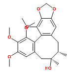
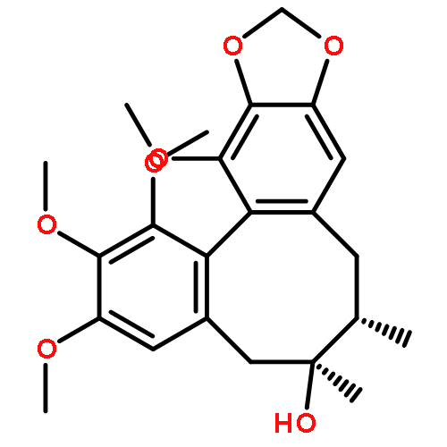
![13,14-dimethoxy-6,7-dimethyl-5,6,7,8-tetrahydro[1,3]benzodioxolo[5',6':3,4]cycloocta[1,2-f][1,3]benzodioxole (non-preferred name)](http://img.cochemist.com/ccimg/61400/61301-33-5.png)
![13,14-dimethoxy-6,7-dimethyl-5,6,7,8-tetrahydro[1,3]benzodioxolo[5',6':3,4]cycloocta[1,2-f][1,3]benzodioxole (non-preferred name)](http://img.cochemist.com/ccimg/61400/61301-33-5_b.png)


![1-(DIMETHYLAMINO)-3-[2-[(E)-2-(3-METHOXYPHENYL)VINYL]PHENOXY]PROP<WBR />AN-2-OL](http://img.cochemist.com/ccimg/400/389-36-6.png)
![1-(DIMETHYLAMINO)-3-[2-[(E)-2-(3-METHOXYPHENYL)VINYL]PHENOXY]PROP<WBR />AN-2-OL](http://img.cochemist.com/ccimg/400/389-36-6_b.png)


![4H-1-Benzopyran-4-one, 5,7-dihydroxy-3-[4-(sulfooxy)phenyl]-](http://img.cochemist.com/ccimg/176100/176045-28-6.png)
![4H-1-Benzopyran-4-one, 5,7-dihydroxy-3-[4-(sulfooxy)phenyl]-](http://img.cochemist.com/ccimg/176100/176045-28-6_b.png)
![3-[(2r,3r)-1-(dimethylamino)-2-methylpentan-3-yl]phenol](http://img.cochemist.com/ccimg/175600/175591-23-8.png)
![3-[(2r,3r)-1-(dimethylamino)-2-methylpentan-3-yl]phenol](http://img.cochemist.com/ccimg/175600/175591-23-8_b.png)


![Acetamide,N-[2-(6-hydroxy-5-methoxy-1H-indol-3-yl)ethyl]-](http://img.cochemist.com/ccimg/2300/2208-41-5.png)
![Acetamide,N-[2-(6-hydroxy-5-methoxy-1H-indol-3-yl)ethyl]-](http://img.cochemist.com/ccimg/2300/2208-41-5_b.png)
![Acetamide,N-[2-[5-methoxy-6-(sulfooxy)-1H-indol-3-yl]ethyl]-](http://img.cochemist.com/ccimg/2300/2208-40-4.png)
![Acetamide,N-[2-[5-methoxy-6-(sulfooxy)-1H-indol-3-yl]ethyl]-](http://img.cochemist.com/ccimg/2300/2208-40-4_b.png)