.jpg)
A new, mechanically elastic biomaterial can be custom 3D-printed, is surgically friendly, and promotes robust bone regeneration.
A new method for complex metallic architecture fabrication is presented, through synthesis and 3D-printing of a new class of 3D-inks into green-body structures followed by thermochemical transformation into sintered metallic counterparts. Small and large volumes of metal-oxide, metal, and metal compound 3D-printable inks are synthesized through simple mixing of solvent, powder, and the biomedical elastomer, polylactic-co-glycolic acid (PLGA). These inks can be 3D-printed under ambient conditions via simple extrusion at speeds upwards of 150 mm s–1 into millimeter- and centimeter-scale thin, thick, high aspect ratio, hollow and enclosed, and multi-material architectures. The resulting 3D-printed green-bodies can be handled immediately, are remarkably robust, and may be further manipulated prior to metallic transformation. Green-bodies are transformed into metallic counterparts without warping or cracking through reduction and sintering in a H2 atmosphere at elevated temperatures. It is shown that primary metal and binary alloy structures can be created from inks comprised of single and mixed oxide powders, and the versatility of the process is illustrated through its extension to more than two dozen additional metal-based materials. A potential application of this new system is briefly demonstrated through cyclic reduction and oxidation of 3D-printed iron oxide constructs, which remain intact through numerous redox cycles.
Bioactive, in situ forming materials have the potential to complement minimally invasive surgical procedures and enhance tissue healing. For such biomaterials to be adopted in the clinic, they must be cost-effective, easily handled by the surgeon and have a history of biocompatibility. To this end, we report a novel and facile self-assembling strategy to create membranes and encapsulating structures using collagen and hyaluronic acid (HA). Unlike membranes built by layer-by-layer deposition of oppositely charged biomolecules, the collagen–HA membranes described here form a diffusion barrier upon electrostatic interaction of the oppositely charged biomolecules, which is further driven by osmotic pressure imbalances. The resulting membranes have a nanofibrous architecture, a thicknesses of 130 μm and a tensile modulus (0.59 ± 0.06 MPa) that can increase 7-fold using carbodiimide chemistry (4.42 ± 1.46 MPa). Collagen–HA membranes support mesenchymal stem cell proliferation and have a slow and steady protein release profile (7% at day 28), offering opportunities for targeted tissue regeneration. We demonstrate the capacity to encapsulate cells by injecting HA into the collagen solution, and enhance allograft and implant biocompatibility through a coating technique. This study describes a novel mechanism of collagen–HA membrane formation and provides the groundwork to apply these membranes in a variety of tissue engineering applications.
Increasing interest in using soy biomaterials for tissue engineering applications has prompted investigation into the in vivo biocompatibility of soy implants. In this study, the biocompatibility of soy protein scaffolds fabricated using freeze-drying and 3-D printing was assessed using a subcutaneous implant model in BALB/c mice. The main objectives of this study were: (1) to compare soy protein with bovine collagen, a well-characterized natural protein implant, by implanting scaffolds of the same protein weight, and (2) to observe the effects of soy scaffold microstructure and amount of protein loading, which also alters the degradation properties, on the acute and humoral immune responses towards soy. Results showed that freeze-dried soy scaffolds fully degraded after 14 days, whereas collagen scaffolds (of the same protein weight) remained intact for 56 days. Furthermore, Masson’s trichrome staining showed little evidence of damage or fibrosis at the soy implant site. Scaffolds of higher soy protein content, however, were still present after 56 days. H&E staining revealed that macrophage infiltration was hindered in the denser bioplotted soy scaffolds, causing slower degradation. Analysis of soy-specific antibodies in mouse serum after implantation revealed levels of IgG1 that correlated with higher scaffold weight and protein density. However, no soy-specific IgE was detected, indicating the absence of an allergic response to the soy implants. These results demonstrate that soy protein could be an acceptable biocompatible implant for tissue regeneration, and that scaffold porosity, soy protein density and scaffold degradation rate significantly affect the acute and humoral immune response.
Soy protein modified with heat treatment and enzyme crosslinking using transglutaminase in maltodextrin was used to fabricate novel, porous three-dimensional scaffolds through lyophilization. Physical properties of scaffolds were characterized using scanning electron microscopy, mercury intrusion porosimetry, moisture content analysis and mechanical testing. Human mesenchymal stem cells (hMSC) were seeded and cultured in vitro on the scaffolds for up to 2 weeks, and changes in stem cell growth and morphology were examined. The resulting scaffolds had rough surfaces, irregular pores with size distributions between 10 and 125 μm, <5% moisture content and compressive moduli ranging between 50 and 100 Pa. Enzyme treatment significantly lowered the moisture content. Increasing amounts of applied enzyme units lowered the median pore size. Although enzyme treatment did not affect the mechanical properties of the scaffolds, it did increase the degradation time by at least 1 week. These changes in scaffold degradation altered the growth and morphology of seeded hMSC. Cell proliferation was observed in scaffolds containing 3% soy protein isolate treated with 1 U of transglutaminase. These results demonstrate that controlling scaffold degradation rates is crucial for optimizing hMSC growth on soy protein scaffolds and that soy protein scaffolds have the potential to be used in tissue engineering applications.
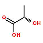
![Poly[oxy[(1S)-1-methyl-2-oxo-1,2-ethanediyl]]](http://img.cochemist.com/ccimg/26200/26161-42-2.png)
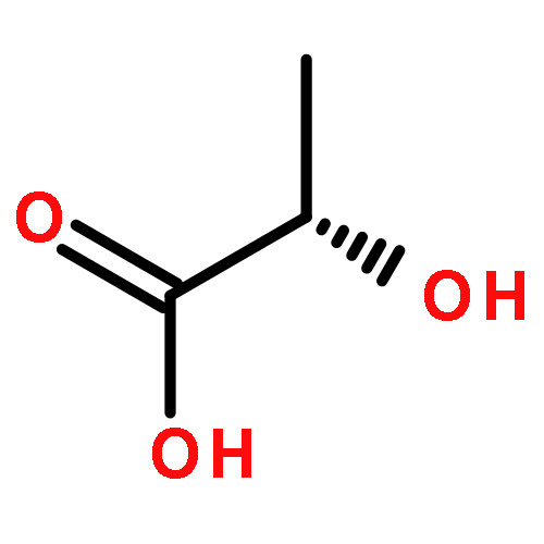
![Poly[oxy[(1S)-1-methyl-2-oxo-1,2-ethanediyl]]](http://img.cochemist.com/ccimg/26200/26161-42-2_b.png)