Co-reporter:Lyulin Hu;Sijie Ren;Qing Shen;Jianchu Chen;Xingqian Ye;Jiangang Ling
RSC Advances (2011-Present) 2017 vol. 7(Issue 44) pp:27496-27505
Publication Date(Web):2017/05/22
DOI:10.1039/C7RA03408C
The effect of different cooking methods on protein and lipid oxidation of farmed sturgeon (Acipenser gueldenstaedtii) was investigated. In this study, the fish fillets were cooked using five different methods (boiling, steaming, microwaving, roasting and deep-frying), to an internal temperature of approximately 85 °C. General indexes for protein oxidation (carbonyls, free thiols and Schiff bases) and lipid oxidation were evaluated by spectrophotometric and fluorimetric methods. Side-chain modifications of amino acid residues were pinpointed by gel- and MS-based proteomic technology. The results are as follows: (1) cooking, especially roasting and frying, significantly increased the levels of carbonyls and Schiff bases and decreased the amounts of free thiols. (2) Oxidation of aromatic amino acids and extensive oxidation of lysine to carbonyl-containing compounds were characterized in all the cooked samples. (3) Indirect protein oxidation caused by interacting with lipid-derived aldehydes like malondialdehyde (MDA) and 4-hydroxynonenal (HNE) was also observed and much more pronounced in roasted and fried samples, giving a footprint of lipid–protein interaction during cooking. In summary, cooking treatment can induce protein oxidation in sturgeon fillets with a variety of modifications occurring at the protein primary structure. Molecular mapping of protein oxidation is conducive to understand the mechanism of cooking-induced protein oxidation and allows better control of the nutritional properties of protein-based products.
Co-reporter:Zhongxiang Fang, Dan Wu, Dong Yü, Xinqian Ye, Donghong Liu, Jianchu Chen
Food Chemistry 2011 Volume 128(Issue 4) pp:943-948
Publication Date(Web):15 October 2011
DOI:10.1016/j.foodchem.2011.03.123
Phenolic compounds and their changes during vacuum frying were investigated for a Chinese purple yam. Three cyanidin derivatives and one peonidin derivative were tentatively identified by HPLC–DAD–ESIMS analysis; sinapic acid and ferulic acid were identified by HPLC–DAD analysis with authentic chemicals. There were 31.0 mg/100 g (dry weight, DW) of total anthocyanin (ACN) and 478 mg/100 g DW of total phenolic content (TPC) in the fresh yam. Sinapic acid and ferulic acid were 135 and 31.3 mg/100 g DW respectively. The blanching process caused about 60% of ACN, and 30–50% of phenolic acids and TPC to be lost, which showed that anthocyanins were most vulnerable during blanching. The retention rate of the phenolic compounds during vacuum frying was 60–69%, indicating it was a practical technology for purple yam processing, on account of its impact on the phenolic compounds stability.Highlights► We tentatively identified 4 anthocyanins in a Chinese purple yam. ► Sinapic acid and ferulic acid were the major phenolic acids in the purple yam. ► Techniques of HPLC–DAD–MS and HPLC–DAD were used for identification. ► Vacuum frying was proved to be a practical technology for purple yam processing.
Co-reporter:Haihua Yang, Xingqian Ye, Donghong Liu, Jianchu Chen, Jinjie Zhang, Yan Shen, and Dong Yu
Journal of Agricultural and Food Chemistry 2011 Volume 59(Issue 5) pp:1622-1629
Publication Date(Web):February 14, 2011
DOI:10.1021/jf103918v
Extractable and unextractable proanthocyanidins (EPAs and UEPAs) from leaves of bayberry were characterized. Both EPAs and UEPAs were analyzed by acid catalysis in the presence of excess phloroglucinol. The main cleavage product, epigallocatechin-3-O-gallate-(4β→2)-phloroglucinol, was successfully identified. The EPAs were of the prodelphinidin type. In fact, epigallocatechin-3-O-gallate (EGCG) and traces of epigallocatechin (EGC) were detected as the extension units, but only EGCG was present in the terminal units. All of the compounds exhibited a 2,3-cis configuration, and >98% of them were galloylated. The mean degree of polymerization (mDP) of bayberry leaf EPAs was 6.5, and the most abundant EPAs were the polymers, with mDP values of 9.5−26.7. The UEPAs were highly polymerized prodelphinidins consisting of EGCG and traces of EGC. In addition, EGCG, three EPA dimers, and two trimers were identified. The EPAs and UEPAs consisted mostly of EGCG, which is unusual in the plant kingdom.


![Bicyclo[4.1.0]hept-2-ene,4,7,7-trimethyl-](http://img.cochemist.com/ccimg/29100/29050-33-7.png)
![Bicyclo[4.1.0]hept-2-ene,4,7,7-trimethyl-](http://img.cochemist.com/ccimg/29100/29050-33-7_b.png)


![2,6-dimethyl-6-(4-methyl-3-pentenyl)bicyclo[3.1.1]hept-2-ene](http://img.cochemist.com/ccimg/17700/17699-05-7.png)
![2,6-dimethyl-6-(4-methyl-3-pentenyl)bicyclo[3.1.1]hept-2-ene](http://img.cochemist.com/ccimg/17700/17699-05-7_b.png)
![Bicyclo[3.1.1]hept-2-ene, 2,6-dimethyl-6-(4-methyl-3-pentenyl)-](http://img.cochemist.com/ccimg/14100/14087-02-6.png)
![Bicyclo[3.1.1]hept-2-ene, 2,6-dimethyl-6-(4-methyl-3-pentenyl)-](http://img.cochemist.com/ccimg/14100/14087-02-6_b.png)


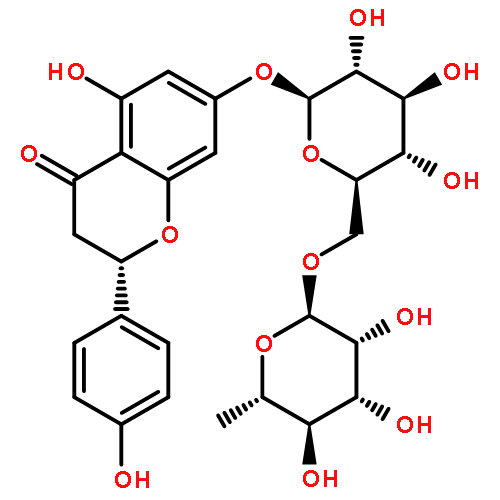
![Didymin with HPLC [14259-47-3]](http://img.cochemist.com/ccimg/14300/14259-47-3.png)
![Didymin with HPLC [14259-47-3]](http://img.cochemist.com/ccimg/14300/14259-47-3_b.png)