Co-reporter:Zejun Wang, Yao Fu, Zhengzhong Kang, Xiaoguo Liu, Nan Chen, Qi Wang, Yaoquan Tu, Lihua Wang, Shiping Song, Daishun Ling, Haiyun Song, Xueqian Kong, and Chunhai Fan
Journal of the American Chemical Society November 8, 2017 Volume 139(Issue 44) pp:15784-15784
Publication Date(Web):October 12, 2017
DOI:10.1021/jacs.7b07895
DNA has proven of high utility to modulate the surface functionality of metal–organic frameworks (MOFs) for various biomedical applications. Nevertheless, current methods for preparing DNA–MOF nanoparticles rely on either inefficient covalent conjugation or specific modification of oligonucleotides. In this work, we report that unmodified oligonucleotides can be loaded on MOFs with high density (∼2500 strands/particle) via intrinsic, multivalent coordination between DNA backbone phosphate and unsaturated zirconium sites on MOFs. More significantly, surface-bound DNA can be efficiently released in either bulk solution or specific organelles in live cells when free phosphate ions are present. As a proof-of-concept for using this novel type of DNA–MOFs in immunotherapy, we prepared a construct of immunostimulatory DNA–MOFs (isMOFs) by intrinsically coordinating cytosine–phosphate–guanosine (CpG) oligonucleotides on biocompatible zirconium MOF nanoparticles, which was further armed by a protection shell of calcium phosphate (CaP) exoskeleton. We demonstrated that isMOFs exhibited high cellular uptake, organelle specificity, and spatiotemporal control of Toll-like receptors (TLR)-triggered immune responses. When isMOF reached endolysosomes via microtubule-mediated trafficking, the CaP exoskeleton dissolved in the acidic environment and in situ generated free phosphate ions. As a result, CpG was released from isMOFs and stimulated potent immunostimulation in living macrophage cells. Compared with naked CpG–MOF, isMOFs exhibited 83-fold up-regulation in stimulated secretion of cytokines. We thus expect this isMOF design with soluble CaP exoskeleton and an embedded sequential “protect–release” program provides a highly generic approach for intracellular delivery of therapeutic nucleic acids.
Co-reporter:Xingjie Hu, Xiaojiao Li, Min Yin, Ping Li, Ping Huang, Lihua Wang, Yiguo Jiang, Hui Wang, Nan Chen, Chunhai Fan, and Haiyun Song
ACS Applied Materials & Interfaces June 7, 2017 Volume 9(Issue 22) pp:18575-18575
Publication Date(Web):May 16, 2017
DOI:10.1021/acsami.7b04788
Introduction of exogenous biomacromolecules into living systems is of great interest in genome editing, cancer immunotherapy, and stem cell reprogramming. Whereas current strategies generally depend on nucleic acids transfection, direct delivery of functional proteins that provides enhanced specificity, increased safety, and fast and temporal regulation is highly desirable. Nevertheless, intracellular delivery of intact and bioactive proteins, especially in vivo, remains poorly explored. In this study, we developed a nanodiamonds (NDs)-based protein delivery system in cultured cells and in Drosophila that showed high adsorption, high efficiency, and effective cytosolic release of fully functional proteins. Through live-cell imaging, we observed a novel phenomenon wherein a substantial amount of internalized NDs–protein complex rejected fusion with the early endosome, thereby evading protein degradation in the lysosome. More significantly, we demonstrated that dietary NDs–RNase induced apoptosis in enterocytes, stimulating regenerative divisions in intestinal stem cells and increasing the number of stem cells and precursor cells in Drosophila intestine. As stem cells are poorly accessible by exogenous agents in vivo, NDs-mediated oral delivery of proteins provides a new approach to modulate the stem cell microenvironment for intestinal remodeling, which has important implications for colorectal cancer therapy and regenerative medicine.Keywords: intestinal remodeling; nanodiamonds; oral delivery; protein delivery; stem cells microenvironment;
Co-reporter:Xiangmeng Qu, Shaopeng Wang, Zhilei Ge, Jianbang Wang, Guangbao Yao, Jiang Li, Xiaolei Zuo, Jiye Shi, Shiping Song, Lihua Wang, Li Li, Hao Pei, and Chunhai Fan
Journal of the American Chemical Society August 2, 2017 Volume 139(Issue 30) pp:10176-10176
Publication Date(Web):July 17, 2017
DOI:10.1021/jacs.7b04040
Programmable remodelling of cell surfaces enables high-precision regulation of cell behavior. In this work, we developed in vitro constructed DNA-based chemical reaction networks (CRNs) to program on-chip cell adhesion. We found that the RGD-functionalized DNA CRNs are entirely noninvasive when interfaced with the fluidic mosaic membrane of living cells. DNA toehold with different lengths could tunably alter the release kinetics of cells, which shows rapid release in minutes with the use of a 6-base toehold. We further demonstrated the realization of Boolean logic functions by using DNA strand displacement reactions, which include multi-input and sequential cell logic gates (AND, OR, XOR, and AND-OR). This study provides a highly generic tool for self-organization of biological systems.
Co-reporter:Chunhai Fan;Xiaohong Fang
Science China Chemistry 2017 Volume 60( Issue 10) pp:1265-1266
Publication Date(Web):19 September 2017
DOI:10.1007/s11426-017-9135-x
Co-reporter:Jie Tian;Qinglong Yan;Ying Zhu;Jichao Zhang;Jiao Li;Ben Shi;Ge Xu;Chunchang Zhao
Chinese Journal of Chemistry 2017 Volume 35(Issue 11) pp:1711-1716
Publication Date(Web):2017/11/01
DOI:10.1002/cjoc.201700248
A fluorescent turn-on probe for specifically targeting γ-glutamyltranspeptidase (GGT) was designed and synthesized by integrating boron-dipyrromethene (BODIPY) as a chromophore and glutathione (GSH) as the GGT substrate. GGT-catalyzed the cleavage of the γ-glutamyl bond and generated the aromatic hydrocarbon transfer between the sulfur and the nitrogen atom in BODIPY, leading to distinct optical changes. Such specific responsiveness provides an easily distinguishable fluorescence signal to visualize the GGT activity in living cells and differentiate GGT-positive cancer cells from GGT-negative cells.
Co-reporter:Jianlei Shen;Binquan Luan;Hao Pei;Zaixing Yang;Xiaolei Zuo;Gang Liu;Jiye Shi;Lihua Wang;Ruhong Zhou;Wenlong Cheng
Advanced Materials 2017 Volume 29(Issue 35) pp:
Publication Date(Web):2017/09/01
DOI:10.1002/adma.201606796
2D materials possess many interesting properties, and have shown great application potentials. In this work, the development of humidity-responsive, 2D plasmonic nanostructures with switchable chromogenic properties upon wetting–dewetting transitions is reported. By exploiting DNA hybridization-directed anchoring of gold nanoparticles (AuNPs) on substrates, a series of single-nanoparticle-layer (SNL) plasmonic films is fabricated. Due to the collective plasmonic responses in SNL, these ultrathin 2D films display rapid and reversible red-blue color change upon the wetting–dewetting transition, suggesting that hydration-induced microscopic plasmonic coupling between AuNPs is replicated in the macroscopic, centimeter-scale films. It is also found that hydration finely tunes the electric field distribution between AuNPs in the SNL film, based on which responsive surface-enhanced Raman scattering substrates with spatially homogeneous hot spots are developed. Thus it is expected that DNA-mediated 2D SNL structures open new avenues for designing miniaturized plasmonic nanodevices with various applications.
Co-reporter:Ying Zhu, Jichao Zhang, Aiguo Li, Yuanqing Zhang, Chunhai Fan
Current Opinion in Chemical Biology 2017 Volume 39(Volume 39) pp:
Publication Date(Web):1 August 2017
DOI:10.1016/j.cbpa.2017.04.016
•Synchrotron-based X-ray microscopy holds great promise for nanoscale cell imaging.•X-ray imaging in the ‘water window’ offers natural contrast.•Development of X-ray-sensitive nanoprobes for cell labeling is required.Microscopic imaging provides a straightforward approach to deepen our understanding of cellular events. While the resolution of optical microscopes is generally limited to 200–300 nm due to the diffraction limit, there has been ever growing interest in studying cells at the sub-100 nm regime. By exploiting the short wavelength, long penetration depth and elemental specificity of X-rays, synchrotron-based X-ray microscopy (XRM) has demonstrated its power in exploring the structure and function of cells at the nanometer resolution. Here we summarize recent advances in using XRM for imaging ultrastructure of organelles and specific biomolecular locations in cells, and provide a perspective on potentials and applications of XRM.
Co-reporter:Yu Zhang;Zhifen Cui;Huating Kong;Kai Xia;Liang Pan;Jiang Li;Yanhong Sun;Jiye Shi;Lihua Wang;Ying Zhu
Advanced Materials 2016 Volume 28( Issue 14) pp:2699-2708
Publication Date(Web):
DOI:10.1002/adma.201506232
Co-reporter:Yi Zhang;Zhuyao Wang;Xiaojiao Li;Lu Wang;Min Yin;Lihua Wang;Nan Chen;Haiyun Song
Advanced Materials 2016 Volume 28( Issue 7) pp:1387-1393
Publication Date(Web):
DOI:10.1002/adma.201503893
Co-reporter:Xiuhai Mao, Anna J. Simon, Hao Pei, Jiye Shi, Jiang Li, Qing Huang, Kevin W. Plaxco and Chunhai Fan
Chemical Science 2016 vol. 7(Issue 2) pp:1200-1204
Publication Date(Web):26 Oct 2015
DOI:10.1039/C5SC03705K
Recognition of the fundamental importance of allosteric regulation in biology dates back to not long after its discovery in the 1960s. Our ability to rationally engineer this potentially useful property into normally non-allosteric catalysts, however, remains limited. In response we report a DNA nanotechnology-enabled approach for introducing allostery into catalytic nucleic acids. Specifically, we have grafted one or two copies of a peroxidase-like DNAzyme, hemin-bound G-quadruplex (hemin-G), onto a DNA tetrahedral nanostructure in such a manner as to cause them to interact, modulating their catalytic activity. We achieve allosteric regulation of these catalysts by incorporating dynamically responsive oligonucleotides that respond to specific “effector” molecules (complementary oligonucleotides or small molecules), altering the spacing between the catalytic sites and thus regulating their activity. This designable approach thus enables subtle allosteric modulation in DNAzymes that is potentially of use for nanomedicine and nanomachines.
Co-reporter:Lifeng Xu, Geng Wang, Jianlei Shen, Heping Geng, Wenqin Li, Longlong Wu, Shanshan Gao, Jianing Wang, Lihua Wang, Chunhai Fan and Gang Chen
Nanoscale 2016 vol. 8(Issue 17) pp:9337-9342
Publication Date(Web):05 Apr 2016
DOI:10.1039/C6NR00193A
The broken symmetry of Janus nanostructures (JNs) provides a distinctive means to express drastically different chemical and physical characters within a single particle and acquire emergent properties usually inconceivable for homogeneous or symmetric nanostructures. In spite of their tremendous application potential, considerable challenges are encountered in identifying pathways to synthesize or assemble JNs with a controllable geometry and morphology. Here, we exploit the reverse process of growth, i.e. silver etching, to quantitatively control the structural and optical properties of the DNA-mediated Au–Ag JNs. The transmission electron microscopy and optical measurements, along with numerical simulations, present a comprehensive view of the etching dynamics and a detailed analysis of the influencing factors that provide handles for regulating the silver etching rate and progress. In addition, a novel type of composite JN is proposed and a model system is designed and engineered through dynamical control of the etching and DNA-hybridization processes.
Co-reporter:Dan Zhu, Ping Song, Juwen Shen, Shao Su, Jie Chao, Ali Aldalbahi, Ziang Zhou, Shiping Song, Chunhai Fan, Xiaolei Zuo, Yang Tian, Lianhui Wang, and Hao Pei
Analytical Chemistry 2016 Volume 88(Issue 9) pp:4949
Publication Date(Web):April 8, 2016
DOI:10.1021/acs.analchem.6b00891
Understanding the behavior of biomolecules on nanointerface is critical in bioanalysis, which is great challenge due to the instability and the difficulty to control the orientation and loading density of biomolecules. Here, we investigated the thermodynamics and kinetics of DNA hybridization on gold nanoparticle, with the aim to improve the efficiency and speed of DNA analysis. We achieved precise and quantitative surface control by applying a recently developed poly adenines (polyA)-based assembly strategy on gold nanoparticles (DNA-AuNPs). PolyA served as an effective anchoring block based on the preferential binding with the AuNP surface and the appended recognition block adopted an upright conformation that favors DNA hybridization. The lateral spacing and surface density of DNA on AuNPs can be systematically modulated by adjusting the length of polyA block. We found the stability of duplex on AuNP was enhanced with the increasing length of polyA block. When the length of polyA block reached to 40 bases, the thermodynamic properties were more similar to that of duplex in solution. Fast hybridization rate was observed on the diblock DNA-AuNPs and was increased along with the length of polyA block. We consider the high stability and excellent hybridization performance come from the minimization of the DNA–DNA and DNA-AuNP interactions with the use of polyA block. This study provides better understanding of the behavior of biomolecules on the nanointerface and opens new opportunities to construct high-efficiency and high-speed biosensors for DNA analysis.
Co-reporter:Shixing Chen, Yanzhi Dou, Zhihan Zhao, Fuwu Li, Jing Su, Chunhai Fan, and Shiping Song
Analytical Chemistry 2016 Volume 88(Issue 7) pp:3476
Publication Date(Web):March 4, 2016
DOI:10.1021/acs.analchem.6b00230
DNA hydroxymethylation (5-hmC) is a kind of new epigenetic modification, which plays key roles in DNA demethylation, genomic reprogramming, and the gene expression in mammals. For further exploring the functions of 5-hmC, it is necessary to develop sensitive and selective methods for detecting 5-hmC. Herein, we developed a novel multiplexing electrochemical (MEC) biosensor for 5-hmC detection based on the glycosylation modification of 5-hmC and enzymatic signal amplification. The 5-hmC was first glycosylated by T4 β-glucosyltransferase and then oxidated by sodium periodate. The resulting glucosyl-modified 5-hmC (5-ghmC) was incubated with ARP-biotin and was bound to avidin-HRP. The 5-hmC can be detected at the subnanogram level. Finally, we performed 5-hmC detection for mouse tissue samples and cancer cell lines. The limit of detection of the MEC biosensor is 20 times lower than that of commercial kits based on optical meaurement. Also, the biosensor presented high detection specificity because the chemical reaction for 5-hmC modification can not happen at any other unhydroxymethylated nucleic acid bases. Importantly, benefited by its multiplexing capacity, the developed MEC biosensor showed excellent high efficiency, which was time-saving and cost less.
Co-reporter:Yanzhi Dou, Zhineng Jiang, Wangping Deng, Jing Su, Shixing Chen, Haiyun Song, Ali Aldalbahi, Xiaolei Zuo, Shiping Song, Jiye Shi, Chunhai Fan
Journal of Electroanalytical Chemistry 2016 Volume 781() pp:339-344
Publication Date(Web):15 November 2016
DOI:10.1016/j.jelechem.2016.04.022
We have developed a fast, highly sensitive and low-cost biosensing system for the detection of clenbuterol (CLB), using a homemade mobile electrochemical device with an electric field-driven acceleration strategy. This system consists of an embedded circuit in smartphone for signal processing and a screen-printed carbon electrode (SPCE) modified with multi-walled carbon nanotubes (MWNTs) and goat anti mouse-immunoglobulin G (IgG) sensing layer (MWNTs-I-layer). CLB monoclonal antibody was assembled through its binding to the surface-confined antibody. Such modified electrodes were used for rapid and sensitive amperometric immunosensing detection of CLB. Horseradish peroxidase-coupled CLB (CLB–HRP) competed with free CLB in the samples to bind the monoclonal antibody. By using this mobile system, we could detect CLB ranging from 0.3 ng ⋅ mL− 1 to 100 ng ⋅ mL− 1 with the detection limit of 0.076 ng ⋅ mL− 1. The whole competitive-type detection process was finished within 6 min. We expect this device can meet the requirements for field detection of various food security-related species.
Co-reporter:Jie Chao;Yinan Zhang;Dan Zhu;Bing Liu;Chengjun Cui;Shao Su
Science China Chemistry 2016 Volume 59( Issue 6) pp:730-734
Publication Date(Web):2016 June
DOI:10.1007/s11426-016-5596-x
Hetero-assembling of spherical building blocks with well-defined spatial distribution holds great significance in developing chiral nanostructures. Herein, a strategy for hetero-assembling of gold nanoparticles (AuNPs) was demonstrated using rigid bifacial DNA origami as templates. By tuning the sizes and the fixed location of AuNPs on DNA origami, right-handed and left-handed AuNPs nanostructures were respectively constructed. Gel electrophoresis indicated the formation of the DNA origami- AuNPs complex and transmission electron microscopy (TEM) visually displayed the arrangement of AuNPs in these two chiral structures. The spatial configuration and 3D geometry of AuNPs were further illustrated by the stereographic TEM with tilting angles from -30° to 30°. This strategy provides a universal approach to construct the asymmetrical 3D geometries, which may have potential applications in biomimicking and nanophotonics.
Co-reporter:Yongxi Zhao, Feng Chen, Qian Li, Lihua Wang, and Chunhai Fan
Chemical Reviews 2015 Volume 115(Issue 22) pp:12491
Publication Date(Web):November 9, 2015
DOI:10.1021/acs.chemrev.5b00428
Isothermal amplification of nucleic acids is a simple process that rapidly and efficiently accumulates nucleic acid sequences at constant temperature. Since the early 1990s, various isothermal amplification techniques have been developed as alternatives to polymerase chain reaction (PCR). These isothermal amplification methods have been used for biosensing targets such as DNA, RNA, cells, proteins, small molecules, and ions. The applications of these techniques for in situ or intracellular bioimaging and sequencing have been amply demonstrated. Amplicons produced by isothermal amplification methods have also been utilized to construct versatile nucleic acid nanomaterials for promising applications in biomedicine, bioimaging, and biosensing. The integration of isothermal amplification into microsystems or portable devices improves nucleic acid-based on-site assays and confers high sensitivity. Single-cell and single-molecule analyses have also been implemented based on integrated microfluidic systems. In this review, we provide a comprehensive overview of the isothermal amplification of nucleic acids encompassing work published in the past two decades. First, different isothermal amplification techniques are classified into three types based on reaction kinetics. Then, we summarize the applications of isothermal amplification in bioanalysis, diagnostics, nanotechnology, materials science, and device integration. Finally, several challenges and perspectives in the field are discussed.
Co-reporter:Jie Chao, Yunfeng Lin, Huajie Liu, Lianhui Wang, Chunhai Fan
Materials Today 2015 Volume 18(Issue 6) pp:326-335
Publication Date(Web):July–August 2015
DOI:10.1016/j.mattod.2015.01.018
Plasmonic nanostructures have rapidly emerged as a type of optical material possessing many novel physical properties and holding great promise for a wide range of applications. One of the key challenges in this area lies in the bottom-up construction of precise plasmonic nanostructures with novel optical properties. By exploiting the unparalleled self-recognition properties of DNA molecules, researchers in the area of DNA nanotechnology have worked to make complex and hierarchical DNA nanostructures in a highly controllable and programmable manner, which offers unprecedented opportunities for developing self-assembled plasmonic nanostructures. In this review, we will summarize recent advances on design and fabrication of static and dynamic DNA nanostructures, and their use as linkers or templates for the assembly of plasmonic nanostructures.
Co-reporter:Dan Zhu, Jie Chao, Hao Pei, Xiaolei Zuo, Qing Huang, Lianhui Wang, Wei Huang, and Chunhai Fan
ACS Applied Materials & Interfaces 2015 Volume 7(Issue 20) pp:11047
Publication Date(Web):April 22, 2015
DOI:10.1021/acsami.5b03066
DNA-decorated metal nanoparticles have found numerous applications, most of which rely on thiolated DNA (SH-DNA)-modified gold nanoparticles (AuNPs). Whereas silver nanoparticles (AgNPs) are known to have stronger plasmonic properties than AuNPs, modification of AgNPs with SH-DNA is technically challenging, partially due to the instability of Ag–S bonding. Here we demonstrate a facile approach to self-assemble unmodified DNA on AgNPs by exploiting intrinsic silver–cytosine (Ag–C) coordination. The strong Ag–C coordination allows for the ready formation of DNA-AgNP conjugates, which show favorable stability under conditions of high ionic strength and high temperature. These nanoconjugates possess much higher efficient molecular recognition capability and faster hybridization kinetics than thiolated DNA-modified AgNPs. More importantly, we could programmably tune the DNA density on AgNPs with the regulation of silver–cytosine coordination numbers, which in turn modulated their hybridizability. We further demonstrated that these DNA-AgNP conjugates could serve as excellent building blocks for assembling silver and hybrid silver–gold nanostructures with superior plasmonic properties.Keywords: coordination; plasmonic; programmable assembly; silver nanoparticle; umodified DNA;
Co-reporter:Degao Wang, Huaican Chen, Guoliang Chang, Xiao Lin, Yuying Zhang, Ali Aldalbahi, Cheng Peng, Jianqiang Wang, and Chunhai Fan
ACS Applied Materials & Interfaces 2015 Volume 7(Issue 25) pp:14072
Publication Date(Web):June 8, 2015
DOI:10.1021/acsami.5b03298
Doping elements in hematite nanostructures is a promising approach to improve the photoelectrochemical (PEC) water-splitting performance of hematite photoanodes. However, uniform doping with precise control on doping amount and morphology is the major challenge for quantitatively investigating the PEC water-splitting enhancement. Here, we report on the design and synthesis of uniform titanium (Ti)-doped hematite nanorods with precise control of the Ti amount and morphology for highly effective PEC water splitting using an atomic layer deposition assisted solid-state diffusion method. We found that Ti doping promoted band bending and increased the carrier density as well as the surface state. Remarkably, these uniformly doped hematite nanorods exhibited high PEC performance with a pronounced photocurrent density of 2.28 mA/cm2 at 1.23 V vs reversible hydrogen electrode (RHE) and 4.18 mA/cm2 at 1.70 V vs RHE, respectively. Furthermore, as-prepared Ti-doping hematite nanorods performed excellent repeatability and durability; over 80% of the as-fabricated photoanodes reproduced the steady photocurrent density of 1.9–2.2 mA/cm2 at 1.23 V vs RHE at least 3 h in a strong alkaline electrolyte solution.Keywords: atomic layer deposition; hematite nanorod; photoelectrochemical; solid-state diffusion; uniform Ti doping;
Co-reporter:Degao Wang;Yuying Zhang;Cheng Peng;Jianqiang Wang;Qing Huang;Shao Su;Lianhui Wang;Wei Huang
Advanced Science 2015 Volume 2( Issue 4) pp:
Publication Date(Web):
DOI:10.1002/advs.201500005
Co-reporter:Dr. Jiang Li;Dr. Jie Chao;Jiye Shi; Dr. Chunhai Fan
ChemBioChem 2015 Volume 16( Issue 1) pp:39-41
Publication Date(Web):
DOI:10.1002/cbic.201402627
Abstract
A very attractive goal in nanotechnology is to manufacture smart nanodevices that integrate multiple biological/biomedical functions and autonomously function in vivo in a predefined and well-controlled manner. For decades, researchers have been developing many different ways toward this target, using bottom-up assembly of types of nanomaterials or top-down fabrication of devices with nanometer-scale precision. However, the practical application of these nanodevices remains challenging. One possible barrier lies in the spatiotemporal separation between fabrication and use, which poses a great challenge for the non-invasive delivery of fully functional nanodevice into live cells. Indeed, cells themselves are highly complex natural machines with membrane barriers and finely regulated pathways for intracellular delivery. However, there is plenty of evidence that nanomaterials or nanodevices are easily aggregated or trapped inside of the cells.
Co-reporter:Guangbao Yao;Dr. Jiang Li;Dr. Jie Chao;Dr. Hao Pei;Dr. Huajie Liu; Yun Zhao;Dr. Jiye Shi;Dr. Qing Huang; Lianhui Wang; Wei Huang; Chunhai Fan
Angewandte Chemie International Edition 2015 Volume 54( Issue 10) pp:2966-2969
Publication Date(Web):
DOI:10.1002/anie.201410895
Abstract
DNA origami has rapidly emerged as a powerful and programmable method to construct functional nanostructures. However, the size limitation of approximately 100 nm in classic DNA origami hampers its plasmonic applications. Herein, we report a jigsaw-puzzle-like assembly strategy mediated by gold nanoparticles (AuNPs) to break the size limitation of DNA origami. We demonstrated that oligonucleotide-functionalized AuNPs function as universal joint units for the one-pot assembly of parent DNA origami of triangular shape to form sub-microscale super-origami nanostructures. AuNPs anchored at predefined positions of the super-origami exhibited strong interparticle plasmonic coupling. This AuNP-mediated strategy offers new opportunities to drive macroscopic self-assembly and to fabricate well-defined nanophotonic materials and devices.
Co-reporter:Guangbao Yao;Dr. Jiang Li;Dr. Jie Chao;Dr. Hao Pei;Dr. Huajie Liu; Yun Zhao;Dr. Jiye Shi;Dr. Qing Huang; Lianhui Wang; Wei Huang; Chunhai Fan
Angewandte Chemie 2015 Volume 127( Issue 10) pp:3009-3012
Publication Date(Web):
DOI:10.1002/ange.201410895
Abstract
DNA origami has rapidly emerged as a powerful and programmable method to construct functional nanostructures. However, the size limitation of approximately 100 nm in classic DNA origami hampers its plasmonic applications. Herein, we report a jigsaw-puzzle-like assembly strategy mediated by gold nanoparticles (AuNPs) to break the size limitation of DNA origami. We demonstrated that oligonucleotide-functionalized AuNPs function as universal joint units for the one-pot assembly of parent DNA origami of triangular shape to form sub-microscale super-origami nanostructures. AuNPs anchored at predefined positions of the super-origami exhibited strong interparticle plasmonic coupling. This AuNP-mediated strategy offers new opportunities to drive macroscopic self-assembly and to fabricate well-defined nanophotonic materials and devices.
Co-reporter:Jing-Juan Xu, Wei-Wei Zhao, Shiping Song, Chunhai Fan and Hong-Yuan Chen
Chemical Society Reviews 2014 vol. 43(Issue 5) pp:1601-1611
Publication Date(Web):17 Dec 2013
DOI:10.1039/C3CS60277J
With the rapidly increasing demands for ultrasensitive biodetection, the design and applications of functional nanoprobes have attracted substantial interest for biosensing with optical, electrochemical, and various other means. In particular, given the comparable sizes of nanomaterials and biomolecules, there exists plenty of opportunities to develop functional nanoprobes with biomolecules for highly sensitive and selective biosensing. Over the past decade, numerous nanoprobes have been developed for ultrasensitive bioaffinity sensing of proteins and nucleic acids in both laboratory and clinical applications. In this review, we provide an update on the recent advances in this direction, particularly in the past two years, which reflects new progress since the publication of our last review on the same topic in Chem. Soc. Rev. The types of probes under discussion include: (i) nanoamplifier probes: one nanomaterial loaded with multiple biomolecules; (ii) quantum dots probes: fluorescent nanomaterials with high brightness; (iii) superquenching nanoprobes: fluorescent background suppression; (iv) nanoscale Raman probes: nanoscale surface-enhanced Raman resonance scattering; (v) nanoFETs: nanomaterial-based electrical detection; and (vi) nanoscale enhancers: nanomaterial-induced metal deposition.
Co-reporter:Hui Xu, Qian Li, Lihua Wang, Yao He, Jiye Shi, Bo Tang and Chunhai Fan
Chemical Society Reviews 2014 vol. 43(Issue 8) pp:2650-2661
Publication Date(Web):06 Jan 2014
DOI:10.1039/C3CS60309A
Nanomaterials with unique optical properties have shown great promise as probes for cellular imaging. Based on these properties, a wide range of plasmonic, fluorescent and Raman probes have been designed and prepared. Nanomaterials of different sizes and shapes have also been functionalized with various types of biomolecules, such as antibodies, DNA or RNA, which are actively exploited to realize targeted imaging. In this review, we will summarize recent advances in using functional nanomaterials for imaging, primarily cellular imaging. These nanomaterials are categorized based on their conducting properties, i.e. conductors, semiconductors and insulators.
Co-reporter:Nan Chen, Jiang Li, Haiyun Song, Jie Chao, Qing Huang, and Chunhai Fan
Accounts of Chemical Research 2014 Volume 47(Issue 6) pp:1720-1730
Publication Date(Web):March 3, 2014
DOI:10.1021/ar400324n
DNA and DNA structures can also form hybrids with inorganic NMs. Notably, DNA anchored at the interface of inorganic NMs behaves differently from that at the macroscopic interface. Several types of DNA–NM conjugates have exerted beneficial effects for bioassays and in vitro translation of proteins. Even more interestingly, hybrid nanoconjugates demonstrate distinct properties under the context of biological systems such as cultured cells or animal models. These unprecedented properties not only arouse great interest in studying such interfaces but also open new opportunities for numerous applications in artificial and living systems.
Co-reporter:Hao Pei, Xiaolei Zuo, Dan Zhu, Qing Huang, and Chunhai Fan
Accounts of Chemical Research 2014 Volume 47(Issue 2) pp:550
Publication Date(Web):December 31, 2013
DOI:10.1021/ar400195t
There has been tremendous interest in constructing nanostructures by exploiting the unparalleled ability of DNA molecules in self-assembly. We have seen the appearance of many fantastic, “art-like” DNA nanostructures in one, two, or three dimensions during the last two decades. More recently, much attention has been directed to the use of these elegant nanoobjects for applications in a wide range of areas. Among them, diagnosis and therapy (i.e., theranostics) are of particular interest given the biological nature of DNA.One of the major barricades for the biosensor design lies in the restricted target accessibility at the solid–water interface. DNA nanotechnology provides a convenient approach to well control the biomolecule-confined surface to increase the ability of molecular recognition at the biosensing interface. For example, tetrahedral DNA nanostructures with thiol modifications can be self-assembled at the gold surface with high reproducibility. Since DNA tetrahedra are highly rigid and well-defined structures with atomic precision and versatile functionality, they provide scaffolds for anchoring of a variety of biomolecular probes (DNA, aptamers, peptides, and proteins) for biosensing. Significantly, this DNA nanostructure-based biosensing platform greatly increases target accessibility and improves the sensitivity for various types of molecular targets (DNA, RNA, proteins, and small molecules) by several orders of magnitude.In an alternative approach, DNA nanostructures provide a framework for the development of dynamic nanosensors that can function inside the cell. DNA tetrahedra are found to be facilely cell permeable and can sense and image specific molecules in cells. More importantly, these DNA nanostructures can be efficient drug delivery nanocarriers. Since they are DNA molecules by themselves, they have shown excellent cellular biocompatibility with minimal cytotoxicity. As an example, DNA tetrahedra tailored with CpG oligonucleotide drugs have shown greatly improved immunostimulatory effects that makes them a highly promising nanomedicine. By taking them together, we believe these functionalized DNA nanostructures can be a type of intelligent theranostic nanodevice for simultaneous sensing, diagnosis, and therapy inside the cell.
Co-reporter:Ying Zhu;Thomas Earnest;Qing Huang;Xiaoqing Cai;Zhili Wang;Ziyu Wu
Advanced Materials 2014 Volume 26( Issue 46) pp:7889-7895
Publication Date(Web):
DOI:10.1002/adma.201304281
It is one of the ultimate goals in cell biology to understand the complex spatio–temporal interplay of biomolecules in the cellular context. To this end, there have been great efforts on the development of various probes to detect and localize specific biomolecules in cells with a variety of microscopic imaging techniques. In this Research News, we first summarize several types of microscopy for visualizing specific biomolecular targets. Then we focus on recent advances in the design of X-ray sensitive nanoprobes for applications in synchrotron-based cellular imaging. With the availability of advanced synchrotron techniques, there has been rapid progress toward high-resolution and multi-color X-ray imaging in cells with various types of functional nanoprobes.
Co-reporter:Fan Yang;Xiaolei Zuo;Zhenhua Li;Wangping Deng;Jiye Shi;Guojun Zhang;Qing Huang;Shiping Song
Advanced Materials 2014 Volume 26( Issue 27) pp:4671-4676
Publication Date(Web):
DOI:10.1002/adma.201400451
Co-reporter:Chunhai Fan;Jun Hu ;Zhentang Zhao
Advanced Materials 2014 Volume 26( Issue 46) pp:7685-7687
Publication Date(Web):
DOI:10.1002/adma.201404134
No abstract is available for this article.
Co-reporter:Yongxi Zhao, Feng Chen, Qing Zhang, Yue Zhao, Xiaolei Zuo and Chunhai Fan
NPG Asia Materials 2014 6(9) pp:e131
Publication Date(Web):2014-09-01
DOI:10.1038/am.2014.84
Herein, we propose a novel and universal biosensing platform based on a polymerase-nicking enzyme synergetic isothermal quadratic DNA machine (ESQM). This platform tactfully integrates two signal amplification modules, strand displacement amplification (SDA) and nicking enzyme signal amplification (NESA), into a one-step system. A bifunctional DNA probe with a stem-loop structure was designed to be partly complementary to the SDA product and digested substrate of NESA for bridging SDA and NESA. ESQM can be performed by using only the enzymes and buffer involved in the SDA module. In the presence of a target, this DNA machine is activated to afford a high quadratic amplified signal. Using Pb2+ and DNA adenine methylation (Dam) methyltransferase (MTase) as analytes, a sensitive biosensing platform is demonstrated. Low detection limits of 30 fM Pb2+ and 0.05 U ml−1 Dam MTase were achieved within a short assay time (40 min), which were each superior to those of most previously reported methods. This DNA machine exhibited high selectivity for Pb2+. Furthermore, the successful detection of complex environmental water samples demonstrated the applicability of the proposed strategy in real samples, holding great potential for its application in environmental monitoring, biomedical research and clinical diagnosis.
Co-reporter:Alireza Abi, Meihua Lin, Hao Pei, Chunhai Fan, Elena E. Ferapontova, and Xiaolei Zuo
ACS Applied Materials & Interfaces 2014 Volume 6(Issue 11) pp:8928
Publication Date(Web):May 7, 2014
DOI:10.1021/am501823q
Nanomechanical switching of functional three-dimensional (3D) DNA nanostructures is crucial for nanobiotechnological applications such as nanorobotics or self-regulating sensor and actuator devices. Here, DNA tetrahedral nanostructures self-assembled onto gold electrodes were shown to undergo the electronically addressable nanoswitching due to their mechanical reconfiguration upon external chemical stimuli. That enables construction of robust surface-tethered electronic nanodevices based on 3D DNA tetrahedra. One edge of the tetrahedron contained a partially self-complementary region with a stem-loop hairpin structure, reconfigurable upon hybridization to a complementary DNA (stimulus DNA) sequence. A non-intercalative ferrocene (Fc) redox label was attached to the reconfigurable tetrahedron edge in such a way that reconfiguration of this edge changed the distance between the electrode and Fc.Keywords: 3D nanostructures; DNA nanotechnology; DNA tetrahedron; electromechanical devices; nanomechanical switching; self-assembly;
Co-reporter:Xiaoming Li;Nan Chen;Yuanyuan Su;Yao He;Min Yin;Min Wei;Lianhui Wang;Wei Huang;Qing Huang
Advanced Healthcare Materials 2014 Volume 3( Issue 3) pp:354-359
Publication Date(Web):
DOI:10.1002/adhm.201300294
Co-reporter:Bing Liu;Xiangyuan Ouyang;Jie Chao;Huajie Liu;Yun Zhao
Chinese Journal of Chemistry 2014 Volume 32( Issue 2) pp:137-141
Publication Date(Web):
DOI:10.1002/cjoc.201300827
Abstract
During the development of structural DNA nanotechnology, the emerging of scaffolded DNA origami is marvelous. It utilizes DNA double helix inherent specificity of Watson-Crick base pairing and structural features to create self-assembling structures at the nanometer scale exhibiting the addressable character. However, the assembly of DNA origami is disorderly and unpredictable. Herein, we present a novel strategy to assemble the DNA origami using rolling circle amplification based DNA nanoribbons as the linkers. Firstly, long single-stranded DNA from Rolling Circle Amplification is annealed with several staples to form kinds of DNA nanoribbons with overhangs. Subsequently, the rectangle origami is formed with overhanged staple strands at any edge that would hybridize with the DNA nanoribbons. By mixing them up, we illustrate the one-dimensional even two-dimensional assembly of DNA origami with good orientation.
Co-reporter:Dr. Jianlei Shen;Lifeng Xu;Dr. Chunpeng Wang;Dr. Hao Pei;Dr. Renzhong Tai;Dr. Shiping Song;Dr. Qing Huang;Dr. Chunhai Fan;Dr. Gang Chen
Angewandte Chemie 2014 Volume 126( Issue 32) pp:8478-8482
Publication Date(Web):
DOI:10.1002/ange.201402937
Abstract
Reproducible and controllable growth of nanostructures with well-defined physical and chemical properties is a longstanding problem in nanoscience. A key step to address this issue is to understand their underlying growth mechanism, which is often entangled in the complexity of growth environments and obscured by rapid reaction speeds. Herein, we demonstrate that the evolution of size, surface morphology, and the optical properties of gold plasmonic nanostructures could be quantitatively intercepted by dynamic and stoichiometric control of the DNA-mediated growth. By combining synchrotron-based small-angle X-ray scattering (SAXS) with transmission electron microscopy (TEM), we reliably obtained quantitative structural parameters for these fine nanostructures that correlate well with their optical properties as identified by UV/Vis absorption and dark-field scattering spectroscopy. Through this comprehensive study, we report a growth mechanism for gold plasmonic nanostructures, and the first semiquantitative revelation of the remarkable interplay between their morphology and unique plasmonic properties.
Co-reporter:Shao Su;Haofan Sun;Fei Xu;Lihui Yuwen;Lianhui Wang
Microchimica Acta 2014 Volume 181( Issue 13-14) pp:1497-1503
Publication Date(Web):2014 October
DOI:10.1007/s00604-014-1178-9
An electrochemical glucose biosensor was developed by immobilizing glucose oxidase (GOx) on a glass carbon electrode that was modified with molybdenum disulfide (MoS2) nanosheets that were decorated with gold nanoparticles (AuNPs). The electrochemical performance of the modified electrode was investigated by cyclic voltammetry, and it is found that use of the AuNPs-decorated MoS2 nanocomposite accelerates the electron transfer from electrode to the immobilized enzyme. This enables the direct electrochemistry of GOx without any electron mediator. The synergistic effect the MoS2 nanosheets and the AuNPs result in excellent electrocatalytic activity. Glucose can be detected in the concentration range from 10 to 300 μM, and down to levels as low as 2.8 μM. The biosensor also displays good reproducibility and long-term stability, suggesting that it represents a promising tool for biological assays.
Co-reporter:Le Liang;Dr. Jiang Li;Dr. Qian Li; Qing Huang;Dr. Jiye Shi; Hao Yan; Chunhai Fan
Angewandte Chemie International Edition 2014 Volume 53( Issue 30) pp:
Publication Date(Web):
DOI:10.1002/anie.201405099
Co-reporter:Le Liang;Dr. Jiang Li;Dr. Qian Li; Qing Huang;Dr. Jiye Shi; Hao Yan; Chunhai Fan
Angewandte Chemie International Edition 2014 Volume 53( Issue 30) pp:7745-7750
Publication Date(Web):
DOI:10.1002/anie.201403236
Abstract
DNA is typically impermeable to the plasma membrane due to its polyanionic nature. Interestingly, several different DNA nanostructures can be readily taken up by cells in the absence of transfection agents, which suggests new opportunities for constructing intelligent cargo delivery systems from these biocompatible, nonviral DNA nanocarriers. However, the underlying mechanism of entry of the DNA nanostructures into the cells remains unknown. Herein, we investigated the endocytotic internalization and subsequent transport of tetrahedral DNA nanostructures (TDNs) by mammalian cells through single-particle tracking. We found that the TDNs were rapidly internalized by a caveolin-dependent pathway. After endocytosis, the TDNs were transported to the lysosomes in a highly ordered, microtubule-dependent manner. Although the TDNs retained their structural integrity within cells over long time periods, their localization in the lysosomes precludes their use as effective delivery agents. To modulate the cellular fate of the TDNs, we functionalized them with nuclear localization signals that directed their escape from the lysosomes and entry into the cellular nuclei. This study improves our understanding of the entry into cells and transport pathways of DNA nanostructures, and the results can be used as a basis for designing DNA-nanostructure-based drug delivery nanocarriers for targeted therapy.
Co-reporter:Ying Zhu, Xiaoqing Cai, Jiang Li, Zengtao Zhong, Qing Huang, Chunhai Fan
Nanomedicine: Nanotechnology, Biology and Medicine 2014 Volume 10(Issue 3) pp:515-524
Publication Date(Web):April 2014
DOI:10.1016/j.nano.2013.11.005
There have been increasing interests in studying biological effects of nanomaterials, which are nevertheless faced up with many challenges due to the nanoscale dimensions and unique chemical properties of nanomaterials. Synchrotron-based X-ray microscopy, an advanced imaging technology with high spatial resolution and excellent elemental specificity, provides a new platform for studying interactions between nanomaterials and living systems. In this article, we review the recent progress of X-ray microscopic studies on bioeffects of nanomaterials in several living systems including cells, model organisms, animals and plants. We aim to provide an overview of the state of the art, and the advantages of using synchrotron-based X-ray microscopy for characterizing in vitro and in vivo behaviors and biodistribution of nanomaterials. We also expect that the use of a combination of new synchrotron techniques should offer unprecedented opportunities for better understanding complex interactions at the nano-biological interface and accounting for unique bioeffects of nanomaterials.From the Clinical EditorSynchrotron-based X-ray microscopy is a non-destructive imaging technique that enables high resolution spatial mapping of metals with elemental level detection methods. This review summarizes the current use and perspectives of this novel technique in studying the biology and tissue interactions of nanomaterials.Studies on bioeffects of nanomaterials by using synchrotron-based X-ray microscopy are an exciting new area. In this review, we summarize recent interesting X-ray microscopic studies on bioeffects of nanomaterials in several living systems including cells, model organisms, animals and plants. We aim to provide an overview of the state of the art, and the advantages of using synchrotron-based X-ray microscopy for characterizing in vitro and in vivo behaviors and biodistribution of nanomaterials.
Co-reporter:Yongxi Zhao, Feng Chen, Manli Lin, Chunhai Fan
Biosensors and Bioelectronics 2014 Volume 54() pp:565-570
Publication Date(Web):15 April 2014
DOI:10.1016/j.bios.2013.11.055
•A simple label-free colorimetric strategy was developed for the sensitive and selective detection of DNA methytransferase activity and inhibition.•This approach is based on methylation-blocked cascade amplification of G-riched DNAzyme.•It significantly improved the sensing performance and was able to detect target in complex biological matrix with excellent performance.•This proposed strategy might be an alternative tool for anticancer drugs and antibiotics screening.DNA methyltransferase (MTase), catalyzing DNA methylation in both eukaryotes and prokaryotes, is closely related with cancer and bacterial diseases. Although there are various methods focusing on DNA MTase detection, most of them share common defects such as complicated setup, laborious operation and requirement of expensive analytical instruments. In this work, a simple strategy based on methylation-blocked cascade amplification is developed for label-free colorimetric assay of MTase activity. When DNA adenine methylation (Dam) MTase is introduced, the hairpin probe is methylated. This blocks the amplified generation of G-riched DNAzyme by nicking endonuclease and DNA polymerase, and inhibits the DNAzyme-catalyzed colorimetric reaction. Contrarily, an effective colorimetric reaction is initiated and high color signal is clearly observed by the naked eye in the absence of Dam MTase. A satisfying sensitivity and high selectivity are readily achieved within a short assay time of 77 min, which are superior to those of some existing approaches. Additionally, the application of the sensing system in human serum is successfully verified with good recovery and reproducibility, indicating great potential for the practicality in high concentrations of interfering species. By using several anticancer and antimicrobial drugs as model, the inhibition of Dam MTase is well investigated. Therefore, the proposed method is not only promising and convenient in visualized analysis of MTase, but also useful for further application in fundamental biological research, early clinical diagnosis and drug discovery.
Co-reporter:Dr. Jianlei Shen;Lifeng Xu;Dr. Chunpeng Wang;Dr. Hao Pei;Dr. Renzhong Tai;Dr. Shiping Song;Dr. Qing Huang;Dr. Chunhai Fan;Dr. Gang Chen
Angewandte Chemie International Edition 2014 Volume 53( Issue 32) pp:8338-8342
Publication Date(Web):
DOI:10.1002/anie.201402937
Abstract
Reproducible and controllable growth of nanostructures with well-defined physical and chemical properties is a longstanding problem in nanoscience. A key step to address this issue is to understand their underlying growth mechanism, which is often entangled in the complexity of growth environments and obscured by rapid reaction speeds. Herein, we demonstrate that the evolution of size, surface morphology, and the optical properties of gold plasmonic nanostructures could be quantitatively intercepted by dynamic and stoichiometric control of the DNA-mediated growth. By combining synchrotron-based small-angle X-ray scattering (SAXS) with transmission electron microscopy (TEM), we reliably obtained quantitative structural parameters for these fine nanostructures that correlate well with their optical properties as identified by UV/Vis absorption and dark-field scattering spectroscopy. Through this comprehensive study, we report a growth mechanism for gold plasmonic nanostructures, and the first semiquantitative revelation of the remarkable interplay between their morphology and unique plasmonic properties.
Co-reporter:Meihua Lin;Hao Pei;Fan Yang;Xiaolei Zuo
Advanced Materials 2013 Volume 25( Issue 25) pp:3490-3496
Publication Date(Web):
DOI:10.1002/adma.201301333
Abstract
The situation of infectious diseases and biothreats all over the world remains serious. The effective identification of such diseases plays a very important role. In recent years, gold nanoparticles have been widely used in biosensor design to improve the performance for the detection of infectious diseases and biothreats. Here, recent advances of gold-nanoparticle-based biosensors in this field are summarized.
Co-reporter:Na Wu ; Daniel M. Czajkowsky ; Jinjin Zhang ; Jianxun Qu ; Ming Ye ; Dongdong Zeng ; Xingfei Zhou ; Jun Hu ; Zhifeng Shao ; Bin Li
Journal of the American Chemical Society 2013 Volume 135(Issue 33) pp:12172-12175
Publication Date(Web):August 1, 2013
DOI:10.1021/ja403863a
The DNA origami technology holds great promise for the assembly of nanoscopic technological devices and studies of biochemical reactions at the single-molecule level. For these, it is essential to establish well controlled attachment of functional materials to predefined sites on the DNA origami nanostructures for reliable measurements and versatile applications. However, the two-sided nature of the origami scaffold has shown limitations in this regard. We hypothesized that holes of the commonly used two-dimensional DNA origami designs are large enough for the passage of single-stranded (ss)-DNA. Sufficiently long ssDNA initially located on one side of the origami should thus be able to “thread” to the other side through the holes in the origami sheet. By using an origami sheet attached with patterned biotinylated ssDNA spacers and monitoring streptavidin binding with atomic force microscopic (AFM) imaging, we provide unambiguous evidence that the biotin ligands positioned on one side have indeed threaded through to the other side. Our finding reveals a previously overlooked critical design feature that should provide new interpretations to previous experiments and new opportunities for the construction of origami structures with new functional capabilities.
Co-reporter:Fan Li;Hao Pei;Lihua Wang;Jianxin Lu;Jimin Gao;Bowei Jiang;Xingchun Zhao
Advanced Functional Materials 2013 Volume 23( Issue 33) pp:4140-4148
Publication Date(Web):
DOI:10.1002/adfm.201203816
Abstract
A variety of nanomaterials have shown extraordinarily high quenching ability toward a broad range of fluorophores. Recently, there has been intense interest in developing new tools for fluorescent DNA analysis in solution or inside the cell based on this property, and by exploiting interactions between these nanoscale “superquenchers” and DNA molecules in the single-stranded (ss-) or double-stranded (ds-) forms. Here, a comparative study on the nanoqueching effects is performed by using a series of nanomaterials with different dimensions, i.e., gold nanoparticles (AuNPs, 0D), carbon nanotubes (CNTs, 1D), and graphene oxide (GO, 2D). The quenching efficiency, kinetics, differentiation ability, and influencing factors such as concentration and ionic strength are studied. Interestingly, GO exhibits superior quenching abilities to the other two materials in both the quenching efficiency and kinetics. As a result, a GO-based fluorescent sensor, designed in a simple mix-and-detect format, can detect concentrations of DNA as low as 0.2 nM, which is better than either CNTs or AuNPs by an order of magnitude. This sensor can also differentiate single-base mismatches much better than either CNTs- or AuNPs- based sensors. This study paves the way to better choice of nanomaterials for bioanalysis and elaborate design of biosensors for both in vitro diagnosis and in vivo bioimaging.
Co-reporter:Hao Pei, Xiaolei Zuo, Dun Pan, Jiye Shi, Qing Huang and Chunhai Fan
NPG Asia Materials 2013 5(6) pp:e51
Publication Date(Web):2013-06-01
DOI:10.1038/am.2013.22
In addition to its fundamental function as a genetic code carrier, the utilization of DNA in various material applications has been actively explored over the past several decades. DNA is intrinsically an excellent type of self-assembly nanomaterial owing to its predictable base-pairing, high chemical stability and the convenience it possesses for synthesis and modification. Because of these unparalleled properties, DNA is widely used as excellent recognition elements in biosensors and as unique building blocks in nanodevices. A critical challenge in surface-based DNA biosensors lies in the reduced accessibility of target molecules to the DNA probes arranged on heterogeneous surfaces, especially when compared to probe–target recognition in homogeneous solutions. To improve the recognition abilities of these heterogeneous surface-confined DNA probes, much effort has been devoted to controlling the surface chemistry, conformation and packing density of the probe molecules, as well as the size and geometry of the surface. In this review, we aim to summarize the recent progress on the improvement of the probe–target recognition properties by introducing DNA nanostructure scaffolds. A range of new strategies have proven to provide a significantly enhanced range in the spatial positioning and the accessibility of the probes to the surface over previously reported linear structures. We will also describe the applications of DNA nanostructure scaffold-based biosensors.
Co-reporter:Xiuhai Mao, Ming Wei, Chengfeng Zhu, Jianxin Lu, Jimin Gao, Anna J. Simon, Jiye Shi, Qing Huang, and Chunhai Fan
ACS Applied Materials & Interfaces 2013 Volume 5(Issue 7) pp:2604
Publication Date(Web):March 12, 2013
DOI:10.1021/am3033052
DNA methylation, catalyzed by methylases, plays a critical role in many biological processes, and many methylases have been regarded as promising targets for antimicrobial drugs. In this work, we report a stimulus responsive, self-regulating anticancer drug release platform, comprising a multifunctional DNA that upon methylation by methyltransferase (MTase) releases 5-fluorouracil (5-Fu) and in turn inhibits subsequent expression of MTase. The multifunctional DNA with anticancer drug are first methylated by DNA adenine methylation (DAM) methyltransferase (MTase) and then cut by the methylation-sensitive restriction endonuclease Dpn I. Removal of duplex from the functional DNA by the methylation/cleavage process will release the anticancer drug, resulting in inhibition of the activity of DAM in turn. Consequently, the enzyme activity of DAM MTase can be self-regulated. Furthermore, we found that the inhibition efficiency of 5-Fu significantly increase as it is functionalized with DNA.Keywords: anticancer drug; functional DNA; nicking endonuclease; self-regulation; signal amplification;
Co-reporter:Jinming Zhao;Bo Deng;Min Lv;Jingye Li;Yujie Zhang;Haiqing Jiang;Cheng Peng;Jiang Li;Jiye Shi;Qing Huang
Advanced Healthcare Materials 2013 Volume 2( Issue 9) pp:1259-1266
Publication Date(Web):
DOI:10.1002/adhm.201200437
Abstract
Graphene oxide (GO) is an excellent bacteria-killing nanomaterial. In this work, macroscopic applications of this promising nanomaterial by fixing GO sheets onto cotton fabrics, which possess strong antibacterial property and great laundering durability, are reported. The GO-based antibacterial cotton fabrics are prepared in three ways: direct adsorption, radiation-induced crosslinking, and chemical crosslinking. Antibacterial tests show that all these GO-containing fabrics possess strong antibacterial property and could inactivate 98% of bacteria. Most significantly, these fabrics can still kill >90% bacteria even after being washed for 100 times. Also importantly, animal tests show that GO-modified cotton fabrics cause no irritation to rabbit skin. Hence, it is believed that these flexible, foldable, and re-usable GO-based antibacterial cotton fabrics have high promise as a type of new nano-engineered antibacterial materials for a wide range of applications.
Co-reporter:Kun Li;Weiwei Qin;Fan Li; Xingchun Zhao;Dr. Bowei Jiang;Dr. Kun Wang;Dr. Suhui Deng; Chunhai Fan; Di Li
Angewandte Chemie 2013 Volume 125( Issue 44) pp:11756-11759
Publication Date(Web):
DOI:10.1002/ange.201305980
Co-reporter:YanMing Fu;Jie Chao;HuaJie Liu
Science Bulletin 2013 Volume 58( Issue 21) pp:2646-2650
Publication Date(Web):2013 July
DOI:10.1007/s11434-012-5530-3
Anisotropic nanopatterns have potentials in constructing novel plasmonic structures which have various applications in such as super-resolution microscopy, medicine, and sensors. However, it remains challenging to build big anisotropic nanopatterns that are suitable for big noble metal nanoparticles. Herein, we report a simple and reliable strategy for constructing DNA origami-based big anisotropic nanopatterns with controlled size and shape, nanoscale resolution, and fully addressability. Two kinds of basic DNA origami nanoblocks — cross-shaped and rectangular DNA origami units were used. We have demonstrated that by encoding nanoblocks’ edges, anisotropic higher-order nanopatterns, such as dimer, trimer, tetramer and mini “windmill” like pentamer nanopatterns could be constructed. To show the potential use as template to direct the assembly of anisotropic nanoparticles arrays, a proof of concept work was conducted by anchoring streptavidin nanoparticles on the “windmill” template to form a chiral array. Significantly, these nanopatterns have the sizes of hundreds of nanometers, which are in principle also suitable for big noble metal nanoparticles arrays.
Co-reporter:Kun Li;Weiwei Qin;Fan Li; Xingchun Zhao;Dr. Bowei Jiang;Dr. Kun Wang;Dr. Suhui Deng; Chunhai Fan; Di Li
Angewandte Chemie International Edition 2013 Volume 52( Issue 44) pp:11542-11545
Publication Date(Web):
DOI:10.1002/anie.201305980
Co-reporter:Anran Gao, Na Lu, Yuchen Wang, Pengfei Dai, Tie Li, Xiuli Gao, Yuelin Wang, and Chunhai Fan
Nano Letters 2012 Volume 12(Issue 10) pp:5262-5268
Publication Date(Web):September 17, 2012
DOI:10.1021/nl302476h
Silicon nanowire (SiNW) field effect transistors (FETs) have emerged as powerful sensors for ultrasensitive, direct electrical readout, and label-free biological/chemical detection. The sensing mechanism of SiNW-FET can be understood in terms of the change in charge density at the SiNW surface after hybridization. So far, there have been limited systematic studies on fundamental factors related to device sensitivity to further make clear the overall effect on sensing sensitivity. Here, we present an analytical result for our triangle cross-section wire for predicting the sensitivity of nanowire surface-charge sensors. It was confirmed through sensing experiments that the back-gated SiNW-FET sensor had the highest percentage current response in the subthreshold regime and the sensor performance could be optimized in low buffer ionic strength and at moderate probe concentration. The optimized SiNW-FET nanosensor revealed ultrahigh sensitivity for rapid and reliable detection of target DNA with a detection limit of 0.1 fM and high specificity for single-nucleotide polymorphism discrimination. In our work, enhanced sensing of biological species by optimization of operating parameters and fundamental understanding for SiNW FET detection limit was obtained.
Co-reporter:Hao Pei ; Fan Li ; Ying Wan ; Min Wei ; Huajie Liu ; Yan Su ; Nan Chen ; Qing Huang
Journal of the American Chemical Society 2012 Volume 134(Issue 29) pp:11876-11879
Publication Date(Web):July 16, 2012
DOI:10.1021/ja304118z
Conjugates of DNA and gold nanoparticles (AuNPs) typically exploit the strong Au–S chemistry to self-assemble thiolated oligonucleotides at AuNPs. However, it remains challenging to precisely control the orientation and conformation of surface-tethered oligonucleotides and finely tune the hybridization ability. We herein report a novel strategy for spatially controlled functionalization of AuNPs with designed diblock oligonucleotides that are free of modifications. We have demonstrated that poly adenine (polyA) can serve as an effective anchoring block for preferential binding with the AuNP surface, and the appended recognition block adopts an upright conformation that favors DNA hybridization. The lateral spacing and surface density of DNA on AuNPs can also be systematically modulated by adjusting the length of the polyA block. Significantly, this diblock oligonucleotide strategy results in DNA–AuNPs nanoconjugates with high and tunable hybridization ability, which form the basis of a rapid plasmonic DNA sensor.
Co-reporter:Na Lu ; Hao Pei ; Zhilei Ge ; Chad R. Simmons ; Hao Yan
Journal of the American Chemical Society 2012 Volume 134(Issue 32) pp:13148-13151
Publication Date(Web):July 18, 2012
DOI:10.1021/ja302447r
Three-dimensional (3D) DNA nanostructures have shown great promise for various applications including molecular sensing and therapeutics. Here we report kinetic studies of DNA-mediated charge transport (CT) within a 3D DNA nanostructure framework. A tetrahedral DNA nanostructure was used to investigate the through-duplex and through-space CT of small redox molecules (methylene blue (MB) and ferrocene (Fc)) that were bound to specific positions above the surface of the gold electrode. CT rate measurements provide unambiguous evidence that the intercalative MB probe undergoes efficient mediated CT over longer distances along the duplex, whereas the nonintercalative Fc probe tunnels electrons through the space. This study sheds new light on DNA-based molecular electronics and on designing high-performance biosensor devices.
Co-reporter:Yanming Fu ; Dongdong Zeng ; Jie Chao ; Yanqiu Jin ; Zhao Zhang ; Huajie Liu ; Di Li ; Hongwei Ma ; Qing Huang ; Kurt V. Gothelf
Journal of the American Chemical Society 2012 Volume 135(Issue 2) pp:696-702
Publication Date(Web):December 14, 2012
DOI:10.1021/ja3076692
Self-assembled DNA origami nanostructures have shown great promise for bottom-up construction of complex objects with nanoscale addressability. Here we show that DNA origami-based 1D nanoribbons and nanotubes are one-pot assembled with controllable sizes and nanoscale addressability with high speed (within only 10–20 min), exhibiting extraordinarily high cooperativity that is often observed in assembly of natural molecular machines in cells (e.g. ribosome). By exploiting the high specificity of DNA-based self-assembly, we can precisely anchor proteins on these DNA origami nanostructures with sub-10 nm resolution and at the single-molecule level. We attach a pair of enzymes (horseradish peroxidase and glucose oxidase) at the inner side of DNA nanotubes and observe high coupling efficiency of enzyme cascade within this confined nanospace. Hence, DNA nanostructures with such unprecedented properties shed new light on the design of nanoscale bioreactors and nanomedicine and provide an artificial system for studying enzyme activities and cascade in highly organized and crowded cell-mimicking environments.
Co-reporter:Shao Su, Wenhe Wu, Jimin Gao, Jianxin Lu and Chunhai Fan
Journal of Materials Chemistry A 2012 vol. 22(Issue 35) pp:18101-18110
Publication Date(Web):27 Jun 2012
DOI:10.1039/C2JM33284A
Nanomaterials are well known to possess excellent electrical, optical, thermal, catalytic properties and strong mechanical strength, which offer great opportunities to construct nanomaterials-based sensors or devices for monitoring environmental contaminations in air, water and soil. Various nanomaterials, such as carbon nanotubes, gold nanoparticles, silicon nanowires and quantum dots, have been extensively explored in detecting and measuring toxic metal ions, toxic gases, pesticides, and hazardous industrial chemicals with high sensitivity, selectivity and simplicity. In the feature article, we reviewed recent advances in this direction, by classifying nanomaterials into five categories to illustrate the applications of nanomaterials in environmental monitoring.
Co-reporter:Yongxi Zhao, Lin Qi, Feng Chen, Yanhua Dong, Yu Kong, Yayan Wu and Chunhai Fan
Chemical Communications 2012 vol. 48(Issue 27) pp:3354-3356
Publication Date(Web):07 Feb 2012
DOI:10.1039/C2CC17422G
An ultrasensitive fluorescence assay for nicotinamide adenine dinucleotide (NAD+) was developed by target-triggered ligation–rolling circle amplification (L-RCA). This novel approach can detect as low as 1 pM NAD+, much lower than those of previously reported biosensors, and exhibits high discrimination ability even against 200 times excess of NAD+ analogs.
Co-reporter:Honglu Zhang, Jie Chao, Dun Pan, Huajie Liu, Qing Huang and Chunhai Fan
Chemical Communications 2012 vol. 48(Issue 51) pp:6405-6407
Publication Date(Web):24 Apr 2012
DOI:10.1039/C2CC32204H
A 26 kilobase single strand DNA fragment was obtained from long-range PCR amplification and subsequent enzymatic digestion, which we folded into a super-sized DNA origami nanostructure by using ∼800 staple strands.
Co-reporter:Peng Wang, Degao Wang, Jun Lin, Xiaolong Li, Cheng Peng, Xingyu Gao, Qing Huang, Jianqiang Wang, Hongjie Xu, and Chunhai Fan
ACS Applied Materials & Interfaces 2012 Volume 4(Issue 4) pp:2295
Publication Date(Web):March 27, 2012
DOI:10.1021/am300395p
Nanostructured hematite photoanodes have been intensively studied in photoelectrochemical (PEC) water splitting for sustainable hydrogen production. Whereas many previous efforts have been focused on doping elements in nanostructured hematite (α-Fe2O3), we herein demonstrated an alternative approach to enhance the PEC performance by exploiting intrinsic nanostructuring properties of hematite. We found that the introduction of lattice defects effectively decreased the flatband potential and increased the charge transport mobility of nanostructured hematite, hence enhance the light harvest for more efficient hydrogen production via PEC. The nanostructured hematite photoanodes with lattice defects yielded water-splitting photocurrent density of 1.2 mA/cm2 at 1.6 V vs reversible hydrogen electrode (RHE), which excelled defect-free ones by approximately 1.5 folds. This study thus provides a new strategy for finely tuning properties of nanostructured hematite photoanodes and enhancing the water-splitting ability of PEC.Keywords: hydrogen; lattice defect; light harvest; nanostructured hematite; photoelectrochemical; water splitting;
Co-reporter:Dongdong Zeng, Weijie Luo, Jiang Li, Huajie Liu, Hongwei Ma, Qing Huang and Chunhai Fan
Analyst 2012 vol. 137(Issue 19) pp:4435-4439
Publication Date(Web):09 Aug 2012
DOI:10.1039/C2AN35900F
We have coupled gold nanoparticles with horseradish peroxidase (HRP) to assemble catalytic nanoconjugates (HRP-AuNPs) for glucose detection. We found that a proper mixing ratio of HRP/AuNPs can significantly improve catalytic activity for the cascade reaction, an effect arising from increased spatial coupling between enzymes. Such gold nanoparticle-based nanoconjugates are shown to be promising nanosensors for glucose.
Co-reporter:Dun Pan, Lijuan Mi, Qing Huang, Jun Hu and Chunhai Fan
Integrative Biology 2012 vol. 4(Issue 10) pp:1155-1163
Publication Date(Web):17 Jul 2012
DOI:10.1039/C2IB20076G
Polymerase chain reaction (PCR) has become a standard and important molecular biological technique with numerous applications in genetic analysis, forensics and in vitro diagnostics. Since its invention in the 1980s, there has been dramatic performance improvement arising from long-lasting efforts to optimize amplification conditions in both academic studies and commercial applications. More recently, a range of nanometer-sized materials including metal nanoparticles, semiconductor quantum dots, carbon nanomaterials and polymer nanoparticles, have shown unique effects in tuning amplification processes of PCR. It is proposed that these artificial nanomaterials mimic protein components in the natural DNA replication machinery in vivo. These so-called nanomaterials-assisted PCR (nanoPCR) strategies shed new light on powerful PCR with unprecedented sensitivity, selectivity and extension rate. In this review, we aim to summarize recent progress in this direction and discuss possible mechanisms for such performance improvement and potential applications in genetic analysis (particularly gene typing and haplotyping) and diagnostics.
Co-reporter:Hao Pei;Le Liang;Guangbao Yao;Dr. Jiang Li; Qing Huang ; Chunhai Fan
Angewandte Chemie International Edition 2012 Volume 51( Issue 36) pp:9020-9024
Publication Date(Web):
DOI:10.1002/anie.201202356
Co-reporter:Hao Pei;Le Liang;Guangbao Yao;Dr. Jiang Li; Qing Huang ; Chunhai Fan
Angewandte Chemie International Edition 2012 Volume 51( Issue 36) pp:
Publication Date(Web):
DOI:10.1002/anie.201206132
Co-reporter:Hao Pei;Le Liang;Guangbao Yao;Dr. Jiang Li; Qing Huang ; Chunhai Fan
Angewandte Chemie 2012 Volume 124( Issue 36) pp:9154-9158
Publication Date(Web):
DOI:10.1002/ange.201202356
Co-reporter:Hao Pei;Le Liang;Guangbao Yao;Dr. Jiang Li; Qing Huang ; Chunhai Fan
Angewandte Chemie 2012 Volume 124( Issue 36) pp:
Publication Date(Web):
DOI:10.1002/ange.201206132
Co-reporter:Min Wei;Dr. Nan Chen;Dr. Jiang Li;Min Yin;Le Liang; Yao He; Haiyun Song; Chunhai Fan; Qing Huang
Angewandte Chemie 2012 Volume 124( Issue 5) pp:1228-1232
Publication Date(Web):
DOI:10.1002/ange.201105187
Co-reporter:Min Wei;Dr. Nan Chen;Dr. Jiang Li;Min Yin;Le Liang; Yao He; Haiyun Song; Chunhai Fan; Qing Huang
Angewandte Chemie International Edition 2012 Volume 51( Issue 5) pp:1202-1206
Publication Date(Web):
DOI:10.1002/anie.201105187
Co-reporter:Shao Su, Xinpan Wei, Yiling Zhong, Yuanyuan Guo, Yuanyuan Su, Qing Huang, Shuit-Tong Lee, Chunhai Fan, and Yao He
ACS Nano 2012 Volume 6(Issue 3) pp:2582
Publication Date(Web):February 13, 2012
DOI:10.1021/nn2050449
Nanomaterial-based molecular beacons (nanoMBs) have been extensively explored due to unique merits of nanostructures, including gold nanoparticle (AuNP)-, carbon nanotube (CNT)-, and graphene-based nanoMBs. Those nanoMBs are well-studied; however, they possess relatively poor salt stability or low specificity, limiting their wide applications. Here, we present a novel kind of multicolor silicon-based nanoMBs by using AuNP-decorated silicon nanowires as high-performance quenchers. Significantly, the nanoMBs feature robust stability in high-concentration (0.1 M) salt solution and wide-ranging temperature (10–80 °C), high quenching efficiency (>90%) for various fluorophores (e.g., FAM, Cy5, and ROX), and large surfaces for simultaneous assembly of different DNA strands. We further show that silicon-based nanoMBs are highly effective for sensitive and specific multidetection of DNA targets. The unprecedented advantages of silicon-based multicolor nanoMBs would bring new opportunities for challenging bioapplications, such as allele discrimination, early cancer diagnosis, and molecular engineering, etc.Keywords: DNA; gold nanoparticles; molecular beacons; multianalysis; silicon nanowires
Co-reporter:YuJie Zhang;MaKe Geng;Huan Zhang;Yao He;Cheng Peng
Science Bulletin 2012 Volume 57( Issue 23) pp:3086-3092
Publication Date(Web):2012 August
DOI:10.1007/s11434-012-5333-6
Graphene and its derivative, graphene oxide (GO) have been substantively used as the main framework for dispersing or building nanoarchitectures because of their excellent properties in electronics and catalysis. The requirement to obtain superior graphene-metal hybrid nanomaterials has led us to explore a facile way to design 4-aminobenzenethiol/1-hexanethiolate-protected gold nanoparticles (aAuNPs)-functionalized graphene oxide composite (aAuNPs-GO) in solution. We demonstrate that when aAuNPs with amino groups are exposed to GO, well-dispersed coverage of Au nanoparticles are mainly observed on the edge of GO sheet. In contrast, when 1-hexanethiolate-protected gold nanoparticles (hAuNPs) without amino groups are exposed to GO, hAuNPs simply aggregate on the surface of GO. This indicates that amino groups located on the surface of Au nanoparticles are an essential prerequisite for attachment of nearly monodispersed aAuNPs. The strategy described here for the fabrication of aAuNPs-GO provides a straightforward approach to develop graphene-based nanocomposites with undamaged sheets structure and good solubility and also improve the conductivity of GO sheets evidently.
Co-reporter:Cheng Peng, Bowei Jiang, Qing Liu, Zhi Guo, Zijian Xu, Qing Huang, Hongjie Xu, Renzhong Tai and Chunhai Fan
Energy & Environmental Science 2011 vol. 4(Issue 6) pp:2035-2040
Publication Date(Web):15 Apr 2011
DOI:10.1039/C0EE00495B
Graphene is a two-dimensional nanomaterial with exceptionally interesting physical and chemical properties, which has been actively explored in nanoelectronics, nanodevices and nanoscale catalysis. Here we report a graphene-templated route toward mild, solution-phase synthesis of ultrathin single crystal lepidocrocite (γ-FeOOH) nanosheets with high aspect ratio. We find that when reduced graphene oxide (rGO) was incubated with FeCl3 of 20 wt% at 80 °C and in the presence of reducing reagents (e.g.hydrazine hydrate), close-spaced 2D nanosheets of γ-FeOOH was formed on the surface of rGO, with the average thickness of 2.1 nm. This ultrathin nanomaterial was characterized via a range of complementary techniques, transmission electron microscopy (TEM), high resolution transmission electron microscopy (HRTEM), X-ray diffraction (XRD), Mössbauer spectroscopy and synchrotron-based scanning transmission X-ray microscopy (STXM), which confirmed the formation of γ-FeOOH 2D nanosheets. Importantly, we explore the application of this novel nanomaterial as an efficient and stable catalyst for phenol treatment in wastewater.
Co-reporter:Lihua Wang;Kan-Yi Pu;Jing Li;Xiaoying Qi;Hai Li;Hua Zhang;Bin Liu
Advanced Materials 2011 Volume 23( Issue 38) pp:4386-4391
Publication Date(Web):
DOI:10.1002/adma.201102227
Co-reporter:Anran Gao, Na Lu, Pengfei Dai, Tie Li, Hao Pei, Xiuli Gao, Yibin Gong, Yuelin Wang, and Chunhai Fan
Nano Letters 2011 Volume 11(Issue 9) pp:3974-3978
Publication Date(Web):August 17, 2011
DOI:10.1021/nl202303y
We herein report the design of a novel semiconducting silicon nanowire field-effect transistor (SiNW-FET) biosensor array for ultrasensitive label-free and real-time detection of nucleic acids. Highly responsive SiNWs with narrow sizes and high surface-to-volume-ratios were “top-down” fabricated with a complementary metal oxide semiconductor compatible anisotropic self-stop etching technique. When SiNWs were covalently modified with DNA probes, the nanosensor showed highly sensitive concentration-dependent conductance change in response to specific target DNA sequences. This SiNW-FET nanosensor revealed ultrahigh sensitivity for rapid and reliable detection of 1 fM of target DNA and high specificity single-nucleotide polymorphism discrimination. As a proof-of-concept for multiplex detection with this small-size and mass producible sensor array, we demonstrated simultaneous selective detection of two pathogenic strain virus DNA sequences (H1N1 and H5N1) of avian influenza.
Co-reporter:Yanqin Wen, Cheng Peng, Di Li, Lin Zhuo, Shijiang He, Lihua Wang, Qing Huang, Qing-Hua Xu and Chunhai Fan
Chemical Communications 2011 vol. 47(Issue 22) pp:6278-6280
Publication Date(Web):19 Apr 2011
DOI:10.1039/C1CC11486G
We investigate interactions between graphene oxide and a Pb2+-dependent DNAzyme, based on which a Pb2+ sensor with high sensitivity, selectivity and tunable dynamic range is developed.
Co-reporter:Hao Pei, Ying Wan, Jiang Li, Haiyan Hu, Yan Su, Qing Huang and Chunhai Fan
Chemical Communications 2011 vol. 47(Issue 22) pp:6254-6256
Publication Date(Web):04 May 2011
DOI:10.1039/C1CC11660F
A regenerable electrochemical immunosensor with novel 3D DNA nanostructure-decorated gold surfaces was developed by taking advantage of DNA-directed antibody conjugation and high resistance to non-specific protein adsorption.
Co-reporter:Xiaoyong Zhang, Jilei Yin, Cheng Peng, Weiqing Hu, Zhiyong Zhu, Wenxin Li, Chunhai Fan, Qing Huang
Carbon 2011 Volume 49(Issue 3) pp:986-995
Publication Date(Web):March 2011
DOI:10.1016/j.carbon.2010.11.005
We determined the distribution and biocompatibility of graphene oxide (GO) in mice by using radiotracer technique and a series of biological assays. Results showed that GO was predominantly deposited in the lungs, where it was retained for a long time. Compared with other carbon nanomaterials, GO exhibited long blood circulation time (half-time 5.3 ± 1.2 h), and low uptake in reticuloendothelial system. No pathological changes were observed in examined organs when mice were exposed to 1 mg kg−1 body weight of GO for 14 days. Moreover, GO showed good biocompatibility with red blood cells. These results suggested that GO might be a promising material for biomedical applications, especially for targeted drug delivery to the lung. However, due to its high accumulation and long time retention, significant pathological changes, including inflammation cell infiltration, pulmonary edema and granuloma formation were found at the dosage of 10 mg kg−1 body weight. More attention should be paid to the toxicity of GO.Graphical abstractResearch highlights► GO can be effectively labeled with 188Re. ► 188Re–GO was predominantly deposited in the lungs. ► GO shows good biocompatibility to targeted organs. ► GO shows good biocompatibility to RBC. ► Provided basic information for toxicity assessment and biomedical applications.
Co-reporter:Yanli Wen, Hao Pei, Ying Wan, Yan Su, Qing Huang, Shiping Song, and Chunhai Fan
Analytical Chemistry 2011 Volume 83(Issue 19) pp:7418
Publication Date(Web):August 19, 2011
DOI:10.1021/ac201491p
The sensitivity of aptamer-based electrochemical sensors is often limited by restricted target accessibility and surface-induced perturbation of the aptamer structure, which arise from imperfect packing of probes on the heterogeneous and locally crowded surface. In this study, we have developed an ultrasensitive and highly selective electrochemical aptamer-based cocaine sensor (EACS), based on a DNA nanotechnology-based sensing platform. We have found that the electrode surface decorated with an aptamer probe-pendant tetrahedral DNA nanostructure greatly facilitates cocaine-induced fusion of the split anticocaine aptamer. This novel design leads to a sensitive cocaine sensor with a remarkably low detection limit of 33 nM. It is also important that the tetrahedra-decorated surface is protein-resistant, which not only suits the enzyme-based signal amplification scheme employed in this work, but ensures high selectivity of this sensor when deployed in sera or other adulterated samples.
Co-reporter:Gang LIU, Ying WAN, Zi-Ying ZOU, Shu-Zhen REN, Chun-Hai FAN
Chinese Journal of Analytical Chemistry 2011 Volume 39(Issue 7) pp:953-962
Publication Date(Web):July 2011
DOI:10.1016/S1872-2040(10)60449-5
This review presents a novel kind of electrochemical biosensors based on DNA configuration switch. Special DNA probes (stem-loop or aptamers etc.) fixed on an electrode surface dramatically change their configuration on the combination of the target molecule giving birth to a detectable electrochemical signal. This strategy attracts enormous research interest because of its easier operation and higher specificity. To this day, its analysis performance has been markedly developed with an expanding application. We here summarize the development of these sensors.
Co-reporter:Jiang Li, Hao Pei, Bing Zhu, Le Liang, Min Wei, Yao He, Nan Chen, Di Li, Qing Huang, and Chunhai Fan
ACS Nano 2011 Volume 5(Issue 11) pp:8783
Publication Date(Web):October 11, 2011
DOI:10.1021/nn202774x
Designed oligonucleotides can self-assemble into DNA nanostructures with well-defined structures and uniform sizes, which provide unprecedented opportunities for biosensing, molecular imaging, and drug delivery. In this work, we have developed functional, multivalent DNA nanostructures by appending unmethylated CpG motifs to three-dimensional DNA tetrahedra. These small-sized functional nanostructures are compact, mechanically stable, and noncytotoxic. We have demonstrated that DNA nanostructures are resistant to nuclease degradation and remain substantially intact in fetal bovine serum and in cells for at least several hours. Significantly, these functional nanostructures can noninvasively and efficiently enter macrophage-like RAW264.7 cells without the aid of transfection agents. After they are uptaken by cells, CpG motifs are recognized by the Toll-like receptor 9 (TLR9) that activates downstream pathways to induce immunostimulatory effects, producing high-level secretion of various pro-inflammatory cytokines including tumor necrosis factor (TNF)-α, interleukin (IL)-6, and IL-12. We also show that multivalent CpG motifs greatly enhance the immunostimulatory effect of the nanostructures. Given the high efficacy of these functional nanostructures and their noncytotoxic nature, we expect that DNA nanostructures will become a promising tool for targeted drug delivery.Keywords: DNA nanotechnology; immunostimulation; nanomedicine; tetrahedral; transfection
Co-reporter:Wenbing Hu, Cheng Peng, Min Lv, Xiaoming Li, Yujie Zhang, Nan Chen, Chunhai Fan, and Qing Huang
ACS Nano 2011 Volume 5(Issue 5) pp:3693
Publication Date(Web):April 18, 2011
DOI:10.1021/nn200021j
Graphene is a single layer of sp2-bonded carbons that has unique and highly attractive electronic, mechanical, and thermal properties. Consequently, the potential impact of graphene and its derivatives (e.g., graphene oxide, GO) on human and environmental health has raised considerable concerns. In this study, we have carried out a systematic investigation on cellular effects of GO nanosheets and identified the effect of fetal bovine serum (FBS), an often-employed component in cell culture medium, on the cytotoxicity of GO. At low concentrations of FBS (1%), human cells were sensitive to the presence of GO and showed concentration-dependent cytotoxicity. Interestingly, the cytotoxicity of GO was greatly mitigated at 10% FBS, the concentration usually employed in cell medium. Our studies have demonstrated that the cytotoxicity of GO nanosheets arises from direct interactions between the cell membrane and GO nanosheets that result in physical damage to the cell membrane. This effect is largely attenuated when GO is incubated with FBS due to the extremely high protein adsorption ability of GO. The observation of this FBS-mitigated GO cytotoxicity effect may provide an alternative and convenient route to engineer nanomaterials for safe biomedical and environmental applications.Keywords: cell membrane damage; cytotoxicity; graphene oxide; protein adsorbability
Co-reporter:Yao He, Haoting Lu, Yuanyuan Su, Liman Sai, Mei Hu, Chunhai Fan, Lianhui Wang
Biomaterials 2011 32(8) pp: 2133-2140
Publication Date(Web):
DOI:10.1016/j.biomaterials.2010.11.034
Co-reporter:ZhiLei Ge;Hao Pei;LiHua Wang;ShiPing Song
Science China Chemistry 2011 Volume 54( Issue 8) pp:
Publication Date(Web):2011 August
DOI:10.1007/s11426-011-4327-6
An electrochemical assay for single nucleotide polymorphisms (SNPs) genotyping is reported. Although electrochemical method is sensitive for DNA detection on surfaces, the ability of surface assay to precisely recognize DNA hybridization event is sacrificed to some extent due to the crowded confined surfaces environments that disfavor DNA hybridization. In the present study, we employed branched tetrahedron structure probes (TSPs) to replace regular linear single stranded DNA capture probes that were immobilized on solid surfaces. This three-dimensional DNA nanostructure lowers the density of immobilized DNA probes on confined surfaces, providing a hybridization environment that is similar to homogenous solution. This TSP-based electrochemical assay reveals excellent performance for SNPs genotyping with concentration as low as 1 nM.
Co-reporter:Yao He, Shao Su, Tingting Xu, Yiling Zhong, J. Antonio Zapien, Jiang Li, Chunhai Fan, Shuit-Tong Lee
Nano Today 2011 Volume 6(Issue 2) pp:122-130
Publication Date(Web):April 2011
DOI:10.1016/j.nantod.2011.02.004
A nanostructured complex, silicon nanowires (SiNWs) coated with in situ grown AgNPs (SiNWs@AgNPs), is developed as a well-defined surface-enhanced Raman Scattering (SERS)-active platform for ultrasensitive DNA detection. Such SiNWs@AgNPs nanostructure possesses an extremely high SERS enhancement factor of ∼1010. We design a DNA sensor based on the nanostructure via immobilization of capture probe DNA at the surface of AgNPs, and a sandwich strategy for the detection of target DNA that brings a reporter probe labeled with a dye to the proximity of the surface, leading to high SERS signals. This silicon nanostructure-based biosensor platform can detect a remarkably low DNA concentration at ∼1 fM, which is comparable to the lowest DNA concentration ever detected via SERS. We expect this highly sensitive and robust SiNWs-based SERS platform may serve as a practical and powerful tool for biomolecular sensing and biomedical analysis.Graphical abstractResearch highlights► A new silicon nanowires (SiNWs)-based SERS-active platform is developed for ultrahigh-sensitive DNA detection. ► This SiNWs-based platform possesses an exceptionally high SERS enhancement factor (∼1010), which is developed to a high-performance DNA biosensor. ► This silicon-based biosensor can readily detect a remarkably low DNA concentration at 1 fM, which is the lowest DNA concentration ever detected via SERS.
Co-reporter:Xiaoxue Zheng;Qing Liu;Chao Jing; Yang Li; Di Li;Weijie Luo;Yanqin Wen;Dr. Yao He; Qing Huang; Yi-Tao Long; Chunhai Fan
Angewandte Chemie International Edition 2011 Volume 50( Issue 50) pp:11994-11998
Publication Date(Web):
DOI:10.1002/anie.201105121
Co-reporter:Yuanyuan Su, Fei Peng, Ziyun Jiang, Yiling Zhong, Yimei Lu, Xiangxu Jiang, Qing Huang, Chunhai Fan, Shuit-Tong Lee, Yao He
Biomaterials 2011 Volume 32(Issue 25) pp:5855-5862
Publication Date(Web):September 2011
DOI:10.1016/j.biomaterials.2011.04.063
Fluorescent Ⅱ–Ⅳ Quantum dots (QDs) have demonstrated to be highly promising biological probes for various biological and biomedical applications due to their many attractive merits, such as robust photostabilty, strong photoluminescence, and size-tunable fluorescence. Along with wide ranging bioapplications, concerns about their biosafety have attracted increasingly intensive attentions. In comparison to full investigation of in vitro toxicity, there has been only scanty information regarding in vivo toxicity of the QDs. Particularly, while in vivo toxicity of organic synthesized QDs (orQDs) have been investigated recently, there exist no comprehensive studies concerning in vivo behavior of aqueous synthesized QDs (aqQDs) up to present. Herein, we investigate short- and long-term in vivo biodistribution, pharmacokinetics, and toxicity of the aqQDs. Particularly, the aqQDs are initially accumulated in liver after short-time (0.5–4 h) post-injection, and then are increasingly absorbed by kidney during long-time (15–80 days) blood circulation. Moreover, obviously size-dependent biodistribution is observed: aqQDs with larger sizes are more quickly accumulated in the spleen. Furthermore, histological and biochemical analysis, and body weight measurement demonstrate that there is no overt toxicity of aqQDs in mice even at long-time exposure time. Our studies provide invaluable information for the design and development of aqQDs for biological and biomedical applications.
Co-reporter:Shiping Song, Yu Qin, Yao He, Qing Huang, Chunhai Fan and Hong-Yuan Chen
Chemical Society Reviews 2010 vol. 39(Issue 11) pp:4234-4243
Publication Date(Web):24 Sep 2010
DOI:10.1039/C000682N
There has been great interest in developing new nucleic acid and protein detection methods for both clinical and numerous non-clinical applications. In a long-lasting effort to improve the detection ability of bioassays, functional nanomaterials have been actively explored to greatly enhance the sensitivity during the last two decades. This tutorial review focuses on recent progress in biosensor development by exploiting several unique optical, electronic and catalytic properties of a range of nanomaterials, such as gold nanoparticles, quantum dots, silicon nanowires, carbon nanotubes and graphene. In addition, a perspective on new opportunities offered by emerging technologies (e.g. DNA nanotechnology) is provided.
Co-reporter:Di Li, Shiping Song and Chunhai Fan
Accounts of Chemical Research 2010 Volume 43(Issue 5) pp:631
Publication Date(Web):March 11, 2010
DOI:10.1021/ar900245u
Interest in the development of sensitive, selective, rapid, and cost-effective biosensors for biomedical analysis, environmental monitoring, and the detection of bioterrorism agents is rapidly increasing. A classic biosensor directly transduces ligand−target binding events into a measurable physical readout. More recently, researchers have proposed novel biosensing strategies that couple ligand-induced structural switching of biomolecules with advanced optical and electronic transducers. This approach has proven to be a highly general platform for the development of new biosensors. In this Account, we describe a series of electrochemical and optical nucleic acid sensors that use target-responsive DNA structures. By employing surface-confined DNA structures with appropriate redox labels, we can monitor target-induced structural switching of DNA or aptamer-specific small molecule probes by measuring electrochemical currents that are directly associated with the distance between the redox label and the electrode surface. We have also demonstrated significant improvements in sensing performance through optimization of the DNA self-assembly process at electrode surfaces or the introduction of nanomaterial-based signal amplification. Alternatively, gold nanoparticles interact differently with folded and unfolded DNA structures, which provides a visual method for detecting target-induced structural switching based on the plasmonic change of gold nanoparticles. This novel method using gold nanoparticles has proven particularly suitable for the detection of a range of small-molecule targets (e.g., cocaine) and environmentally toxic metal ions (e.g., Hg2+). Rational sequence design of DNA aptamers improves the sensitivity and increases the reaction kinetics. Recently, we have also designed microfluidic devices that allow rapid and portable mercury detection with the naked eye. This Account focuses on the use of bulk and nanoscale gold and DNA/aptamer molecules. We expect that researchers will further expand the analyte spectrum and improve the sensitivity and selectivity of nucleic acid sensors using functional biomolecules, such as DNAzymes, peptide aptamers and engineered proteins, and nanomaterials of different sizes, dimensions and compositions, such as carbon nanotubes, graphene, silicon nanowires, and metal nanoparticles or nanorods.
Co-reporter:Zhao Zhang;Ying Wang;Can Li;You Li;Lulu Qian;Yanming Fu;Yongyong Shi;Jun Hu;Lin He
Advanced Materials 2010 Volume 22( Issue 24) pp:2672-2675
Publication Date(Web):
DOI:10.1002/adma.201000151
Co-reporter:Yang Yang, Gang Liu, Huajie Liu, Di Li, Chunhai Fan and Dongsheng Liu
Nano Letters 2010 Volume 10(Issue 4) pp:1393-1397
Publication Date(Web):March 10, 2010
DOI:10.1021/nl100169p
In this Letter, we have realized the electrical actuation of a DNA molecular device in a rapid and reliable manner with a microfabricated chip. The three-electrode chip containing Ir, IrO2, and Ag electrodes deposited in designed shapes and positions on the SiO2 surface was made by photolithography and magnetron reaction sputter deposition technology. In this design, the negative feedback property enabled the system to rapidly change and maintain the solution pH at arbitrary value by water electrolysis. As a proof-of-concept, we can drive a DNA switch based on the opening and close of an i-motif structure by switching the potential between the working and reference electrodes between −304 and −149 mV. We have demonstrated that DNA can be electrically switched within seconds, without obvious decay of the fluorescence amplitudes for at least 30 cycles, suggesting that this DNA switch is rapid in response and fairly robust. We have also demonstrated that this device could manipulate the DNA switch automatically by using chronoamperometry.
Co-reporter:Shijiang He;Bo Song;Di Li;Changfeng Zhu;Wenpeng Qi;Yanqin Wen;Lihua Wang;Shiping Song;Haiping Fang
Advanced Functional Materials 2010 Volume 20( Issue 3) pp:453-459
Publication Date(Web):
DOI:10.1002/adfm.200901639
Abstract
Coupling nanomaterials with biomolecular recognition events represents a new direction in nanotechnology toward the development of novel molecular diagnostic tools. Here a graphene oxide (GO)-based multicolor fluorescent DNA nanoprobe that allows rapid, sensitive, and selective detection of DNA targets in homogeneous solution by exploiting interactions between GO and DNA molecules is reported. Because of the extraordinarily high quenching efficiency of GO, the fluorescent ssDNA probe exhibits minimal background fluorescence, while strong emission is observed when it forms a double helix with the specific targets, leading to a high signal-to-background ratio. Importantly, the large planar surface of GO allows simultaneous quenching of multiple DNA probes labeled with different dyes, leading to a multicolor sensor for the detection of multiple DNA targets in the same solution. It is also demonstrated that this GO-based sensing platform is suitable for the detection of a range of analytes when complemented with the use of functional DNA structures.
Co-reporter:Fan Li, Yan Huang, Qing Yang, Zentao Zhong, Di Li, Lihua Wang, Shiping Song and Chunhai Fan
Nanoscale 2010 vol. 2(Issue 6) pp:1021-1026
Publication Date(Web):11 May 2010
DOI:10.1039/B9NR00401G
In this work, we report the design of a novel graphene-based molecular beacon (MB) that could sensitively and selectively detect specific DNA sequences. The ability of water-soluble graphene oxide (GO) to differentiated hairpin and dsDNA offered a new approach to detect DNA. We found that the background fluorescence of MB was significantly suppressed in the presence of GO, which increased the signal-to-background ratio, hence the sensitivity. Moreover, the single-mismatch differentiation ability of hairpin DNA was maintained, leading to high selectivity of this new method.
Co-reporter:Mei Hu, Yao He, Shiping Song, Juan Yan, Hao-Ting Lu, Li-Xing Weng, Lian-Hui Wang and Chunhai Fan
Chemical Communications 2010 vol. 46(Issue 33) pp:6126-6128
Publication Date(Web):27 Jul 2010
DOI:10.1039/C0CC01608J
A DNA-bridged strategy is used to facilely conjugate streptavidin (STV) to luminescent semiconductor quantum dots (QDs), which leads to convenient and stable QD–DNA–biotin–STV conjugates that serve as fluorescent nanoprobes for ultrasensitive detection of cancer biomarkers with a microfluidic protein chip.
Co-reporter:Yanqin Wen, Feifei Xing, Shijiang He, Shiping Song, Lihua Wang, Yitao Long, Di Li and Chunhai Fan
Chemical Communications 2010 vol. 46(Issue 15) pp:2596-2598
Publication Date(Web):05 Feb 2010
DOI:10.1039/B924832C
A fluorescence sensor for Ag(I) ions is developed based on the target-induced conformational change of a silver-specific cytosine-rich oligonucleotide (SSO) and the interactions between the fluorogenic SSO probe and graphene oxide.
Co-reporter:Ying Wan, Ruojun Lao, Gang Liu, Shiping Song, Lihua Wang, Di Li and Chunhai Fan
The Journal of Physical Chemistry B 2010 Volume 114(Issue 19) pp:6703-6706
Publication Date(Web):April 23, 2010
DOI:10.1021/jp100871u
In this work, we report a multiplexed electrochemical DNA sensor for highly specific single-nucleotide polymorphism (SNP) detection by using oligonucleotide-incorporated nonfouling surfaces (ONS). A typical “sandwich” scheme was employed to perform the SNP assay. The presence of the target DNA templated the ligation between the capture probe DNA anchored on gold electrodes and the tandem reporter probe tagged with a biotin moiety, which could be transduced to peroxidase-based catalyzed amperometric signals. This method could effectively differentiate SNP sites with only one-base mismatch. Importantly, the differentiation ratio was significantly higher than that with surfaces without the nonfouling property, which clearly demonstrated the superiority of the ONS strategy.
Co-reporter:Mei Hu, Juan Yan, Yao He, Haoting Lu, Lixing Weng, Shiping Song, Chunhai Fan and Lianhui Wang
ACS Nano 2010 Volume 4(Issue 1) pp:488
Publication Date(Web):December 30, 2009
DOI:10.1021/nn901404h
Sensitive and selective detection for cancer biomarkers are critical in cancer clinical diagnostics. Here we developed a microfluidic protein chip for an ultrasensitive and multiplexed assay of cancer biomarkers. Aqueous-phase-synthesized CdTe/CdS quantum dots (aqQDs) were employed as fluorescent signal amplifiers to improve the detection sensitivity. Secondary antibodies (goat anti-mouse IgG) were conjugated to luminescent CdTe/CdS QDs to realize a versatile fluorescent probe that could be used for multiplexed detection in both sandwich and reverse phase immunoassays. We found that our microfluidic protein chip not only possessed ultrahigh femtomolar sensitivity for cancer biomarkers, but was selective enough to be directly used in serum. This protein chip thus combines the high-throughput capabilities of a microfluidic network with the high sensitivity and multicolor imaging ability offered by highly fluorescent QDs, which can become a promising diagnostic tool in clinical applications.Keywords: cancer biomarkers detection; microfluidic network; multiplex target detection; protein chip; quantum dots
Co-reporter:Bo Song Dr.;Di Li Dr.;Wenpeng Qi;Marcus Elstner ;Haiping Fang
ChemPhysChem 2010 Volume 11( Issue 3) pp:585-589
Publication Date(Web):
DOI:10.1002/cphc.200900743
Abstract
Based on numerical simulations and experimental studies, we show that a composite material which consists of a sheet of graphene on a Au(111) surface exhibits both an excellent conductivity and the ability to stably adsorb biomolecules. If we use this material as a substrate, the signal-to-noise ratios can be greatly enhanced. The key to this unique property is that graphene can stably adsorb carbon-based rings, which are widely present in biomolecules, due to π-stacking interactions while the substrate retains the excellent conductivity of gold. Remarkably, the signal-to-noise ratio is found to be so high that the signal is clearly distinguishable for different nucleobases when an ssDNA is placed on this graphene-on-Au(111) material. Our finding opens opportunities for a range of bio/nano-applications including single-DNA-molecule-based biodevices and biosensors, particularly, high-accuracy sequencing of DNA strands with repeating segments.
Co-reporter:Bo Song Dr.;Di Li Dr.;Wenpeng Qi;Marcus Elstner ;Haiping Fang
ChemPhysChem 2010 Volume 11( Issue 3) pp:
Publication Date(Web):
DOI:10.1002/cphc.201090011
Co-reporter:Dr. Chan Luo;Hua Zheng;Dr. Lei Wang; Haiping Fang; Jun Hu; Chunhai Fan; Yong Cao; Jian Wang
Angewandte Chemie International Edition 2010 Volume 49( Issue 48) pp:9145-9148
Publication Date(Web):
DOI:10.1002/anie.201002470
Co-reporter:Xiaolei Zuo, Shijiang He, Di Li, Cheng Peng, Qing Huang, Shiping Song and Chunhai Fan
Langmuir 2010 Volume 26(Issue 3) pp:1936-1939
Publication Date(Web):August 20, 2009
DOI:10.1021/la902496u
Graphene is a particularly useful nanomaterial that has shown great promise in nanoelectronics. Because of the ultrahigh electron mobility of graphene and its unique surface properties such as one-atom thickness and irreversible protein adsorption at surfaces, graphene-based materials might serve as an ideal platform for accommodating proteins and facilitating protein electron transfer. In this work, we demonstrate that graphene oxide (GO) supports the efficient electrical wiring the redox centers of several heme-containing metalloproteins (cytochrome c, myoglobin, and horseradish peroxidase) to the electrode. Importantly, proteins retain their structural intactness and biological activity upon forming mixtures with GO. These important features imply the promising applications of GO/protein complexes in the development of biosensors and biofuel cells.
Co-reporter:Dr. Chan Luo;Hua Zheng;Dr. Lei Wang; Haiping Fang; Jun Hu; Chunhai Fan; Yong Cao; Jian Wang
Angewandte Chemie 2010 Volume 122( Issue 48) pp:9331-9334
Publication Date(Web):
DOI:10.1002/ange.201002470
Co-reporter:Yao He, Chunhai Fan, Shuit-Tong Lee
Nano Today 2010 Volume 5(Issue 4) pp:282-295
Publication Date(Web):August 2010
DOI:10.1016/j.nantod.2010.06.008
There has been rapidly increasing interest in design and synthesis of silicon-based nanostructured materials for bioapplications. In this review, we focus on recent research progress in design, synthesis and bioapplications of two silicon-based nanostructures, zero-dimensional silicon quantum dots and one-dimensional silicon nanowires. These two low-dimensional silicon nanomaterials have found important applications in ultrasensitive biomolecular detection and fluorescent cellular imaging. We further highlight major challenges and promises in this area.
Co-reporter:Weijie Luo, Changfeng Zhu, Shao Su, Di Li, Yao He, Qing Huang, and Chunhai Fan
ACS Nano 2010 Volume 4(Issue 12) pp:7451
Publication Date(Web):November 30, 2010
DOI:10.1021/nn102592h
Size and shape of nanoparticles are generally controlled by external influence factors such as reaction temperature, time, precursor, and/or surfactant concentration. Lack of external influence may eventually lead to unregulated growth of nanoparticles and possibly loss of their nanoscale properties. Here we report a gold nanoparticle (AuNPs)-based self-catalyzed and self-limiting system that exploits the glucose oxidase-like catalytic activity of AuNPs. We find that the AuNP-catalyzed glucose oxidation in situ produces hydrogen peroxide (H2O2) that induces the AuNPs’ seeded growth in the presence of chloroauric acid (HAuCl4). This crystal growth of AuNPs is internally regulated via two negative feedback factors, size-dependent activity decrease of AuNPs and product (gluconic acid)-induced surface passivation, leading to a rapidly self-limiting system. Interestingly, the size, shape, and catalytic activities of AuNPs are simultaneously controlled in this system. We expect that it provides a new method for controlled synthesis of novel nanomaterials, design of “smart” self-limiting nanomedicine, as well as in-depth understanding of self-limiting systems in nature.Keywords: catalytic activity; crystal growth; glucose oxidase; gold nanoparticles; self-limiting
Co-reporter:Wenbing Hu, Cheng Peng, Weijie Luo, Min Lv, Xiaoming Li, Di Li, Qing Huang and Chunhai Fan
ACS Nano 2010 Volume 4(Issue 7) pp:4317
Publication Date(Web):July 1, 2010
DOI:10.1021/nn101097v
Graphene is a monolayer of tightly packed carbon atoms that possesses many interesting properties and has numerous exciting applications. In this work, we report the antibacterial activity of two water-dispersible graphene derivatives, graphene oxide (GO) and reduced graphene oxide (rGO) nanosheets. Such graphene-based nanomaterials can effectively inhibit the growth of E. coli bacteria while showing minimal cytotoxicity. We have also demonstrated that macroscopic freestanding GO and rGO paper can be conveniently fabricated from their suspension via simple vacuum filtration. Given the superior antibacterial effect of GO and the fact that GO can be mass-produced and easily processed to make freestanding and flexible paper with low cost, we expect this new carbon nanomaterial may find important environmental and clinical applications.Keywords: antibacterial activity; graphene oxide; graphene oxide paper; minimal cytotoxicity; reduced graphene oxide
Co-reporter:Lanyong Zhang, Di Li, Weili Meng, Qing Huang, Yan Su, Lihua Wang, Shiping Song, Chunhai Fan
Biosensors and Bioelectronics 2009 Volume 25(Issue 2) pp:368-372
Publication Date(Web):15 October 2009
DOI:10.1016/j.bios.2009.07.019
We herein report a DNA sensor for the sequence-specific DNA detection by using glucose oxidase-based biocatalyzed electrochemiluminescence and non-fouling surfaces. In this design, a glucose oxidase-labeled sandwich-type DNA sensor was built on a non-fouling surfaces made of a mixed self-assembled monolayers (SAMs) incorporating thiolated oligonucleotides and oligo(ethylene glycol) (OEG) thiols (SH-DNA/OEG). The sequence-specific DNA sensing was accomplished by the electrochemiluminesce (ECL) signal of luminol with the in-situ generated H2O2. The protein-resistant non-fouling surfaces significantly suppressed the non-specific adsorption of enzyme label on electrode and reduced the background noise of this sensor. This sensor was able to detect as little as 1 pM of target DNA in pure buffer matrix. In complicated biological fluids such as human serum, this non-fouling platform-based sensor also revealed superior performance over conventional sandwich-type DNA sensors with mercaptohexanol (MCH)-coated surfaces. This strategy combines the high sensitivity of enzymatic amplified ECL and protein-resistant non-fouling platform, which is expected to be extended to a range of biological detection.
Co-reporter:Xiaolei Zuo;Cheng Peng;Qing Huang;Shiping Song;Lihua Wang;Di Li
Nano Research 2009 Volume 2( Issue 8) pp:617-623
Publication Date(Web):2009 August
DOI:10.1007/s12274-009-9062-3
We report a novel nanotechnology-based approach for the highly efficient catalytic oxidation of phenols and their removal from wastewater. We use a nanocomplex made of multi-walled carbon nanotubes (MWNTs) and magnetic nanoparticles (MNPs). This nanocomplex retains the magnetic properties of individual MNPs and can be effectively separated under an external magnetic field. More importantly, the formation of the nanocomplex enhances the intrinsic peroxidase-like activity of the MNPs that can catalyze the reduction of hydrogen peroxide (H2O2). Significantly, in the presence of H2O2, this nanocomplex catalyzes the oxidation of phenols with high efficiency, generating insoluble polyaromatic products that can be readily separated from water.
Co-reporter:Shiping Song ;Zhiqiang Liang Dr.;Juan Zhang;Lihua Wang Dr.;Genxi Li
Angewandte Chemie International Edition 2009 Volume 48( Issue 46) pp:8670-8674
Publication Date(Web):
DOI:10.1002/anie.200901887
Co-reporter:Changfeng Zhu;Yanqin Wen;Di Li ;Lihua Wang ;Shiping Song ;Itamar Willner
Chemistry - A European Journal 2009 Volume 15( Issue 44) pp:11898-11903
Publication Date(Web):
DOI:10.1002/chem.200901275
Abstract
DNA replication plays a central role in living organisms. Unregulated or uncontrollable DNA replication is well known to result in many pathological states, such as cancer, autoimmune diseases, and viral/bacterial infections. We report that an aptamer–protein complex could indirectly inhibit in vitro replication of DNA. An isothermal DNA machine based on the strand-displacement amplification is employed to support our assumption. An antithrombin aptamer sequence is rationally encoded into the DNA replication template. Once thrombin binds to the template, the as-formed aptamer–protein complexes can, in turn, become a barrier to the polymerase and inhibit the DNA replication activities in both static and dynamic modes. The inhibition is successfully confirmed by both fluorescence and gel-electrophoresis experiments. Considering the availability of a broad library of aptamers and the existence of various DNA/protein interactions, our results imply the possibility for the rational regulation of DNA replication in vivo.
Co-reporter:Ying Wan, Jiong Zhang, Gang Liu, Dun Pan, Lihua Wang, Shiping Song, Chunhai Fan
Biosensors and Bioelectronics 2009 Volume 24(Issue 5) pp:1209-1212
Publication Date(Web):1 January 2009
DOI:10.1016/j.bios.2008.07.004
We herein report an electrochemical biosensor for the sequence-specific detection of DNA with high discrimination ability for single-nucleotide polymorphisms (SNPs). This DNA sensor was constructed by a pair of flanking probes that “sandwiched” the target. A 16-electrode electrochemical sensor array was employed, each having one individual DNA capture probe immobilized at gold electrodes via gold-thiol chemistry. By coupling with a biotin-tagged detection probe, we were able to detect multiple DNA targets with a single array. In order to realize SNP detection, a ligase-based approach was employed. In this method, both the capture probe and the detection probe were in tandem upon being hybridized with the target. Importantly, we employed a ligase that specifically could ligate tandem sequences only in the absence of mismatches. As a result, when both probes were complementary to the target, they were ligated in the presence of the ligase, thus being retained at the surface during the subsequent stringent washing steps. In contrast, if there existed 1-base mismatch, which could be efficiently recognized by the ligase, the detection probe was not ligated and subsequently washed away. A conjugate of avidin–horseradish peroxidase was then attached to the biotin label at the end of the detection probe via the biotin–avidin bridge. We then electrochemically interrogated the electrical current for the peroxidase-catalyzed reduction of hydrogen peroxide. We demonstrated that the electrochemical signal for the wild-type DNA was significantly larger than that for the sequence harboring the SNP.
Co-reporter:Yan-Qin Huang, Xing-Fen Liu, Qu-Li Fan, Lihua Wang, Shiping Song, Lian-Hui Wang, Chunhai Fan, Wei Huang
Biosensors and Bioelectronics 2009 Volume 24(Issue 10) pp:2973-2978
Publication Date(Web):15 June 2009
DOI:10.1016/j.bios.2009.03.003
Three cationic conjugated polymers (CCPs) exhibiting different backbone geometries and charge densities were used to investigate how their conjugated backbone and side chain properties, together with the transitions of DNA amphiphilic properties, interplay in the CCP/DNA-C* (DNA-C*: fluorophore-labeled DNA) complexes to influence the optical signal amplification of fluorescent DNA detection based on Förster resonance energy transfer (FRET). By examining the FRET efficiencies to dsDNA-C* (dsDNA: double-stranded DNA) and ssDNA-C* (ssDNA: single-stranded DNA) for each CCP, twisted conjugated backbones and higher charge densities were proved to facilitate electrostatic attraction in CCP/dsDNA-C* complexes, and induced improved sensitivity to DNA hybridization. Especially, by using the CCP with twisted conjugated backbone and the highest charge density, a more than 7-fold higher efficiency of FRET to dsDNA-C* was found than to ssDNA-C*, indicating a high signal amplification for discriminating between dsDNA and ssDNA. By contrast, linear conjugated backbones and lower charge density were demonstrated to favor hydrophobic interactions in CCP/ssDNA-C* complexes. These findings provided guidelines for the design of novel sensitive CCP, which can be useful to recognize many other important DNA activities involving transitions of DNA amphiphilic properties like DNA hybridization, such as specific DNA binding with ions, some secondary or tertiary structural changes of DNA, and so forth.
Co-reporter:Chan Luo, Xiaolei Zuo, Lei Wang, Ergang Wang, Shiping Song, Jing Wang, Jian Wang, Chunhai Fan and Yong Cao
Nano Letters 2008 Volume 8(Issue 12) pp:4454-4458
Publication Date(Web):November 5, 2008
DOI:10.1021/nl802411d
CNT/Nafion nanocomposite film made by solution process exhibits high conductivity and superhydrophobicity. The highest water contact angle reaches 165.3 ± 1.9°. The wettability of the film can be controlled by simply varying the filtering rate and the content ratio of Nafion to CNT. We also develop a novel optical method to directly observe the air−solid−liquid interface for the first time. The extraordinary mechanical strength provided by the polymer helps the film retain its conductivity and superhydrobicity after 1000 bending cycles.
Co-reporter:Shijiang He, Di Li, Changfeng Zhu, Shiping Song, Lihua Wang, Yitao Long and Chunhai Fan
Chemical Communications 2008 (Issue 40) pp:4885-4887
Publication Date(Web):11 Aug 2008
DOI:10.1039/B811528A
A gold nanoprobe that can respond colorimetrically to Hg2+ is designed and coupled with a power-free PDMS device; the system can be used for rapid and visual detection of low micromolar Hg2+ in real environmental samples.
Co-reporter:Jiong Zhang, Ruojun Lao, Shiping Song, Zhuoyan Yan and Chunhai Fan
Analytical Chemistry 2008 Volume 80(Issue 23) pp:9029
Publication Date(Web):October 31, 2008
DOI:10.1021/ac801424y
In this work, we report a novel electrochemical DNA sensor based on a nonfouling monolayer structure self-assembled at gold surfaces. Self-assembled monolayers (SAMs) with oligo(ethylene glycol) (OEG)-terminated thiols are known to be highly protein-resistant and effectively repel nonspecific adsorption. We found that a mixed SAM structure incorporating thiolated oligonucleotides and OEG thiols (SH−DNA/OEG) exhibited the similar nonfouling feature. More importantly, it allowed facile electron transfer across the monolayer and thus was fully compatible with electrochemical detection. On the basis of this SH−DNA/OEG platform, we developed a sandwich-type electrochemical sensor for the sequence-specific detection of DNA targets. This sensor was able to detect as little as 1 pM target DNA even in the presence of complicated biological fluids such as human serum. We also employed this sensor to directly detect a polymerase chain reaction (PCR) amplicon from the genomic DNA of Escherichia coli K12, which led to a very low detect limit of 60 fg (∼10 copies).
Co-reporter:Lanyong Zhang;Huan Sun;Di Li;Shiping Song;Shu Wang
Macromolecular Rapid Communications 2008 Volume 29( Issue 19) pp:
Publication Date(Web):
DOI:10.1002/marc.200800476
No abstract is available for this article.
Co-reporter:Lanyong Zhang;Huan Sun;Di Li;Shiping Song;Shu Wang
Macromolecular Rapid Communications 2008 Volume 29( Issue 17) pp:1489-1494
Publication Date(Web):
DOI:10.1002/marc.200800260
Co-reporter:Haode Chen, Xiaolei Zuo, Shao Su, Zhuzhao Tang, Aibo Wu, Shiping Song, Dabing Zhang and Chunhai Fan
Analyst 2008 vol. 133(Issue 9) pp:1182-1186
Publication Date(Web):21 Jul 2008
DOI:10.1039/B805334K
There has been urgent demand for rapid, sensitive and cost-effective pesticide assay technologies due to the global attention of environmental and food-safety problems. Acetycholinesterase (AChE)-based electrochemical sensors have attracted significant interest toward this goal. In this contribution, we introduced multiwalled carbon nanotubes (MWNTs) into our sensor design, where they played dual enhancement roles; first is that MWNTs loaded on glassy carbon (GC) electrodes significantly increase surface areas, facilitating the electrochemical polymerization of prussian blue (PB), a redox mediator for the electrochemical oxidation of the enzymatic product, thiocholine (TCh). Second, MWNTs enhance the enzymatic activity of AChE, as manifested by the decreased Michaelis–Menten constant (Km). As a result of these two important enhancement factors offered by MWNTs, our electrochemical pesticide sensor exhibited rapid response and high sensitivity toward the detection of a series of pesticides. Moreover, we demonstrated that this sensor was stable, reproducible and selective enough for detection in real samples.
Co-reporter:Lihua Wang;Juan Zhang;Xun Wang;Qing Huang;Dun Pan;Shiping Song
Gold Bulletin 2008 Volume 41( Issue 1) pp:37-41
Publication Date(Web):2008 March
DOI:10.1007/BF03215621
In this work, we report the use of unmodified gold nanoparticles (AuNPs) as an optical probe for the detection of target-responsive structural variations of DNA. By employing two DNA structures, i.e., a pH-responsive i-motif oligonucleotide and a mercury-specific oligonucleotide (MSO), we demonstrated that AuNPs could selectively distinguish target-free and target-bound oligonucleotides via the characteristic surface plasmon resonance-associated red-to-blue color change. Based on these observations, we developed a convenient “mix-and-detect” approach that could selectively detect environmentally toxic mercury ions.
Co-reporter:Yao He Dr.;Zhen-Hui Kang Dr.;Quan-Song Li;ChiHimA. Tsang;Chun-Hai Fan ;Shuit-Tong Lee
Angewandte Chemie International Edition 2008 Volume 48( Issue 1) pp:128-132
Publication Date(Web):
DOI:10.1002/anie.200802230
Co-reporter:Yao He Dr.;Zhen-Hui Kang Dr.;Quan-Song Li;ChiHimA. Tsang;Chun-Hai Fan ;Shuit-Tong Lee
Angewandte Chemie International Edition 2008 Volume 48( Issue 1) pp:
Publication Date(Web):
DOI:10.1002/anie.200890277
Co-reporter:Juan Yan;Mei Hu;Di Li;Yao He;Rui Zhao;Xingyu Jiang;Shiping Song
Nano Research 2008 Volume 1( Issue 6) pp:490-496
Publication Date(Web):2008 December
DOI:10.1007/s12274-008-8052-1
A novel nano- and micro-integrated protein chip (NMIPC) that can detect proteins with ultrahigh sensitivity has been fabricated. A microfluidic network (µFN) was used to construct the protein chips, which allowed facile patterning of proteins and subsequent biomolecular recognition. Aqueous phase-synthesized, water-soluble fluorescent CdTe/CdS core-shell quantum dots (aqQDs), having high quantum yield and high photostability, were used as the signaling probe. Importantly, it was found that aqQDs were compatible with microfluidic format assays, which afforded highly sensitive protein chips for cancer biomarker assays.
Co-reporter:Yao He Dr.;Zhen-Hui Kang Dr.;Quan-Song Li;ChiHimA. Tsang;Chun-Hai Fan ;Shuit-Tong Lee
Angewandte Chemie 2008 Volume 121( Issue 1) pp:134-138
Publication Date(Web):
DOI:10.1002/ange.200802230
Co-reporter:Yao He Dr.;Zhen-Hui Kang Dr.;Quan-Song Li;ChiHimA. Tsang;Chun-Hai Fan ;Shuit-Tong Lee
Angewandte Chemie 2008 Volume 121( Issue 1) pp:
Publication Date(Web):
DOI:10.1002/ange.200890326
Co-reporter:X. Liu;Y. Tang;L. Wang;J. Zhang;S. Song;C. Fan;S. Wang
Advanced Materials 2007 Volume 19(Issue 13) pp:
Publication Date(Web):26 JUN 2007
DOI:10.1002/adma.200790049
Co-reporter:X. Liu;Y. Tang;L. Wang;S. Song;J. Zhang;C. Fan;S. Wang
Advanced Materials 2007 Volume 19(Issue 11) pp:1471-1474
Publication Date(Web):9 MAY 2007
DOI:10.1002/adma.200602578
A conjugated-polymer-based “mix-and-detect” optical sensor for mercury ions is fabricated by using a water-soluble poly[3-(3′-N,N,N-triethylamino-1′-propyloxy)-4-methyl-2,5-thiophene hydrochloride] (PMNT) and a label-free, mercury-specific oligonucleotide (MSO) probe. PMNT binds to the Hg2+-free MSO and the Hg2+–MSO complex in different ways, and exhibits distinguishable and specific optical responses to the target-induced conformation change.
Co-reporter:
Nature Protocols 2007 2(11) pp:
Publication Date(Web):2007-11-08
DOI:10.1038/nprot.2007.419
Sequence-specific detection of nucleic acid targets has become increasingly important in molecular diagnostics, pathogen detection and antibioterrorism1, 2. Although polymerase chain reaction is undoubtedly the most popular technology, its relatively high cost and poor portability have to some extent limited its application in resource-limited conditions, such as in small clinics in developing countries or in field detection3. Electrochemical DNA sensors offer a promising alternative as a rapid and sensitive sensing technology with low cost/mass/power requirements4, 5. As an example, electrochemistry-based glucose sensors (glucose monitor) have gained great commercial success, partially due to their small size (handheld) and low cost (<100 USD). Electrochemistry also has many advantages compared to other existing technologies. First, it does not require the use of hazardous radioactive labels that are indispensable in traditional hybridization-based isotopic detection methods such as Southern blot (DNA blot). Second, electrochemical detection is usually more specific than label-free methods (e.g., surface plasmon resonance or piezoelectric). Third, unlike fluorescent dyes, electroactive (redox) labels are relatively stable and usually insensitive to photobleaching.Three components are required to construct electrochemical DNA sensors: (i) a solid electrode that can be made of either a metal (e.g., gold) or a nonmetal (e.g., carbon), (ii) capture DNA probes that are confined to the electrode surface via physical absorption, self-assembly or covalent conjugation and (iii) electroactive (redox) labels that are bound either intercalatively to the hybridized double-stranded DNA (dsDNA) or covalently to the reporter DNA probes. In a preliminary approach, Millan and Mikkelsen6 first proposed the use of exogenous, redox-active intercalators that bind preferably to dsDNA to electrochemically detect DNA hybridization. The detection sensitivity of this approach was later significantly improved by the elegant coupling of electrocatalytic species for amplification4. Despite its simplicity, the use of hybridization indicators often leads to high background signals that arise from their nonspecific binding to unhybridized DNA. Consequently, sandwich-type strategies were developed, which involved the use of an exogenous, redox-labeled reporter DNA probe that was complementary to a different segment of the target DNA7. In a typical sandwich-based assay, the electrode loaded with capture probes is challenged with samples containing target DNA, as well as a redox-labeled reporter DNA7, 8, 9, which results in the generation of electrochemical current signals at a potential specific for the reduction or oxidation of the label. More recently, Fan et al.8 developed a reagent-less, sensitive and selective E-DNA sensor that integrated the capturing part (oligonucleotides) and the signaling part (redox labels) within a single surface-confined stem–loop DNA structure.Although electrochemical DNA sensors have attracted intense research interest, their applications in the real world are still limited. To some extent, this reflects the lack of robust protocols that define the key factors and steps involved in electrode modification as well as signal amplification. Recently, we developed a nanoparticle-based electrochemical DNA sensor that exploits the signal amplification capabilities of AuNPs and relies on the controlled interfacial assembly of DNA probes at gold electrodes. Inorganic nanoparticles were employed as a signal amplification element because of their high stability, low cost and labeling convenience10, 11. In this paper, we describe a detailed protocol for the preparation of DNA-modified gold electrodes, DNA–AuNP conjugates, DNA hybridization at surfaces and chronocoulometry (CC) measurements.Note that in the present protocol, a single AuNP is loaded with several hundreds of reporter DNA strands; thus, a single hybridization event might be translated into hundreds of RuHex redox conversions, offering a significant amplification for the detection of target DNA. CC is employed to interrogate such hybridization events and measure the time-dependent flow of charge in response to an applied potential step waveform, which is a more sensitive and reliable approach for the detection of surface-confined electroactive species than current-based electrochemical techniques14. More importantly, it is easy to distinguish between the diffusion-based redox process (i.e., RuHex diffused to the electrode) and the surface-confined redox process (i.e., RuHex localized at the electrode) in CC by using the Cottrell plot (charge Q as a function of time t; see ref. 12); thus, one can accurately measure redox charges of RuHex confined at the electrode surface.It is important to control the quality of self-assembled monolayers of DNA at gold electrode surfaces. Thiolated DNA strands are expected to form self-assembled monolayers via Au–S bonding and stay in a conformation that is nearly perpendicular to surfaces. However, DNA strands are known to interact with gold via the nitrogen atoms of the bases as well, thus leading to multiple contacts at gold surfaces15. We employ 6-mercapto-1-hexanol (MCH) to displace such nonspecific adsorption and help DNA 'stand up' on gold surface, favoring the competing DNA hybridization. Besides this orientation problem, the density of the probe also significantly affects the hybridization efficiency and hence the performance of DNA sensors (see Box 1 for directions on how to estimate probe density on gold surfaces). We have previously demonstrated that precise control of DNA assembly at gold electrode surfaces can be achieved by optimizing probe concentration, time course for self-assembly and ionic strength of the immobilization buffer (I-buffer)16.Immobilization of capture probe DNA at gold electrodes: Steps 1 and 2, 30 min; Steps 3–5, 30 min; Steps 6–9, 3 h 30 min; total time ~4 h 30 minCC measurements: Steps 10 and 11, 15 min; Steps 12 and 13, 10 min; total time ~25 minFunctionalization of AuNPs with reporter probe DNA: Steps 14 and 15, 45 min; Step 16, 16 h; Step 17, 2 h 30 min; Step 18, 40 h; Steps 19 and 20, 2 h; Step 21, 45 min; total time ~62 hDNA hybridization and CC detection: Step 22, 30 min; Steps 23 and 24, 1 h 10 min; Steps 25–27, 20 min; total time ~2 hTroubleshooting advice can be found in Table 2.This protocol allows sensitive and selective detection of DNA samples. In Figure 4, typical CC curves that afford estimations of Q values corresponding to capture probe DNA alone, hybridized dsDNA (in the absence of AuNPs) and AuNP-amplified hybridization are reported. As can be seen, the presence of AuNPs significantly increases the signal intensity. Figure 5 shows the DNA target concentration-dependent curve for the CDS. The detection limit for DNA detection is estimated to be 10 fM (three times the signal-to-noise ratio). The CDS can also effectively differentiate single-nucleotide polymorphisms (see Fig. 6). We employ a sequence associated with a breast cancer gene, BRCA-1, and demonstrate that CC signals for the single-mismatched targets are significantly lower than those for the correct target (Fig. 6). The sensor can also be regenerated for at least three times (Fig. 7). Additional data can be seen in ref. 16.
Co-reporter:Jiong Zhang, Lihua Wang, Dun Pan, Shiping Song and Chunhai Fan
Chemical Communications 2007 (Issue 11) pp:1154-1156
Publication Date(Web):10 Jan 2007
DOI:10.1039/B615742D
The efficiency of electrocatalysis occurring at DNA-modified gold electrodes is highly dependently on the density of DNA monolayers, as a result, DNA hybridization can “turn on” electrocatalysis by increasing the DNA surface density.
Co-reporter:Shiping Song, Bin Li, Lihua Wang, Haiping Wu, Jun Hu, Minqian Li and Chunhai Fan
Molecular BioSystems 2007 vol. 3(Issue 2) pp:151-158
Publication Date(Web):02 Nov 2006
DOI:10.1039/B608973A
Antibody microarrays have shown great potential for measurement of either a spectrum of target proteins in proteomics or disease-associated antigens in molecular diagnostics. Despite its importance, the applications of antibody microarrays are still limited by a variety of fundamental problems. Among them, cross-reactivity significantly limits the multiplexing ability in parallel sandwich immunoassays. As a result, it is very important to design new capture probes in order to incorporate a universal label into the assay configuration. In this report, an antibody fragments (F(ab′)2) microarray platform for serum tumor markers was developed. Each antigen was detected at different concentrations to assemble its calibration curve, and combinations of different markers were tested to examine the specificity of simultaneous detection based on the F(ab′)2 microarrays. Diagnostics of serum samples with this cancer antibody microarray platform and immunoradiometric assays (IRMA) were also performed. Wide range calibration curves (0–1280 U mL–1) were obtained for each tumor marker. Comparative studies demonstrated that such F(ab′)2 microarrays exhibited both moderately improved sensitivity and better specificity than full-sized monoclonal antibody microarrays. It is also demonstrated that this microarray platform is quantitative, highly specific and reasonably sensitive. More importantly, clinical applications of our F(ab′)2 microarray platform for upwards of 100 patient serum samples clearly show its potential in cancer diagnostics.
Co-reporter:Z.Q. Zhu, B. Zhu, J. Zhang, J.Z. Zhu, C.H. Fan
Biosensors and Bioelectronics 2007 Volume 22(9–10) pp:2351-2355
Publication Date(Web):15 April 2007
DOI:10.1016/j.bios.2006.08.007
A novel oligonucleotide array sensor has been developed with nanocrystalline Si (ncSi) substrates. The ncSi was prepared by electrochemical etching technique. Our study indicated that both the binding capacity and the hybridization efficiency are dependent upon the particle size of ncSi. In contrary, the chips developed with Si substrates exhibit the lower binding capacity and hybridization efficiency. The improved performances of the sensor chips are attributed to the large specific surface area of ncSi compared to the existing conventional techniques. The sensor chips with the ncSi substrate of 13 nm-sized particle can be regenerated and reused for at least 12 times. The oligonucleotide array sensor also shows high stability, which can bear relatively the stringent conditions (e.g. 80 °C, 75% of relative humidity and 3.6 klx of irradiation).
Co-reporter:Lihua Wang, Xingfen Liu, Xiaofang Hu, Shiping Song and Chunhai Fan
Chemical Communications 2006 (Issue 36) pp:3780-3782
Publication Date(Web):08 Aug 2006
DOI:10.1039/B607448K
Unmodified gold nanoparticles effectively differentiate unfolded and folded DNA, thus providing a novel approach to colorimetrically probe aptamer-based recognition processes.
Co-reporter:Xi Chen, Yifei Wang, Qiang Liu, Zhizhou Zhang, Chunhai Fan,Lin He
Angewandte Chemie International Edition 2006 45(11) pp:1759-1762
Publication Date(Web):
DOI:10.1002/anie.200502511
Co-reporter:Xi Chen;Yifei Wang;Qiang Liu;Zhizhou Zhang Dr. Dr.;Lin He Dr.
Angewandte Chemie 2006 Volume 118(Issue 11) pp:
Publication Date(Web):10 FEB 2006
DOI:10.1002/ange.200502511
DNA-Logikgatter, die mithilfe eines Cu2+-abhängigen DNA-spaltenden Desoxyribozyms hergestellt wurden, arbeiten ähnlich zuverlässig wie die relativ instabilen Ribonucleotidsysteme. Als Eingabe- (Effektoren), Gatter- (Desoxyribozyme) und Ausgabespezies (Substrate) dienen chemisch robuste DNA-Oligonucleotide, die die logischen Operationen „JA“, „NEIN“ und „UND(A,NICHT(B), NICHT(C))“ ausführen (siehe Bild).
Co-reporter:Wu Hai-Ping;Xu Hui;Fan Chun-Hai
Chinese Journal of Chemistry 2005 Volume 23(Issue 7) pp:
Publication Date(Web):16 AUG 2005
DOI:10.1002/cjoc.200590925
Layer-by-layer fluorescent conjugated polyelectrolyte films have been studied. The photoluminescence of conjugate polyelectrolytes was observed to be highly tunable during this film assembly process. Efficient photoinduced electron transfer from thus prepared highly luminescent film to a natural electron-transfer protein cytochrome c has also been observed.
Co-reporter:Haikuo Li;Jiehuan Huang;Junhong Lv Dr.;Hongjie An;Xiaodong Zhang Dr.;Zhizhou Zhang Dr. Dr.;Jun Hu Dr.
Angewandte Chemie International Edition 2005 Volume 44(Issue 32) pp:
Publication Date(Web):8 AUG 2005
DOI:10.1002/anie.200590108
Co-reporter:Haikuo Li;Jiehuan Huang;Junhong Lv Dr.;Hongjie An;Xiaodong Zhang Dr.;Zhizhou Zhang Dr. Dr.;Jun Hu Dr.
Angewandte Chemie International Edition 2005 Volume 44(Issue 32) pp:
Publication Date(Web):8 JUN 2005
DOI:10.1002/anie.200500403
Nanogold may not glitter, but its presence significantly improves the specificity and yield of PCR reactions owing to the greater affinity of gold nanoparticles to single-stranded DNA than to double-stranded DNA which helps to reduce mispairing. Indeed, gel electrophoresis shows a single predominant band for the target DNA obtained by this method, in contrast to streaking bands for products obtained by conventional PCR (lanes 1 and 2; see image).
Co-reporter:Haikuo Li;Jiehuan Huang;Junhong Lv Dr.;Hongjie An;Xiaodong Zhang Dr.;Zhizhou Zhang Dr. Dr.;Jun Hu Dr.
Angewandte Chemie 2005 Volume 117(Issue 32) pp:
Publication Date(Web):8 JUN 2005
DOI:10.1002/ange.200500403
Nanogold glänzt vielleicht nicht, aber seine Gegenwart verbessert Spezifität und Ausbeute von PCR-Reaktionen erheblich. Grund ist die größere Affinität von Goldnanopartikeln zu Einzelstrang- als zu Doppelstrang-DNA, was zu weniger Fehlpaarungen führt. Entsprechend tritt bei der Gelelektrophorese eine einzige Bande für die mit diesem Verfahren erhaltene Ziel-DNA auf, während konventionelle PCR verwaschene Produktbanden liefert (Spuren 1, 2 im Bild).
Co-reporter:Haikuo Li;Jiehuan Huang;Junhong Lv Dr.;Hongjie An;Xiaodong Zhang Dr.;Zhizhou Zhang Dr. Dr.;Jun Hu Dr.
Angewandte Chemie 2005 Volume 117(Issue 32) pp:
Publication Date(Web):8 AUG 2005
DOI:10.1002/ange.200590107
Co-reporter:Na Lu, Anran Gao, Pengfei Dai, Tie Li, Yi Wang, Xiuli Gao, Shiping Song, Chunhai Fan, Yuelin Wang
Methods (October 2013) Volume 63(Issue 3) pp:212-218
Publication Date(Web):1 October 2013
DOI:10.1016/j.ymeth.2013.07.012
Silicon nanowire field-effect transistors (SiNW-FETs) have recently emerged as a type of powerful nanoelectronic biosensors due to their ultrahigh sensitivity, selectivity, label-free and real-time detection capabilities. Here, we present a protocol as well as guidelines for detecting DNA with complementary metal oxide semiconductor (CMOS) compatible SiNW-FET sensors. SiNWs with high surface-to-volume ratio and controllable sizes were fabricated with an anisotropic self-stop etching technique. Probe DNA molecules specific for the target DNA were covalently modified onto the surface of the SiNWs. The SiNW-FET nanosensors exhibited an ultrahigh sensitivity for detecting the target DNA as low as 1 fM and good selectivity for discrimination from one-base mismatched DNA.
Co-reporter:Xiangyuan Ouyang, Jiang Li, Huajie Liu, Bin Zhao, Juan Yan, Dannong He, Chunhai Fan, Jie Chao
Methods (15 May 2014) Volume 67(Issue 2) pp:198-204
Publication Date(Web):15 May 2014
DOI:10.1016/j.ymeth.2013.05.024
DNA nanostructures have recently emerged as a type of drug delivery nanocarriers due to their suitable sizes, well-defined structures and low-toxicity. Here, we present a protocol for the assembly of DNA nanoribbon structures with rolling circle amplification (RCA) and delivery of CpG oligonucleotide. DNA nanoribbons with different dimensions and patterns were assembled with long RCA strands and several short staples. Significantly, we demonstrated they exhibited high-efficiency cellular uptake and improved immunostimulatory activity compared with ss- or ds- DNA.
Co-reporter:Yanli Wen, Gang Liu, Hao Pei, Lanying Li, Qin Xu, Wen Liang, Yan Li, Li Xu, Suzhen Ren, Chunhai Fan
Methods (15 December 2013) Volume 64(Issue 3) pp:276-282
Publication Date(Web):15 December 2013
DOI:10.1016/j.ymeth.2013.07.035
MicroRNAs (miRNAs) are key regulators of a wide range of cellular processes, and have been identified as promising cancer biomarkers due to their stable presence in serum. As an surface-based electrochemical biosensors which offer great opportunities for low-cost, point-of-care tests (POCTs) of disease-associated miRNAs. Nevertheless, the sensitivity of miRNA sensors is often limited by mass transport and the surface crowding effect at the water-electrode interface. Here, we present a protocol as well as guidelines for ultrasensitive detection of miRNA with DNA nanostructure-based electrochemical miRNA biosensor. By employing the three-dimensional DNA nanostructure-based interfacial engineering approach, we can directly detect as few as attomolar (<1000 copies) miRNAs with high single-base discrimination ability. Since this ultrasensitive electrochemical miRNA sensor (EMRS) is highly reproducible and essentially free of prior target labeling and PCR amplification, it can conveniently and reliably analyze miRNA expression levels in clinical samples from esophageal squamous cell carcinoma (ESCC) patients.
Co-reporter:Hao Pei ; Jiang Li ; Min Lv ; Jingyan Wang ; Jimin Gao ; Jianxin Lu ; Yongping Li ; Qing Huang ; Jun Hu
Journal of the American Chemical Society () pp:
Publication Date(Web):July 31, 2012
DOI:10.1021/ja305814u
In this work, we report a new concept of adaptive “ensemble aptamers” (ENSaptamers) that exploits the collective recognition abilities of a small set of rationally designed, nonspecific DNA sequences to identify molecular or cellular targets discriminatively. In contrast to in vitro-selected aptamers, which possess specific “lock-and-key” recognition, ENSaptamers rely on pattern recognition that mimics natural olfactory or gustatory systems. Nanographene oxide was employed to provide a low-background and highly reproducible fluorescent assay system. We demonstrate that this platform provides a highly discriminative and adaptive tool for high-precision identification of a wide range of targets for diagnostic and proteomic applications with a nearly unlimited supply of ENSaptamer receptors.
Co-reporter:Shijiang He;Di Li;Changfeng Zhu;Shiping Song;Lihua Wang;Yitao Long
Chemical Communications 2008(Issue 40) pp:
Publication Date(Web):2008/10/16
DOI:10.1039/B811528A
A gold nanoprobe that can respond colorimetrically to Hg2+ is designed and coupled with a power-free PDMS device; the system can be used for rapid and visual detection of low micromolar Hg2+ in real environmental samples.
Co-reporter:Jing-Juan Xu, Wei-Wei Zhao, Shiping Song, Chunhai Fan and Hong-Yuan Chen
Chemical Society Reviews 2014 - vol. 43(Issue 5) pp:NaN1611-1611
Publication Date(Web):2013/12/17
DOI:10.1039/C3CS60277J
With the rapidly increasing demands for ultrasensitive biodetection, the design and applications of functional nanoprobes have attracted substantial interest for biosensing with optical, electrochemical, and various other means. In particular, given the comparable sizes of nanomaterials and biomolecules, there exists plenty of opportunities to develop functional nanoprobes with biomolecules for highly sensitive and selective biosensing. Over the past decade, numerous nanoprobes have been developed for ultrasensitive bioaffinity sensing of proteins and nucleic acids in both laboratory and clinical applications. In this review, we provide an update on the recent advances in this direction, particularly in the past two years, which reflects new progress since the publication of our last review on the same topic in Chem. Soc. Rev. The types of probes under discussion include: (i) nanoamplifier probes: one nanomaterial loaded with multiple biomolecules; (ii) quantum dots probes: fluorescent nanomaterials with high brightness; (iii) superquenching nanoprobes: fluorescent background suppression; (iv) nanoscale Raman probes: nanoscale surface-enhanced Raman resonance scattering; (v) nanoFETs: nanomaterial-based electrical detection; and (vi) nanoscale enhancers: nanomaterial-induced metal deposition.
Co-reporter:Shiping Song, Yu Qin, Yao He, Qing Huang, Chunhai Fan and Hong-Yuan Chen
Chemical Society Reviews 2010 - vol. 39(Issue 11) pp:NaN4243-4243
Publication Date(Web):2010/09/24
DOI:10.1039/C000682N
There has been great interest in developing new nucleic acid and protein detection methods for both clinical and numerous non-clinical applications. In a long-lasting effort to improve the detection ability of bioassays, functional nanomaterials have been actively explored to greatly enhance the sensitivity during the last two decades. This tutorial review focuses on recent progress in biosensor development by exploiting several unique optical, electronic and catalytic properties of a range of nanomaterials, such as gold nanoparticles, quantum dots, silicon nanowires, carbon nanotubes and graphene. In addition, a perspective on new opportunities offered by emerging technologies (e.g. DNA nanotechnology) is provided.
Co-reporter:Shao Su, Wenhe Wu, Jimin Gao, Jianxin Lu and Chunhai Fan
Journal of Materials Chemistry A 2012 - vol. 22(Issue 35) pp:NaN18110-18110
Publication Date(Web):2012/06/27
DOI:10.1039/C2JM33284A
Nanomaterials are well known to possess excellent electrical, optical, thermal, catalytic properties and strong mechanical strength, which offer great opportunities to construct nanomaterials-based sensors or devices for monitoring environmental contaminations in air, water and soil. Various nanomaterials, such as carbon nanotubes, gold nanoparticles, silicon nanowires and quantum dots, have been extensively explored in detecting and measuring toxic metal ions, toxic gases, pesticides, and hazardous industrial chemicals with high sensitivity, selectivity and simplicity. In the feature article, we reviewed recent advances in this direction, by classifying nanomaterials into five categories to illustrate the applications of nanomaterials in environmental monitoring.
Co-reporter:Hui Xu, Qian Li, Lihua Wang, Yao He, Jiye Shi, Bo Tang and Chunhai Fan
Chemical Society Reviews 2014 - vol. 43(Issue 8) pp:NaN2661-2661
Publication Date(Web):2014/01/06
DOI:10.1039/C3CS60309A
Nanomaterials with unique optical properties have shown great promise as probes for cellular imaging. Based on these properties, a wide range of plasmonic, fluorescent and Raman probes have been designed and prepared. Nanomaterials of different sizes and shapes have also been functionalized with various types of biomolecules, such as antibodies, DNA or RNA, which are actively exploited to realize targeted imaging. In this review, we will summarize recent advances in using functional nanomaterials for imaging, primarily cellular imaging. These nanomaterials are categorized based on their conducting properties, i.e. conductors, semiconductors and insulators.
Co-reporter:Xiuhai Mao, Anna J. Simon, Hao Pei, Jiye Shi, Jiang Li, Qing Huang, Kevin W. Plaxco and Chunhai Fan
Chemical Science (2010-Present) 2016 - vol. 7(Issue 2) pp:
Publication Date(Web):
DOI:10.1039/C5SC03705K
Co-reporter:Jiong Zhang, Lihua Wang, Dun Pan, Shiping Song and Chunhai Fan
Chemical Communications 2007(Issue 11) pp:NaN1156-1156
Publication Date(Web):2007/01/10
DOI:10.1039/B615742D
The efficiency of electrocatalysis occurring at DNA-modified gold electrodes is highly dependently on the density of DNA monolayers, as a result, DNA hybridization can “turn on” electrocatalysis by increasing the DNA surface density.
Co-reporter:Mei Hu, Yao He, Shiping Song, Juan Yan, Hao-Ting Lu, Li-Xing Weng, Lian-Hui Wang and Chunhai Fan
Chemical Communications 2010 - vol. 46(Issue 33) pp:NaN6128-6128
Publication Date(Web):2010/07/27
DOI:10.1039/C0CC01608J
A DNA-bridged strategy is used to facilely conjugate streptavidin (STV) to luminescent semiconductor quantum dots (QDs), which leads to convenient and stable QD–DNA–biotin–STV conjugates that serve as fluorescent nanoprobes for ultrasensitive detection of cancer biomarkers with a microfluidic protein chip.
Co-reporter:Honglu Zhang, Jie Chao, Dun Pan, Huajie Liu, Qing Huang and Chunhai Fan
Chemical Communications 2012 - vol. 48(Issue 51) pp:NaN6407-6407
Publication Date(Web):2012/04/24
DOI:10.1039/C2CC32204H
A 26 kilobase single strand DNA fragment was obtained from long-range PCR amplification and subsequent enzymatic digestion, which we folded into a super-sized DNA origami nanostructure by using ∼800 staple strands.
Co-reporter:Yanqin Wen, Feifei Xing, Shijiang He, Shiping Song, Lihua Wang, Yitao Long, Di Li and Chunhai Fan
Chemical Communications 2010 - vol. 46(Issue 15) pp:NaN2598-2598
Publication Date(Web):2010/02/05
DOI:10.1039/B924832C
A fluorescence sensor for Ag(I) ions is developed based on the target-induced conformational change of a silver-specific cytosine-rich oligonucleotide (SSO) and the interactions between the fluorogenic SSO probe and graphene oxide.
Co-reporter:Yanqin Wen, Cheng Peng, Di Li, Lin Zhuo, Shijiang He, Lihua Wang, Qing Huang, Qing-Hua Xu and Chunhai Fan
Chemical Communications 2011 - vol. 47(Issue 22) pp:NaN6280-6280
Publication Date(Web):2011/04/19
DOI:10.1039/C1CC11486G
We investigate interactions between graphene oxide and a Pb2+-dependent DNAzyme, based on which a Pb2+ sensor with high sensitivity, selectivity and tunable dynamic range is developed.
Co-reporter:Hao Pei, Ying Wan, Jiang Li, Haiyan Hu, Yan Su, Qing Huang and Chunhai Fan
Chemical Communications 2011 - vol. 47(Issue 22) pp:NaN6256-6256
Publication Date(Web):2011/05/04
DOI:10.1039/C1CC11660F
A regenerable electrochemical immunosensor with novel 3D DNA nanostructure-decorated gold surfaces was developed by taking advantage of DNA-directed antibody conjugation and high resistance to non-specific protein adsorption.
Co-reporter:Yongxi Zhao, Lin Qi, Feng Chen, Yanhua Dong, Yu Kong, Yayan Wu and Chunhai Fan
Chemical Communications 2012 - vol. 48(Issue 27) pp:NaN3356-3356
Publication Date(Web):2012/02/07
DOI:10.1039/C2CC17422G
An ultrasensitive fluorescence assay for nicotinamide adenine dinucleotide (NAD+) was developed by target-triggered ligation–rolling circle amplification (L-RCA). This novel approach can detect as low as 1 pM NAD+, much lower than those of previously reported biosensors, and exhibits high discrimination ability even against 200 times excess of NAD+ analogs.

![1H,5H,11H,15H-Xantheno[2,3,4-ij:5,6,7-i'j']diquinolizin-18-ium,9-(2,5-dicarboxyphenyl)-2,3,6,7,12,13,16,17-octahydro-, inner salt](/data/chemimg/104900/194785-18-7.png)
![1H,5H,11H,15H-Xantheno[2,3,4-ij:5,6,7-i'j']diquinolizin-18-ium,9-(2,5-dicarboxyphenyl)-2,3,6,7,12,13,16,17-octahydro-, inner salt](/data/chemimg/104900/194785-18-7_b.png)
![1H-Thieno[3,4-d]imidazole-4-pentanoicacid, hexahydro-2-oxo-, 2-[2-(aminooxy)acetyl]hydrazide, (3aS,4S,6aR)-](http://img.cochemist.com/ccimg/139600/139585-03-8.png)
![1H-Thieno[3,4-d]imidazole-4-pentanoicacid, hexahydro-2-oxo-, 2-[2-(aminooxy)acetyl]hydrazide, (3aS,4S,6aR)-](http://img.cochemist.com/ccimg/139600/139585-03-8_b.png)


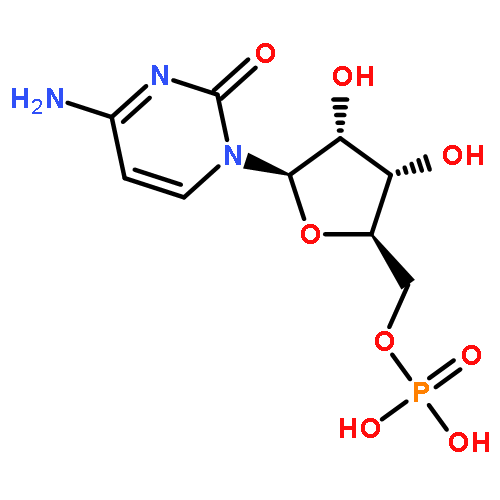
![Poly[imino[(1R)-1-(4-aminobutyl)-2-oxo-1,2-ethanediyl]]](http://img.cochemist.com/ccimg/27000/26913-90-6.png)
![Poly[imino[(1R)-1-(4-aminobutyl)-2-oxo-1,2-ethanediyl]]](http://img.cochemist.com/ccimg/27000/26913-90-6_b.png)


![[1,3]Dioxolo[4',5':4,5]benzo[1,2-c]phenanthridinium,2,3-dimethoxy-12-methyl-](http://img.cochemist.com/ccimg/6900/6872-57-7.png)
![[1,3]Dioxolo[4',5':4,5]benzo[1,2-c]phenanthridinium,2,3-dimethoxy-12-methyl-](http://img.cochemist.com/ccimg/6900/6872-57-7_b.png)


![Methyl (3s,4r)-3-benzoyloxy-8-methyl-8-azabicyclo[3.2.1]octane-4-carboxylate](http://img.cochemist.com/ccimg/100/50-36-2.png)
![Methyl (3s,4r)-3-benzoyloxy-8-methyl-8-azabicyclo[3.2.1]octane-4-carboxylate](http://img.cochemist.com/ccimg/100/50-36-2_b.png)