Co-reporter:Shujie Liu, Bing Jia, Ruirui Qiao, Zhi Yang, Zilin Yu, Zhaofei Liu, Kan Liu, Jiyun Shi, Han Ouyang, Fan Wang and Mingyuan Gao
Molecular Pharmaceutics August 3, 2009 Volume 6(Issue 4) pp:
Publication Date(Web):June 15, 2009
DOI:10.1021/mp900143a
A novel dual-modality molecular probe composed of biocompatible Fe3O4 nanocrystal, monoclonal antibody and radionuclide was designed and prepared. All functional components in the dual-modality molecular probe, i.e., Fe3O4, PEG, mAb 3H11 and 125I, were chemically bonded together for forming a stable molecular probe. Systematic in vitro experiments were carried out for evaluating the biological activity of the antibody in the targeting probe. A series of in vivo experiments were performed based on the dual-modality imaging probe for detecting xenografted tumors in nude mice by MRI and γ-imaging techniques. The pharmacokinetics of the dual-modality molecular probe in tumor-bearing nude mice was studied.Keywords: Dual-modality molecular probe; Fe3O4 nanocrystals; MRI; nuclear imaging; tumor;
Co-reporter:Zhenyu Gao;Yi Hou;Jianfeng Zeng;Lei Chen;Chunyan Liu;Wensheng Yang
Advanced Materials 2017 Volume 29(Issue 24) pp:
Publication Date(Web):2017/06/01
DOI:10.1002/adma.201701095
A tumor microenvironment responsive nanoprobe is developed for enhanced tumor imaging through in situ crosslinking of the Fe3O4 nanoparticles modified with a responsive peptide sequence in which a tumor-specific Arg-Gly-Asp peptide for tumor targeting and a self-peptide as a “mark of self” are linked through a disulfide bond. Positioning the self-peptide at the outmost layer is aimed at delaying the clearance of the nanoparticles from the bloodstream. After the self-peptide is cleaved by glutathione within tumor microenvironment, the exposed thiol groups react with the remaining maleimide moieties from adjacent particles to crosslink the particles in situ. Both in vitro and in vivo experiments demonstrate that the aggregation substantially improves the magnetic resonance imaging (MRI) contrast enhancement performance of Fe3O4 particles. By labeling the responsive particle probe with 99mTc, single-photon emission computed tomography is enabled not only for verifying the enhanced imaging capacity of the crosslinked Fe3O4 particles, but also for achieving sensitive dual modality imaging of tumors in vivo. The novelty of the current probe lies in the combination of tumor microenvironment-triggered aggregation of Fe3O4 nanoparticles for boosting the T2 MRI effect, with antiphagocytosis surface coating, active targeting, and dual-modality imaging, which is never reported before.
Co-reporter:Mingxia Jiao;Lihong Jing;Xiaojun Wei;Chunyan Liu;Xiliang Luo
Nanoscale (2009-Present) 2017 vol. 9(Issue 47) pp:18609-18612
Publication Date(Web):2017/12/07
DOI:10.1039/C7NR06291E
The present study reports a continuous flow synthesis of differently sized Fe3O4 nanocrystals stabilized by oleylamine and oleic acid. Oleylamine and oleic acid are particularly investigated to elucidate their roles in tailoring the size and magnetic properties of the resulting particles potentially useful for magnetic resonance imaging.
Co-reporter:Stephen V. Kershaw;Lihong Jing;Xiaodan Huang;Andrey L. Rogach
Materials Horizons (2014-Present) 2017 vol. 4(Issue 2) pp:155-205
Publication Date(Web):2017/03/06
DOI:10.1039/C6MH00469E
Semiconductor nanocrystal quantum dots are at last starting to emerge in commercial applications such as flat panel displays. Meanwhile, they are also showing seriously attractive performance levels in other types of optoelectronic devices. This maturing has been driven to a large degree by a deep level of understanding of materials related aspects of semiconductor nanocrystals themselves and in combination with other materials in the form of composite films to build optoelectronic components. We examine the synthetic and post synthetic chemical strategies that have led to semiconductor quantum dot structures with robust and strong device performance, and review the current work-in-progress as well as longer term developments in the areas of photovoltaics and photoelectrochemical cells, photodetectors, solid state lighting, photocatalysts, and sensing applications.
Co-reporter:Ruirui Qiao, Hongyu Qiao, Yan Zhang, Yabin Wang, Chongwei Chi, Jie Tian, Lifang Zhang, Feng Cao, and Mingyuan Gao
ACS Nano 2017 Volume 11(Issue 2) pp:
Publication Date(Web):January 25, 2017
DOI:10.1021/acsnano.6b07842
Owing to the high mortality rate of cardiovascular diseases, developing novel noninvasive diagnostic methods becomes urgent and mandatory. It is well-known that the rupture of vulnerable plaques directly leads to deadly consequences. However, differentiating vulnerable plaques from stable plaques remains challenging in the clinic. In the current study, osteopontin (OPN), a secreted biomarker associated with macrophages and foamy macrophages, was selected as a target for identifying the vulnerable plaques. A dual modality imaging probe was constructed by covalently attaching an OPN antibody to NaGdF4:Yb,Er@NaGdF4 upconversion nanoparticles. Upon intravenous injection of the resulting probes, upconversion optical imaging was performed to visualize the plaques induced by altering the shear stress in carotid arteries of a mouse model. The imaging studies revealed that the signals of vulnerable and stable plagues induced by lowered shear stress and oscillatory shear stress, respectively, presented significantly different signal intensities, implying that the current probe and imaging strategy are potentially useful for a precise diagnosis of atherosclerosis plaques.Keywords: macrophage; molecular imaging; OPN; upconversion nanoparticle; vulnerable plaques;
Co-reporter:Lihong Jing, Stephen V. Kershaw, Yilin Li, Xiaodan Huang, Yingying Li, Andrey L. Rogach, and Mingyuan Gao
Chemical Reviews 2016 Volume 116(Issue 18) pp:10623-10730
Publication Date(Web):September 2, 2016
DOI:10.1021/acs.chemrev.6b00041
This review summarizes traditional and recent nonconventional, bioinspired, methods for the aqueous synthesis of colloidal semiconductor quantum dots (QDs). The basic chemistry concepts are critically emphasized at the very beginning as these are strongly correlated with the selection of ligands and the optimal formation of aqueous QDs and their more sophisticated structures. The synergies of biomimetic and biosynthetic methods that can combine biospecific reactivity with the robust and strong optical responses of QDs have also resulted in new approaches to the synthesis of the nanoparticles themselves. A related new avenue is the recent extension of QD synthesis to form nanoparticles endowed with chiral optical properties. The optical characteristics of QD materials and their advanced forms such as core/shell heterostructures, alloys, and doped QDs are discussed: from the design considerations of optical band gap tuning, the control and reduction of the impact of surface traps, the consideration of charge carrier processes that affect emission and energy and charge transfer, to the impact and influence of lattice strain. We also describe the considerable progress in some selected QD applications such as in bioimaging and theranostics. The review concludes with future strategies and identification of key challenges that still need to be resolved in reaching very attractive, scalable, yet versatile aqueous syntheses that may widen the scope of commercial applications for semiconductor nanocrystals.
Co-reporter:Ling Wen;Ling Chen;Shimin Zheng;Jianfeng Zeng;Guangxin Duan;Yong Wang;Guanglin Wang;Zhifang Chai;Zhen Li
Advanced Materials 2016 Volume 28( Issue 25) pp:5072-5079
Publication Date(Web):
DOI:10.1002/adma.201506428
Co-reporter:Xiuyu Wang; Yi Hou; Li Yao; Mingyuan Gao;Maofa Ge
Journal of the American Chemical Society 2016 Volume 138(Issue 7) pp:2090-2093
Publication Date(Web):February 2, 2016
DOI:10.1021/jacs.5b12149
Hierarchically structured magnetic single-hole hollow spheres (MSHS) have been successfully obtained via a facile self-assembly strategy. This methodology allows the double emulsions generated via the combined effect of self-emulsification and phase separation to provide confinement for directing the self-assembly of magnetic nanoparticles (MNPs). The resulting MSHS fully capitalize on both the multifunctional properties of MNPs and container features of single-hole hollow spheres. Moreover, the magnetic properties showed obvious improvement and can be tuned by modulating the assembled structure. Thus, MSHS can be used as a smart platform with multiple functionalities including image contrast enhancement, selective encapsulation for biomacromolecules, on-demand release, and magnetically guided transport. This strategy is very promising in the design of hierarchically structured assemblies for desired applications in biomedicine and other fields.
Co-reporter:Yong Wang;Yongyou Wu;Yujing Liu;Jia Shen;Ling Lv;Liubing Li;Liecheng Yang;Jianfeng Zeng;Yangyun Wang;Leshuai W. Zhang;Zhen Li;Zhifang Chai
Advanced Functional Materials 2016 Volume 26( Issue 29) pp:5335-5344
Publication Date(Web):
DOI:10.1002/adfm.201601341
Fabrication of ultrasmall single-component omnipotent nanotheranostic agents integrated with multimodal imaging and multiple therapeutic functions becomes more and more practically relevant but challenging. In this article, sub 10 nm Bi2S3 biocompatible particles are prepared through a bovine serum albumin (BSA)-mediated biomineralization process under ambient aqueous conditions. Owing to the ultrasmall size and colloidal stability, the resulting nanoparticles (NPs) present outstanding blood circulation behavior and excellent tumor targeting ability. Toward theranostic applications, the biosafety profile is carefully investigated. In addition, photothermal conversion is characterized for both photoacoustic imaging and photothermal treatment of cancers. Upon radiolabeling, the performance of the resulting particles for SPECT/CT imaging in vivo is also carried out. Additionally, different combinations of treatments are applied for evaluating the performance of the as-prepared Bi2S3 NPs in photothermal- and radiotherapy of tumors. Due to the remarkable photothermal conversion efficiency and large X-ray attenuation coefficient, the implanted tumors are completely eradicated through combined therapies, which highlights the potential of BSA-capped Bi2S3 NPs as a novel multifunctional nanotheranostic agent.
Co-reporter:Chunyan Liu, Yifei Qi, Ruirui Qiao, Yi Hou, Kaying Chan, Ziqian Li, Jiayi Huang, Lihong Jing, Jun Du and Mingyuan Gao
Nanoscale 2016 vol. 8(Issue 25) pp:12579-12587
Publication Date(Web):30 Nov 2015
DOI:10.1039/C5NR07858J
Early detection and diagnosis of cancers is extremely beneficial for improving the survival rate of cancer patients and molecular imaging techniques are believed to be relevant for offering clinical solutions. Towards early cancer detection, we developed a primary animal colorectal cancer model and constructed a tumor-specific imaging probe by using biocompatible NaGdF4:Yb,Er@NaGdF4 upconversion luminescent NPs for establishing a sensitive early tumor imaging method. The primary animal tumor model, which can better mimic the human colorectal cancer, was built upon continual administration of 1,2-dimethylhydrazine in Kunming mice and the tumor development was carefully monitored through histopathological and immunohistochemical analyses to reveal the pathophysiological processes and molecular features of the cancer microenvironment. The upconversion imaging probe was constructed through covalent coupling of PEGylated core–shell NPs with folic acid whose receptor is highly expressed in the primary tumors. Upon 980 nm laser excitation, the primary colorectal tumors in the complex abdominal environment were sensitively imaged owing to the ultralow background of the upconversion luminescence and the high tumor-targeting specificity of the nanoprobe. We believe that the current studies provide a highly effective and potential approach for early colorectal cancer diagnosis and tumor surgical navigation.
Co-reporter:Mingxia Jiao, Lihong Jing, Chunyan Liu, Yi Hou, Jiayi Huang, Xiaojun Wei and Mingyuan Gao
Chemical Communications 2016 vol. 52(Issue 34) pp:5872-5875
Publication Date(Web):22 Mar 2016
DOI:10.1039/C6CC01686C
The present study reports a novel approach for synthesizing differently sized magnetic/upconversion luminescent NaGdF4:Yb,Er nanocrystals through flow chemistry. The solvent effects on the control of particle size were particularly investigated for tailoring the size and physical properties of the resulting particles potentially useful for bioimaging.
Co-reporter:Jianfeng Zeng;Ming Cheng;Yong Wang;Ling Wen;Ling Chen;Zhen Li;Yongyou Wu;Zhifang Chai
Advanced Healthcare Materials 2016 Volume 5( Issue 7) pp:772-780
Publication Date(Web):
DOI:10.1002/adhm.201500898
Co-reporter:Lihong Jing; Stephen V. Kershaw; Tobias Kipp; Sergii Kalytchuk; Ke Ding; Jianfeng Zeng; Mingxia Jiao; Xiaoyu Sun; Alf Mews; Andrey L. Rogach
Journal of the American Chemical Society 2015 Volume 137(Issue 5) pp:2073-2084
Publication Date(Web):January 16, 2015
DOI:10.1021/ja5127352
The impact of strain on the optical properties of semiconductor quantum dots (QDs) is fundamentally important while still awaiting detailed investigation. CdTe/CdS core/shell QDs represent a typical strained system due to the substantial lattice mismatch between CdTe and CdS. To probe the strain-related effects, aqueous CdTe/CdS QDs were synthesized by coating different sized CdTe QD cores with CdS shells upon the thermal decomposition of glutathione as a sulfur source under reflux. The shell growth was carefully monitored by both steady-state absorption and fluorescence spectroscopy and transient fluorescence spectroscopy. In combination with structural analysis, the band alignments as a consequence of the strain were modified based on band deformation potential theory. By further taking account of these strain-induced band shifts, the effective mass approximation (EMA) model was modified to simulate the electronic structure, carrier spatial localization, and electron–hole wave function overlap for comparing with experimentally derived results. In particular, the electron/hole eigen energies were predicted for a range of structures with different CdTe core sizes and different CdS shell thicknesses. The overlap of electron and hole wave functions was further simulated to reveal the impact of strain on the electron–hole recombination kinetics as the electron wave function progressively shifts into the CdS shell region while the hole wave function remains heavily localized in CdTe core upon the shell growth. The excellent agreement between the strain-modified EMA model with the experimental data suggests that strain exhibits remarkable effects on the optical properties of mismatched core/shell QDs by altering the electronic structure of the system.
Co-reporter:Jiayi Huang, Yi Hou, Chunyan Liu, Lihong Jing, Tiancong Ma, Xiaodan Sun, and Mingyuan Gao
Chemistry of Materials 2015 Volume 27(Issue 23) pp:7918
Publication Date(Web):November 6, 2015
DOI:10.1021/acs.chemmater.5b02875
The development of T1/T2 dual-modality contrast agents for magnetic resonance imaging (MRI) is beneficial for eliminating artifacts. However, T1 and T2 agents in the case of direct contact will interfere with each other, and the intrinsic mechanism remains unclear yet. Gadolinium(III) is widely used as T1 contrast agents, while dysprosium(III) exhibits the strongest magnetic moment among rare earth (RE) elements due to the spin–orbital coupling and can potentially be used as T2 contrast agents. The very similar chemical properties of RE elements allow integration of Gd(III) and Dy(III) within single nanocrystals for investigating the interactions between T1 and T2 components so as to achieve T1/T2 dual-modality contrast agents with optimized performance. Therefore, core–shell–shell structured NaDyF4@NaREF4@NaGdF4:Yb,Er nanocrystals were prepared via a seed-mediated growth approach and Gd(III), Er(III), and Y(III) were chosen to construct NaREF4 spacer layer for mediating the interactions between Dy(III) and Gd(III) ions. It was found out that, with the decrease of the electron cloud distortion ability from Gd(III) to Y(III), the PEGylated core–shell–shell particles present increasing longitudinal relaxivity values from 0.50 to 1.82 mM–1 s–1. On the basis of the longitudinal relaxivity theory and polarizability of different RE elements, the above tendency can be interpreted by the distortion tendency of the electron cloud of RE(III) in the spacer layer which strongly mediates the interactions between electron clouds of Dy(III) and Gd(III) ions. Further upconversion luminescence spectroscopy study in combination with the relaxivity measurements on nanocrystals with thicker spacer (3.5 vs 1.9 nm) also supported the above explanation.
Co-reporter:Mingxia Jiao, Jianfeng Zeng, Lihong Jing, Chunyan Liu, and Mingyuan Gao
Chemistry of Materials 2015 Volume 27(Issue 4) pp:1299
Publication Date(Web):February 2, 2015
DOI:10.1021/cm504313c
PEGylated Fe3O4 nanoparticles were prepared through flow synthesis upon the pyrolysis of ferric acetylacetonate (Fe(acac)3) in anisole at 250 °C under pressure of 33 bar, in the presence of α,ω-dicarboxyl-terminated polyethylene glycol (HOOC–PEG–COOH) and oleylamine. In combination with theoretical analysis, the effects of linear velocity, residence time, and reactor dimension on particle size distribution were systematically investigated. In addition, the impact of Ostwald ripening on particle size distribution was also revealed. In particular, the impacts of monomer concentration distributions along both axial and radial directions of the tube reactor on the particle size distribution were carefully investigated. Under optimized conditions, PEGylated Fe3O4 nanoparticles with the relative standard deviation of particle size down to 10.6% were thus obtained. The resulting 4.6 nm particles exhibited excellent colloidal stability and high longitudinal relaxivity (r1) up to 11.1 mM–1·s–1, which manifested the reliability of flow synthesis in preparing PEGylated Fe3O4 nanoparticles as contrast agents for magnetic resonance imaging applications.
Co-reporter:Ke Ding, Jianfeng Zeng, Lihong Jing, Ruirui Qiao, Chunyan Liu, Mingxia Jiao, Zhen Li and Mingyuan Gao
Nanoscale 2015 vol. 7(Issue 25) pp:11075-11081
Publication Date(Web):15 May 2015
DOI:10.1039/C5NR02180D
By integrating high imaging sensitivity and high resolution in a single modality, photoacoustic (PA) imaging emerges as a promising diagnostic tool for clinical applications. Benefiting from the absorption in the near-infrared region (NIR), copper sulfide nanoparticles (NPs) as a contrast agent are potentially useful for increasing the sensitivity of PA imaging. However, the aqueous synthesis of size-tunable, biocompatible and colloidally stable copper sulfide NPs remains challenging due to the intrinsic dipole–dipole interactions among particles. In this work, aqueous synthesis of PEGylated copper sulfide NPs with controllable size between 3 and 7 nm was developed. The particle size-dependent contrast enhancement effect of the copper sulfide NPs for PA imaging was carefully studied both in vitro and in vivo. Although the contrast enhancement effect of the copper sulfide NPs is proportional to particle size, the in vivo studies revealed that copper sulfide NPs smaller than 5 nm presented higher tumor imaging performance, especially at the tumor boundary site, which was further discussed in combination with the pharmacokinetic behaviors of differently sized particles.
Co-reporter:Ruirui Qiao, Changhao Liu, Muhan Liu, Hao Hu, Chunyan Liu, Yi Hou, Kaichun Wu, Yenan Lin, Jimin Liang, and Mingyuan Gao
ACS Nano 2015 Volume 9(Issue 2) pp:2120
Publication Date(Web):January 20, 2015
DOI:10.1021/nn507433p
Lymphatic metastasis is an important prognostic factor regarding long-term survival rate of gastric cancer (GC) patients. Pretreatment knowledge of lymph node status is extremely helpful for planning treatment and prognosis. However, to date, no imaging method has been demonstrated to be effective for detecting lymphatic metastasis in GC. Molecular imaging probes based on upconversion nanoparticles with unique optical and magnetic properties have provided great hope for early tumor detection. Herein we report highly sensitive detection of lymphatic spread using core@shell structured NaGdF4:Yb,Er@NaGdF4 upconversion nanoparticles coated with polyethylene glycol (PEG). A GC-specific probe was constructed through “click” reaction between the maleimide moiety of PEG ligand and the thiol group from partly reduced antigastric cancer antibody MGb2. The primary tumor and adjacent lymphatic metastasis site were clearly differentiated by upconversion luminescence imaging after the GC-specific probe was delivered through tail vein injection into tumor-bearing mice. Moreover, lymphatic metastases smaller than 1 mm were successfully detected, owing to the ultralow background under 980 nm excitation. It has been demonstrated that both primary and lymphatic metastatic sites in an orthotopic mouse model of human gastric cancer can be optically detected by using GC-specific upconversion luminescence nanoprobes. The current studies may therefore provide a highly effective approach for GC diagnosis.Keywords: gastric cancer; lymphatic metastasis; molecular imaging; ultrasensitive detection; upconversion nanoparticles;
Co-reporter:Yi Hou, Jin Zhou, Zhenyu Gao, Xiaoyu Sun, Chunyan Liu, Dihua Shangguan, Wensheng Yang, and Mingyuan Gao
ACS Nano 2015 Volume 9(Issue 3) pp:3199
Publication Date(Web):February 11, 2015
DOI:10.1021/acsnano.5b00276
A protease-activated ratiometric fluorescent probe based on fluorescence resonance energy transfer between a pH-sensitive fluorescent dye and biocompatible Fe3O4 nanocrystals was constructed. A peptide substrate of MMP-9 served as a linker between the particle quencher and the chromophore that was covalently attached to the antitumor antibody. The optical response of the probe to activated MMP-9 and gastric cell line SGC7901 tumor cells was investigated, followed by in vivo tumor imaging. Based on the ratiometric pH response to the tumor microenvironment, the resulting probe was successfully used to image the pH of subcutaneous tumor xenografts.Keywords: Fe3O4 nanocrystals; pH mapping; protease-activated; ratiometric fluorescence; smart probe;
Co-reporter:Lihong Jing;Ke Ding;Stephen V. Kershaw;Ivan M. Kempson;Andrey L. Rogach
Advanced Materials 2014 Volume 26( Issue 37) pp:6367-6386
Publication Date(Web):
DOI:10.1002/adma.201402296
Light-emitting semiconductor quantum dots (QDs) combined with magnetic resonance imaging contrast agents within a single nanoparticle platform are considered to perform as multimodal imaging probes in biomedical research and related clinical applications. The principles of their rational design are outlined and contemporary synthetic strategies are reviewed (heterocrystalline growth; co-encapsulation or assembly of preformed QDs and magnetic nanoparticles; conjugation of magnetic chelates onto QDs; and doping of QDs with transition metal ions), identifying the strengths and weaknesses of different approaches. Some of the opportunities and benefits that arise through in vivo imaging using these dual-mode probes are highlighted where tumor location and delineation is demonstrated in both MRI and fluorescence modality. Work on the toxicological assessments of QD/magnetic nanoparticles is also reviewed, along with progress in reducing their toxicological side effects for eventual clinical use. The review concludes with an outlook for future biomedical imaging and the identification of key challenges in reaching clinical applications.
Co-reporter:Jianfeng Zeng;Lihong Jing;Yi Hou;Mingxia Jiao;Ruirui Qiao;Qiaojuan Jia;Chunyan Liu;Fang Fang;Hao Lei
Advanced Materials 2014 Volume 26( Issue 17) pp:2694-2698
Publication Date(Web):
DOI:10.1002/adma.201304744
Co-reporter:Chunyan Liu;Yi Hou
Advanced Materials 2014 Volume 26( Issue 40) pp:6922-6932
Publication Date(Web):
DOI:10.1002/adma.201305535
Rare earth (RE) nanoparticles have attracted considerable attention due to their unique optical and magnetic properties associated with f-electrons. The recent accomplishments in RE nanoparticle synthesis have aroused great interest of scientists to further explore their biomedical applications. This Research News summarizes recent achievements in controlled synthesis of magnetic and luminescent RE nanoparticles, surface modification, and toxicity studies of RE nanomaterials, and highlights state-of-the-art in in vivo applications of RE nanoparticles.
Co-reporter:Hongwei Zhang, Lihong Jing, Jianfeng Zeng, Yi Hou, Zhen Li and Mingyuan Gao
Nanoscale 2014 vol. 6(Issue 11) pp:5918-5925
Publication Date(Web):02 Apr 2014
DOI:10.1039/C4NR00761A
By pyrolyzing manganese(II) acetate in 1-octadecene in the presence of oleylamine and oleic acid, manganese oxide nanocrystals were prepared. It was observed that both MnO and Mn3O4 nanocrystals were simultaneously formed by quickly heating the reaction mixture up to 250 °C, while a preheating procedure carried out at 100 °C led to uniform MnO nanocubes that developed into eight-arm MnO nanocrystals upon prolonged reaction. To understand the mechanisms for forming these two different kinds of manganese oxide nanocrystals, i.e., Mn3O4 and MnO, the coordination between oleic acid/oleylamine and Mn2+ was investigated. The detailed investigations suggest that Mn2+–oleylamine coordination is kinetically driven and favorable for the formation of Mn3O4 nanocrystals due to the relatively low electronegativity of N from oleylamine, while Mn2+–oleate coordination is thermodynamically driven and can prevent the central metal ion (Mn2+) from being oxidized owing to the relatively high electronegativity of O from the oleate ligand. Following these new insights, by properly balancing the coordination of oleic acid and oleylamine to Mn2+, the selective synthesis of MnO and Mn3O4 nanocrystals with uniform shapes was successfully achieved.
Co-reporter:Chunyan Liu, Wei Ma, Zhenyu Gao, Jiayi Huang, Yi Hou, Chuanlai Xu, Wensheng Yang and Mingyuan Gao
Journal of Materials Chemistry A 2014 vol. 2(Issue 45) pp:9637-9642
Publication Date(Web):30 Sep 2014
DOI:10.1039/C4TC02034K
Because of the near infrared excitation, upconversion luminescence (UCL) rare-earth nanoparticles are very suitable for biological applications in terms of low background interference. NaGdF4:Yb,Er nanoparticles were firstly synthesized through a replacement reaction at high temperature. To improve the upconversion luminescence efficiency, core–shell NaGdF4:Yb,Er@NaGdF4 nanoparticles were then prepared by a seed-mediated method. The core–shell architecture significantly improved the upconversion luminescence and more effectively retained the luminescence during the following phase transfer process by ligand exchange. Upon further coupling with anti-cephalexin monoclonal antibody via “click” reaction, the resultant upconversion luminescence probe was obtained and used in a lateral flow immunochromatographic assay (LFIA) to detect the antibiotic residue of cephalexin. The results were compared with those achieved by a nanoprobe assay based on gold nanoparticles. More quantitative results were extracted by luminescent intensity analysis. Under optimized conditions, the detection limit of UCL nanoparticles-based LFIA was considerably comparable with gold nanoparticles-based LFIA.
Co-reporter:Jianfeng Zeng, Bing Jia, Ruirui Qiao, Chao Wang, Lihong Jing, Fan Wang and Mingyuan Gao
Chemical Communications 2014 vol. 50(Issue 17) pp:2170-2172
Publication Date(Web):02 Jan 2014
DOI:10.1039/C3CC48948E
The present study reports a new approach for synthesizing 111In-radiolabeled biocompatible Fe3O4 nanoparticles. Radioactive 111In is doped in situ into the lattice of Fe3O4 nanoparticles to achieve robust radiolabeling for accurately tracing PEGylated Fe3O4 particles in vivo.
Co-reporter:Yilin Li, Lihong Jing, Ke Ding, Jing Gao, Zhi Peng, Yanyan Li, Lin Shen and Mingyuan Gao
RSC Advances 2014 vol. 4(Issue 43) pp:22545-22550
Publication Date(Web):13 May 2014
DOI:10.1039/C4RA02277G
By using denatured bovine serum albumin (dBSA) molecules as multidentate ligands, surface-functionalized CdTe quantum dots (QDs) were synthesized and further evaluated for preparing fluorescent probes capable of detecting Epstein–Barr virus infection. The optical studies revealed that the dBSA coating effectively improved the optical stability of the resulting QDs (CdTe@dBSA). The interaction between CdTe@dBSA and the membrane of carcinoma cells further suggested that the nonspecific adsorption of CdTe QDs stabilized by thioglycolic acid (TGA) was significantly eliminated upon the dBSA coating. Streptavidin was covalently conjugated to CdTe@dBSA to endow the QDs with specific binding affinity in detecting the serum level of anti-Epstein–Barr virus (EBV) capsid antigen IgA (VCA-IgA) in nasopharyngeal carcinoma patients for early screening and diagnosing EBV-associated cancers.
Co-reporter:Ke Ding, Lihong Jing, Chunyan Liu, Yi Hou, Mingyuan Gao
Biomaterials 2014 35(5) pp: 1608-1617
Publication Date(Web):
DOI:10.1016/j.biomaterials.2013.10.078
Co-reporter:Wentao Wang, Yuchun Han, Maozhang Tian, Yaxun Fan, Yongqiang Tang, Mingyuan Gao, and Yilin Wang
ACS Applied Materials & Interfaces 2013 Volume 5(Issue 12) pp:5709
Publication Date(Web):May 31, 2013
DOI:10.1021/am4011226
A novel synthetic approach was developed for creating versatile hollow Au nanostructures by stepwise reductions of AuIII upon the use of cationic gemini surfactant hexamethylene-1,6-bis(dodecyl dimethylammonium bromide) (C12C6C12Br2) as a template agent. It was observed that the AuI ions obtained from the reduction of AuIII by ascorbic acid can assist the gemini surfactant to form vesicles, capsule-like, and tube-like aggregates that subsequently act as soft templates for hollow Au nanostructures upon further reduction of AuI to Au0 by NaBH4. It was demonstrated that the combination of C12C6C12Br2 and AuI plays a key role in regulating the structure of the hollow precursors not only because C12C6C12Br2 has a stronger aggregation ability in comparison with its single chain counterpart but also because the electrostatic repulsion between head groups of C12C6C12Br2 is greatly weakened after AuIII is converted to AuI, which is in favor of the construction of vesicles, capsule-like, and tube-like aggregates. Compared with solid Au nanospheres, the resultant hollow nanostructures exhibit enhanced electrocatalytic activities in methanol oxidation, following the order of elongated nanocapsule > nanocapsule > nanosphere. Benefiting from balanced interactions between the gemini surfactant and AuI, this soft-template method may present a facile and versatile approach for the controlled synthesis of Au nanostructures potentially useful for fuel cells and other Au nanodevices.Keywords: electrocatalytic oxidation; gemini surfactant; hollow Au nanostructures; stepwise reduction; template;
Co-reporter:Wentao Wang, Yuchun Han, Mingyuan Gao and Yilin Wang
CrystEngComm 2013 vol. 15(Issue 14) pp:2648-2656
Publication Date(Web):17 Jan 2013
DOI:10.1039/C3CE26527G
Snowflake-like two-dimensional highly branched gold nanostructures (2DHBNs) have been constructed in the presence of hexamethylene-1,6-bis (dodecyl dimethylammonium bromide) (C12C6C12Br2) through the reduction of HAuCl4 by ascorbic acid. High resolution transmission electron microscopy results indicate that the 2DHBNs are uniform in crystallographic orientation, where the branched nanostructures grow along the <211> direction and present multiple twin planes stacked along the <111> direction. An ex situ kinetic study, followed by TEM observations, shows that the 2DHBNs are evolved from triangular nanoprisms through overgrowth along the <211> direction. The gemini surfactant C12C6C12Br2 is found to be the most essential factor for the formation of the 2DHBNs. C12C6C12Br2 may selectively absorb onto the (111) planes, which promotes the construction of 2D nanostructures. Moreover, C12C6C12Br2 can act as an excellent kinetic modifier to control the growth of the nanostructures, which also affects the generation of branched nanostructures. The as-prepared 2DHBNs exhibit an efficient surface-enhanced Raman scattering (SERS) property which has potential application in constructing sensitive SERS substrates.
Co-reporter:Lihong Jing, Ke Ding, Sergii Kalytchuk, Yu Wang, Ruirui Qiao, Stephen V. Kershaw, Andrey L. Rogach, and Mingyuan Gao
The Journal of Physical Chemistry C 2013 Volume 117(Issue 36) pp:18752-18761
Publication Date(Web):August 28, 2013
DOI:10.1021/jp406342m
Core/shell CdTe/ZnS colloidal quantum dots with varying dopant levels (4.7–9.7%) of paramagnetic manganese ions spatially distributed within the thin ZnS shell are synthesized by the aqueous approach. They exhibit both strong fluorescence originating from the CdTe core (up to 45% room temperature emission quantum yield) and high ionic relaxivity in the range of 10.7–5.4 mM–1 s–1, which render them promising dual fluorescent/paramagnetic probes.
Co-reporter:Xiaoyu Sun ; Ke Ding ; Yi Hou ; Zhenyu Gao ; Wensheng Yang ; Lihong Jing
The Journal of Physical Chemistry C 2013 Volume 117(Issue 40) pp:21014-21020
Publication Date(Web):September 11, 2013
DOI:10.1021/jp408036p
Monodispersed superparticles (SPs) consisting of hydrophobic CuInS2@ZnS quantum dots (QDs) were prepared through a self-assembling process induced by introducing ethanol into the cyclohexane solution of the QDs stabilized by 1-dodecanethiol. The solvent polarity-dependent size and size distribution of the resultant SPs were carefully characterized by using scanning electron microscopy, transmission electron microscopy, and dynamic light scattering. The formation and the growth mechanism of the SPs were investigated. By manipulating the nucleation and the growth kinetics through the addition rate of ethanol, the size was effectively tuned without broadening the size distribution of the SPs. By overcoating the SPs with amphibious Fe3O4 nanocrystals (NCs) stabilized by poly(4-vinylpyrollidone), aqueous dispersible SPs were obtained. Inheriting the fluorescent and magnetic properties from the corresponding mother particles, the resultant SPs presented excellent fluorescent and magnetic properties and, thus, may potentially be useful for bioseparation, detection, etc.
Co-reporter:Yi Hou, Ruirui Qiao, Fang Fang, Xuxia Wang, Chengyan Dong, Kan Liu, Chunyan Liu, Zhaofei Liu, Hao Lei, Fan Wang, and Mingyuan Gao
ACS Nano 2013 Volume 7(Issue 1) pp:330
Publication Date(Web):November 30, 2012
DOI:10.1021/nn304837c
Differently sized NaGdF4 nanocrystals with narrow particle size distributions were synthesized by a high temperature approach. Upon ligand exchange, the as-prepared hydrophobic NaGdF4 nanocrystals were transferred into water by using asymmetric PEGs simultaneously bearing phosphate and maleimide groups. Further investigations demonstrated that the water-soluble NaGdF4 nanocrystals, coated by PEG bearing two phosphate groups on the same side, exhibit not only excellent colloidal stability in water and PBS buffer, but also higher T1 relaxivity than Gd-DTPA (Magnevist). Through “click” reaction between the maleimide residue on particle surface and thiol group from the partly reduced anti-EGFR monoclonal antibody (mAb), NaGdF4–PEG-mAb nanoprobes were constructed, and their biocompatibility and binding specificity were evaluated through in vitro experiments. A series of in vivo experiments were then carried out for detecting intraperitoneal tumor xenografts in nude mice by using magnetic resonance (MR) imaging technique. The results revealed that the NaGdF4–PEG-mAb probes possessed satisfying tumor-specific targeting ability and strong MR contrast enhancement effects.Keywords: intraperitoneal tumor xenografts; ligand exchange; MR molecular imaging probe; NaGdF4 nanoparticles; T1 contrast agent
Co-reporter:Chunyan Liu, Zhenyu Gao, Jianfeng Zeng, Yi Hou, Fang Fang, Yilin Li, Ruirui Qiao, Lin Shen, Hao Lei, Wensheng Yang, and Mingyuan Gao
ACS Nano 2013 Volume 7(Issue 8) pp:7227
Publication Date(Web):July 23, 2013
DOI:10.1021/nn4030898
Detection of early malignant tumors remains clinically difficult; developing ultrasensitive imaging agents is therefore highly demanded. Owing to the unusual magnetic and optical properties associated with f-electrons, rare-earth elements are very suitable for creating functional materials potentially useful for tumor imaging. Nanometer-sized particles offer such a platform with which versatile unique properties of the rare-earth elements can be integrated. Yet the development of rare-earth nanoparticle-based tumor probes suitable for imaging tiny tumors in vivo remains difficult, which challenges not only the physical properties of the nanoparticles but also the rationality of the probe design. Here we report new approaches for size control synthesis of magnetic/upconversion fluorescent NaGdF4:Yb,Er nanocrystals and their applications for imaging tiny tumors in vivo. By independently varying F–:Ln3+ and Na+:Ln3+ ratios, the size and shape regulation mechanisms were investigated. By replacing the oleic acid ligand with PEG2000 bearing a maleimide group at one end and two phosphate groups at the other end, PEGylated NaGdF4:Yb,Er nanoparticles with optimized size and upconversion fluorescence were obtained. Accordingly, a dual-modality molecular tumor probe was prepared, as a proof of concept, by covalently attaching antitumor antibody to PEGylated NaGdF4:Yb,Er nanoparticles through a “click” reaction. Systematic investigations on tumor detections, through magnetic resonance imaging and upconversion fluorescence imaging, were carried out to image intraperitoneal tumors and subcutaneous tumors in vivo. Owing to the excellent properties of the molecular probes, tumors smaller than 2 mm was successfully imaged in vivo. In addition, pharmacokinetic studies on differently sized particles were performed to disclose the particle size dependent biodistributions and elimination pathways.Keywords: dual-modal molecular probes; in vivo tumor imaging; magnetic/upconversion fluorescent nanoparticles; pharmacokinetics; tiny tumor
Co-reporter:Enyu Zhao, Zhixia Zhao, Jiancheng Wang, Chunhui Yang, Chengjun Chen, Lingyan Gao, Qiang Feng, Wenjie Hou, Mingyuan Gao and Qiang Zhang
Nanoscale 2012 vol. 4(Issue 16) pp:5102-5109
Publication Date(Web):13 Jun 2012
DOI:10.1039/C2NR31290E
Cellular uptake, endosomal/lysosomal escape, and the effective dissociation from the carrier are a series of hurdles for specific genes to be delivered both in vitro and in vivo. To construct siRNA delivery systems, poly(allylamine hydrochloride) (PAH) and siRNA were alternately assembled on the surface of 11.8 ± 0.9 nm Au nanoparticles (GNP), stabilized by denatured bovine serum albumin, by the ionic layer-by-layer (LbL) self-assembly method. By manipulating the outmost PAH layer, GNP–PAH vectors with different surface electric potentials were prepared. Then, the surface potential-dependent cytotoxicity of the resultant GNP–PAH particles was evaluated via sulforhodamine B (SRB) assay, while the surface potential-dependent cellular uptake efficiency was quantitatively analyzed by using the flow cytometry method based on carboxyfluorescein (FAM)-labeled siRNA. It was revealed that the GNP–PAH particles with surface potential of +25 mV exhibited the optimal cellular uptake efficiency and cytotoxicity for human breast cancer MCF-7 cells. Following these results, two more positively charged polyelectrolytes with different protonating abilities in comparison with PAH, i.e., polyethylenimine (PEI), and poly(diallyl dimethyl ammonium chloride) (PDDA), were chosen to fabricate similarly structured vectors. Confocal fluorescence microscopy studies indicated that siRNA delivered by GNP–PAH and GNP–PEI systems was better released than that delivered by the GNP–PDDA system. Further flow cytometric assays based on immunofluorescence staining of the epidermal growth factor receptor (EGFR) revealed that EGFR siRNA delivered by GNP–PAH and GNP–PEI exhibited similar down-regulation effects on EGFR expression in MCF-7 cells. The following dual fluorescence flow cytometry assays by co-staining phosphatidylserine and DNA suggested the EGFR siRNA delivered by GNP–PAH exhibited an improved silencing effect in comparison with that delivered by the commercial transfection reagent Lipofectamine 2000.
Co-reporter:Luoxin Yi, Dan Wang and Mingyuan Gao
CrystEngComm 2012 vol. 14(Issue 2) pp:401-404
Publication Date(Web):28 Nov 2011
DOI:10.1039/C1CE06153D
Orthorhombic Cu3SnS4 nano-cuboids and nanosheets were synthesized by using monoclinic Cu31S16 nanocrystals as seeds. Transmission electron microscopy and powder X-ray diffractometry were adopted to investigate the resultant nanocrystals and the structural transformation of monoclinic Cu31S16 to orthorhombic Cu3SnS4.
Co-reporter:Ruirui Qiao, Qiaojuan Jia, Sabine Hüwel, Rui Xia, Ting Liu, Fabao Gao, Hans-Joachim Galla, and Mingyuan Gao
ACS Nano 2012 Volume 6(Issue 4) pp:3304
Publication Date(Web):March 25, 2012
DOI:10.1021/nn300240p
A brain delivery probe was prepared by covalently conjugating lactoferrin (Lf) to a poly(ethylene glycol) (PEG)-coated Fe3O4 nanoparticle in order to facilitate the transport of the nanoparticles across the blood–brain barrier (BBB) by receptor-mediated transcytosis via the Lf receptor present on cerebral endothelial cells. The efficacy of the Fe3O4-Lf conjugate to cross the BBB was evaluated in vitro using a cell culture model for the blood–brain barrier as well as in vivo in SD rats. For an in vitro experiment, a well-established porcine BBB model was used based on the primary culture of cerebral capillary endothelial cells grown on filter supports, thus allowing one to follow the transfer of nanoparticles from the apical (blood) to the basolateral (brain) side. For in vivo experiments, SD rats were used as animal model to detect the passage of the nanoparticles through the BBB by MRI techniques. The results of both in vitro and in vivo experiments revealed that the Fe3O4-Lf probe exhibited an enhanced ability to cross the BBB in comparison to the PEG-coated Fe3O4 nanoparticles and further suggested that the Lf-receptor-mediated transcytosis was an effective measure for delivering the nanoparticles across the BBB.Keywords: blood−brain barrier; lactoferrin; magnetic nanoparticle; MRI; receptor-mediated
Co-reporter:Qiaojuan Jia ; Jianfeng Zeng ; Ruirui Qiao ; Lihong Jing ; Liang Peng ; Fenglong Gu
Journal of the American Chemical Society 2011 Volume 133(Issue 48) pp:19512-19523
Publication Date(Web):October 27, 2011
DOI:10.1021/ja2081263
Biocompatible Fe3O4 nanocrystals were synthesized through the pyrolysis of ferric acetylacetonate (Fe(acac)3) in diphenyl oxide, in the presence of α,ω-dicarboxyl-terminated polyethylene glycol (HOOC–PEG–COOH) and oleylamine. Unusual gelification phenomena were observed from the aliquots extracted at different reaction stages after they were cooled to room temperature. By reaction time, the average size of the Fe3O4 nanocrystals was tuned from 5.8 to 11.7 nm with an equilibrium size around 11.3 nm. By increasing the gelification degree of the stock solution, the equilibrium size of the Fe3O4 nanocrystals was further increased from 11.3 to 18.9 nm. The underlying gel formation mechanism was investigated by using ultraviolet–visible absorption spectroscopy and Fourier transform infrared spectroscopy. The results suggest that the complexation between HOOC–PEG–COOH and Fe(acac)3, with the help of oleylamine, results in large molecular networks, which are responsible for the gelification of the stock solution, while the interaction between the fragment of the molecular network and Fe3O4 nanocrystal is responsible for the second gelification process observed during the early stage of reflux. To further investigate the particle growth behavior, small molecules released during the preparation were collected and analyzed by using photoelectron spectroscopy/photoionization mass spectroscopy (PES/PIMS). It was demonstrated that the pyrolysis of the Fe precursor is strongly correlated with the particle growth process. Further numerical simulations reveal that the first gelification process induced by the complexation between HOOC–PEG–COOH and Fe(acac)3 largely alters the pyrolysis behavior of the Fe precursor; consequently, the equilibrium size of the resultant Fe3O4 nanocrystals can effectively be tuned by the gelification degree of the stock solution.
Co-reporter:Yilin Li, Lihong Jing, Ruirui Qiao and Mingyuan Gao
Chemical Communications 2011 vol. 47(Issue 33) pp:9293-9311
Publication Date(Web):31 May 2011
DOI:10.1039/C1CC11331C
As an alternative choice to CdSe nanocrystals synthesized in organic phase, CdTe nanocrystals synthesized in aqueous solution, have attracted increasing research interests in recent years. This feature article summarizes the aqueous solution-based syntheses, some optoelectronic applications, and bioapplications of CdTe nanocrystals, with emphasis on the recent progresses in related fields.
Co-reporter:Chunyan Liu, Qiaojuan Jia, Chunhui Yang, Ruirui Qiao, Lihong Jing, Libing Wang, Chuanlai Xu, and Mingyuan Gao
Analytical Chemistry 2011 Volume 83(Issue 17) pp:6778
Publication Date(Web):July 27, 2011
DOI:10.1021/ac201462d
Magnetic Fe3O4 particle aggregates were prepared by cross-linking Fe3O4 nanoparticles bearing surface carbonyl groups with poly-l-lysine. Upon further coupling with antiparaoxon methyl polyclonal antibody, the resultant particle aggregate-based probes were used in a lateral flow immunochromatographic assay (LFIA) of pesticide residue of paraoxon methyl. The results were compared with that achieved by using the mother Fe3O4 nanoparticles. More quantitative results on the signal amplification effect endowed by the controlled aggregation of Fe3O4 nanoparticles were extracted by relative optical density analysis. Under optimized conditions, a detection limit of 1.7 ng/mL for paraoxon methyl was achieved by using the particle aggregates, which is almost 40-fold lower than that based on the mother Fe3O4 nanoparticles.
Co-reporter:Luoxin Yi and Mingyuan Gao
Crystal Growth & Design 2011 Volume 11(Issue 4) pp:1109
Publication Date(Web):March 16, 2011
DOI:10.1021/cg101322s
Sn-doped ultrathin copper sulfide nanosheets of 209 ± 33 nm and nanoplates of 36.0 ± 5.9 nm were synthesized by pyrolyzing copper(II) acetylacetonate (Cu(acac)2) in dodecanethiol in the presence of different amounts of SnCl4·5H2O. The large nanosheets appeared in hexagonal and quasi-triangular shapes, while the small nanoplates presented mainly triangular shapes. Transmission electron microscopy (TEM) studies revealed that both nanosheets and nanoplates tended to form face-to-face stacking, which was further confirmed by X-ray diffraction studies. Such a self-assembling tendency became so strong for the small nanoplates that they formed one-dimensional (1D) self-assembled nanorods of 365 ± 145 nm. Atomic force microscopy studies revealed that the thickness of nanosheets was around 6.4−6.6 Å. The powder X-ray diffraction and high resolution TEM investigations demonstrated that the resultant two-dimensional (2D) nanocrystals are of monoclinic djurleite (Cu31S16). Further investigations on different control samples revealed that Sn could partly replace Cu in forming lamellar supramolecular structures which actually acted as the precursors for the ultrathin 2D djurleite nanocrystals. Because of the excellent thermal stability and protective effects, the Sn-dodecanethiol complexes survived the pyrolysis of Cu(acac)2 and preferentially attached on {100} facets of the resultant djurleite nanocrystals. Consequently, the growth of djurleite nanocrystals along the [100] direction was blocked, resulting in 2D Cu31S16 nanocrystals. The dipole−dipole interaction along the [100] direction and the hydrophobic interaction between the nanoplates were the main driving force for the formation of 1D superstructures of the nanoplates.
Co-reporter:Yilin Li, Xin Duan, Lihong Jing, Chunhui Yang, Ruirui Qiao, Mingyuan Gao
Biomaterials 2011 32(7) pp: 1923-1931
Publication Date(Web):
DOI:10.1016/j.biomaterials.2010.11.024
Co-reporter:Lihong Jing, Chunhui Yang, Ruirui Qiao, Mu Niu, Meihong Du, Dayang Wang and Mingyuan Gao
Chemistry of Materials 2010 Volume 22(Issue 2) pp:420
Publication Date(Web):December 30, 2009
DOI:10.1021/cm9029962
Following on from our previous investigations, fluorescent core/shell CdTe@SiO2 particles were prepared via the water-in-oil (W/O) reverse microemulsion method. It was found out that incubating the as-prepared aqueous CdTe quantum dots stabilized by thioglycolic acid in ammoniacal solution not only increased the fluorescence quantum yield of CdTe quantum dots but also gave rise to high retention of fluorescence throughout the silica coating. Under optimized conditions, the fluorescence quantum yield of CdTe quantum dots encapsulated in silica particles reached 47%. Both absorption and fluorescence spectroscopy were used in combination with X-ray photoelectron spectroscopy and electrophoresis to investigate the fluorescence enhancement effect occurring during incubation, the high retention of fluorescence quantum yield, as well as the formation of multicore/shell CdTe@SiO2 particles as a result of the incubation process imposed on the as-prepared CdTe quantum dots.
Co-reporter:Mu Niu;Meihong Du;Zhenyu Gao;Chunhui Yang;Xianyong Lu;Ruirui Qiao
Macromolecular Rapid Communications 2010 Volume 31( Issue 20) pp:1805-1810
Publication Date(Web):
DOI:10.1002/marc.201000293
Co-reporter:Luoxin Yi, Aiwei Tang, Mu Niu, Wei Han, Yanbing Hou and Mingyuan Gao
CrystEngComm 2010 vol. 12(Issue 12) pp:4124-4130
Publication Date(Web):29 Jul 2010
DOI:10.1039/C0CE00026D
Cu1.94S–ZnS heterostructured nanorods are prepared by injecting Zn(acac)2-dodecanethiol solution into hot reaction systems containing Cu1.94S nanocrystals which catalyze the pyrolysis of Zn(acac)2 and the following epitaxial growth of ZnS on Cu1.94S seeds. Crystallographic analysis suggests that the built-in p–n type heterostructures allow the dipole moment vectors of both the Cu1.94S “head” and ZnS “tail” to overlap, consequently giving rise to unexpected zipper-like self-assembled structures formed by the heterostructured Cu1.94S–ZnS nanorods.
Co-reporter:Shujie Liu ; Yuchun Han ; Ruirui Qiao ; Jianfeng Zeng ; Qiaojuan Jia ; Yilin Wang
The Journal of Physical Chemistry C 2010 Volume 114(Issue 49) pp:21270-21276
Publication Date(Web):November 16, 2010
DOI:10.1021/jp1086653
Three types of magnetic iron oxide nanoparticles with various kinds of surface modifications were synthesized, and the interactions between the nanoparticles and two types of high abundant plasma proteins were investigated by isothermal titration calorimetry and dynamic light scattering (DLS) methods. It was found that these interactions were strongly dependent on the surface properties of the nanoparticles. Enthalpy−entropy analysis suggested that poly(ethylene glycol) (PEG) modification on the particle surface could effectively reduce the interactions between the magnetic nanoparticles and the plasma proteins. DLS investigations further implied that electrostatic attractions could either increase or decrease the colloidal stability of the nanoparticles, depending on the particle surface properties, which will give rise to different in vivo biodistributions for the intravenously injected nanoparticles, according to literature reports. Proper surface modifications, upon the use of PEG in combination with various types of small molecules for reducing surface charges, were found to be effective for eliminating the strong interactions between nanoparticles and proteins, which is of the utmost importance for developing iron oxide magnetic nanoparticles with long blood circulation time for in vivo applications.
Co-reporter:XianYong Lu;Mu Niu;ChunHui Yang;LuoXin Yi;RuiRui Qiao
Science Bulletin 2010 Volume 55( Issue 30) pp:3472-3478
Publication Date(Web):2010 October
DOI:10.1007/s11434-010-4054-y
Novel poly(N-vinyl-2-pyrrolidone) (PVP)-coated nickel ferrite nanocrystals were prepared by simultaneously pyrolyzing nickel(II) acetylacetonate (Ni(acac)2) and iron(III) acetylacetonate (Fe(acac)3) in N-vinyl-2-pyrrolidone (NVP). The PVP coating was formed in situ through polymerization of NVP. The crystalline structure of the resultant nickel ferrite was analyzed by high-resolution transmission electron microscopy, electron diffraction patterns, and powder X-ray diffraction. In addition, the valence state of Ni and the metal contents of Ni and Fe in different valence states were analyzed by X-ray photoelectron spectroscopy (XPS), atomic absorption and the phenanthroline method. The surface coating layer of PVP and its binding states were characterized by Fourier transform infrared spectroscopy in combination with XPS. Colloidal stability experiments revealed that the nanocrystals could be dispersed well in both phosphate-buffered saline and Dulbecco’s Modified Eagle Medium.
Co-reporter:Nan Zhao
Advanced Materials 2009 Volume 21( Issue 2) pp:184-187
Publication Date(Web):
DOI:10.1002/adma.200800570
Co-reporter:Ruirui Qiao, Chunhui Yang and Mingyuan Gao
Journal of Materials Chemistry A 2009 vol. 19(Issue 35) pp:6274-6293
Publication Date(Web):19 May 2009
DOI:10.1039/B902394A
Superparamagnetic iron oxide nanoparticles have received great attention due to their applications as contrast agents for magnetic resonance imaging (MRI). This feature article briefly introduces the concepts of MRI and MRI contrast agents, and then mainly discusses the synthesis, surface modification, surface functionalization, colloidal stability and biocompatibility of iron oxide particles, followed by their MRI applications.
Co-reporter:Chifeng Tu, Jingjie Du, Li Yao, Chunhui Yang, Maofa Ge, Chuanlai Xu and Mingyuan Gao
Journal of Materials Chemistry A 2009 vol. 19(Issue 9) pp:1245-1251
Publication Date(Web):12 Dec 2008
DOI:10.1039/B816568H
The Ni/SiO2 composite microcapsules were prepared by a one-pot synthesis via thermally decomposing Ni(acac)2·2H2O in 2-pyrrolidone in the presence of tetraethyloxysilane and 3-aminopropyltrimethoxysilane (APS). Transmission electron microscopy and powder X-ray diffraction were used to characterize the general morphology of the resultant capsules as well as the crystalline phases of the Ni nanocrystals incorporated. Ultraviolet photoelectron spectroscopy and ultraviolet photoionization mass spectroscopy were adopted to analyze the gaseous species released during the reaction to elucidate the mechanism behind the formation of the Ni/SiO2 composite microcapsules. Preliminary experiments were designed to show the catalytic ability of the resultant capsules which were collectable by applying an external magnetic field.
Co-reporter:Lan Liu, Lei Fu, Yong Liu, Yaling Liu, Peng Jiang, Shaoqin Liu, Mingyuan Gao and Zhiyong Tang
Crystal Growth & Design 2009 Volume 9(Issue 11) pp:4793
Publication Date(Web):September 9, 2009
DOI:10.1021/cg900634t
We developed a green, low-cost, bioinspired aqueous synthesis method to fabricate one-dimensional (1D) vertically aligned ZnO nanorod arrays on zinc substrates at room temperature. Scanning electron microscopy images, transmission electron microscopy images, and energy-dispersive X-ray spectrum show densely well-aligned single-crystal ZnO nanorod arrays with a length of ∼400 nm fabricated by a simple method. Further, we reveal a growth mechanism that continuously deposits the freshly formed ZnO onto the zinc substrates, which results from the reaction between Zn2+ coordinated with cysteine and its derivatives and OH− in base solution, and leads to forming ZnO nanorod arrays. More importantly, the simple preparation can grow hierarchical structures of ZnO nanorod arrays as well as fabricating the arrays on different types of zinc-based substrates. Finally, the paper reports as-prepared ZnO nanorod arrays repeatedly photobleach Rhodamine 6G, and the results suggest the photobleach efficiency increases significantly due to the arrays.
Co-reporter:Nan Zhao, Wei Ma, Zhimin Cui, Weiguo Song, Chuanlai Xu and Mingyuan Gao
ACS Nano 2009 Volume 3(Issue 7) pp:1775
Publication Date(Web):June 3, 2009
DOI:10.1021/nn900364g
Highly uniformed polyhedral γ-Fe2O3 nanocrystals are prepared via a flame synthesis method by directly combusting the organic solution of iron-precursor. Systematic investigations reveal that the polyhedral γ-Fe2O3 nanocrystals are 26-facet particles enclosed by six {100}, eight {111}, and twelve {110} facets, and these 26-facet γ-Fe2O3 nanocrystals present excellent catalytic activities associated with their polyhedral structures.Keywords: catalytic properties; flame synthesis; morphology; polyhedral maghemite nanocrystals
Co-reporter:Chunhui Yang, Jingjie Du, Qian Peng, Ruirui Qiao, Wei Chen, Chuanlai Xu, Zhigang Shuai and Mingyuan Gao
The Journal of Physical Chemistry B 2009 Volume 113(Issue 15) pp:5052-5058
Publication Date(Web):March 20, 2009
DOI:10.1021/jp811125k
Polyaniline/Fe3O4 nanoparticle composite was prepared by polymerizing aniline in the presence of Fe3O4 nanoparticles upon the use of H2O2 as oxidant. The polymerization was monitored by ultraviolet−visible absorption spectroscopy. The microstructure of the resultant composite was characterized by transmission electron microscopy. The molecular structure of the resultant polyaniline in the composite was investigated by both Fourier transform infrared spectroscopy and X-ray photoelectron spectroscopy, while the magnetic property of the composite was characterized by vibrating sample magnetometer. Furthermore, the microwave absorption property of the resultant composite was measured in a frequency range of 2−18 GHz. Systematic investigations revealed that carboxylic acid in the buffer presented a determined role in the polymerization of aniline. To discover the role of carboxylic acid in the polymerization of aniline, more control experiments were designed and carried out by theoretical calculation in combination with electron spin resonance measurements. It was for the first time found out that carboxylic acid such as acetic acid and succinic acid can not only catalyze the polymerization of aniline but also facilitate the generation of hydroxyl radical via the decomposition of H2O2.
Co-reporter:Wei Han and Mingyuan Gao
Crystal Growth & Design 2008 Volume 8(Issue 3) pp:1023
Publication Date(Web):February 9, 2008
DOI:10.1021/cg701075u
Two-dimensional (2D) magnetic pyrrhotite (Fe7S8) and greigite (Fe3S4) nanosheets were synthesized by pyrolyzing, respectively, single-source precursors such as Fe(Ddtc)2(Phen) (Phen = 1,10-phenanthroline; Ddtc = diethyldithiocarbamate) and Fe(Ddtc)3 in oleylamine. Under optimized reaction temperature of 280 °C, both monoclinic Fe7S8 and cubic Fe3S4 obtained presented hexagonal sheet structure with sizes of 500–800 nm and 100–500 nm, respectively. AFM measurements revealed that both of these two types of nanosheets had a nearly identical thickness of 50 nm. Further XRD measurements revealed that the reaction temperature played a critical role in determining the crystalline structure as well as the chemical composition of the resultant nanosheets. In the reaction temperature range of 240−320 °C, a higher reaction temperature generally favored the transformations of monoclinic Fe7S8 to hexagonal FeS, and cubic Fe3S4 to hexagonal Fe7S8, respectively. The magnetic properties of the resultant nanosheets were accordingly altered against the reaction temperature. Further experimental results obtained by combining different types of solvents suggested that oleylamine was necessary for the formation of hexagonal Fe7S8 nanosheets. In contrast, it only helped prevent the hexagonal Fe3S4nanosheets from agglomeration. It was also found that both pyrrhotite (Fe7S8) and greigite (Fe3S4) nanosheets were metastable under ambient conditions. Systematic XPS measurements were carried out to investigate the air stabilities of the resultant nanosheets.
Co-reporter:Aiwei Tang;Feng Teng;Yan Wang;Yanbing Hou;Wei Han
Nanoscale Research Letters 2008 Volume 3( Issue 12) pp:
Publication Date(Web):2008 December
DOI:10.1007/s11671-008-9187-4
In this paper, we synthesized a novel type II cuprous sulfide (Cu2S)–indium sulfide (In2S3) heterostructure nanocrystals with matchstick-like morphology in pure dodecanethiol. The photovoltaic properties of the heterostructure nanocrystals were investigated based on the blends of the nanocrystals and poly(2-methoxy-5-(2′-ethylhexoxy)-p-phenylenevinylene) (MEH-PPV). In comparison with the photovoltaic properties of the blends of Cu2S or In2S3nanocrystals alone and MEH-PPV, the power conversion efficiency of the hybrid device based on blend of Cu2S–In2S3and MEH-PPV is enhanced by ~3–5 times. This improvement is consistent with the improved exciton dissociation or separation and better charge transport abilities in type II heterostructure nanocrystals.
Co-reporter:Xianyong Lu, Mu Niu, Ruirui Qiao and Mingyuan Gao
The Journal of Physical Chemistry B 2008 Volume 112(Issue 46) pp:14390-14394
Publication Date(Web):July 31, 2008
DOI:10.1021/jp8025072
Poly(N-vinyl-2-pyrrolidone) (PVP)-coated Fe3O4 nanocrystals were prepared by a “one-pot” synthesis through the pyrolysis of ferric triacetylacetonate (Fe(acac)3) in N-vinyl-2-pyrrolidone (NVP). The polymerization of NVP was followed by measuring the shear viscosity of the reaction mixture. The PVP molecules formed in the reaction mixture was investigated by gel permeation chromatography. As the resultant Fe3O4 nanocrystals presented superdispersibility in 10 different types of organic solvents and aqueous solutions with different pH, including 0.01 M phosphate-buffered saline buffer, their hydrodynamic properties in both organic and aqueous systems were investigated by dynamic light-scattering. The results indicated that the PVP-coated Fe3O4 nanocrystals can completely be dispersed forming stable colloidal solutions in both organic solvents and water. Fourier transform infrared spectroscopy results suggested that PVP interacted with Fe3O4 via its carbonyl groups. Further surface analysis by X-ray photoelectron spectroscopy revealed that there were both coordinating and noncoordinating segments of PVP on the particle surface; the molar ratio between them was of 1:2.6.
Co-reporter:Yunhua Yang, Chifeng Tu and Mingyuan Gao
Journal of Materials Chemistry A 2007 vol. 17(Issue 28) pp:2930-2935
Publication Date(Web):25 Apr 2007
DOI:10.1039/B703060F
Herein we report a general synthetic approach, relying on the use of polymerizable surfactants as both phase transfer agents and emulsifiers, for encapsulating various types of aqueous colloidal NPs, independent of their chemical composition, in polystyrene microbeads.
Co-reporter:Fengqin Hu, Zhen Li, Chifeng Tu, Mingyuan Gao
Journal of Colloid and Interface Science 2007 Volume 311(Issue 2) pp:469-474
Publication Date(Web):15 July 2007
DOI:10.1016/j.jcis.2007.03.023
By one-pot reaction, biocompatible magnetite nanocrystals with surface reactive moieties were prepared through the thermal decomposition of Fe(acac)3 in 2-pyrrolidone using α,ωα,ω-dicarboxyl-terminated poly(ethylene glycol) as surface capping molecule. The successful conjugation between the magnetite nanocrystals and 9-amino acridine on the one hand demonstrates the existence of free carboxylic groups from PEG binding on the particle surface, on the other hand may also lead to a new type of magneto-optical materials as well as magneto-drugs.By one-pot reaction, through the thermal decomposition of Fe(acac)3 in 2-pyrrolidone, biocompatible magnetite nanocrystals with surface reactive moieties were prepared using α,ωα,ω-dicarboxyl-terminated poly(ethylene glycol) as a surface capping agent. An EDC-mediated coupling reaction was conducted to link 9-amino acridine (9-AA) with Fe3O4 nanocrystals to detect the existence of the carboxylic groups on the particle surface.
Co-reporter:Haijun Niu
Angewandte Chemie 2007 Volume 119(Issue 5) pp:
Publication Date(Web):12 JAN 2007
DOI:10.1002/ange.200790012
Co-reporter:Haijun Niu
Angewandte Chemie International Edition 2007 Volume 46(Issue 5) pp:
Publication Date(Web):12 JAN 2007
DOI:10.1002/anie.200790012
Co-reporter:F. Q. Hu;Z. Zhou;L. Wei;Y. L. Ran;Z. Li;M. Y. Gao
Advanced Materials 2006 Volume 18(Issue 19) pp:2553-2556
Publication Date(Web):14 SEP 2006
DOI:10.1002/adma.200600385
Biocompatible Fe3O4 magnetic nanocrystals with reactive moieties on the surface can be prepared via a “one-pot” reaction and used straightforwardly in cancer detection by being coupled with a specific cancer-targeting antibody. It is demonstrated that the as-prepared Fe3O4 nanocrystals can potentially be used as effective magnetic resonance imaging (MRI) contrast agents for cancer diagnosis (see figure).
Co-reporter:Xiaojuan Wang and Mingyuan Gao
Journal of Materials Chemistry A 2006 vol. 16(Issue 14) pp:1360-1365
Publication Date(Web):23 Jan 2006
DOI:10.1039/B517179B
Lanthanum orthophosphate nanorods with typical dimensions of 8 nm in diameter and 80 nm in length were prepared simply by mixing NaH2PO4 and LaCl3 aqueous solutions in an oil bath of 100 °C. Eu3+- and Ce3+-doped nanorods were also be prepared in a similar way by introducing Eu3+ and Ce3+ ions, respectively, into the reaction systems. TEM (transmission electron microscope) and XRD (X-ray diffraction) results revealed that the above-mentioned lanthanide orthophosphate nanorods possessed a rhabdophane-type structure. Compositional analysis by X-ray photoelectron spectroscopy demonstrated that LaPO4 nanorods had an anion-rich surface, which might be the main reason for their colloidal stabilities. The appearance of radiating aggregates during the early stage of the precipitation reactions and their pH dependent morphologies indicated that the formation of lanthanide orthophosphate nanorods was mainly governed by the growth nature of hexagonal LaPO4. The optical measurements indicated that LaPO4
∶ Ce3+ with the rhabdophane structure has stronger electron–phonon coupling than monazite-type. The fluorescence quantum yield of LaPO4
∶ Ce3+ nanorods was estimated to be 35%. In comparison with rhabdophane nanoparticles, the strongest emission of the Eu3+-doped nanorods originates from the 5D0 to 7F1 transition rather than 5D0 to 7F2. This may be attributed to the nature of the 1D structure as well as the lattice distortions in the nanorods.
Co-reporter:Haijun Niu
Angewandte Chemie International Edition 2006 Volume 45(Issue 39) pp:
Publication Date(Web):9 AUG 2006
DOI:10.1002/anie.200601779
Self-sacrifice: Poly(acrylic acid) was used to tune the diameter of 1D nanowires prepared from linear Cd2+/thioglycolic acid coordination polymer chains. The nanowires were then employed as sacrificial templates for preparing long CdTe nanotubes of different diameters (see the TEM images).
Co-reporter:Haijun Niu
Angewandte Chemie 2006 Volume 118(Issue 39) pp:
Publication Date(Web):9 AUG 2006
DOI:10.1002/ange.200601779
Opfergang: Polyacrylsäure diente zur Einstellung des Durchmessers von Nanodrähten, die aus linearen Cd2+-Thioglycolsäure-Koordinationspolymerketten hergestellt wurden. Die Nanodrähte wurden dann als Opfertemplate für die Synthese langer CdTe-Nanoröhren mit unterschiedlichen Durchmessern genutzt (siehe TEM-Bilder).
Co-reporter:Y. Yang;M. Y. Gao
Advanced Materials 2005 Volume 17(Issue 19) pp:
Publication Date(Web):16 AUG 2005
DOI:10.1002/adma.200500403
Highly fluorescent CdTe@SiO2 core–shell-structured spheres (see Figure) are prepared via hydrolysis and condensation of tetraethyl orthosilicate in water-in-oil emulsions. Systematic results reveal that the electrostatic interactions between negatively charged CdTe nanocrystals (NCs) and silica intermediates play a critical role in determining the final structures of the resultant particles. Following this mechanism, the number of CdTe NCs encapsulated in silica spheres is successfully tuned.
Co-reporter:Z. Li;L. Wei;M. Y. Gao;H. Lei
Advanced Materials 2005 Volume 17(Issue 8) pp:
Publication Date(Web):7 APR 2005
DOI:10.1002/adma.200401545
Biocompatible magnetite nanocrystals (see Figure) are prepared by thermal decomposition of ferric triacetylacetonate in 2-pyrrolidone in the presence of monocarboxyl-terminated poly(ethylene glycol) (MPEG-COOH). The results reveal that MPEG is covalently bound to the nanocrystal surface by COOH groups. Magnetic resonance imaging (MRI) experiments indicate that the coated nanocrystals are potential MRI contrast agents due to their excellent water solubility and biocompatibility.
Co-reporter:M. Kuang;D. Wang;H. Bao;M. Gao;H. Möhwald;M. Jiang
Advanced Materials 2005 Volume 17(Issue 3) pp:
Publication Date(Web):4 FEB 2005
DOI:10.1002/adma.200400818
Single- or multicolor-encoded microspheres are created by confining water-soluble CdTe nanocrystals with different sizes within hydrogel microspheres of N-isopropylacrylamide and 4-vinylpyridine copolymer based on the pH-responsive swelling behavior of the gel (see Figure). The emission intensity level and color of the resulting fluorescent spheres can be manipulated simply by changing the concentrations of the different-sized NCs and their molar ratios.
Co-reporter:Zhen Li;Qiao Sun
Angewandte Chemie International Edition 2005 Volume 44(Issue 1) pp:
Publication Date(Web):15 DEC 2004
DOI:10.1002/anie.200460715
Differently sized and shaped nanocrystals of water-soluble magnetite were prepared by refluxing FeCl3⋅6 H2O in 2-pyrrolidone for different periods of time. The picture (scale bar: 100 nm) shows how first spherical nanoparticles appear (1 h, a), grow (10 h, b), and finally form cubic particles (24 h, c). The mechanism leading to Fe3O4 nanocrystals is discussed.
Co-reporter:Zhen Li;Qiao Sun
Angewandte Chemie 2005 Volume 117(Issue 1) pp:
Publication Date(Web):15 DEC 2004
DOI:10.1002/ange.200460715
In Größe und Form unterschiedliche wasserlösliche Magnetit-Nanokristalle entstehen, je nachdem wie lange eine Lösung von FeCl3⋅6 H2O in 2-Pyrrolidon erhitzt wird. Die Bilder (Balkenlänge: 100 nm) zeigen die anfänglich gebildeten sphärischen Nanopartikel (1 h, a), die wachsen (10 h, b) und schließlich kubische Partikel ergeben (24 h, c). Es wird ebenfalls diskutiert, nach welchem Mechanismus die Fe3O4-Nanokristalle gebildet werden.
Co-reporter:Hongyu Qiao, Yabin Wang, Ruohan Zhang, Quansheng Gao, Xiao Liang, Lei Gao, Zhenhua Jiang, Ruirui Qiao, Dong Han, Yan Zhang, Ya Qiu, Jie Tian, Mingyuan Gao, Feng Cao
Biomaterials (January 2017) Volume 112() pp:336-345
Publication Date(Web):January 2017
DOI:10.1016/j.biomaterials.2016.10.011
Rupture of vulnerable atherosclerotic plaque is the major pathological cause of luminal thrombosis in acute coronary syndromes. Since foamy macrophages have been identified as a prominent component in vulnerable atherosclerotic lesions and osteopontin (OPN) is reported to be highly expressed in foamy macrophages, OPN could be a potential target for vulnerable atherosclerotic plaque imaging. The current study designed an OPN-specific MRI/optical dual-modality probe to detect vulnerable plaques. Fluorescence imaging revealed that 24 h after injection of the Cy5.5-OPN-DMSA-MNPs (COD-MNPs), the atherosclerotic plaques in carotid artery exhibited significant higher signals in high fat diet (HFD) fed mice in comparison to the group injected with Cy5.5-IgG-DMSA-MNPs (CID-MNPs) or normal diet fed group injected with COD-MNPs (1.87 ± 0.19 × 10¹⁰ vs. 0.74 ± 0.04 × 10¹⁰, 0.73 ± 0.03 × 10¹⁰ p/sec/cm2/sr, P < 0.05). Meanwhile, MRI displayed stronger T2 contrast enhancement 24 h post-injection at the area of atherosclerotic plaques in the carotid of HFD fed group injected with COD-MNPs than group injected with CID-MNPs or normal diet fed group injected with COD-MNPs (post/pre signal ratio: 0.64 ± 0.04 vs. 0.95 ± 0.02, 0.98 ± 0.01, P < 0.05). As a dual-modality molecular probe, the resulting COD-MNPs conjugates exhibit promising potentials for noninvasive detection of vulnerable atherosclerotic plaque in vivo.
Co-reporter:Hongyu Qiao, Yabin Wang, Ruohan Zhang, Quansheng Gao, Xiao Liang, Lei Gao, Zhenhua Jiang, Ruirui Qiao, Dong Han, Yan Zhang, Ya Qiu, Jie Tian, Mingyuan Gao, Feng Cao
Biomaterials (January 2017) Volume 112() pp:336-345
Publication Date(Web):January 2017
DOI:10.1016/j.biomaterials.2016.10.011
Co-reporter:Yunhua Yang, Chifeng Tu and Mingyuan Gao
Journal of Materials Chemistry A 2007 - vol. 17(Issue 28) pp:NaN2935-2935
Publication Date(Web):2007/04/25
DOI:10.1039/B703060F
Herein we report a general synthetic approach, relying on the use of polymerizable surfactants as both phase transfer agents and emulsifiers, for encapsulating various types of aqueous colloidal NPs, independent of their chemical composition, in polystyrene microbeads.
Co-reporter:Ruirui Qiao, Chunhui Yang and Mingyuan Gao
Journal of Materials Chemistry A 2009 - vol. 19(Issue 35) pp:NaN6293-6293
Publication Date(Web):2009/05/19
DOI:10.1039/B902394A
Superparamagnetic iron oxide nanoparticles have received great attention due to their applications as contrast agents for magnetic resonance imaging (MRI). This feature article briefly introduces the concepts of MRI and MRI contrast agents, and then mainly discusses the synthesis, surface modification, surface functionalization, colloidal stability and biocompatibility of iron oxide particles, followed by their MRI applications.
Co-reporter:Chifeng Tu, Jingjie Du, Li Yao, Chunhui Yang, Maofa Ge, Chuanlai Xu and Mingyuan Gao
Journal of Materials Chemistry A 2009 - vol. 19(Issue 9) pp:NaN1251-1251
Publication Date(Web):2008/12/12
DOI:10.1039/B816568H
The Ni/SiO2 composite microcapsules were prepared by a one-pot synthesis via thermally decomposing Ni(acac)2·2H2O in 2-pyrrolidone in the presence of tetraethyloxysilane and 3-aminopropyltrimethoxysilane (APS). Transmission electron microscopy and powder X-ray diffraction were used to characterize the general morphology of the resultant capsules as well as the crystalline phases of the Ni nanocrystals incorporated. Ultraviolet photoelectron spectroscopy and ultraviolet photoionization mass spectroscopy were adopted to analyze the gaseous species released during the reaction to elucidate the mechanism behind the formation of the Ni/SiO2 composite microcapsules. Preliminary experiments were designed to show the catalytic ability of the resultant capsules which were collectable by applying an external magnetic field.
Co-reporter:Mingxia Jiao, Lihong Jing, Chunyan Liu, Yi Hou, Jiayi Huang, Xiaojun Wei and Mingyuan Gao
Chemical Communications 2016 - vol. 52(Issue 34) pp:NaN5875-5875
Publication Date(Web):2016/03/22
DOI:10.1039/C6CC01686C
The present study reports a novel approach for synthesizing differently sized magnetic/upconversion luminescent NaGdF4:Yb,Er nanocrystals through flow chemistry. The solvent effects on the control of particle size were particularly investigated for tailoring the size and physical properties of the resulting particles potentially useful for bioimaging.
Co-reporter:Jianfeng Zeng, Bing Jia, Ruirui Qiao, Chao Wang, Lihong Jing, Fan Wang and Mingyuan Gao
Chemical Communications 2014 - vol. 50(Issue 17) pp:NaN2172-2172
Publication Date(Web):2014/01/02
DOI:10.1039/C3CC48948E
The present study reports a new approach for synthesizing 111In-radiolabeled biocompatible Fe3O4 nanoparticles. Radioactive 111In is doped in situ into the lattice of Fe3O4 nanoparticles to achieve robust radiolabeling for accurately tracing PEGylated Fe3O4 particles in vivo.
Co-reporter:Yilin Li, Lihong Jing, Ruirui Qiao and Mingyuan Gao
Chemical Communications 2011 - vol. 47(Issue 33) pp:NaN9311-9311
Publication Date(Web):2011/05/31
DOI:10.1039/C1CC11331C
As an alternative choice to CdSe nanocrystals synthesized in organic phase, CdTe nanocrystals synthesized in aqueous solution, have attracted increasing research interests in recent years. This feature article summarizes the aqueous solution-based syntheses, some optoelectronic applications, and bioapplications of CdTe nanocrystals, with emphasis on the recent progresses in related fields.
Co-reporter:Chunyan Liu, Wei Ma, Zhenyu Gao, Jiayi Huang, Yi Hou, Chuanlai Xu, Wensheng Yang and Mingyuan Gao
Journal of Materials Chemistry A 2014 - vol. 2(Issue 45) pp:NaN9642-9642
Publication Date(Web):2014/09/30
DOI:10.1039/C4TC02034K
Because of the near infrared excitation, upconversion luminescence (UCL) rare-earth nanoparticles are very suitable for biological applications in terms of low background interference. NaGdF4:Yb,Er nanoparticles were firstly synthesized through a replacement reaction at high temperature. To improve the upconversion luminescence efficiency, core–shell NaGdF4:Yb,Er@NaGdF4 nanoparticles were then prepared by a seed-mediated method. The core–shell architecture significantly improved the upconversion luminescence and more effectively retained the luminescence during the following phase transfer process by ligand exchange. Upon further coupling with anti-cephalexin monoclonal antibody via “click” reaction, the resultant upconversion luminescence probe was obtained and used in a lateral flow immunochromatographic assay (LFIA) to detect the antibiotic residue of cephalexin. The results were compared with those achieved by a nanoprobe assay based on gold nanoparticles. More quantitative results were extracted by luminescent intensity analysis. Under optimized conditions, the detection limit of UCL nanoparticles-based LFIA was considerably comparable with gold nanoparticles-based LFIA.


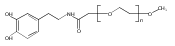


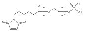


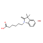


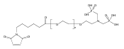


![dodecyl-[6-[dodecyl(dimethyl)azaniumyl]hexyl]-dimethylazanium,bromide](http://img.cochemist.com/ccimg/18600/18507-15-8.png)
![dodecyl-[6-[dodecyl(dimethyl)azaniumyl]hexyl]-dimethylazanium,bromide](http://img.cochemist.com/ccimg/18600/18507-15-8_b.png)