Co-reporter:Dan Zhang, Chao Huang, Wenyu Xin, Xiaowei Ma, Weiku Zhang, Tiantai Zhang, Guanhua Du
Journal of Pharmaceutical and Biomedical Analysis 2015 Volume 109() pp:1-10
Publication Date(Web):10 May 2015
DOI:10.1016/j.jpba.2015.02.022
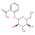
•Methyl salicylate-2-O-β-D-lactoside and the metabolite of salicylic acid could be quantified by UPLC–MS/MS method in biological samples.•Evaluate the pharmacokinetic, distribution and excretion of methyl salicylate-2-O-β-d-lactoside in rats.•Hydrolysed, sulfated and glucuronidated products are the main metabolites.Methyl salicylate-2-O-β-d-lactoside (MSL) is a natural salicylate derivative from the traditional Chinese medicine of Gaultheria yunnanensis (Franch.) Rehder (G. yunnanensis). As a non-steroidal anti-inflammatory drug (NSAID), MSL exerts a significant anti-arthritis effect but hardly has any gastrointestinal toxicity. In this paper, the pharmacokinetics, distribution, excretion and identification of MSL and its metabolites are described following rat oral and intravenous administration. The biological samples were quantified by UPLC–MS/MS and the metabolites in urine and feces were identified by using Q-TOF-MS. These results will support future investigations leading to clinical development of this drug.
Co-reporter:Wenyu Xin;Chao Huang;Xue Zhang;Sheng Xin;Yiming Zhou;Xiaowei Ma;Dan Zhang;Yongjie Li;Sibai Zhou;Dongming Zhang;Guanhua Du
British Journal of Pharmacology 2014 Volume 171( Issue 14) pp:3526-3538
Publication Date(Web):
DOI:10.1111/bph.12715
Background and Purpose
Methyl salicylate 2-O-β-d-lactoside (MSL), whose chemical structure is similar to that of salicylic acid, is a natural product derivative isolated from a traditional Chinese herb. The aim of this study was to investigate the therapeutic effect of MSL in mice with collagen-induced arthritis (CIA) and explore its underlying mechanism.
Experimental Approach
The anti-arthritic effects of MSL were evaluated on human rheumatoid fibroblast-like synoviocytes (FLS) in vitro and CIA in mice in vivo by obtaining clinical scores, measuring hind paw thickness and inflammatory cytokine levels, radiographic evaluations and histopathological assessments.
Key Results
Treatment with MSL after the onset of arthritis significantly prevented the progression and development of rheumatoid arthritis (RA) in CIA mice without megascopic gastric mucosa damage. In addition, MSL inhibited the production of pro-inflammatory mediators, the phosphorylation and translocation of NF-κB, and cell proliferation induced by TNF-α in FLS. MSL non-selectively inhibited the activity of COX in vitro, but was a more potent inhibitor of COX-2 than COX-1. MSL also inhibited the phosphorylation of inhibitor of NF-κB kinase, IκBα and p65, thus blocking the nuclear translocation of NF-κB in TNF-α-stimulated FLS.
Conclusion and Implications
MSL exerts therapeutic effects on CIA mice, suppressing the inflammatory response and joint destruction by non-selectively inhibiting the activity of COX and suppressing activation of the NF-κB signalling pathway, but without damaging the gastric mucosa. Therefore, MSL has great potential to be developed into a novel therapeutic agent for the treatment of RA.
Co-reporter:Xue Zhang, Jialin Sun, Wenyu Xin, Yongjie Li, Lin Ni, Xiaowei Ma, Dan Zhang, Dongming Zhang, Tiantai Zhang, Guanhua Du
International Immunopharmacology (March 2015) Volume 25(Issue 1) pp:88-95
Publication Date(Web):1 March 2015
DOI:10.1016/j.intimp.2015.01.024
•MSL significantly inhibited the arthritis progression in AIA rats.•MSL inhibited the activity of COX in LPS-induced RAW264.7.•MSL prominently blocked phosphorylation of p38 and ERK in LPS-induced RAW264.7.Methyl salicylate 2-O-β-D-lactoside (MSL) is a derivative of natural salicylate isolated from Gaultheria yunnanensis (Franch.) Rehder, which is widely used for treating rheumatoid arthritis (RA), swelling and pain. The aim of the present study was to investigate the effect of MSL on the progression of adjuvant-induced arthritis (AIA) in rat in vivo and explore the anti-inflammatory effects and mechanism of MSL in lipopolysaccharide (LPS)-treated murine macrophages RAW264.7 cells in vitro. Our results showed that MSL significantly inhibited the arthritis progression in AIA rats, decreasing the right hind paw swelling and ankle diameter, attenuating histopathological changes and suppressing the plasma levels of TNF-α and IL-1β in AIA rats. Besides, MSL had potent anti-inflammatory effects on the LPS-activated RAW264.7. MSL dose-dependently inhibited the activity of COX-1, and COX-2. Moreover, MSL prominently inhibited LPS-induced activation of MAPK in RAW264.7 cells by blocking phosphorylation of p38 and ERK. Our study suggests that MSL may be effective in the treatment of inflammatory diseases by inhibiting the pro-inflammatory cytokine production and regulating the MAPK signal pathway.
Co-reporter:Rui Liu, Jin-ze Li, Jun-ke Song, Dan Zhou, Chao Huang, Xiao-yu Bai, Tao Xie, Xue Zhang, Yong-jie Li, Cai-xia Wu, Lan Zhang, Lin Li, Tian-tai Zhang, Guan-hua Du
Neurobiology of Aging (June 2014) Volume 35(Issue 6) pp:1275-1285
Publication Date(Web):1 June 2014
DOI:10.1016/j.neurobiolaging.2013.12.031
Amyloid-β (Aβ) peptides accumulate in the brain and initiate a cascade of pathologic events in Alzheimer's disease. The receptor for advanced glycation end products (RAGE) has been implicated to mediate Aβ-induced perturbations in the neurovascular unit (NVU). We demonstrated that pinocembrin exhibits neuroprotection through inhibition of the Aβ and/or RAGE pathway, but the therapeutic role and mechanism involved are not ascertained. Here, we report that a 3-month treatment with pinocembrin prevents the cognition decline in APP/PS1 transgenic mice without altering Aβ burden and oxidative stress. Instead, pinocembrin is effective in conferring neurovascular protection through maintenance of neuropil ultrastructure, reduction of glial activation and levels of inflammatory mediators, preservation of microvascular function, improving the cholinergic system by conserving the ERK-CREB-BDNF pathway, and modulation of RAGE-mediated transduction. Furthermore, in an in vitro model, pinocembrin provides the NVU protection against fibrillar Aβ1–42, accompanied by regulation of neurovascular RAGE pathways. Our findings indicate that pinocembrin improves cognition, at least in part, attributable to the NVU protection, and highlights pinocembrin as a potential therapeutic strategy for the prevention and/or treatment of Alzheimer's disease.
Co-reporter:Yongjie Li, Sibai Zhou, Jinze Li, Yuhua Sun, Hamlati Hasimu, Rui Liu, Tiantai Zhang
Acta Pharmaceutica Sinica B (January 2015) Volume 5(Issue 1) pp:
Publication Date(Web):1 January 2015
DOI:10.1016/j.apsb.2014.12.003
Amyloid beta-peptides (Aβ) are known to undergo active transport across the blood-brain barrier, and cerebral amyloid angiopathy has been shown to be a prominent feature in the majority of Alzheimer׳s disease. Quercetin is a natural flavonoid molecule and has been demonstrated to have potent neuroprotective effects, but its protective effect on endothelial cells under Aβ-damaged condition is unclear. In the present study, the protective effects of quercetin on brain microvascular endothelial cells injured by fibrillar Aβ1–40 (fAβ1–40) were observed. The results show that fAβ1–40-induced cytotoxicity in human brain microvascular endothelial cells (hBMECs) can be relieved by quercetin treatment. Quercetin increases cell viability, reduces the release of lactate dehydrogenase, and relieves nuclear condensation. Quercetin also alleviates intracellular reactive oxygen species generation and increases superoxide dismutase activity. Moreover, it strengthens the barrier integrity through the preservation of the transendothelial electrical resistance value, the relief of aggravated permeability, and the increase of characteristic enzyme levels after being exposed to fAβ1–40. In conclusion, quercetin protects hBMECs from fAβ1–40-induced toxicity.Quercetin increased TEER and permeability efficient (Pe) values of fluorescein sodium and FITC-albumin. From the results of this study, quercetin improved barrier function of hBMECs against fAβ1–40-induced toxicity. Download full-size image
Co-reporter:Wenyu Xin, Chao Huang, Xue Zhang, Guidong Zhang, Xiaowei Ma, Lan Sun, Chao Wang, Dongming Zhang, Tiantai Zhang, Guanhua Du
International Immunopharmacology (February 2013) Volume 15(Issue 2) pp:303-308
Publication Date(Web):1 February 2013
DOI:10.1016/j.intimp.2012.11.014
Ethyl salicylate 2-O-β-d-glucoside (ESG) is a derivative of natural salicylate isolated from Gaultheria yunnanensis (Franch.) Rehder, it has been used for the treatments of rheumatoid arthritis, swelling and pain. The aim of this study was to evaluate the anti-inflammatory effects of ESG and explore the anti-inflammatory mechanisms. We found that ESG had potent anti-inflammatory effects on the lipopolysaccharide (LPS)-activated murine macrophages RAW264.7. ESG exerted a dose-dependent inhibition of the LPS-stimulated release of the pro-inflammatory cytokines TNF-α and IL-1β. Moreover, it significantly inhibited LPS-stimulated the production of NO and PGE2 by repressing the expression of iNOS and COX protein respectively. Western blot analysis showed that ESG prominently inhibited LPS-induced activation of NF-κB in RAW264.7 cells by blocking phosphorylation of inhibitor IκBα and p65. Consistent with these results, we found that ESG prevented the nuclear translocation of NF-κB induced by LPS. Our study suggests that ESG may be effective in the treatment of inflammatory diseases by inhibiting the pro-inflammatory cytokine production and regulating the NF-κB signal pathway.Highlights► ESG had potent anti-inflammatory effects on the LPS-activated RAW264.7 macrophages. ► ESG inhibited the PGE2 release and COX protein expression. ► ESG suppresses the production of pro-inflammatory cytokines TNF-α and IL-1β. ► ESG inhibited the LPS-stimulated expression of iNOS and NO generation. ► ESG inhibited LPS-induced activation of NF-κB by blocking phosphorylation of IκBα.