Co-reporter:Takuya Miyagawa;Tat Thang Vo Doan;Hirotaka Sato; Ferdinandus;Toshinori Fujie
ACS Applied Materials & Interfaces December 14, 2016 Volume 8(Issue 49) pp:33377-33385
Publication Date(Web):August 29, 2016
DOI:10.1021/acsami.6b06075
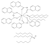
In this paper, a microthermograph, temperature mapping with high spatial resolution, was established using luminescent molecules embedded ultrathin polymeric films (nanosheets), and demonstrated in a living small animal to map out and visualize temperature shift due to animal’s muscular activity. Herein, we report super flexible and self-adhesive (no need of glue) nanothermosensor consisting of stacked two different polymeric nanosheets with thermosensitive (Eu-tris (dinaphthoylmethane)-bis-trioctylphosphine oxide: EuDT) and insensitive (Rhodamine 800) dyes being embedded. Such stacked nanosheets allow for the ratiometric thermometry, with which the undesired luminescence intensity shift due to focal drift or animal’s z-axis displacement is eliminated and the desired intensity shift solely due to the temperature shift of the sample (living muscle) can be acquired. With the stacked luminescent nanosheets, we achieved the first-ever demonstration of video filming of chronologically changing temperature-shift distribution from the rest state to the active state of the muscles in the living animal. The polymer nanosheet engineering and in vivo microthermography presented in the paper are promising technologies to microscopically explore the heat production and heat transfer in living cells, tissues, and organisms with high spatial resolution beyond what existing thermometric technologies such as infrared thermography have ever achieved.Keywords: heat production; luminescent; optical sensor; thermometry; ultrathin film (nanosheet);
Co-reporter:Tianshu Li, Takuya Amari, Kentaro Semba, Tadashi Yamamoto, Shinji Takeoka
Nanomedicine: Nanotechnology, Biology and Medicine 2017 Volume 13, Issue 3(Issue 3) pp:
Publication Date(Web):1 April 2017
DOI:10.1016/j.nano.2016.11.018
1,5-Dihexadecyl N,N-diglutamyl-lysyl-L-glutamate (GGLG) liposomes were previously developed to enhance drug delivery efficiency in tumor cells owing to its pH-responsive properties. Herein, we report the modification of GGLG liposomes by conjugating a Fab′ fragment of an ErbB2 antibody to the terminus of PEG (polyethylene glycol)-lipid (Fab′-GGLG liposomes). The conjugation of Fab′ fragments did not affect the antibody activity, drug (doxorubicin, DOX) encapsulation efficiency, stability during storage or pH-sensitivity. However, the binding affinity of Fab′-GGLG liposomes was enhanced to ErbB2-overexpressing HCC1954 cells specifically, and the cell association increased 10-fold in comparison to GGLG liposomes. Consequently, intracellular DOX delivery was enhanced, with an increased cytotoxicity in HCC1954 cells (i.e., IC50 of 1.17 and 3.08 μg/mL for Fab′-GGLG-DOX and GGLG-DOX liposomes, respectively). Further, a significantly enhanced tumor growth inhibition was obtained in an ErbB2-overexpressing breast cancer-bearing mouse model. Therefore, a potent anticancer drug delivery system was constructed by the immunological modification of pH-sensitive liposomes.Anti-ErB2 antibody-modified pH-sensitive (i.e., Fab′-GGLG) liposomes enhanced the cellular delivery and anticancer effect of doxorubicin (DOX) to ErB2-overexpressing breast cancer cells both in vitro and in vivo in comparison with unmodified liposomes.Download high-res image (250KB)Download full-size image
Co-reporter:Hong Zhang, Mao Fujii, Yosuke Okamura, Li Zhang, and Shinji Takeoka
ACS Applied Materials & Interfaces 2016 Volume 8(Issue 25) pp:16296-16302
Publication Date(Web):June 7, 2016
DOI:10.1021/acsami.6b03788
We present a facile method to fabricate polymer thin films with tens of nanometers thickness and several micrometers size (also called “microdiscs” herein) by applying phase separation of polymer blend. A water-soluble supporting layer is employed to obtain a freestanding microdisc suspension. Owing to their miniaturized size, microdiscs can be injected through a syringe needle. Herein, poly(d,l-lactic acid) microdiscs were fabricated with various thicknesses and sizes, in the range from ca. 10 to 60 nm and from ca. 1.0 to 10.0 μm, respectively. Magnetic nanoparticles were deposited on polymer microdiscs with a surface coating method. The magnetic manipulation of microdiscs in a liquid environment under an external magnetic field was achieved with controllable velocity by adjusting the microdisc dimensions and the loading amount of magnetic components. Such biocompatible polymer microdiscs are expected to serve as injectable vehicles for targeted drug delivery.
Co-reporter:Keisuke Ito;Akihiro Saito;Toshinori Fujie;Hiromi Miyazaki;Manabu Kinoshita;Daizoh Saitoh;Shinya Ohtsubo
Journal of Biomedical Materials Research Part B: Applied Biomaterials 2016 Volume 104( Issue 3) pp:585-593
Publication Date(Web):
DOI:10.1002/jbm.b.33429
Abstract
Ultra-thin polymer films (nanosheets) fabricated by a layer-by-layer (LbL) method possess unique properties such as high flexibility, adhesive strength, and transparency, and can be peeled off from a substrate and attached to various surfaces via a water-soluble supporting film. Therefore, flexible and transferrable LbL nanosheets are convenient tools as coating materials. Here, we fabricated a novel antimicrobial coating material by embedding silver nanoparticles (AgNPs) in an LbL nanosheet composed of layers of chitosan and sodium alginate (Ag-LbL nanosheet) by means of a photo-reduction method. Optimizing the amount of irradiated energy applied led to robust antimicrobial efficacy against methicillin-resistant Staphylococcus aureus (MRSA), sufficient to meet ISO standards (ISO 22196), while maintaining the flexibility and adhesive potency of the LbL nanosheet. Thus, the Ag-LbL nanosheet is a promising coating material that can provide antimicrobial efficacy to various surfaces. © 2015 Wiley Periodicals, Inc. J Biomed Mater Res Part B: Appl Biomater, 104B: 585–593, 2016.
Co-reporter:Kento Yamagishi, Kazuaki Sawaki, Atsushi Murata and Shinji Takeoka
Chemical Communications 2015 vol. 51(Issue 37) pp:7879-7882
Publication Date(Web):24 Mar 2015
DOI:10.1039/C4CC09947H
We synthesized a novel cyclooctyne-based clickable fluorescent probe with versatile properties such as high cell-membrane permeability and free diffusibility in the cell. Our probe “FC-DBCO” was conjugated to an azide-modified mannose via a Cu-free click reaction in living HeLa cells and displayed intracellular specific fluorescence imaging with low background signals.
Co-reporter:Mitsumasa Homma, Yoshiaki Takei, Atsushi Murata, Takafumi Inoue and Shinji Takeoka
Chemical Communications 2015 vol. 51(Issue 28) pp:6194-6197
Publication Date(Web):26 Feb 2015
DOI:10.1039/C4CC10349A
Mitochondrial thermodynamics is the key to understand cellular activities related to homeostasis and energy balance. Here, we report the first ratiometric fluorescent molecular probe (Mito-RTP) that is selectively localized in the mitochondria and visualize the temperature. We confirmed that Mito-RTP could work as a ratiometric thermometer in a cuvette and living cells.
Co-reporter:Keisuke Ito, Akihiro Saito, Toshinori Fujie, Keisuke Nishiwaki, Hiromi Miyazaki, Manabu Kinoshita, Daizoh Saitoh, Shinya Ohtsubo, Shinji Takeoka
Acta Biomaterialia 2015 Volume 24() pp:87-95
Publication Date(Web):15 September 2015
DOI:10.1016/j.actbio.2015.05.035
Abstract
Partial-thickness burn injury has the potential for reepithelialization and heals within 3 weeks. If the wound is infected by bacteria before reepithelization, however, the depth of disruption increases and the lesion easily progresses to the full-thickness dermal layers. In the treatment of partial-thickness burn injury, it is important to prevent the wound area from bacterial infection with an antimicrobial dressing. Here, we have tested the antimicrobial properties of polymeric ultra-thin films composed of poly(lactic acid) (termed “PLA nanosheets”), which have high flexibility, adhesive strength and transparency, and silver sulfadiazine (AgSD), which exhibits antimicrobial efficacy. The AgSD-loaded nanosheet released Ag+ for more than 3 days, and exerted antimicrobial efficacy against methicillin-resistant Staphylococcus aureus (MRSA) in an in vitro Kirby–Bauer test. By contrast, a cell viability assay indicated that the dose of AgSD used in the PLA nanosheets did not show significant cytotoxicity toward fibroblasts. In vivo evaluation using a mouse model of infection in a partial-thickness burn wound demonstrated that the nanosheet significantly reduced the number of MRSA bacteria on the lesion (more than 105-fold) and suppressed the inflammatory reaction, thereby preventing a protracted wound healing process.
Co-reporter:Kenta Kakiuchi;Kenichi Matsuda;Norikazu Harii
Journal of Artificial Organs 2015 Volume 18( Issue 3) pp:220-227
Publication Date(Web):2015 September
DOI:10.1007/s10047-015-0835-z
Micro/nano-bubbles are practical nanomaterials designed to increase the gas content in liquids. We attempted to use oxygen micro/nano-bubble dispersions as an oxygen-rich liquid as a means for total liquid ventilation. To determine the oxygen content in the bubble dispersion, a new method based on a spectrophotometric change between oxy- and deoxy-hemoglobin was established. The oxygen micro/nano-bubble dispersion was supplied to an experimental total ventilation liquid in anesthetic rats. Though the amount of dissolving oxygen was as low as 6 mg/L in physiological saline, the oxygen content in the oxygen micro/nano-bubble dispersion was increased to 45 mg/L. The positive correlation between the oxygen content and the life-saving time under liquid ventilation clearly indicates that the life-saving time is prolonged by increasing the oxygen content in the oxygen micro/nano-bubble dispersion. This is the first report indicating that the oxygen micro/nano-bubbles containing a sufficient amount of oxygen are useful in producing oxygen-rich liquid for the process of liquid ventilation.
Co-reporter:Satya Ranjan Sarker, Ryosuke Hokama, and Shinji Takeoka
Molecular Pharmaceutics 2014 Volume 11(Issue 1) pp:164-174
Publication Date(Web):November 13, 2013
DOI:10.1021/mp400363z
An amino acid-based cationic lipid having a TFA counterion (trifluoroacetic acid counterion) in the lysine headgroup was used to deliver functional proteins into human cervical cancer cells, HeLa, in the presence of serum. Proteins used in the study were fluorescein isothiocyanate (FITC) labeled bovine serum albumin, mouse anti-F actin antibody [NH3], and goat anti mouse IgG conjugated with FITC. The formation of liposome/protein complexes was confirmed using native polyacrylamide gel electrophoresis. Furthermore, the complexes were characterized in terms of their size and zeta potential at different pH values and found to be responsive to changes in pH. The highest delivery efficiency of the liposome/albumin complexes was 99% at 37 °C. The liposomes effectively delivered albumin and antibodies as confirmed by confocal laser scanning microscopy (CLSM). Inhibition studies showed that the cellular uptake mechanism of the complexes was via caveolae-mediated endocytosis, and the proteins were subsequently released from either the early endosomes or the caveosomes as suggested by CLSM. Thus, lysine-based cationic liposomes can be a useful tool for intracellular protein delivery.Keywords: amino acid-based cationic liposome; antibody; endocytosis; fluorescent labeled protein; intracellular protein delivery;
Co-reporter:Yoshiaki Takei, Satoshi Arai, Atsushi Murata, Masao Takabayashi, Kotaro Oyama, Shin’ichi Ishiwata, Shinji Takeoka, and Madoka Suzuki
ACS Nano 2014 Volume 8(Issue 1) pp:198
Publication Date(Web):December 19, 2013
DOI:10.1021/nn405456e
The homeostasis of body temperature and energy balance is one of the major principles in biology. Nanoscale thermometry of aqueous solutions is a challenging but crucial technique to understand the molecular basis of this essential process. Here, we developed a ratiometric nanothermometer (RNT) for intracellular temperature measurement in real time. Both the thermosensitive fluorophore, β-diketonate chelate europium(III) thenoyltrifluoroacetonate, and the thermoinsensitive fluorophore, rhodamine 101, which was used as a self-reference, are embedded in a polymeric particle that protects the fluorophores from intracellular conditions. The ratiometric measurement of single RNT spots is independent of the displacement of the RNT along the z-axis. The temperature is therefore determined at the location of each RNT under an optical microscope regardless of the dynamic movement of living cells. As a demonstration of the spot-by-spot intracellular thermometry, we successfully followed the temperature change in individual RNT spots in a single cell together with the Ca2+ burst induced by the Ca2+ ionophore ionomycin. The temperature increases differently among different spots, implying heterogeneous heat production in the cell. We then show that, in some spots, the temperature gradually decreases, while in others it remains high. The average temperature elevation within a cell is positively correlated to the increase in Ca2+, suggesting that the activity and/or number of heat sources are dependent on the Ca2+ concentration.Keywords: Ca2+-ATPase; fluorescent nanoparticle; polymer nanoparticle; ratiometry; rhodamine 101; thermogenesis; thermometry
Co-reporter:Yoshiaki Takei, Atsushi Murata, Kento Yamagishi, Satoshi Arai, Hideki Nakamura, Takafumi Inoue and Shinji Takeoka
Chemical Communications 2013 vol. 49(Issue 66) pp:7313-7315
Publication Date(Web):16 May 2013
DOI:10.1039/C3CC42489H
The powerful strategy of “intracellular click reaction” was used to retain a chemical Ca2+ indicator in the cytosol. Specifically, a novel clickable Ca2+ indicator “N3-fura-2 AM” was coupled with dibenzylcyclooctyl-modified biomacromolecules via copper-free click reaction in living cells and Ca2+ oscillation was observed for an extended period of time.
Co-reporter:Yumiko Aoshima, Ryosuke Hokama, Keitaro Sou, Satya Ranjan Sarker, Kabuto Iida, Hideki Nakamura, Takafumi Inoue, and Shinji Takeoka
ACS Chemical Neuroscience 2013 Volume 4(Issue 12) pp:1514
Publication Date(Web):October 2, 2013
DOI:10.1021/cn400036j
The delivery of specific genes into neurons offers a potent approach for treatment of diseases as well as for the study of neuronal cell biology. Here we investigated the capabilities of cationic amino acid based lipid assemblies to act as nonviral gene delivery vectors in primary cultured neurons. An arginine-based lipid, Arg-C3-Glu2C14, and a lysine-based lipid, Lys-C3-Glu2C14, with two different types of counterion, chloride ion (Cl–) and trifluoroacetic acid (TFA–), were shown to successfully mediate transfection of primary cultured neurons with plasmid DNA encoding green fluorescent protein. Among four types of lipids, we optimized their conditions such as the lipid-to-DNA ratio and the amount of pDNA and conducted a cytotoxicity assay at the same time. Overall, Arg-C3-Glu2C14 with TFA– induced a rate of transfection in primary cultured neurons higher than that of Lys-C3-Glu2C14 using an optimal weight ratio of lipid-to-plasmid DNA of 1. Moreover, it was suggested that Arg-C3-Glu2C14 with TFA– showed the optimized value higher than that of Lipofectamine2000 in experimental conditions. Thus, Arg-C3-Glu2C14 with TFA– is a promising candidate as a reliable transfection reagent for primary cultured neurons with a relatively low cytotoxicity.Keywords: Cationic amino acid based lipids; gene delivery; green fluorescent protein; liposomes; primary cultured neurons; transfection
Co-reporter:Hong Zhang, Yukio Honda, and Shinji Takeoka
Langmuir 2013 Volume 29(Issue 5) pp:1333-1339
Publication Date(Web):January 16, 2013
DOI:10.1021/la304280h
The morphological stability of polymer films is critically important to their application as functional materials. The deformation of polymer surfaces on the nanoscale may be significantly influenced by geometrical confinement. Herein, we constructed a mechanically heterogeneous polymer surface by phase separation in a thin polymer film and investigated the deformation behavior of its nanostructure (∼30 nm thickness and ∼100 nm average diameter) with tapping-mode atomic force microscopy. By changing different scan parameters, we could induce deformation localized to the nanostructure in a controllable manner. A quantity called the deformation index is defined and shown to be correlated to energy dissipation by tip–sample interaction. We clarified that the plastic deformation of a polymer on the nanoscale is energy-dependent and is related to the glass-to-rubber transition. The mobility of polymer chains beneath the tapping tip is enhanced, and in the corresponding region a rubberlike deformation with the lateral motion of the tip is performed. The method we developed can provide insight into the geometrical confinement effects on polymer behavior.
Co-reporter:Daisuke Niwa;Masatsugu Koide;Toshinori Fujie;Nobuhito Goda
Journal of Biomedical Materials Research Part B: Applied Biomaterials 2013 Volume 101( Issue 7) pp:1251-1258
Publication Date(Web):
DOI:10.1002/jbm.b.32937
Abstract
Postoperative adhesion often causes serious adverse effects such as bowl obstruction, chronic abdominal pain, pelvic pain, and infertility. We previously reported that a poly-L-lactic acid (PLLA) nanosheet can efficiently seal a surgical incision without scarring. In this report, we examined whether the PLLA nanosheet can form an effective anti-adhesion barrier in partial hepatectomy accompanied by severe hemorrhaging in rats. To evaluate the anti-adhesive property of the nanosheet, the liver wound surface was covered with TachoComb®, a well-known hemostat material used in clinical procedures, and then with the PLLA nanosheet. Dressing the wound surface with TachoComb® alone caused severe adhesion with omentum and/or residual parts of the liver. By contrast, combinational usage of TachoComb® and the PLLA nanosheet significantly reduced such adhesion, presumably by inhibiting the permeation of oozing blood cells and the infiltration of fibroblastic cells. Moreover, the nanosheet displayed low permeability against serum proteins as well as cells in vitro, supporting the notion that the PLLA nanosheet has anti-adhesive properties in vivo. These results strongly suggested that the PLLA nanosheet is a promising material for reducing unwanted postoperative adhesion. © 2013 Wiley Periodicals, Inc. J Biomed Mater Res Part B: Appl Biomater 101B: 1251–1258, 2013.
Co-reporter:Toshinori Fujie, Yuko Kawamoto, Hiroki Haniuda, Akihiro Saito, Koki Kabata, Yukio Honda, Eriko Ohmori, Toru Asahi, and Shinji Takeoka
Macromolecules 2013 Volume 46(Issue 2) pp:395-402
Publication Date(Web):December 31, 2012
DOI:10.1021/ma302081e
Most polymers solidify below a glass transition temperature (Tg), which is important for the fabrication of polymeric materials. The glass transition dynamics (GTD) of polymers alters their physical properties and therefore the range of applications suitable for the particular materials. In this regard, most GTD studies were oriented to the thermodynamics of amorphous polymer systems, while little studies were known for semicrystalline polymers. Here, we focus on the glassy and crystalline properties of semicrystalline polymers such as poly(l-lactic acid) (PLLA) and envisage to control the nanostructure of free-standing PLLA ultrathin films (referred as “PLLA nanosheets”), via thermodynamic rearrangement of polymer chains entangled in a quasi-two-dimensional interface during the GTD process. The annealing process on the PLLA nanosheets (<100 nm thick) resulted in the formation of semicrystalline domains and microscopic apertures with polymer chains (∼100 nm in size). Such nanostructure surprisingly induced selective molecular permeability, which was controlled as a function of film thickness and inherent crystallinity. The present methodology demonstrates the direct conversion of thermodynamic properties of semicrystalline polymers into the functional nanostructured polymeric materials.
Co-reporter:Shota Hirosawa, Satoshi Arai and Shinji Takeoka
Chemical Communications 2012 vol. 48(Issue 40) pp:4845-4847
Publication Date(Web):29 Mar 2012
DOI:10.1039/C2CC30603D
We report a mitochondrial targeted redox probe (MitoRP) that comprises a nitroxide radical (TEMPO) moiety and coumarin 343. Using isolated mitochondria in the presence/absence of substrates and inhibitors of oxidative phosphorylation, we demonstrated that MitoRP is a useful probe to monitor the electron flow associated with complex I.
Co-reporter:Akihiro Saito, Hiromi Miyazaki, Toshinori Fujie, Shinya Ohtsubo, Manabu Kinoshita, Daizoh Saitoh, Shinji Takeoka
Acta Biomaterialia 2012 Volume 8(Issue 8) pp:2932-2940
Publication Date(Web):August 2012
DOI:10.1016/j.actbio.2012.04.019
Polymeric ultra-thin films (nanosheets) possess unique properties that make them suitable materials for various biomedical applications. In our previous study, we assessed the use of an antibiotic (tetracycline, TC)-loaded nanosheet (or “TC-nanosheet”) for the treatment of gastrointestinal tissue defects. The nanosheet consisted of three functional layers: layer-by-layer nanosheet as a stable platform, TC as an antimicrobial agent with autofluorescence for tracing, and a poly(vinyl acetate) nanosheet to act as a protecting layer. The TC-nanosheet has high flexibility, adhesive strength and transparency. Here, we evaluated the effectiveness of the TC-nanosheet in preventing full thickness burn-wound infections. In an in vivo study, murine dorsal skin was injured by full-thickness burns and then infected with Pseudomonas aeruginosa (P. aeruginosa), a common bacterium causing burn-associated infections. The wound site was treated either with a TC-nanosheet, TC-unloaded nanosheet or left untreated. Wound management was facilitated by the high transparency of the TC-nanosheet. The TC-nanosheet significantly improved burn-wound infection by P. aeruginosa in mice. Indeed, all mice treated with the TC-nanosheet survived, whereas the other treatment groups displayed increased rates of mortality due to bacterial infection. According to histological analyses and viable bacterial counting in the liver (bacterial translocation), the TC-nanosheets were able to prevent not only the local inflammation but also systemic inflammation. We conclude that the TC-nanosheet can act as an effective treatment for full-thickness burn-wound infection. Hence, the TC-nanosheet is a promising therapeutic tool for burn-wound management in severely burn-injured patients.
Co-reporter:Satoshi Arai, Shota Hirosawa, Yusuke Oguchi, Madoka Suzuki, Atsushi Murata, Shin’ichi Ishiwata, and Shinji Takeoka
ACS Combinatorial Science 2012 Volume 14(Issue 8) pp:451
Publication Date(Web):July 18, 2012
DOI:10.1021/co300028n
This paper describes a convenient screening method using ion trap electrospray ionization mass spectrometry to classify ligands to a target molecule in terms of kinetic parameters. We demonstrate this method in the screening of ligands to a hexahistidine tag from a pooled library synthesized by click chemistry. The ion trap mass spectrometry analysis revealed that higher stabilities of ligand-target complexes in the gas phase were related to lower dissociation rate constants, i.e., off-rates in solution. Finally, we prepared a fluorescent probe utilizing the ligand with lowest off-rate and succeeded in performing single molecule observations of hexahistidine-tagged myosin V walking on actin filaments.Keywords: click chemistry; fluorescent probes; high-throughput screening; mass spectrometry
Co-reporter:Hong Zhang and Shinji Takeoka
Macromolecules 2012 Volume 45(Issue 10) pp:4315-4321
Publication Date(Web):May 10, 2012
DOI:10.1021/ma3005394
Polymer phase separation has established a series of bottom-up nanofabrication methods. Owing to the intrinsic immiscibility of most polymer blends, phase separation is typically produced by rapid quenching during spin-casting. However, the full sequence of events that occur during this process is still unclear, especially in the case of ultrathin polymer blend films. Herein, a freestanding method is first introduced to obtain morphological information from the bottom side of the film. We demonstrate that when the thickness of ultrathin film is comparable to the dimensional scale of the phase separation domains, it is feasible to prepare perforated films with uniform nanopores via selective solvent etching. Our results also provide direct evidence that the spinodal decomposition mechanism plays an important role in determining the final morphology within the ultrathin polymer blend films. These findings are of practical value in the fabrication of desired nanostructures by polymer phase separation.
Co-reporter:Chihiro Urata ; Hironori Yamada ; Ryutaro Wakabayashi ; Yuko Aoyama ; Shota Hirosawa ; Satoshi Arai ; Shinji Takeoka ; Yusuke Yamauchi ;Kazuyuki Kuroda
Journal of the American Chemical Society 2011 Volume 133(Issue 21) pp:8102-8105
Publication Date(Web):May 3, 2011
DOI:10.1021/ja201779d
Aqueous colloidal mesoporous nanoparticles with ethenylene-bridged silsesquioxane frameworks with a uniform diameter of ∼20 nm were prepared from bis(triethoxysilyl)ethenylene in a basic aqueous solution containing cationic surfactants. The nanoparticles, which had higher hydrolysis resistance under aqueous conditions, showed lower hemolytic activity toward bovine red blood cells than colloidal mesoporous silica nanoparticles.
Co-reporter:Toshinori Fujie, Hiroki Haniuda and Shinji Takeoka
Journal of Materials Chemistry A 2011 vol. 21(Issue 25) pp:9112-9120
Publication Date(Web):27 May 2011
DOI:10.1039/C1JM10156K
A convenient method for surface modification is crucially important for the preparation of a functional interface. The precise control of surface properties by chemical and physical approaches is expected to enhance the potential application of innovative nanomaterials. In this study, we propose a methodology for convenient surface modification using a freestanding anti-biofouling ultra-thin film (nanosheet) bearing poly(2-methacryloyloxyethyl phosphorylcholine: MPC) (i.e., pMPC-nanosheets). The physicochemical properties of the pMPC-nanosheet such as physiological stability, surface wettability and anti-biofouling were precisely controlled by means of thermal crosslinking of the nanosheet, the grafting amount of MPC and tuning the thickness of pMPC brushes. These approaches revealed physicochemical insight into the pMPC-nanosheet and its remarkable biological response. The pMPC-nanosheet was prepared via a spin-coating assisted layer-by-layer method in combination with atom transfer radical polymerization of MPC. The freestanding pMPC-nanosheet, comprising an 11 nm-thick pMPC brushed layer with a hydrophilic surface, was easily transferred by tweezers, cut by scissors, patterned by a needle and patched onto various interfaces with the aid of a water-soluble supporting film. The pMPC-nanosheets patched on cell culture substrates displayed anti-biofouling properties such as anti-coagulant activity against human whole blood as well as the potential to microscopically pattern murine fibroblast cells. The present study not only reveals physicochemical properties of the surface-functionalized nanosheet for biological application, but also constitutes an alternative approach to conventional lithographic techniques using photoresists and micro-patterned molds; thereby providing a new tool in the field of nanobiotechnology.
Co-reporter:Yosuke Okamura, Shunsuke Katsuno, Hidenori Suzuki, Hitomi Maruyama, Makoto Handa, Yasuo Ikeda, Shinji Takeoka
Journal of Controlled Release 2010 Volume 148(Issue 3) pp:373-379
Publication Date(Web):20 December 2010
DOI:10.1016/j.jconrel.2010.09.013
We have constructed phospholipid vesicles with hemostatic activity as a platelet substitute. The vesicles were conjugated with a dodecapeptide (HHLGGAKQAGDV, H12), which is a fibrinogen γ-chain carboxy-terminal sequence (γ400–411). We have recently exploited these vesicles as a potential drug delivery system by encapsulation of adenosine 5'-diphosphate (ADP) (H12-(ADP)-vesicles).Here we explore the relationship between the ADP release from H12-(ADP)-vesicles with different membrane properties and their hemostatic effects. In total, we prepared five kinds of H12-(ADP)-vesicles with different lamellarities and membrane flexibilities. By radioisotope-labeling, we directly show that H12-(ADP)-vesicles were capable of augmenting platelet aggregation by releasing ADP in an aggregation-dependent manner. The amount of ADP released from the vesicles was dependent on their membrane properties. Specifically, the amount of ADP released increased with decreasing lamellarity and tended to increase with increasing membrane flexibility. Our in vivo results clearly demonstrated that H12-(ADP)-vesicles with the ability to release ADP exert considerable hemostatic action in terms of correcting prolonged bleeding time in a busulphan-induced thrombocytopenic rat model.We propose a recipe to control the hemostatic abilities of H12-(ADP)-vesicles by modulating ADP release based on membrane properties. We believe that this concept will be invaluable to the development of platelet substitutes and other drug carriers.
Co-reporter:Toshinori Fujie, Sho Furutate, Daisuke Niwa and Shinji Takeoka
Soft Matter 2010 vol. 6(Issue 19) pp:4672-4676
Publication Date(Web):20 Aug 2010
DOI:10.1039/C0SM00527D
We report an artificially fabricated extracellular matrix (ECM)-like nanostructure, which utilizes self-assembly of collagen and hyaluronic acid in a quasi two-dimensional space of tens-of-nm thickness, displaying different cellular adhesive characteristics depending on the structural properties between the fibril and non-fibril elements. This nano-fibrous assembly will facilitate a mechano-biological approach for the development of tissue-engineering scaffolds.
Co-reporter:Satoshi Arai, Toshiya Okamura, Shinji Takeoka
Tetrahedron Letters 2010 Volume 51(Issue 39) pp:5177-5180
Publication Date(Web):29 September 2010
DOI:10.1016/j.tetlet.2010.07.139
An efficient procedure through deprotection of 2,4,6-trimethoxypyrimidine derivative afforded porphyrinato zinc bearing multi-barbiturates acting as multiple hydrogen bonding sites at the meso positions. Addition of excess amphiphilic triaminopyrimidine derivative, as a complementary motif to the barbiturate, in an aqueous solution of porphyrin conjugated with multiple barbiturates at the meso positions resulted in precipitation. The cast film from the chloroform solution of the precipitate indicated the formation of a well-defined porphyrin assembly.
Co-reporter:Atsushi Murata, Satoshi Arai, Su-In Yoon, Masao Takabayashi, Miwako Ozaki, Shinji Takeoka
Bioorganic & Medicinal Chemistry Letters 2010 Volume 20(Issue 23) pp:6905-6908
Publication Date(Web):1 December 2010
DOI:10.1016/j.bmcl.2010.10.011
Hexahistidine ((His)6) tags are used to purify genetically engineered proteins. Herein, we describe the construction of a ‘turn-on’ fluorescent probe system that consists of the fluorescence quencher, Dabcyl, conjugated to (His)6, and fluorescent tetramethylrhodamine conjugated to nitrilotriacetic acid, which, in the presence of Ni2+, can bind (His)6. The system is turned off when Dabcyl-(His)6 is bound to the fluorescent nitrilotriacetic acid derivative. The binding strength of this system was assessed using electrospray ionization mass spectrometry, fluorescence correlation spectroscopy, and fluorescence intensity distribution analysis-polarization. Although there was no significant enhancement in fluorescence after addition of an equimolar amount of ubiquitin, the fluorescence increased from 14% to 40% of its initial intensity when an equimolar amount of (His)6-ubiquitin was added. Therefore, this system should be able to specifically recognize (His)6-proteins with good resolution and has the additional advantage that a washing step is not required to remove fluorescent probe, that is, not bound to the (His)6-protein.A new type of a ‘turn-on’ fluorescent probe for His-tagged proteins was constructed using tetramethylrhodamine-conjugated nitrilotriacetate derivative and Dabcyl-conjugated hexahistidine peptide.
Co-reporter:Yosuke Okamura, Kaoruko Eto, Hitomi Maruyama, Makoto Handa, Yasuo Ikeda, Shinji Takeoka
Nanomedicine: Nanotechnology, Biology and Medicine 2010 Volume 6(Issue 2) pp:391-396
Publication Date(Web):April 2010
DOI:10.1016/j.nano.2009.07.004
We have constructed liposomes with hemostatic activity as a platelet substitute using moderately thrombocytopenic rats. The liposomes were conjugated with the dodecapeptide (HHLGGAKQAGDV: H12), which is a fibrinogen γ-chain C-terminal sequence (γ 400–411). To visualize liposome accumulation at the site of vascular injury by in vivo computed tomography, a water-soluble contrast dye, N,N′-bis[2-hydroxy-1-(hydroxylmethyl)ethyl]-5-[(2S)-2-hydroxylpropanoylamino]-2,4,6-triiodoisophthalamide (iopamidol), was encapsulated into the H12-conjugated liposomes. We achieved direct visualization of specific accumulation of the H12-(iopamidol)liposomes at the jugular vein injured by ferric chloride and succeeded in semiquantitative analyses of the accumulated amount of H12-liposomes in the injured site. We therefore propose that H12-liposomes that are specifically recruited to, and exert their hemostatic activity at the site of vascular injury, have a significant potential as a carrier and/or as an ideal platelet substitute. Furthermore, the H12-(iopamidol)liposomes would also be clinically useful as diagnostic agents for pathological thrombus detection and as contrast dyes for hepatosplenography.From the Clinical EditorThe authors have constructed liposomes with hemostatic activity as a platelet substitute using moderately thrombocytopenic rats. They propose that H12-liposomes that are specifically recruited to, and exert their hemostatic activity at the site of vascular injury, have a significant potential as a carrier and/or as an ideal platelet substitute. Furthermore, the H12-(iopamidol) liposomes would also be clinically useful as diagnostic agents for thrombus detection and as contrast dyes for hepatosplenography.
Co-reporter:Yosuke Obata, Gianni Ciofani, Vittoria Raffa, Alfred Cuschieri, Arianna Menciassi, Paolo Dario, Shinji Takeoka
Nanomedicine: Nanotechnology, Biology and Medicine 2010 Volume 6(Issue 1) pp:70-77
Publication Date(Web):February 2010
DOI:10.1016/j.nano.2009.04.005
We investigated the ability of cationic liposomes composed of 1,5-dihexadecyl N-arginyl-L-glutamate (Arg-Glu2C16) to carry nucleic acids into neuronal cells. Such liposomes have been shown to have a remarkable capacity for transfecting immortalized cell lines. Lipoplexes between the Arg-Glu2C16 liposomes and plasmid DNA encoding green fluorescent protein (GFP) were analyzed in terms of lipoplex formation, intracellular DNA trafficking, transfection efficiency, and cytotoxicity in neuronal SH-SY5Y cells. A maximum number of cells expressing GFP was obtained with lipoplexes at a lipid-to-DNA ratio of 15. With these lipoplexes, 16% of the cells were GFP-positive, which was approximately fourfold higher than the level obtained with a commercially available transfection reagent, Lipofectamine 2000. Furthermore, as a result of the low cytotoxicity of the Arg-Glu2C16 lipoplexes, the proportion of GFP-positive cells could be increased to 25% by increasing the concentration of lipoplexes that was applied to the cells. We have demonstrated that Arg-Glu2C16, as a model cationic amino acid–based lipid, has a high capability as a gene carrier, even for neuronal transfection.From the Clinical EditorIn this study, specific cationic liposomes were characterized as nucleic acid transfection agents for neuronal cells. A fourfold higher transfection rate with low cytotoxicity was reported compared to Lipofectamine 2000, a commercial reagent. The authors conclude that the studied cationic liposomes have a high capability as a gene carrier for neuronal transfection. This may become clinically significant in future gene therapy efforts of neuronal diseases.
Co-reporter:Toshinori Fujie, Akihiro Saito, Manabu Kinoshita, Hiromi Miyazaki, Shinya Ohtsubo, Daizoh Saitoh, Shinji Takeoka
Biomaterials 2010 31(24) pp: 6269-6278
Publication Date(Web):
DOI:10.1016/j.biomaterials.2010.04.051
Co-reporter:Yosuke Okamura;Koki Kabata;Manabu Kinoshita;Daizoh Saitoh
Advanced Materials 2009 Volume 21( Issue 43) pp:4388-4392
Publication Date(Web):
DOI:10.1002/adma.200901035
Co-reporter:Toshinori Fujie;Noriyuki Matsutani;Manabu Kinoshita;Yosuke Okamura;Akihiro Saito
Advanced Functional Materials 2009 Volume 19( Issue 16) pp:2560-2568
Publication Date(Web):
DOI:10.1002/adfm.200900103
Abstract
Recent developments in nanotechnology have led to a method for producing free-standing polymer nanosheets as a macromolecular organization. Compared with bulk films, the large aspect ratio of such nanosheets leads to unique physical properties, such as transparency, noncovalent adhesion, and high flexibility. Here, a biomedical application of polymer nanosheets consisting of biocompatible and biodegradable polysaccharides is reported. Micro-scratch and bulge tests indicate that the nanosheets with a thickness of tens of nanometers have sufficient physical adhesiveness and mechanical strength for clinical use. A nanosheet of 75 nm thickness, a critical load of 9.1 × 10⁴ N m−1, and an elastic modulus of 9.6 GPa is used for the minimally invasive repair of a visceral pleural defect in beagle dogs without any pleural adhesion caused by wound repair. For the first time, clinical benefits of sheet-type nano-biomaterials based on molecular organization are demonstrated, suggesting that novel therapeutic tools for overlapping tissue wounds will be possible without the need for conventional surgical interventions.
Co-reporter:Toshinori Fujie, Jin Young Park, Atsushi Murata, Nicel C. Estillore, Maria Celeste R. Tria, Shinji Takeoka and Rigoberto C. Advincula
ACS Applied Materials & Interfaces 2009 Volume 1(Issue 7) pp:1404
Publication Date(Web):June 25, 2009
DOI:10.1021/am900111r
Freestanding quasi-two-dimensional ultrathin films (e.g., 41 nm thick polymer nanosheets) were produced, on which stimuli-responsive 47 nm thick polymer brushes were constructed by atom transfer radical polymerization (ATRP) of poly(N-isopropylacrylamide). The resulting surfaces of the multilayered polysaccharide ultrathin films were evaluated by ellipsometry, IR imaging, in situ variable-temperature atomic force microscopy (AFM), and contact angle measurements. The morphological transformation of the freestanding polymer nanosheet bearing thermoresponsive polymer brushes was observed macroscopically through reversible structural color changes at the air−water interface. The dynamic shape change of the nanosheet was also monitored with the addition of a surfactant such as sodium n-dodecylsulfate to reduce the hydrophobicity of the surface. It was then demonstrated that the highly flexible freestanding polymer nanosheet is capable of acting as a unique platform for inducing stimuli-responsive behavior in nanomaterials.Keywords: atom transfer radical polymerization (ATRP); layer-by-layer; polymer nanosheet; polysaccharide; thermoresponsive surface
Co-reporter:Yosuke Okamura, Yoshihito Fukui, Koki Kabata, Hidenori Suzuki, Makoto Handa, Yasuo Ikeda and Shinji Takeoka
Bioconjugate Chemistry 2009 Volume 20(Issue 10) pp:1958
Publication Date(Web):September 29, 2009
DOI:10.1021/bc900325w
We have studied biocompatible spherical carriers carrying a dodecapeptide, HHLGGAKQAGDV (H12), on their surface as platelet substitutes. This peptide is a fibrinogen γ-chain carboxy-terminal sequence (γ400−411) and specifically recognizes the active form of glycoprotein IIb/IIIa on activated platelets. Our purpose is to assess the possibility of making a novel platelet substitute consisting of disk-shaped nanosheets having a large contact area for the targeting site, rather than conventional small contact area spherical carriers. The H12 peptide was conjugated to the surface of the free-standing nanosheets made of biodegradable poly(d,l-lactide-co-glycolide) (PLGA). These H12-PLGA nanosheets were fabricated onto 3 μm disk-shaped patterned hydrophobic octadecyl regions on a SiO2 substrate. By way of comparison, spherical H12-PLGA microparticles with the same surface area and conjugation number of H12 were also prepared. The resulting H12-PLGA nanosheets specifically interacted with the activated platelets adhered on the collagen surface at twice the rate of the H12-PLGA microparticles under flow conditions, and showed platelet thrombus formation in a two-dimensional spreading manner. Thus, H12-PLGA nanosheets might be a suitable candidate novel platelet alternative substitute for infused human platelet concentrates for the treatment of bleeding in patients with severe thrombocytopenia.
Co-reporter:Yosuke Okamura;Takahiro Goto;Daisuke Niwa;Yoshihito Fukui;Masanobu Otsuka;Norikazu Motohashi;Tetsuya Osaka
Journal of Biomedical Materials Research Part A 2009 Volume 89A( Issue 1) pp:233-241
Publication Date(Web):
DOI:10.1002/jbm.a.31934
Abstract
Sheet-shaped carriers, having both obverse and reverse surfaces and thus a large contact area for targeting a site, have several advantages over spherical-shaped carriers, which have an extremely small contact area for targeting sites. Here, we proposed a novel method to prepare a free-standing ultrathin and biocompatible nanosheet having heterosurfaces, by a combination of four processes: (1) specific adsorption of recombinant human serum albumin (rHSA) molecules onto a patterned octadecyltrimethoxysilane self-assembled monolayer region (ODS-SAM), (2) preparation of nanosheets of rHSA molecules bearing thiol groups (SH-rHSA) via two-dimensionally disulfide crosslinking, (3) surface modification of the resulting nanosheet, and (4) preparation of the free-standing nanosheet by detachment from the ODS-SAM. The SH-rHSA molecules at pH 5.0 and a concentration of 1 μg/mL were specifically adsorbed on the patterned ODS-SAM regions by hydrophobic interaction, and were two-dimensionally crosslinked in the presence of copper ion as an oxidant. The rHSA-nanosheets were then simply detached from the ODS-SAM by treatment with surfactant. We succeeded in the preparation of rectangular (10 μm × 30 μm) and ultrathin (4.5 ± 1.0 nm) rHSA-nanosheets on a patterned ODS-SAM, and could also obtain free-standing rHSA-nanosheets having heterosurfaces by surface modification with fluorescent latex beads. Thus, the rHSA-nanosheets having heterosurfaces could be regarded as a new biomaterial for drug carriers, hemostatic reagents, wound dressing for burn injury, and so forth. © 2008 Wiley Periodicals, Inc. J Biomed Mater Res, 2009
Co-reporter:Toshinori Fujie, Yosuke Okamura, Shinji Takeoka
Colloids and Surfaces A: Physicochemical and Engineering Aspects 2009 Volume 334(1–3) pp:28-33
Publication Date(Web):20 February 2009
DOI:10.1016/j.colsurfa.2008.09.056
The chemical and physical modification of a material surface is important for practical applications. In this study, we utilized the bilateral nature of a free-standing polysaccharide nanosheet as a platform for surface modification, selectively modifying the upper and lower surfaces with different sizes of latex beads. By means of a water-soluble sacrificial layer, we adsorbed latex beads with the diameter of 200 nm onto the obverse surface of the polysaccharide nanosheet as well as latex beads with that of 2 μm onto the reverse side. The optical characteristics of the nanosheet were analyzed on the basis of ‘thin film interference theory’ by making stepwise increments in thickness through sequentially overlaying free-standing 30-nm polysaccharide nanosheets onto a SiO2 substrate. The surface of the polysaccharide nanosheet was quite flat and smooth. Having confirmed by optical and scanning electron microscopy that we had achieved selective surface modification of the polysaccharide nanosheet with micro/nano-latex beads, we noted a unique optical property when the nanosheet was adsorbed on the SiO2 substrate. The localization of the beads on the nanosheet surface produced structural colors in the films due to an optical interference. This selective surface modification technique will be utilized to create ‘smart’ nanocomposites possessing different surface functions.
Co-reporter:Yosuke Obata, Daisuke Suzuki and Shinji Takeoka
Bioconjugate Chemistry 2008 Volume 19(Issue 5) pp:1055
Publication Date(Web):April 23, 2008
DOI:10.1021/bc700416u
We synthesized cationic lipids bearing lysine, histidine, or arginine as a cationic headgroup for use in gene transfer studies. The cationic assemblies formed from lysine- or arginine-type lipids gave unilamellar vesicles (∼100 nm diameter), whereas the morphology of the histidine-type lipids was tube-like. The competences of the cationic assemblies were sufficient to form lipoplexes, and the resulting lipoplexes were evaluated in terms of gene expression efficiencies with COS-7 cells. The lysine- or arginine-type lipids exhibited higher gene expression efficiencies than that of Lipofectamine2000, a conventional transgenic reagent, indicating that stable lipoplexes could be prepared between spherical cationic assemblies and plasmid DNA. The gene expression efficiency in relation to the cationic headgroup of the lipids was as follows: lysine ≥ arginine > histidine. In addition, gene expression efficiency was enhanced by decreasing the length of the alkyl chain of the hydrophobic moiety. Unlike Lipofectamine2000, no reduction in transfection efficiency in the presence of fetal bovine serum was observed for the lipoplexes formed using synthetic cationic lipids. Moreover, the synthetic cationic lipids revealed remarkably low cytotoxicity compared with Lipofectamine2000. In conclusion, cationic assemblies formed from 1,5-ditetradecyl-N-lysyl-l-glutamate or 1,5-ditetradecyl-N-arginyl-l-glutamate can be used as an effective plasmid DNA delivery system.
Co-reporter:Shinsuke Ishihara;Yusuke Furuki
Polymers for Advanced Technologies 2008 Volume 19( Issue 8) pp:1097-1104
Publication Date(Web):
DOI:10.1002/pat.1085
Abstract
A hydrogen-bonded helical columnar liquid crystal was synthesized, in which the helical structure is induced by a centered triphenylene derivative bearing chiral side-chains. The triphenylene derivative, 2,6,10-tris(carboxymethoxy)-3,7,11-tris((S)-(-)-2-methyl-1-butanoxy)triphenylene (TPC4(S)), and a dendric amphiphile, 3,5-bis-(3,4-bis-dodecyloxy-benzyloxy)-N-pyridine-4-yl-benzamide (DenC12), were mixed in a 1:3 ratio to obtain a complex, TPC4(S)-DenC12. Analyses by 1H-NMR spectroscopy, diffusion ordered spectroscopy (DOSY), CD spectroscopy, infrared (IR) spectroscopy, polarized optical microscopy (POM), differential scanning calorimetry (DSC), and X-ray diffractometry revealed that TPC4(S)-DenC12 self-assembles to form helical columnar stacks in solution and a helical columnar liquid crystal in bulk. The hydrogen bonding between TPC4(S) and DenC12 is essential for the helical columnar organization, and the preference for a one-handed helical conformation is likely derived from the steric interaction between the chiral side-chains and the dendric amphiphiles in the packing of the hydrogen-bonded columnar assemblies. Copyright © 2008 John Wiley & Sons, Ltd.
Co-reporter:Yosuke Okamura, Saori Utsunomiya, Hidenori Suzuki, Daisuke Niwa, Tetsuya Osaka, Shinji Takeoka
Colloids and Surfaces A: Physicochemical and Engineering Aspects 2008 Volume 318(1–3) pp:184-190
Publication Date(Web):1 April 2008
DOI:10.1016/j.colsurfa.2007.12.049
Sheet-shaped carriers, having obverse and reverse surfaces and thus a large contact area as a targeting site, have several advantages over spherical-shaped carriers, which have an extremely small contact area. Herein is proposed a novel method for the preparation of free-standing nanoparticle-fused nanosheets having uniform micrometer shape, nanometer thickness and heterogenous surfaces, using a water-soluble sacrificial film. This was achieved by combination of four processes: (1) specific adsorption of latex beads at pH 5.0 and a concentration of 1.0 × 10¹¹ mL−1 onto a patterned dodecyltrimethoxysilane self-assembled monolayer (DTS-SAM) region by a conventional dry patterning process, (2) fabrication of the latex bead-sheet via thermal-fusion at 110 °C for 60 s, (3) preparation of the free-standing nanosheet by detachment from the DTS-SAM, and (4) hetero-modification of the resulting nanosheet using a water-soluble poly(acrylic acid) as a sacrificial supporting film. Thus, this sheet-shaped carrier having hetero-surfaces can be regarded as a new material for delivery of drugs, hemostatic reagents and as wound dressings for burn injury, etc.
Co-reporter:Satoshi Arai;Hiroyuki Nishide;Hiroki Ohshiro
Polymers for Advanced Technologies 2007 Volume 18(Issue 6) pp:497-501
Publication Date(Web):10 APR 2007
DOI:10.1002/pat.894
Self-assembled porphyrins via noncovalent bonding have attracted wide-ranging researchers in material science. We reported herein the synthesis of the tetraphenyl porphyrin derivatives bearing uracyl groups as acceptor–donor–acceptor (ADA) type hydrogen bonding units, through the condensation of 5,10- or 5,15-bis (3-amino-4-ethylhexylphenyl) porphyrin derivatives with 6-carboxyuracyl derivatives. When two porphyrins having uracyl groups at the different substituted positions were respectively mixed with a melamine derivative in benzene, 1H NMR spectra showed that the 5,15 substituted uracyl porphyrin formed a hydrogen-bonded suprastructure with the melamine derivative as a complementary molecule to the uracyl moiety, although the other 5,10-substituted uracylporphyrin could not form such a structure. The SEM observation indicated that the mixture with the 5,15-substituted uracyl porphyrin and the melamine with long alkyl chains formed a sheet-like structure. Copyright © 2007 John Wiley & Sons, Ltd.
Co-reporter:Yosuke Obata, Shunsuke Saito, Naoya Takeda, Shinji Takeoka
Biochimica et Biophysica Acta (BBA) - Biomembranes (May 2009) Volume 1788(Issue 5) pp:
Publication Date(Web):May 2009
DOI:10.1016/j.bbamem.2009.02.014
We have synthesized a series of cationic amino acid-based lipids having a spacer between the cationic head group and hydrophobic moieties and examined the influence of the spacer on a liposome gene delivery system. As a comparable spacer, a hydrophobic spacer with a hydrocarbon chain composed of 0, 3, 5, 7, or 11 carbons, and a hydrophilic spacer with an oxyethylene chain (10 carbon and 3 oxygen molecules) were investigated. Plasmid DNA (pDNA)-encapsulating liposomes were prepared by mixing an ethanol solution of the lipids with an aqueous solution of pDNA. The zeta potentials and cellular uptake efficiency of the cationic liposomes containing each synthetic lipid were almost equivalent. However, the cationic lipids with the hydrophobic spacer were subject to fuse with biomembrane-mimicking liposomes. 1,5-Dihexadecyl-N-lysyl-N-heptyl-l-glutamate, having a seven carbon atom spacer, exhibited the highest fusogenic potential among the synthetic lipids. Increased fusion potential correlated with enhanced gene expression efficiency. By contrast, an oxyethylene chain spacer showed low gene expression efficiency. We conclude that a hydrophobic spacer between the cationic head group and hydrophobic moieties is a key component for improving pDNA delivery.
Co-reporter:Yoshiaki Takei, Atsushi Murata, Kento Yamagishi, Satoshi Arai, Hideki Nakamura, Takafumi Inoue and Shinji Takeoka
Chemical Communications 2013 - vol. 49(Issue 66) pp:NaN7315-7315
Publication Date(Web):2013/05/16
DOI:10.1039/C3CC42489H
The powerful strategy of “intracellular click reaction” was used to retain a chemical Ca2+ indicator in the cytosol. Specifically, a novel clickable Ca2+ indicator “N3-fura-2 AM” was coupled with dibenzylcyclooctyl-modified biomacromolecules via copper-free click reaction in living cells and Ca2+ oscillation was observed for an extended period of time.
Co-reporter:Toshinori Fujie, Hiroki Haniuda and Shinji Takeoka
Journal of Materials Chemistry A 2011 - vol. 21(Issue 25) pp:NaN9120-9120
Publication Date(Web):2011/05/27
DOI:10.1039/C1JM10156K
A convenient method for surface modification is crucially important for the preparation of a functional interface. The precise control of surface properties by chemical and physical approaches is expected to enhance the potential application of innovative nanomaterials. In this study, we propose a methodology for convenient surface modification using a freestanding anti-biofouling ultra-thin film (nanosheet) bearing poly(2-methacryloyloxyethyl phosphorylcholine: MPC) (i.e., pMPC-nanosheets). The physicochemical properties of the pMPC-nanosheet such as physiological stability, surface wettability and anti-biofouling were precisely controlled by means of thermal crosslinking of the nanosheet, the grafting amount of MPC and tuning the thickness of pMPC brushes. These approaches revealed physicochemical insight into the pMPC-nanosheet and its remarkable biological response. The pMPC-nanosheet was prepared via a spin-coating assisted layer-by-layer method in combination with atom transfer radical polymerization of MPC. The freestanding pMPC-nanosheet, comprising an 11 nm-thick pMPC brushed layer with a hydrophilic surface, was easily transferred by tweezers, cut by scissors, patterned by a needle and patched onto various interfaces with the aid of a water-soluble supporting film. The pMPC-nanosheets patched on cell culture substrates displayed anti-biofouling properties such as anti-coagulant activity against human whole blood as well as the potential to microscopically pattern murine fibroblast cells. The present study not only reveals physicochemical properties of the surface-functionalized nanosheet for biological application, but also constitutes an alternative approach to conventional lithographic techniques using photoresists and micro-patterned molds; thereby providing a new tool in the field of nanobiotechnology.
Co-reporter:Shota Hirosawa, Satoshi Arai and Shinji Takeoka
Chemical Communications 2012 - vol. 48(Issue 40) pp:NaN4847-4847
Publication Date(Web):2012/03/29
DOI:10.1039/C2CC30603D
We report a mitochondrial targeted redox probe (MitoRP) that comprises a nitroxide radical (TEMPO) moiety and coumarin 343. Using isolated mitochondria in the presence/absence of substrates and inhibitors of oxidative phosphorylation, we demonstrated that MitoRP is a useful probe to monitor the electron flow associated with complex I.
Co-reporter:Kento Yamagishi, Kazuaki Sawaki, Atsushi Murata and Shinji Takeoka
Chemical Communications 2015 - vol. 51(Issue 37) pp:NaN7882-7882
Publication Date(Web):2015/03/24
DOI:10.1039/C4CC09947H
We synthesized a novel cyclooctyne-based clickable fluorescent probe with versatile properties such as high cell-membrane permeability and free diffusibility in the cell. Our probe “FC-DBCO” was conjugated to an azide-modified mannose via a Cu-free click reaction in living HeLa cells and displayed intracellular specific fluorescence imaging with low background signals.
Co-reporter:Mitsumasa Homma, Yoshiaki Takei, Atsushi Murata, Takafumi Inoue and Shinji Takeoka
Chemical Communications 2015 - vol. 51(Issue 28) pp:NaN6197-6197
Publication Date(Web):2015/02/26
DOI:10.1039/C4CC10349A
Mitochondrial thermodynamics is the key to understand cellular activities related to homeostasis and energy balance. Here, we report the first ratiometric fluorescent molecular probe (Mito-RTP) that is selectively localized in the mitochondria and visualize the temperature. We confirmed that Mito-RTP could work as a ratiometric thermometer in a cuvette and living cells.

![1H,5H,11H,15H-Xantheno[2,3,4-ij:5,6,7-i'j']diquinolizin-18-ium, 9-cyano-2,3,6,7,12,13,16,17-octahydro-, perchlorate (1:1)](/data/chemimg/103300/137993-41-0.png)
![1H,5H,11H,15H-Xantheno[2,3,4-ij:5,6,7-i'j']diquinolizin-18-ium, 9-cyano-2,3,6,7,12,13,16,17-octahydro-, perchlorate (1:1)](/data/chemimg/103300/137993-41-0_b.png)

![β-D-Mannopyranose, 2-[(2-azidoacetyl)amino]-2-deoxy-, 1,3,4,6-tetraacetate](/data/chemimg/173100/1094268-54-8.png)
![β-D-Mannopyranose, 2-[(2-azidoacetyl)amino]-2-deoxy-, 1,3,4,6-tetraacetate](/data/chemimg/173100/1094268-54-8_b.png)


![9H-Xanthen-9-one,3,6-bis[[(1,1-dimethylethyl)dimethylsilyl]oxy]-](http://img.cochemist.com/ccimg/121800/121714-18-9.png)
![9H-Xanthen-9-one,3,6-bis[[(1,1-dimethylethyl)dimethylsilyl]oxy]-](http://img.cochemist.com/ccimg/121800/121714-18-9_b.png)

![Poly[oxy[(1S)-1-methyl-2-oxo-1,2-ethanediyl]]](http://img.cochemist.com/ccimg/26200/26161-42-2.png)
![Poly[oxy[(1S)-1-methyl-2-oxo-1,2-ethanediyl]]](http://img.cochemist.com/ccimg/26200/26161-42-2_b.png)

![tris[4,4,4-trifluoro-1-(2-thienyl)butane-1,3-dionato-O,O']europium](http://img.cochemist.com/ccimg/14100/14054-87-6.png)
![tris[4,4,4-trifluoro-1-(2-thienyl)butane-1,3-dionato-O,O']europium](http://img.cochemist.com/ccimg/14100/14054-87-6_b.png)