Co-reporter:Noriko Takahashi, Shunpei Koyama, Shinya Hasegawa, Masahiro Yamasaki, Masahiko Imai
Bioorganic & Medicinal Chemistry Letters 2017 Volume 27, Issue 20(Issue 20) pp:
Publication Date(Web):15 October 2017
DOI:10.1016/j.bmcl.2017.09.005
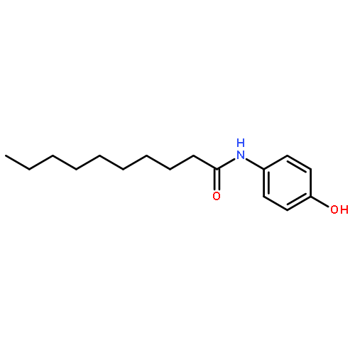
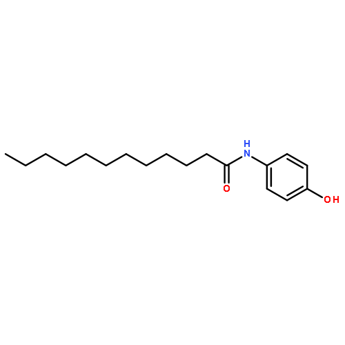
Neuroblastoma is an aggressive and drug-resistant refractory cancer. The human high-risk neuroblastoma cell line, SK-N-AS (non-amplified N-myc) is derived from stromal cells and it is resistant to treatment with retinoic acid (1, RA), which is a chemotherapeutic agent used to induce neuronal cellular differentiation of neuroblastomas. We have developed p-dodecylaminophenol (3, p-DDAP), based on N-(4-hydroxyphenyl)retinamide (2, 4-HPR), a synthetic amide of 1, since 1 and 2 are associated with the side-effect of nyctalopia. In order to evaluate the effects of 3 on high-risk neuroblastomas, we employed SK-N-AS cells as well as a second high-risk human neuroblastoma cell line, IMR-32, which is derived from neuronal cells (amplified N-myc, drug sensitive). Compound 3 suppressed cell growth of SK-N-AS and IMR-32 cells more effectively than 1, 2, p-decylaminophenol (4, p-DAP), N-(4-hydroxyphenyl)dodecananamide (5, 4-HPDD) or N-(4-hydroxyphenyl)decananamide (6, 4-HPD). In SK-N-AS cells, 3 induced G0/G1 arrest and apoptosis to a greater extent than 1 and 2. In IMR-32 cells, 3 induced apoptosis to a similar extent as 1 and 2, potentially by inhibiting N-myc expression. In addition, i.p. administration of 3 suppressed tumor growth in SK-N-AS-implanted mice in vivo. Since 3 showed no effects on blood retinol concentrations, in contrast to reductions following the administration of 2, it exhibited excellent anticancer efficacy against high-risk neuroblastoma SK-N-AS and IMR-32 expressing distinct levels of N-myc. Compound 3 may have potential for clinical use in the treatment of refractory neuroblastoma with reduced side effects.Download high-res image (103KB)Download full-size image
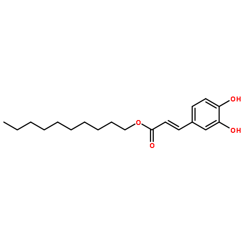
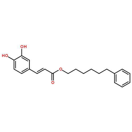
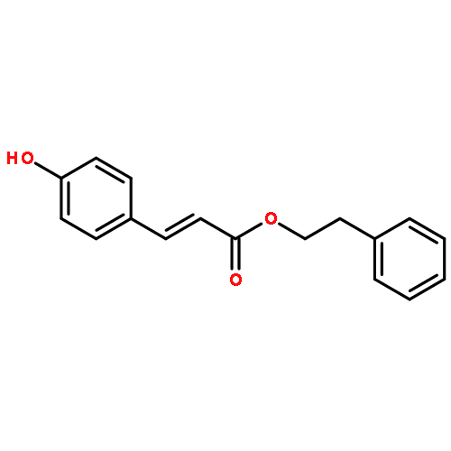
Co-reporter:Masahiko Imai, Takaya Kumaoka, Makiko Hosaka, Yui Sato, Chuan Li, Masashi Sudoh, Yoshiko Tamada, Hiromasa Yokoe, Setsu Saito, Masayoshi Tsubuki, Noriko Takahashi
Bioorganic & Medicinal Chemistry 2015 Volume 23(Issue 13) pp:3788-3795
Publication Date(Web):1 July 2015
DOI:10.1016/j.bmc.2015.03.086
Obesity is a risk factor associated with several lifestyle-related diseases, for example, diabetes, high blood pressure, hyperlipidemia and cancer. Caffeic acid 2-phenylethyl ester (CAPE, 1), a naturally-occurring compound found in various plants and propolis, which exhibits anti-inflammatory, immunomodulatory and cytotoxic activities and inhibits 3T3-L1 differentiation to adipocytes. As part of our efforts to moderate lifestyle-related diseases, we synthesized analogs of 1 and studied their effects on pancreatic lipase activities, lipid absorption, and 3T3-L1 differentiation. We found that catechols 1–4 show inhibitory activities against pancreatic lipase in a dose-dependent manner in vitro. Compounds 1–3 proved to be more potent inhibitors of pancreatic lipase than 5, 6, 8, and 9, which have one hydroxyl group, respectively. Compound 7 has three aromatic hydroxyl groups and restrains greater lipase inhibitory activity than the other compounds. In addition, 7 and 3 significantly suppress a rise in blood triglyceride (TG) levels in mice given corn oil orally. Furthermore, 2 and 3 are more potent at preventing 3T3-L1 differentiation (lipid accumulation) than 1, while 7 is more potent than 3, 8, and 9 in these assays. Compounds 2, 3, and 7 inhibit lipid absorption and accumulation, with new compound 7 being the most potent. These results indicate that 7 may have potential benefits as a health agent with anti-obesity properties.Compound 7 is an excellent inhibitor of lipid absorption and accumulation, being more potent than currently used caffeic acid 2-phenylethyl ester (1).
Co-reporter:Noriko Takahashi, Masahiko Imai, Yu Komori
Bioorganic & Medicinal Chemistry 2014 Volume 22(Issue 17) pp:4677-4683
Publication Date(Web):1 September 2014
DOI:10.1016/j.bmc.2014.07.016
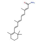
Melanin protects the skin against ultraviolet (UV) rays. It is produced in excess by UV radiation, which causes skin disorders and pigmentation. Retinoic acid (RA) decreases the levels of epidermal melanin by suppressing the expression of melanogenic enzymes including tyrosinase, which is the rate-limiting enzyme in melanin synthesis. However, RA shows inflammatory effects on the skin. In an effort to develop potent inhibitors of melanin synthesis, new aminophenol derivatives were synthesized based on structure–activity relationship studies of N-(4-hydroxyphenyl)retinamide (1), a derivative of RA. We investigated the inhibitory effects of a series of aminophenols on melanogenesis using B16 melanoma cells. p-Decylaminophenol (3) was the most potent agent examined, showing significant inhibition of B16 tyrosinase activities at concentrations less than what was required to achieve a similar level of inhibition by the well-known tyrosinase inhibitor, kojic acid. Compound 3 decreased melanin content and inhibited protein and mRNA expression for the tyrosinase-related protein-1 (TRP-1). It also inhibited the microphthalmia-associated transcription factor (MITF), a master transcription factor in melanogenesis. Compound 3 suppressed MEK/ERK signal pathways involved in the activation and expression of MITF. The data indicate that 3 inhibits TRP-1 expression by decreasing MITF expression through suppressing MEK/ERK signal pathways. This results in the reduction of melanin in B16 cells. Compound 3 might be an alternative to RA as a potent inhibitor of melanogenesis.Compound 3 is an excellent inhibitor of melanogenesis, tyrosinase activity, expressions of TRP-1 and MITF, and MEK/ERK signal pathways.
Co-reporter:Yu Komori, Masahiko Imai, Takayasu Yamauchi, Kimio Higashiyama, Noriko Takahashi
Bioorganic & Medicinal Chemistry 2014 22(15) pp: 3994-4000
Publication Date(Web):
DOI:10.1016/j.bmc.2014.05.073
Co-reporter:Noriko Takahashi, Kotaro Takeda, Masahiko Imai
Bioorganic & Medicinal Chemistry 2013 Volume 21(Issue 19) pp:6015-6021
Publication Date(Web):1 October 2013
DOI:10.1016/j.bmc.2013.07.039
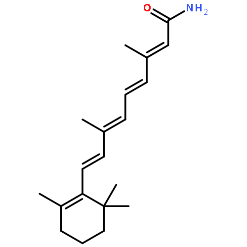
Cancer is a major cause of death, and the development of new anticancer drugs is urgently needed. Invasion and metastasis are the primary causes of death due to cancer rather than growth of the primary tumor. In the current study, we examined the anti-invasive effects of p-dodecylaminophenol (1), which was developed based on N-(4-hydroxyphenyl)retinamide (2), a synthetic amide of all-trans-retinoic acid (3). In HT1080 cells 1 inhibited growth, induced apoptosis and arrested the cell cycle in S phase in a dose-dependent manner. In addition, 1 significantly suppressed cell invasion, and the activity and mRNA expression of matrix metalloproteinase-9 (MMP-9). Furthermore, the expression of the reversion-inducing cysteine-rich protein with Kazal motifs (RECK), which is a negative regulator of MMP-9, was increased by treatment with 1. These results suggest that 1 could be an effective anti-cancer agent that suppresses cell growth through apoptosis induction and cell cycle arrest, which also inhibits cell invasion by decreasing MMP-9 expression due to an increase in RECK. Compound 1 might be useful clinically as a new and potent anticancer agent that could overcome adverse side effects of the retinoids.Compound 1 is an excellent inhibitor of invasion and growth in human metastatic cancer cells.
Co-reporter:Eiko Kato, Noriko Takahashi
Bioorganic & Medicinal Chemistry 2012 Volume 20(Issue 12) pp:3837-3842
Publication Date(Web):15 June 2012
DOI:10.1016/j.bmc.2012.04.029
Sodium dl-α-tocopheryl-6-O-phosphate (1), a water-soluble derivative of vitamin E (dl-α-tocopherol, 2), exhibits protective effects against various type of skin damage. As reported herein, we found that topical application of 1 improves hygroscopicity and water holding capacity in the stratum corneum of hairless mice in vivo by increasing the ceramide content. In normal human epidermal keratinocytes, treatment with 1 increases ceramide levels and enhances gene expression of serine palmitoyltransferase, which catalyzes the first step of ceramide synthesis in vitro. In addition, 1 increases gene expressions of differentiation markers (transglutaminase 1, cytokeratin 10, involucrin and loricrin), and intracellular Ca2+ concentrations. These results suggest that 1 could be an excellent agent for improving skin moisture-retention by enhancing ceramide synthesis through the induction of differentiation.
Co-reporter:Masahiko Imai, Noriko Takahashi
Bioorganic & Medicinal Chemistry 2012 Volume 20(Issue 8) pp:2520-2526
Publication Date(Web):15 April 2012
DOI:10.1016/j.bmc.2012.02.060
Pancreatic cancer and cholangiocarcinoma are aggressive and drug-resistant refractory cancers. Based on N-(4-hydroxyphenyl)retinamide (3), a synthetic amide of all-trans-retinoic acid (RA), p-dodecylaminophenol (1) was developed to be an effective anticancer agent without key side-effects of these agents. Compound 1 suppresses cell growth of pancreatic cancer (MIA Paca2) and cholangiocarcinoma (HuCCT1), potentially by inhibiting ras expression and signaling through ERK pathways in MIA Paca2 cells and both ERK and Akt pathways in HuCCT1 cells. Compound 1 inhibits proliferation of these cells to a greater extent than either RA or 3. Compound 1 may represent a potent and useful anti-cancer drug for use against pancreatic cancer and cholangiocarcinoma that lacks their key side-effects.Compound 1 is an excellent growth inhibitor of refractory human pancreatic cancer and cholangiocarcinoma.
Co-reporter:Eiko Kato, Yuichi Sasaki, Noriko Takahashi
Bioorganic & Medicinal Chemistry 2011 Volume 19(Issue 21) pp:6348-6355
Publication Date(Web):1 November 2011
DOI:10.1016/j.bmc.2011.08.067
The water-soluble vitamin E derivative, sodium dl-α-tocopheryl-6-O-phosphate (1), exhibits protective effects against skin damage. As reported herein, we investigated the actions of 1 on the formation of the inflammatory mediator, prostaglandin E2 (PGE2), as compared to dl-α-tocopheryl acetate (2) and dipotassium glycyrrhizin acid (3). In a three-dimensional (3D) human skin model 1 was converted to α-tocopherol (Toc) to a greater extent than 2. Post-treatment using 2% 1 following ultraviolet B (UVB) irradiation for 2 h significantly reduced photodamage as indicated by UVB-damaged cell formation and PGE2 synthesis. In normal human epidermal keratinocytes stimulated with UVB irradiation, or exposed to interleukin-1beta, tert-butylhydroperoxide or hydrogen peroxide, pre-treatment with 1 (0–2 μM) inhibited PGE2 production in dose-dependent manner to a greater extent than 2 and 3. Increases in stimulator-induced cyclooxygenase 2 mRNA expression and p38 MAPK phosphorylation were suppressed by pre-treatment with 1. The vitamin C derivative, magnesium l-ascorbyl-2-phosphate, significantly and synergistically, enhanced the inhibitory effects of 1 on PGE2 production. These results suggest that 1 is a highly potent protective when compared among the examined commercial human skin care products, and that it might be useful for therapeutic and preventive medicine.Compound 1 is an excellent inhibitor of inflammatory mediators and serves as a protective agent against exogenous stimulants.
Co-reporter:Noriko Takahashi, Yasunori Fujiu
Journal of Investigative Dermatology (May 2010) Volume 130(Issue 5) pp:1258-1267
Publication Date(Web):1 May 2010
DOI:10.1038/jid.2009.386
p-Dodecylaminophenol (p-DDAP) was designed on the basis of structure–activity relationship studies on N-(4-hydroxyphenyl)retinamide (fenretinide, 4-HPR), a synthetic derivative of retinoic acid (RA). p-DDAP exhibits antioxidative activities greater than those of RA and 4-HPR. RA shows biological effects in epidermal cells that include the inhibition of differentiation to the squamous phenotype. In the current study, we examined the effects of topical p-DDAP treatment on the skin of hairless mice as compared with those of RA treatment. p-DDAP caused an increase in epidermal thickness and decreased matrix metalloprotease and hyaluronidase activities in mouse skin tissues to the same extent that RA did. p-DDAP did not induce desquamation, erythema, or inflammatory cytokine expression as observed with RA treatment. Two-dimensional polyacrylamide gel electrophoresis patterns of proteins from skin treated with p-DDAP were distinct from those treated with RA. A protein induced by both p-DDAP and RA was identified as cytokeratin 16. p-DDAP did not elevate transcriptional activities of RA nuclear receptors. These results suggest that p-DDAP improves skin as potently as RA without causing the desquamation and erythema that the latter does. An increase in cytokeratin 16 expression might be essential for the effects of both p-DDAP and RA in skin healing and maintenance.
Co-reporter:Asako Sakai, Masahiko Imai, Katsuhiko Takahashi, Shinya Hasegawa, Masahiro Yamasaki, Toshihiro Ohba, Noriko Takahashi
Biochimica et Biophysica Acta (BBA) - General Subjects (February 2017) Volume 1861(Issue 2) pp:276-285
Publication Date(Web):February 2017
DOI:10.1016/j.bbagen.2016.11.039
Co-reporter:Noriko Takahashi, Toshihiro Ohba, Masahiko Imai, Shinya Hasegawa, Katsuhiko Takahashi, Masahiro Yamasaki, Yuri Kameoka
Biochimica et Biophysica Acta (BBA) - Molecular and Cell Biology of Lipids (December 2016) Volume 1861(Issue 12) pp:
Publication Date(Web):December 2016
DOI:10.1016/j.bbalip.2016.10.001
•Rho GDP dissociation inhibitor β (Rho-GDIβ) is modified by retinoic acid (RA).•RA covalently binds to the Thr2 residue of Rho-GDIβ.•RA increases Rho-GDIβ protein levels, but not its mRNA level or caspase-3 activity.•RA treatment stabilizes Rho-GDIβ protein in cells.•RA-binding to Rho-GDIβ may play a significant role in RA-induced differentiation.Retinoic acid (RA) has a variety of biological effects in mammalian cells and tissues. It is well known that RA induces differentiation of human acute promyelocytic leukemia (APL) HL60 cells, fresh human APL cells, and clinical remission in patients with APL. Retinoylation (acylation of proteins by RA) is a possible pathway for RA action. However, an understanding of the role that retinoylation plays in the actions of RA is lacking. In the current study, several retinoylated proteins were detected in RA-treated HL60 fractions following Mono Q anion exchange chromatography and analysis using two-dimensional polyacrylamide gel electrophoresis. One of the retinoylated proteins was identified as Rho-GDIβ (28 kDa) by TOF-MS and co-migration with Rho-GDIβ (28 kDa). Truncated Rho-GDIβ (23 kDa, N ∆ 19), a product of cleavage by caspase-3, was not retinoylated. RA covalently bound to the Thr2 residue in Rho-GDIβ (5 kDa), which is the second product resulting from the cleavage of Rho-GDIβ (28 kDa) by caspase-3. RA treatment increased the level of Rho-GDIβ (28 kDa) and decreased the level of Rho-GDIβ (23 kDa). RA did not induce caspase-3 activity or Rho-GDIβ mRNA expression. It is likely that retinoylation of Rho-GDIβ increases its metabolic stability.