Co-reporter:Jie Yu, Chan Cao, and Yi-Tao Long
Analytical Chemistry November 7, 2017 Volume 89(Issue 21) pp:11685-11685
Publication Date(Web):October 9, 2017
DOI:10.1021/acs.analchem.7b03133
Detection of DNA methylation in real human serum is of great importance to push the development of clinical research and early diagnosis of human diseases. Herein, taking advantage of stable pore structure of aerolysin in a harsh environment, we distinguish methylated cytosine from cytosine using aerolysin nanopore in human serum. Since wild-type (WT) aerolysin enables high sensitivity detection of DNA, the subtle difference between methylated cytosine and cytosine could be measured directly without any specific designs. Methylated cytosine induced a population of I/I0 = 0.53 while cytosine was focused on I/I0 = 0.56. The dwell time of methylated cytosine (5.3 ± 0.1 ms) was much longer than that of cytosine (3.9 ± 0.1 ms), which improves the accuracy for the discrimination of the two oligomers. Moreover, the pore-membrane system could remain stable for more than 2 h and achieve the detection of methylated cytosine with zero-background signal in the presence of serum. Additionally, event frequency of methylated cytosine is in correspondence with the relative concentration and facilitate the quantification of methylation.
Co-reporter:Rui Gao, Yi-Lun Ying, Yong-Xu Hu, Yuan-Jie Li, and Yi-Tao Long
Analytical Chemistry July 18, 2017 Volume 89(Issue 14) pp:7382-7382
Publication Date(Web):June 27, 2017
DOI:10.1021/acs.analchem.7b00729
Solid-state nanopore-based techniques have become a promising strategy for diverse single molecule detections. Owing to the challenge in well and rapid fabrication of solid-state nanopores with the diameter less than 2 nm, small molecule detection is hard to be addressed by existing label-free nanopore methods. In this work, we for the first time propose a metal-coated wireless nanopore electrode (WNE) which offers a novel and generally accessible detection method for analyzing small molecules and ions at the single molecule/ion level. Here, a silver-coated WNE is developed as a proof-of-principle model which achieves the detection the self-generated H2, the smallest known molecule, and Ag+ at single molecule/ion level by monitoring the enhanced ionic signatures. Under a bias potential of −800 mV, the WNE could accomplish the distinction of as low as 14 H2 molecules and 28 Ag+ from one spike signal. The finite element simulation is introduced to suggest that the generation of H2 at the orifice of the WNE results in the enhanced spike of ionic current. As a proof-of-concept experiment, the WNE is further utilized to directly detect Hg2+ from 100 pM to 100 nM by monitoring the frequency of the spike signals. This novel nanoelectrode provides a brand new label-free, ultrasensitive, and simple detection mechanism for various small molecules/ions detection, especially for redox analytes.
Co-reporter:Ru-Jia Yu, Yi-Lun Ying, Yong-Xu Hu, Rui Gao, and Yi-Tao Long
Analytical Chemistry August 15, 2017 Volume 89(Issue 16) pp:8203-8203
Publication Date(Web):July 24, 2017
DOI:10.1021/acs.analchem.7b01921
The nanopipette has been employed for the single molecule analysis due to its advantage of easy fabrication and controllable diameter. Herein, we present that the single molecule immunoreaction could be monitored by using the quartz nanopipette through the discrimination of characteristic blockade current, which reflect the intrinsic character of the individual unlabeled protein molecules due to its heterogeneous motion in solution. Our methods show the ability to monitor the immunoreaction between single α-fetal protein (AFP) and its specific antibody in aqueous solution without any labeling. Our studies may open a new door to comprehensively understand the single molecule immunoreaction, which gain more insight into the molecular dynamic of elementary steps.
Co-reporter:Zhen Gu, Huifeng Wang, Yi-Lun Ying, Yi-Tao Long
Science Bulletin 2017 Volume 62, Issue 18(Volume 62, Issue 18) pp:
Publication Date(Web):30 September 2017
DOI:10.1016/j.scib.2017.09.005
Nanopore is an ultra-sensitive electrochemical technique for single molecular detection in confined space. To suppress the noise in detection of the weak current of nanopore, we investigated the influence of membrane capacitance and applied voltage on the noise of the current signal by model analysis, simulation and experiment. The obtained results demonstrated that membrane capacitance affects the noise by amplifying the noise of the applied voltage. Therefore, suppression of applied voltage noise is an efficient approach for reducing the noise in nanopore detection. Here, we developed an ultra-low noise instrument system for detecting the single molecule signal in nanopores. As demonstrated by nanopore experiments, the p-p noise of the developed system during the recording is reduced to 3.26 pA using the filter of 5 kHz. Therefore, the developed system could be applied in highly sensitive nanopore detection.By developing the circuit model of nanopore, the influences of membrane capacitance and applied voltage on the noise of current signal were investigated through simulation and experimentation. An ultra-low noise instrument system was designed for detecting the single molecule signal in nanopore.Download high-res image (133KB)Download full-size image
Co-reporter:Yue Cao;Hao Zhou;Ruo-Can Qian;Jingquan Liu;Yi-Lun Ying
Chemical Communications 2017 vol. 53(Issue 42) pp:5729-5732
Publication Date(Web):2017/05/23
DOI:10.1039/C7CC01464C
Carbon quantum dot wrapped gold nanorods were fabricated on an ITO electrode surface via electrostatic interactions. The electron transfer properties of carbon quantum dots on gold nanorod surfaces were systematically investigated by plasmonic resonance scattering spectroscopy.
Co-reporter:Wei Ma;Hui Ma;Jian-Fu Chen;Yue-Yi Peng;Zhe-Yao Yang;Hai-Feng Wang;Yi-Lun Ying;He Tian
Chemical Science (2010-Present) 2017 vol. 8(Issue 3) pp:1854-1861
Publication Date(Web):2017/02/28
DOI:10.1039/C6SC04582K
Single nanoparticle (NP) electrochemical measurements are widely described, both theoretically and experimentally, as they enable visualization of the electrochemical signal of a single NP that is masked in ensemble measurements. However, investigating the behavior of individual NPs using electrochemical signals remains a significant challenge. Here we report experiments and simulations demonstrating that multiple distinct motion trajectories could be discerned from time-resolved current traces by dynamic Monte Carlo simulations. We show that continuous monitoring and quantification of electrochemical oxidation of individual AgNPs using a low-noise electrochemical measurement platform produce significantly distinguished current traces due to the size-dependent motions of AgNPs. Our findings offer a view of the electrochemical signals of individual NPs that are largely different from that in the literature, and underscore the significance of motion behaviors in single NP electrochemistry.
Co-reporter:Tao Xie;Chao Jing
Analyst (1876-Present) 2017 vol. 142(Issue 3) pp:409-420
Publication Date(Web):2017/01/26
DOI:10.1039/C6AN01852A
Plasmonic nanoparticles, especially Au and Ag, exhibit unique and tuneable optical properties due to their localized surface plasmon resonance, which have been widely applied in cell imaging, biocatalysis and biosensing. Notably, dark-field microscopy enables the observation of the scattering spectra of single nanoparticles and therefore has been intensively used in the construction of various nanosensors. In this mini review, we will briefly introduce the recent applications of plasmonic nanoparticles based on dark-field microscopy. Ultrasensitive sensors at the single-nanoparticle level are mainly focused by Rayleigh scattering spectroscopy, which is sensitive to their geometry, plasmon coupling, plasmon resonance energy transfer and assembly. Fabrication methods for the nanomaterials and new analysis techniques will also be discussed in detail.
Co-reporter:Meng LiLei Shi, Tao Xie, Chao Jing, Guangli Xiu, Yi-Tao Long
ACS Sensors - New in 2016 2017 Volume 2(Issue 2) pp:
Publication Date(Web):January 16, 2017
DOI:10.1021/acssensors.6b00769
Glucose is the most common but important aldehyde, and it is necessary to create biosensors with high sensitivity and anti-interference to detect it. Under the existence of silver ions and aldehyde compounds, single gold nanoparticles and freshly formed silver atoms could respectively act as core and shell, which finally form a core–shell structure. By observing the reaction between glucose and Tollens’ reagent, metallic silver was found to be reduced on the surface of gold nanoparticles and formed Au@Ag nanoparticles that lead to a direct wavelength shift. Based on this principle and combined with in situ plasmon resonance scattering spectra, a plasmonic nanosensor was successfully applied in identifying aldehyde compounds with excellent sensitivity and specificity. This ultrasensitive sensor was successfully further utilized to detect blood glucose in mice serum samples, exhibiting good anti-interference ability and great promise for future clinical application.Keywords: anti-interference; glucose sensor; nanoparticles; plasmonic; ultrasensitive;
Co-reporter:Jun-Gang Wang, Xin Hua, Meng LiYi-Tao Long
ACS Applied Materials & Interfaces 2017 Volume 9(Issue 3) pp:
Publication Date(Web):December 27, 2016
DOI:10.1021/acsami.6b14689
Polydopamine functionalized plasmonic nanocomposites with well-distributed catalytically active small gold nanoislands around large gold core were fabricated without using any chemical reductant or surfactant. The optical properties, surface molecular structures, and ensemble catalytic activity of the gold nanocomposites were investigated by time-of-flight secondary ion mass spectrometry and UV–vis spectroscopy, respectively. Moreover, the considerable catalytic activity of the nanocomposites toward 4-nitrophenol reduction was real time monitored by dark-field spectroscopy techniques at the single-nanoparticle level avoiding averaging effects in bulk systems. According to the obtained plasmonic signals from individual nanocomposites, the electron charging and discharging rates for these nanocomposites during the catalytic process were calculated. Our results offer new insights into the design and synthesis of plasmonic nanocomposites for future catalytic applications as well as a further mechanistic understanding of the electron transfer during the catalytic process at the single-nanoparticle level.Keywords: dark-field microscopy; plasmonic nanocomposites; polydopamine; single-particle catalysis; ToF-SIMS;
Co-reporter:Meng Li, Da-Wei Li, Guangli Xiu, Yi-Tao Long
Current Opinion in Electrochemistry 2017 Volume 3, Issue 1(Volume 3, Issue 1) pp:
Publication Date(Web):1 June 2017
DOI:10.1016/j.coelec.2017.08.016
•SPEs are widely used in electrochemical analyzing the environmental pollutants.•Topics include sensing heavy metal ions, pesticides and phenolic compounds since 2013.•Screen-printing technique is promising in POC devices and energy storage devices.Electrochemical sensing allows the reliable, fast and possibly on-site analysis of an ever increasing range of environmental pollutants. Screen-printed electrodes (SPEs) contribute greatly to this task; these electrical sensors are sensitive, selective and have low detection limits while being mass produced and often used as a disposable item. Here we review a selected number of relevant studies published within the last 3 years that focus on the detection of heavy metal ions, pesticides/herbicide and phenolic compounds by SPEs. These developments focused both on the modification of the working electrode surface and on an ever-extending list of target analytes.
Co-reporter:Yi-Lun Ying;Ru-Jia Yu;Yong-Xu Hu;Rui Gao
Chemical Communications 2017 vol. 53(Issue 61) pp:8620-8623
Publication Date(Web):2017/07/27
DOI:10.1039/C7CC03927A
Understanding the single molecular protein–protein interaction has great significance in evaluating the affinity of a specific antibody. Herein, the interaction between single α-fetal protein (AFP) and its antibody was monitored via transient ionic current recording by using the antibody functionalized nanopore sensors. More importantly, the kinetic evaluation was performed at the single molecule level to determine the dissociation constant of this interaction. This method enables the monitoring of the kinetic antigen–antibody interaction in their heterogenetic state without any labelling. Our results provided new insights into the evaluation of the antibody's binding affinity and more into the development of immunoassays for diagnostics.
Co-reporter:Xiao-Yuan Liu;Hao Chen;Ruili Wang;Yuequn Shang;Qiong Zhang;Wei Li;Guozhen Zhang;Juan Su;Cao Thang Dinh;F. Pelayo García de Arquer;Jie Li;Jun Jiang;Qixi Mi;Rui Si;Xiaopeng Li;Yuhan Sun;He Tian;Edward H. Sargent;Zhijun Ning
Advanced Materials 2017 Volume 29(Issue 22) pp:
Publication Date(Web):2017/06/01
DOI:10.1002/adma.201605646
Hydrogen generation via photocatalysis-driven water splitting provides a convenient approach to turn solar energy into chemical fuel. The development of photocatalysis system that can effectively harvest visible light for hydrogen generation is an essential task in order to utilize this technology. Herein, a kind of cadmium free Zn–Ag–In–S (ZAIS) colloidal quantum dots (CQDs) that shows remarkably photocatalytic efficiency in the visible region is developed. More importantly, a nanocomposite based on the combination of 0D ZAIS CQDs and 2D MoS2 nanosheet is developed. This can leverage the strong light harvesting capability of CQDs and catalytic performance of MoS2 simultaneously. As a result, an excellent external quantum efficiency of 40.8% at 400 nm is achieved for CQD-based hydrogen generation catalyst. This work presents a new platform for the development of high-efficiency photocatalyst based on 0D–2D nanocomposite.
Co-reporter:Yi-Lun Ying
Science China Chemistry 2017 Volume 60( Issue 9) pp:1187-1190
Publication Date(Web):17 July 2017
DOI:10.1007/s11426-017-9082-5
Electrochemical analysis of single molecules is a method with the strong ability of the enhanced efficiency and ultra-sensitivity. Here, we demonstrate that the electrochemical confined space could efficiently convert single molecule characteristics into measurable electrochemical signatures with high temporal resolution. The human telomere repeat sequence T8 was used as a probe to determine the electrochemical confined effect in a nanopore. Our results show that the nanopore with comparable confined space of the telomere repeat sequence exhibits the most distinguishable single-molecule signals which suggest the folded conformation of T8. This method will greatly extend the lifetime of a metastable conformation for a single biomolecule by strong analyte-nanopore interactions, which brings the new insight into the understanding of the biomolecule’s function at single-molecule level.
Co-reporter:Tao Xie;Meng Li
Chemical Communications 2017 vol. 53(Issue 55) pp:7768-7771
Publication Date(Web):2017/07/06
DOI:10.1039/C7CC02864D
An intracellular nanosensor was designed and developed to accurately sense mRNA in living cells without false positive results. This nanosensor consists of gold nanoparticles assembled with recognition sequences and dye-labelled nanoflares. By analyzing both the wavelength shifts and intensity recovery of the scattering spectra, this dual-channel nanosensor could efficiently distinguish and avoid the interference induced by other common substances. This novel dual-channel nanosensor would precisely recognize the target mRNA, and it is promising for further cancer diagnoses.
Co-reporter:Yao Lin;Yi-Lun Ying;Xin Shi;Shao-Chuang Liu
Chemical Communications 2017 vol. 53(Issue 84) pp:11564-11567
Publication Date(Web):2017/10/19
DOI:10.1039/C7CC06775E
Here, we show a designed solid-state nanopore sensor for the direct sensing and quantification of prostate-specific antigen (PSA) as cancer biomarker in serum without any pretreatment. This nanopore technique provides a convenient, fast, and low-cost quantification of cancer biomarkers in clinical samples.
Co-reporter:Yi-Lun Ying;Zhifeng Ding;Dongping Zhan
Chemical Science (2010-Present) 2017 vol. 8(Issue 5) pp:3338-3348
Publication Date(Web):2017/05/03
DOI:10.1039/C7SC00433H
Nanoelectrodes, with dimensions below 100 nm, have the advantages of high sensitivity and high spatial resolution. These electrodes have attracted increasing attention in various fields such as single cell analysis, single-molecule detection, single particle characterization and high-resolution imaging. The rapid growth of novel nanoelectrodes and nanoelectrochemical methods brings enormous new opportunities in the field. In this perspective, we discuss the challenges, advances, and opportunities for nanoelectrode fabrication, real-time characterizations and high-performance electrochemical instrumentation.
Co-reporter:Yao Lin;Xin Shi;Shao-Chuang Liu;Yi-Lun Ying;Qiao Li;Rui Gao;Farkhondeh Fathi;He Tian
Chemical Communications 2017 vol. 53(Issue 25) pp:3539-3542
Publication Date(Web):2017/03/23
DOI:10.1039/C7CC00060J
Herein, the unzipping and translocation of DNA duplexes through a sub-2 nm silicon nitride (SiNx) solid-state nanopore have been demonstrated by well-resolved three-level blockades. In order to examine our observations, we applied a simple model which is applicable to the unzipping and translocation processes of DNA duplexes through solid-state nanopores. The generation of these highly recognizable signatures is an important step towards the real applications of solid-state nanopores in complex samples.
Co-reporter:Chao Jing, Zhen Gu, Tao Xie and Yi-Tao Long
Chemical Science 2016 vol. 7(Issue 8) pp:5347-5351
Publication Date(Web):28 Apr 2016
DOI:10.1039/C6SC00903D
Electrochromic materials have attracted increasing attention in the field of smart devices and energy economy due to their excellent reversible chromic properties. Investigating an electrochromic process at the nano-scale is beneficial to the development of functional nano-devices exploiting chromophores. In this study, a new method for real-time imaging of an electrochromic process at the single nanoparticle level is developed based on an ultra-sensitive plasmon resonance energy transfer (PRET) technique. The scattering light intensity of nanoparticles is applied to reveal energy transfer from nanoparticles to chromophores modulated by an electrochromic reaction. This PRET-based technique achieves the detection of hundreds of molecules on the surface of a single nanoparticle. Furthermore, a color-coded amplifying method has been introduced for high-throughput, converting light intensity into easily recognized colors via the Matlab program. Compared with traditional electrochemical imaging techniques, this facile and rapid approach using optical techniques to characterize a real-time electrochemical process significantly enhances detection sensitivity, time and spatial resolution. Notably, the obtained electrochromic behavior of chromophores on a single nanoparticle is in good agreement with the simulated cyclic voltammetry (CV) curves on a nano-electrode. Therefore, this study provides a promising way to simultaneously monitor electrochromic reactions on single nano-electrodes with high-throughput.
Co-reporter:Yong-Xu Hu, Yi-Lun Ying, Zhen Gu, Chan Cao, Bing-Yong Yan, Hui-Feng Wang and Yi-Tao Long
Chemical Communications 2016 vol. 52(Issue 32) pp:5542-5545
Publication Date(Web):17 Mar 2016
DOI:10.1039/C6CC01292B
We employed an α-hemolysin (α-HL) nanopore as a single-molecule tool to investigate the effects of initial structure on the amyloidosis process. The differences in the initial structure of two β-amyloid (Aβ) peptides (Aβ25–35 and Aβ35–25) could be distinguished in real-time due to their characteristic blockades. More importantly, the distinct aggregate dynamics for these two kinds of Aβ fragments can be readily analyzed by monitoring the blockade frequency over time.
Co-reporter:Yue Cao, Yao Lin, Ruo-Can Qian, Yi-Lun Ying, Wei Si, Jingjie Sha, Yunfei Chen and Yi-Tao Long
Chemical Communications 2016 vol. 52(Issue 30) pp:5230-5233
Publication Date(Web):18 Feb 2016
DOI:10.1039/C6CC00694A
This work proposes a gold nanoparticle (AuNP) based probe to study the single-nanoparticle translocation behavior through a silicon nitride (SiNx) solid-state nanopore. The AuNP probe is functionalized with a rhodamine derivative molecule, termed Rhod-DPA, whose fluorescence can be activated in the presence of Cu2+ due to the binding between Rhod-DPA and Cu2+. The Cu2+ triggered configuration change of Rhod-DPA on the probe surface can induce the plasmon resonance energy transfer (PRET) from single AuNPs to the transformed fluorescent molecules, which can be detected by the color change of AuNP probes under dark-field microscopy (DFM) and their scattering spectra recorded on a spectrometer. By analyzing the peak shifts before and after the addition of Cu2+, evidence of single nanoparticle translocation through the nanopore has been obtained, proving the successful establishment of the tracking strategy.
Co-reporter:Duo Xu, Dong Liu, Tao Xie, Yue Cao, Jun-Gang Wang, Zhi-jun Ning, Yi-Tao Long and He Tian
Chemical Communications 2016 vol. 52(Issue 64) pp:9933-9936
Publication Date(Web):11 Jul 2016
DOI:10.1039/C6CC04283J
We demonstrate the electron transfer between gold nanoparticles and perovskite CH3NH3PbI3 at a single nanoparticle level by plasmon resonance Rayleigh scattering spectroscopy. Different mass concentrations of CH3NH3PbI3 show different formations of crystal grains on the gold nanoparticles, which led to different degrees of red-shift.
Co-reporter:Heng Song, Chao Jing, Wei Ma, Tao Xie and Yi-Tao Long
Chemical Communications 2016 vol. 52(Issue 14) pp:2984-2987
Publication Date(Web):11 Jan 2016
DOI:10.1039/C5CC10468H
We demonstrate reversible photoisomerizaton of azobenzene molecules on a single gold nanoparticle surface with alternating UV and visible irradiation, which is monitored by plasmon resonance Rayleigh scattering spectroscopy and stimulated by the discrete-dipole approximation method.
Co-reporter:Xin Shi, Hao-Wen Li, Yi-Lun Ying, Chang Liu, Li Zhang and Yi-Tao Long
Chemical Communications 2016 vol. 52(Issue 5) pp:1044-1047
Publication Date(Web):11 Nov 2015
DOI:10.1039/C5CC09220E
In this communication, we provide a new method for characterizing the kinetics of a catalytic process on multiple sites of a single nanowire by dark-field-assisted surface-enhanced Raman spectroscopy (DFSERS). The differences in the reaction rate and the extent of the photocatalysis between sites of a single nanowire were observed.
Co-reporter:Jun-Gang Wang, John S. Fossey, Meng Li, Tao Xie, and Yi-Tao Long
ACS Applied Materials & Interfaces 2016 Volume 8(Issue 12) pp:8305
Publication Date(Web):March 4, 2016
DOI:10.1021/acsami.6b01029
Direct electrodeposition of mercury onto gold nanorods on an ITO substrate, without reducing agents, is reported. The growth of single gold amalgam nanoalloy particles and subsequent stripping was monitored in real-time monitoring by plasmonic effects and single-nanoparticle dark-field spectroelectrochemistry techniques. Time-dependent scattering spectral information conferred insight into the growth and stripping mechanism of a single nanoalloy particle. Four critical stages were observed: First, rapid deposition of Hg atoms onto Au nanorods; second, slow diffusion of Hg atoms into Au nanorods; third, prompt stripping of Hg atoms from Au nanorods; fourth, moderate diffusion from the inner core of Au nanorods. Under high Hg2+ concentrations, homogeneous spherical gold amalgam nanoalloys were obtained. These results demonstrate that the morphology and composition of individual gold amalgam nanoalloys can be precisely regulated electrochemically. Moreover, gold amalgam nanoalloys with intriguing optical properties, such as modulated plasmonic lifetimes and quality factor Q, could be obtained. This may offer opportunities to extend applications in photovoltaic energy conversion and chemical sensing.Keywords: dark-field spectroelectrochemistry; electrochemical stripping; gold amalgam nanoalloy; plasmon; single nanoparticle
Co-reporter:Li-Jun Zhao, Ruo-Can Qian, Wei Ma, He Tian, and Yi-Tao Long
Analytical Chemistry 2016 Volume 88(Issue 17) pp:8375
Publication Date(Web):August 5, 2016
DOI:10.1021/acs.analchem.6b02365
Electrocatalysis of molecules is a hot research topic in biological and energy-related chemistry. Here, we develop a new system to study the electrocatalytic efficiency of a single catechol molecule for NADH oxidation by single functionalized nanoparticle collision at ultramicroelectrodes (UMEs). The proposed system is composed of gold nanoparticles (AuNPs) functionalized with catechol molecules and a carbon-fiber ultramicroelectrode. In the absence of NADH, when a functionalized AuNP collides with an UME at a suitable voltage, a small current spike is generated due to the oxidation of catechol molecules modified on the surface of AuNP. In the presence of NADH, the current spike is significantly amplified by the combined effects of the oxidation and electrocatalysis for NADH of catechol molecules. By analyzing the variations of the average peak charges and durations without or with NADH, we calculate that around five thousands NADH molecules could be catalyzed per second by a single catechol molecule, suggesting the successful establishment of this novel catalytic system. Thus, the proposed strategy could be used as a promising platform for research of other molecular electrocatalytic systems.
Co-reporter:Chan Cao, Jie Yu, Ya-Qian Wang, Yi-Lun Ying, and Yi-Tao Long
Analytical Chemistry 2016 Volume 88(Issue 10) pp:5046
Publication Date(Web):April 27, 2016
DOI:10.1021/acs.analchem.6b01514
Aerolysin has been used as a biological nanopore for studying peptides, proteins, and oligosaccharides in the past two decades. Here, we report that wild-type aerolysin could be utilized for polynucleotide analysis. Driven a short polynucleotide of four nucleotides length through aerolysin occludes nearly 50% amplitude of the open pore current. Furthermore, the result of total internal reflection fluorescence measurement provides direct evidence for the driven translocation of single polynucleotide through aerolysin.
Co-reporter:Ruo-Can Qian, Yue Cao, and Yi-Tao Long
Analytical Chemistry 2016 Volume 88(Issue 17) pp:8640
Publication Date(Web):August 2, 2016
DOI:10.1021/acs.analchem.6b01804
Abnormal expression of microRNAs (miRNAs) is often associated with tumorigenesis, metastasis, and progression. Among them, miRNA-21 is found to be overexpressed in most of the cancer cells. Here, a binary system is designed for miRNA-21 targeted imaging and photothermal treatment in single cells. The binary system is composed by a pair of probes (probe-1 and probe-2), which are encapsulated in liposomes for cell delivery. Both of the two probes adopt gold nanoparticles (AuNPs) as the core material, and the AuNPs are functionalized with Cy5-marked molecular beacon (MB-1/MB-2 for probe-1/probe-2, respectively). The loop part of MBs are designed to be complementary with miRNA-21. Therefore, after the binary system enters into the cytoplasm, MBs can be opened upon miRNA-21 triggered hybridization, which turns “on” the fluorescence of Cy5 for the localization of miRNA-21. At the same time, a cross-linking between the probes occurs since the far ends of MB-1 and MB-2 are designed to be complementary with each other. The miRNA-induced aggregation shifts the absorption of AuNPs to near-infrared, which can be observed under dark-field microscopy (DFM) and used for the following photothermal therapy. Under near-infrared (NIR) irradiation, MCF-7 breast cancer cells are successfully killed. The proposed system can be further applied in tumor-bearing mice and shows significant therapeutic effect. This work provides a new tool for intracellular miRNA analysis and targeted treatment against cancer.
Co-reporter:Ru-Jia Yu, Wei Ma, Mao-Pan Peng, Zhi-Shan Bai, Yi-Tao Long
Analytica Chimica Acta 2016 Volume 934() pp:226-230
Publication Date(Web):31 August 2016
DOI:10.1016/j.aca.2016.05.054
•A novel kind of chromogenic reaction was demonstrated which could be mediated by redox substance.•The mechanism based on the reaction between HAuCl4 and ABTS.•It has been successfully adapted to the sensitive detection of both catalase and disease biomarkersA novel redox-mediated chromogenic reaction was demonstrated based on the reaction between HAuCl4 and 2,2-azino-bis(3-ethylbenzothiazoline-6-sulfonic acid) (ABTS), which generate various color responses from red to green in the resulting solutions. Various redox substance could be used to mediate the reaction and trigger a distinct color response. We established a sensitive hydrogen peroxide colorimetric sensor based on the redox-mediated chromogenic reaction and depicted the application both in detection of enzyme and in an immunoassay. Combining the traditional chromogenic reagent with gold nanoparticles, our assay has the advantage in short response time (within three minutes), high sensitivity (10−12 g mL−1 for HBsAg) and stability.
Co-reporter:Hai-Xin Gu, Kai Hu, Da-Wei Li and Yi-Tao Long
Analyst 2016 vol. 141(Issue 14) pp:4359-4365
Publication Date(Web):26 Apr 2016
DOI:10.1039/C6AN00319B
A facile approach based on a bare gold nanoparticles (Au NPs) coupled film system as the surface-enhanced Raman scattering (SERS) substrate was developed for the effective detection of polycyclic aromatic hydrocarbons (PAHs). A smooth gold film (Au film) was self-assembled with a hydrophobic layer of an alkyl chain in order to capture the PAHs molecules from bulk solution to its surface. Next, the bare gold nanoparticles, about 60 nm in diameter without functional modification, were paved onto the PAHs-molecule-coated Au film. This was aimed at generating a plasmon coupling effect to illuminate a stronger electromagnetic field within the gaps between particles and film, exactly where the absorbed molecules were located. The effects of the Au film, alkyl chain, and Au NPs on the SERS response to PAHs were respectively investigated. Through utilizing this simple system, a reproducible and interference-free SERS detection was demonstrated. Furthermore, the excellent detection ability to sense a series of PAHs was achieved with low concentrations of 1.2 × 10⁻⁸ M, 2.0 × 10⁻⁸ M, 5.5 × 10⁻⁸ M, and 6.3 × 10⁻⁸ M for benzo[b]fluoranthene, fluoranthene, benzo[a]anthracene, and pyrene, respectively. This method, capable of sample preparation and SERS measurement on a portable carrier, would be an ideal candidate for practical applications under field conditions.
Co-reporter:Jun-Gang Wang, John S. Fossey, Meng Li, Da-Wei Li, Wei Ma, Yi-Lun Ying, Ruo-Can Qian, Chan Cao, Yi-Tao Long
Journal of Electroanalytical Chemistry 2016 Volume 781() pp:257-264
Publication Date(Web):15 November 2016
DOI:10.1016/j.jelechem.2016.10.008
•The electrocatalytic oxidation of glucose at single particle level is investigated for the first time.•Single-nanoparticle dark-field spectroelectrochemistry techniques are introduced.•High quality optical information is correlated with the electrochemical measurement.Changes in the optical properties of single gold nanorods during electrocatalytic oxidation of glucose in alkaline media are monitored in real-time by single-nanoparticle dark-field spectroelectrochemistry. The spectral scattering characteristics under dynamic potential scan conditions are closely related to the electrochemical processes, and the electrochemical catalytic mechanism of the process is discussed. Moreover, changes in free-electron density are evaluated using a Drude dielectric function with charge density-modification. The proposed sensing technique based on plasmonic analysis shows significant promise for the design and research of electrochemical catalytic systems at the single-nanoparticle level.
Co-reporter:Yi-Tao Long;Tudor Luchian;Li-Qun Gu
Microchimica Acta 2016 Volume 183( Issue 3) pp:923
Publication Date(Web):2016 March
DOI:10.1007/s00604-016-1744-4
Co-reporter:Tao Xie, Chao Jing, Meng Li, Wei Ma, Zhifeng Ding, and Yi-Tao Long
The Journal of Physical Chemistry C 2016 Volume 120(Issue 40) pp:23104-23110
Publication Date(Web):September 13, 2016
DOI:10.1021/acs.jpcc.6b05499
Studying chemical reactions at the nanoscale level could aid to better understand and control reaction processes. In this paper, the redox reaction between hydroquinone and Ag+ via gold nanoparticles as a suitable surface was taken as an example, where its thermochemical values were computed by using density functional theory (DFT) methods with the 6-31G(d) basis set. The calculated results exhibited that the reaction kinetics was related to pH values and concluded as three different kinds of processes. Assisted by the gold nanoparticles, we tracked the reduction of Ag+ in real time by plasmon resonance Rayleigh scattering spectroscopy and dark-field microscopy. Moreover, discrete dipole approximation and transmission electron microscope images were also conducted, and the calculated results were in good agreement with the experimental ones. In addition, we utilized the pH response of the redox reaction to distinguish cancer cells from the normal ones, which showed promising applications in promoting the development of disease diagnosis.
Co-reporter:Xin Shi, Rui Gao, Yi-Lun Ying, Wei Si, Yun-Fei Chen, and Yi-Tao Long
ACS Sensors 2016 Volume 1(Issue 9) pp:1086
Publication Date(Web):August 22, 2016
DOI:10.1021/acssensors.6b00408
Optical nanopore methods are widely believed to be practical approaches to address the flicker noise and the throughput limitations of nanopore detection. However, most optical nanopore methods in the literature are based on fluorescent labeling, which perplexes the detection operation and limits their applications. In this paper, we demonstrate a brand new optical nanopore technique named “scattering nanopore method”. This method achieves single nanopore readouts without the introduction of dyes by detecting scattering light through a single subwavelength aperture on Au film. The single particle induced optical fluctuations could be detected simultaneously with the nanopore ionic current readouts. The method we demonstrated here opens a new direction for nanopore sensing of single particles, even single molecules in the near future.Keywords: dark-field microscopy; nanoplamonics; nanopore sensing; scattering nanopore; single particle analysis
Co-reporter:Ruocan Qian;Yue Cao ; Yi-Tao Long
Angewandte Chemie International Edition 2016 Volume 55( Issue 2) pp:719-723
Publication Date(Web):
DOI:10.1002/anie.201510142
Abstract
p53 is a tumor-suppressor protein related to the cell cycle and programmed cell apoptosis. Herein, dual-targeting nanovesicles are designed for in situ imaging of intracellular wild-type p53 (WTp53) and mutant p53 (MUp53). Nanovesicle-encapsulated plasmonic gold nanoparticles (AuNPs) were functionalized with consensus DNA duplexes, and a fluorescein isothiocyanate (FITC)-marked anti-MUp53 antibody was conjugated to the nanovesicle surface. After entering the cytoplasm, the released AuNPs aggregated through recognition of WTp53 and the double-stranded DNA. The color changes of AuNPs were observed using dark-field microscopy, which showed the intracellular WTp53 distribution. The MUp53 location was detected though the immunological recognition between FITC-labeled anti-MUp53 and MUp53. Thus, a one-step incubation method for the in situ imaging of intracellular WTp53 and MUp53 was obtained; this was used to monitor the p53 level under a drug treatment.
Co-reporter:Ruocan Qian;Yue Cao ; Yi-Tao Long
Angewandte Chemie 2016 Volume 128( Issue 2) pp:729-733
Publication Date(Web):
DOI:10.1002/ange.201510142
Abstract
p53 is a tumor-suppressor protein related to the cell cycle and programmed cell apoptosis. Herein, dual-targeting nanovesicles are designed for in situ imaging of intracellular wild-type p53 (WTp53) and mutant p53 (MUp53). Nanovesicle-encapsulated plasmonic gold nanoparticles (AuNPs) were functionalized with consensus DNA duplexes, and a fluorescein isothiocyanate (FITC)-marked anti-MUp53 antibody was conjugated to the nanovesicle surface. After entering the cytoplasm, the released AuNPs aggregated through recognition of WTp53 and the double-stranded DNA. The color changes of AuNPs were observed using dark-field microscopy, which showed the intracellular WTp53 distribution. The MUp53 location was detected though the immunological recognition between FITC-labeled anti-MUp53 and MUp53. Thus, a one-step incubation method for the in situ imaging of intracellular WTp53 and MUp53 was obtained; this was used to monitor the p53 level under a drug treatment.
Co-reporter:Xiao-Peng He, Bi-Wen Zhu, Yi Zang, Jia Li, Guo-Rong Chen, He Tian and Yi-Tao Long
Chemical Science 2015 vol. 6(Issue 3) pp:1996-2001
Publication Date(Web):13 Jan 2015
DOI:10.1039/C4SC03614J
Expression of specific transmembrane receptors by cells frequently represents an important signature of diseases, but this dynamic event can hardly be monitored directly with live cells due to the limitation of current biochemical techniques. Here we develop a pyrenyl glycoanthraquinone construct that can be firmly immobilized on a graphene-spotted screen printed electrode via strong π-interactions. The inherent current signal produced by the surface-confined glycoquinone can be used to detect selective sugar–protein recognitions with simple electrochemical techniques and portable facilities. Importantly, we demonstrate that the level of pathogenic receptors expressed by different types of live cells can be tracked with the electrode system in a label-free manner, providing a useful tool for the on-demand disease diagnosis as well as basic biochemical studies.
Co-reporter:Tao Xie, Chao Jing, Wei Ma, Zhifeng Ding, Andrew James Gross and Yi-Tao Long
Nanoscale 2015 vol. 7(Issue 2) pp:511-517
Publication Date(Web):05 Nov 2014
DOI:10.1039/C4NR05080K
The morphological characteristics of metal nanoparticles, particularly the shape, play an essential role in the optical, physical and chemical properties. In this work, we reported a transverse etching process to investigate the morphological variations of single gold nanorods (GNRs). Dark-field microscopy and Rayleigh scattering spectroscopy were used as complementary technologies to track the transverse etching process. Dark-field imaging with high spatial and temporal resolution could easily monitor the transverse etching process of GNRs in situ and in real time. Interactions between the scattering spectrum and the morphological variations were judiciously calculated within the dipole approximation by the Drude function. The calculated peak shift of GNRs (Δλmax = 17 nm) was obtained via the ratio of the long axis and short axis (aspect ratio) of GNRs from transmission electron microscopy. The average scattering peak shift (Δλmax = 22 nm) from Rayleigh scattering spectroscopy was in good agreement with the calculated peak shift. Monitoring the morphological variations of single GNRs enables us to track the transverse etching of GNRs at arbitrary time. This promises to be a useful method for the study of different nanomaterials and their spectral properties.
Co-reporter:Hao Zhou, Qing Liu, Frankie J. Rawson, Wei Ma, Da-Wei Li, Di Li and Yi-Tao Long
Chemical Communications 2015 vol. 51(Issue 15) pp:3223-3226
Publication Date(Web):31 Dec 2014
DOI:10.1039/C4CC07939F
Real-time optical monitoring of the electron-transfer process was achieved and modulated on individual gold nanoparticles functionalized with graphene. We found that charge accumulation on single gold nanorods (GNRs) depends on the rate of Faradaic reaction, which is synchronized with double-layer charging.
Co-reporter:Fu-Na Meng, Xuyang Yao, Yi-Lun Ying, Junji Zhang, He Tian and Yi-Tao Long
Chemical Communications 2015 vol. 51(Issue 7) pp:1202-1205
Publication Date(Web):04 Nov 2014
DOI:10.1039/C4CC07919A
The self-assembly process from a 1:1 to a 1:2 complex, facilitated by para-sulfonatocalix[6]arenes (SC6) as host and methyl viologen (MV2+) as guest, was analyzed at the single-molecule level through an α-hemolysin nanopore. Especially, the assembled complex structures were discriminated in real time in the mixture of SC6 and MV2+.
Co-reporter:Xi-Le Hu, Hong-Ying Jin, Xiao-Peng He, Tony D. James, Guo-Rong Chen, and Yi-Tao Long
ACS Applied Materials & Interfaces 2015 Volume 7(Issue 3) pp:1874
Publication Date(Web):December 22, 2014
DOI:10.1021/am5076293
This study describes the simple preparation of core–shell glycosyl gold nanoparticles (AuNPs) using stepwise, copper-free click chemistry-promoted self-assembly. The as-formed glyco-AuNPs can be used for the selective detection of sugar–lectin interactions, which are vital to many important physiological and pathological processes. The approach uses AuNPs as bioprobes since they produce, sensitively, changes in both color visible to the naked eye and surface plasmon resonance (SPR), on aggregation. Strain-promoted click reaction of an azido galactoside with a lipid cyclooctyne affords a galactolipid that can be embedded into polyethylene glycol (PEG)-coated AuNP via self-assembly. Subsequently, using naked-eye and plasmon resonance scattering spectroscopy, we were able to observe the colorimetric and plasmonic variations of the glyco-AuNPs, respectively, in the presence of a selective lectin over other proteins.Keywords: Au-NP; click chemistry; lectin; plasmon resonance scattering spectroscopy; SPAAC; sugar
Co-reporter:Hai-Xin Gu, Lin Xue, Yong-Feng Zhang, Da-Wei Li, and Yi-Tao Long
ACS Applied Materials & Interfaces 2015 Volume 7(Issue 4) pp:2931
Publication Date(Web):January 8, 2015
DOI:10.1021/am508403k
A facile approach to fabricating a surface-enhanced Raman scattering (SERS)-active chip by integrating silver dendrites with copper substrate through a one-step process was explored. The structures of dendrites were synthesized and controlled by an AgNO3/PVP aqueous system, and the fabrication parameters amount of AgNO3/PVP and reaction time were systematically investigated. The optimized silver dendrites, closely aggregated on the surface of the copper chip, exhibited high SERS activity for detecting rhodamine 6G at a concentration as low as 3.2 × 10⁻¹¹ M. Meanwhile, the prepared SERS-active chip displayed a high thermal stability and good reproducibility. Moreover, the potential application for analysis of polycyclic aromatic hydrocarbons was demonstrated by detection of fluoranthene at a low concentration of 4.5 × 10⁻¹⁰ M. This SERS-active chip prepared by the convenient and high-yield method would be a promising means for rapid detection under field conditions.Keywords: copper substrate; detection chip; SERS; silver dendrites
Co-reporter:Wei Ma, Hui-Ting Liu, and Yi-Tao Long
ACS Applied Materials & Interfaces 2015 Volume 7(Issue 26) pp:14352
Publication Date(Web):June 12, 2015
DOI:10.1021/acsami.5b03044
Dopamine (DA) quinone-induced dopaminergic neurotoxicity is known to occur due to the interaction between DA quinone and cysteine (Cys) residue, and it may play an important a role in pathological processes associated with neurodegeneration. In this study, we monitored the interaction process of DA to form DA quinone and the subsequent Cys residue using dopamine functionalized quantum dots (QDs). The fluorescence (FL) of the QD bioconjugates changes as a function of the structure transformation during the interaction process, providing a potential FL tool for monitoring dopaminergic neurotoxicity.Keywords: cysteine residue; dopamine quinone; dopaminergic neurotoxicity; fluorescence; quantum dots;
Co-reporter:Dong Liu and Yi-Tao Long
ACS Applied Materials & Interfaces 2015 Volume 7(Issue 43) pp:24063
Publication Date(Web):October 19, 2015
DOI:10.1021/acsami.5b07068
Structure and surface properties of supporting materials are of great importance for the catalytic performance of the catalysts. Herein, we prepared the iron phthalocyanine (FePc) functionalized electrochemically reduced graphene oxide (ERGO) by the electrochemical reduction of FePc/GO. The resultant FePc/ERGO exhibits higher catalytic activity toward ORR than that of FePc/graphene. More importantly, the onset potential for ORR at FePc/ERGO positively shifts by 45 mV compared with commercial Pt/C in alkaline media. Besides, FePc/ERGO displays enhanced durability and selectivity toward ORR. The superior catalytic performance of FePc/ERGO for ORR are ascribed to the self-supported structure of ERGO, uniformly morphology and size of FePc nanoparticles.Keywords: electrochemical reduction; graphene; iron phthalocyanine; oxygen reduction; self-supported structure
Co-reporter:Yue Cao, Ruo-Can Qian, Da-Wei Li and Yi-Tao Long
Chemical Communications 2015 vol. 51(Issue 99) pp:17584-17587
Publication Date(Web):08 Oct 2015
DOI:10.1039/C5CC07697H
Herein, a pH-sensitive probe has been designed based on DNA modified gold nanoparticles (AuNPs) for Raman/fluorescence dual-imaging of the intracellular pH distribution. In the acidic environment of cancer cells, the configuration change of DNA on the probe surface could turn “on” Raman/fluorescence signals simultaneously.
Co-reporter:Hong-Ying Jin, Da-Wei Li, Na Zhang, Zhen Gu, and Yi-Tao Long
ACS Applied Materials & Interfaces 2015 Volume 7(Issue 22) pp:12249
Publication Date(Web):May 19, 2015
DOI:10.1021/acsami.5b02744
We demonstrated a practical method to analyze carbohydrate–protein interaction based on single plasmonic nanoparticles by conventional dark field microscopy (DFM). Protein concanavalin A (ConA) was modified on large sized gold nanoparticles (AuNPs), and dextran was conjugated on small sized AuNPs. As the interaction between ConA and dextran resulted in two kinds of gold nanoparticles coupled together, which caused coupling of plasmonic oscillations, apparent color changes (from green to yellow) of the single AuNPs were observed through DFM. Then, the color information was instantly transformed into a statistic peak wavelength distribution in less than 1 min by a self-developed statistical program (nanoparticleAnalysis). In addition, the interaction between ConA and dextran was proved with biospecific recognition. This approach is high-throughput and real-time, and is a convenient method to analyze carbohydrate–protein interaction at the single nanoparticle level efficiently.Keywords: carbohydrate−protein interaction; dark field microscopy; single plasmonic nanoparticle;
Co-reporter:Xiao-Peng He, Xi-Le Hu, Hong-Ying Jin, Jiemin Gan, Huili Zhu, Jia Li, Yi-Tao Long, and He Tian
Analytical Chemistry 2015 Volume 87(Issue 17) pp:9078
Publication Date(Web):August 3, 2015
DOI:10.1021/acs.analchem.5b02384
While serology represents the forefront technique for cancer diagnosis, current clinical methods for the detection of serum biomarkers have flaws in terms of the need of complicated manipulations, long analytical time, and high cost. Here, we develop a supramolecular glycoprobe for the quick serological detection of a cancer biomarker. The probe formed by agglutination between self-assembled glyco-gold nanoparticles and a lectin shows subtle optical variations upon the competitive recognition of a glycoprotein biomarker secreted by cancer cells, tumor-bearing mice, as well as clinical cancer patients, with no response to a series of controls including the serum of hepatitis patients. This research provides an insight into the development of effective tools for serological diagnosis of cancer.
Co-reporter:Mao-Pan Peng, Wei Ma, and Yi-Tao Long
Analytical Chemistry 2015 Volume 87(Issue 12) pp:5891
Publication Date(Web):May 13, 2015
DOI:10.1021/acs.analchem.5b00287
Here, we reported a strategy-based plasmonic enzyme-linked immunosorbent assay (ELISA) using alcohol dehydrogenase-catalyzed gold nanoparticle seed-mediated growth to serve as a colorimetric signal generation method for detecting disease biomarkers with the naked eye. This system possesses the advantages of outstanding robustness, sensitivity, and universality. By using this strategy, we investigated the hepatitis B surface antigen (HBsAg) and α-fetoprotein (AFP) with the lowest concentration of naked-eye detection down to 1.0 × 10⁻¹² g mL–1. Experiments with real serum samples from HBsAg-infected patients are presented, demonstrating the potential for clinical analysis. Our method eliminates the need for sophisticated instruments and high detection expenses, making it possible to be a reliable alternative in resource-constrained regions.
Co-reporter:Zhen Gu, Yi-Lun Ying, Chan Cao, Pingang He, and Yi-Tao Long
Analytical Chemistry 2015 Volume 87(Issue 2) pp:907
Publication Date(Web):December 16, 2014
DOI:10.1021/ac5028758
Data analysis for nanopore experiments remains a fundamental and technological challenge because of the large data volume, the presence of unavoidable noise, and the filtering effect. Here, we present an accurate and robust data process that recognizes the current blockades and enables evaluation of the dwell time and current amplitude through a novel second-order-differential-based calibration method and an integration method, respectively. We applied the developed data process to analyze both generated blockages and experimental data. Compared to the results obtained using the conventional method, those obtained using the new method provided a significant increase in the accuracy of nanopore measurements.
Co-reporter:Yue Cao, Da-Wei Li, Li-Jun Zhao, Xiao-Yuan Liu, Xiao-Ming Cao, and Yi-Tao Long
Analytical Chemistry 2015 Volume 87(Issue 19) pp:9696
Publication Date(Web):September 1, 2015
DOI:10.1021/acs.analchem.5b01793
A novel nanosensor was explored for the highly selective detection of intracellular carbon monoxide (CO) by surface enhanced Raman spectroscopy (SERS) on the basis of palladacycle carbonylation. By assembling new synthesized palladacycles (PC) on the surface of gold nanoparticles (AuNPs), SERS nanosensors (AuNP/PC) were prepared with good SERS activity and reactivity with CO. When the AuNP/PC nanosensors were incubated with a CO-containing system, carbonylation of the PC assembled on AuNPs was initiated, and the corresponding SERS spectra of AuNP/PC changed significantly, which allowed the carbonylation reaction to be directly observed in situ. Upon SERS observation of CO-dependent carbonylation, this SERS nanosensor was used for the detection of CO under physiological conditions. Moreover, benefiting from the specificity of the reaction coupled with the fingerprinting feature of SERS, the developed nanosensor demonstrated high selectivity over other biologically relevant species. In vivo studies further indicated that CO in normal human liver cells and HeLa cells at concentrations as low as 0.5 μM were successfully detected with the proposed SERS strategy, demonstrating its great promise for the analytical requirements in studies of physiopathological events involved with CO.
Co-reporter:Zhen Gu, Yi-Lun Ying, Chan Cao, Pingang He, and Yi-Tao Long
Analytical Chemistry 2015 Volume 87(Issue 20) pp:10653
Publication Date(Web):September 28, 2015
DOI:10.1021/acs.analchem.5b03225
Co-reporter:Hai-Xin Gu, Da-Wei Li, Lin Xue, Yong-Feng Zhang and Yi-Tao Long
Analyst 2015 vol. 140(Issue 23) pp:7934-7938
Publication Date(Web):09 Oct 2015
DOI:10.1039/C5AN01517K
We presented a facile method for the preparation of a portable detection column integrated with silver nanoparticle (Ag NP) functionalized glass fibers for surface-enhanced Raman scattering (SERS). Ag NPs were immobilized onto the surface of fibers through a two-step self-assembly process, and the cycling of the assembly process was repeated to optimize the SERS activity. The optimized fibers coated with homogeneous and dense Ag NPs were combined with a glass column, displaying good reproducibility. This combination could construct more “hot spots” and the spatial intra-channel structure for high mass transfer, and provide more sufficient interactions between the probing laser and metallic nanoparticles. The capability of the prepared column to have high sensitivity to dyes was demonstrated by the measurements of rhodamine 6G, alizarin red and methyl orange, with low concentrations of 28 pM, 64 pM and 0.36 nM, respectively. The SERS-active column fabricated by a facile, low-cost and high-yield approach is expected to be an effective and practical means for on-site application when rapid separation and detection of analytes in the liquid sample is needed.
Co-reporter:Meng-Ni Zhang, Zhifeng Ding and Yi-Tao Long
Analyst 2015 vol. 140(Issue 17) pp:6054-6060
Publication Date(Web):06 Jul 2015
DOI:10.1039/C5AN01148E
Cisplatin is a widely used anti-cancer agent, which was believed to trigger apoptosis of cancer cells by forming DNA adducts. However, recent studies evidenced a cisplatin-induced extrinsic apoptotic pathway through interaction with plasma membranes. We present quantitative time-course imaging of cisplatin-induced permeation of ferrocenemethanol to single live human bladder cancer cells (T24) using scanning electrochemical microscopy (SECM). Simultaneous quantification of cellular topography and membrane permeability was realized by running SECM in the depth scan mode. It was demonstrated that the acute addition of cisplatin to the outer environment of T24 cells immediately induced membrane permeability change in 5 min, which indicated a loosened structure of the cellular membrane upon cisplatin dosage. The cisplatin-induced permeation of T24 cells might be a one-step action, an extrinsic mechanism, since the cell response was quick, and no continuous increase in the membrane permeability was observed. The time-lapse SECM depth scan method provided a simple and facile way of monitoring cisplatin-induced membrane permeability changes. Our study is anticipated to lead to a methodology of screening anti-cancer drugs through their interactions with live cells.
Co-reporter:Kai Hu, Da-Wei Li, Jing Cui, Yue Cao and Yi-Tao Long
RSC Advances 2015 vol. 5(Issue 118) pp:97734-97737
Publication Date(Web):17 Nov 2015
DOI:10.1039/C5RA20292B
Palladium-catalyzed carbonylation has emerged as one of the most potential approaches for the synthesis of carbonyl-containing molecules, however, the understanding remains challenging in many reactions partly because of a lack of robust monitoring methods. Herein, we report a novel surface-enhanced Raman spectroscopy (SERS) based strategy for the in situ monitoring of palladacycle-mediated carbonylation. The nanoplatforms integrated with SERS activity and reaction mediability were constructed through assembling new synthesized palladacycles (PCs) on the surface of gold nanoparticles. It was shown that, when carbon monoxide (CO) was introduced to the nanoplatform-containing system as a C1 source, palladacycle-mediated carbonylation was initiated, and the SERS spectra of the nanoplatforms changed concomitantly. With this SERS spectrum variation, the reaction mechanism could be investigated facilely, and the corresponding reaction was found to follow a pseudo-first-order kinetics rate law based on the relationship between the relative ratiometric peak intensities of I1319/I1338 and the reaction time. Therefore, using the proposed SERS approach, the carbonylation process could be directly monitored in situ without tedious pretreatments.
Co-reporter:Xiao-Yuan Liu, Yi-Tao Long and He Tian
RSC Advances 2015 vol. 5(Issue 71) pp:57263-57266
Publication Date(Web):23 Jun 2015
DOI:10.1039/C5RA09324D
A ubiquinone-based triphenylamine compound (TPA-CoQ0) was synthesized as a simple model to study photo-induced electron transfer (PET), where the redox processes of ubiquinone could switch the off/on fluorescent of TPA-CoQ0/TPA-H2CoQ0 via controlling the reversible PET processes. The Density Functional Theory (DFT) studies indicated that the switched PET processes rely on the matched molecular orbitals of the TPA group and CoQ0/H2CoQ0 moiety. This work provides a simple model to deeper understand the PET process and a guide for designing new PET-based molecules in the future.
Co-reporter:Na Zhang, Wei Ma, Pin-Gang He, Yi-Tao Long
Journal of Electroanalytical Chemistry 2015 Volume 739() pp:197-201
Publication Date(Web):15 February 2015
DOI:10.1016/j.jelechem.2014.12.007
•A new kind of thiol dopamine derivatives was synthesized.•Polydopamine monolayer as a platform for secondary reaction.•Horseradish peroxidase was adhered on the polydopamine monolayer.•The HRP on the monolayer catalyzed the oxidation of H2O2 obviously.Here, polydopamine derivative monolayer was generated by electro-polymerization of thiol dopamine derivatives modified on gold electrode, and the adhesion of polydopamine made it being a platform for secondary reaction. The influence of pH on formation of polydopamine derivative was investigated. Horseradish peroxidase (HRP) was adhered on the polydopamine derivative monolayer electrode catalyzing the oxidation of H2O2. Cyclic voltammetry (CV) was used to detect the polydopamine monolayer as well as electrochemical catalysis of H2O2. Besides, electrochemical impedance spectroscopy (EIS) and atomic force microscope (AFM) were also employed to characterize the polymeric monolayer.
Co-reporter:Meng Li, Da-Wei Li and Yi-Tao Long
Analytical Methods 2015 vol. 7(Issue 2) pp:472-477
Publication Date(Web):10 Nov 2014
DOI:10.1039/C4AY02475C
We developed a hybrid method combining an electrochemical technique and fluorescence measurement to detect Cd2+ in water samples. The electrochemical deposition and stripping technique provided a clean and selective pretreatment for transferring and concentrating Cd2+ from original turbid samples to blank buffer, followed by fluorescence quantification. Cd2+ in the original samples was co-deposited with Bi3+, which could provide a fresh bismuth film and concentrate a higher amount of Cd2+ onto a portable screen-printed electrode. The water-soluble compound Fura-2 was introduced as the fluorescent indicator for Cd2+ quantification, with the purpose of eliminating the error caused by the transfer to buffer. The detection results using real samples exhibited an excellent anti-interference ability, and showed a good linear correlation (correlation coefficient of R = 0.993). Moreover, the system was highly sensitive as a detection limit of 1.0 nmol L−1 could be achieved through a repeated electrochemical pretreatment process. This hybrid method is selective, sensitive, convenient and portable. It is promising for in field heavy metal ion determination in remote environments.
Co-reporter:Dr. Da-Wei Li;Lu-Lu Qu;Kai Hu;Dr. Yi-Tao Long;Dr. He Tian
Angewandte Chemie 2015 Volume 127( Issue 43) pp:12949-12952
Publication Date(Web):
DOI:10.1002/ange.201505025
Abstract
Hydrogen sulfide (H2S) has emerged as an important gasotransmitter in diverse physiological processes, although many aspects of its roles remain unclear, partly owing to a lack of robust analytical methods. Herein we report a novel surface-enhanced Raman scattering (SERS) nanosensor, 4-acetamidobenzenesulfonyl azide-functionalized gold nanoparticles (AuNPs/4-AA), for detecting the endogenous H2S in living cells. The detection is accomplished with SERS spectrum changes of AuNPs/4-AA resulting from the reaction of H2S with 4-AA on AuNPs. The SERS nanosensor exhibits high selectivity toward H2S. Furthermore, AuNPs/4-AA responds to H2S within 1 min with a 0.1 μM level of sensitivity. In particular, our SERS method can be utilized to monitor the endogenous H2S generated in living glioma cells, demonstrating its great promise in studies of pathophysiological pathways involving H2S.
Co-reporter:Dr. Da-Wei Li;Lu-Lu Qu;Kai Hu;Dr. Yi-Tao Long;Dr. He Tian
Angewandte Chemie International Edition 2015 Volume 54( Issue 43) pp:12758-12761
Publication Date(Web):
DOI:10.1002/anie.201505025
Abstract
Hydrogen sulfide (H2S) has emerged as an important gasotransmitter in diverse physiological processes, although many aspects of its roles remain unclear, partly owing to a lack of robust analytical methods. Herein we report a novel surface-enhanced Raman scattering (SERS) nanosensor, 4-acetamidobenzenesulfonyl azide-functionalized gold nanoparticles (AuNPs/4-AA), for detecting the endogenous H2S in living cells. The detection is accomplished with SERS spectrum changes of AuNPs/4-AA resulting from the reaction of H2S with 4-AA on AuNPs. The SERS nanosensor exhibits high selectivity toward H2S. Furthermore, AuNPs/4-AA responds to H2S within 1 min with a 0.1 μM level of sensitivity. In particular, our SERS method can be utilized to monitor the endogenous H2S generated in living glioma cells, demonstrating its great promise in studies of pathophysiological pathways involving H2S.
Co-reporter:Yuan-Ting Li, Da-Wei Li, Yue Cao, Yi-Tao Long
Biosensors and Bioelectronics 2015 Volume 69() pp:1-7
Publication Date(Web):15 July 2015
DOI:10.1016/j.bios.2015.01.011
•A label-free SERS method was developed for the in-situ monitoring of PTN process.•SERS signals of nitrated peptides were boosted well with sulfate-aggregated Ag NPs.•PTN monitoring was readily achieved with typical SERS spectra of nitrated proteins.•The presented PTN monitoring method can be directly performed on blood serum samples.A novel label-free method for the in-situ monitoring of protein tyrosine nitration (PTN) was explored based on surface-enhanced Raman spectroscopy (SERS). Benefiting from the relative weak binding ability of sulfate to silver surface, the Raman signals of nitrated peptides were boosted well with sulfate-aggregated silver nanoparticles (Ag NPs). The distinction of the SERS spectra between non-nitrated peptides and nitrated peptides was obtained by directly comparing SERS bands at 330–400 cm−1, allowing the rapid identification of PTN. Furthermore, without any pretreatments, the established method was successfully applied in the rapid in-situ dynamic monitoring of the mimic hemin-catalyzed PTN process in synthetic peptide, bovine serum albumin (BSA), and original human blood serum samples. The results indicated that the proposed approach could be a promising in-situ label-free tool for observing PTN process, which may be quite helpful to deeply understand the mechanism of post-translation modification.
Co-reporter:Wei Ma and Yi-Tao Long
Chemical Society Reviews 2014 vol. 43(Issue 1) pp:30-41
Publication Date(Web):27 Aug 2013
DOI:10.1039/C3CS60174A
Quinones/hydroquinones (Q/HQ) are a class of prototypical redox molecules that play a variety of vital roles in biology, particularly in electron transfer and energy conservation systems. Biointerfaces supported on solid substrates or nanomaterials are widely used as biomimetic platforms that connect biological materials with artificial interfaces. Q/HQ-functionalized biointerfaces provide a new means for understanding the complex behaviors of Q/HQ at the interfaces of biological systems, and for further development of novel biomaterials and biosensors. This tutorial review provides a brief introduction to the recent advances in this field. We begin with the current methods used to functionalize Q/HQ on biointerfaces from the macro- to nano-scale (both solid substrates and nanomaterials), and then discuss their wide biological applications. Particularly exciting applications arise when Q/HQ-functionalized biointerface systems are coupled with biomimetic strategies. These systems can be used as models of other functionalized biointerfaces for biological applications, providing new insights into the future development of this area.
Co-reporter:Xing Zhang, Junji Zhang, Yi-Lun Ying, He Tian and Yi-Tao Long
Chemical Science 2014 vol. 5(Issue 7) pp:2642-2646
Publication Date(Web):07 Mar 2014
DOI:10.1039/C4SC00134F
The photo-regulated interactions between an RNA aptamer and photochromic spiropyran were investigated at a single-molecule level via an α-hemolysin nanopore. Upon irradiation of alternating UV/visible light, the translocation process of the RNA aptamer could be optically tuned on account of its different binding affinity with two photoisomers, spiropyran and merocyanine. This provides a general analytic model for understanding the mechanism of a photo-regulated biomolecule conformational change at a single-molecule level.
Co-reporter:Chao Jing, Frankie James Rawson, Hao Zhou, Xin Shi, Wen-Hui Li, Da-Wei Li, and Yi-Tao Long
Analytical Chemistry 2014 Volume 86(Issue 11) pp:5513
Publication Date(Web):April 28, 2014
DOI:10.1021/ac500785u
Gold nanoparticles (GNPs) have been widely applied in industrial catalysis and electrocatalysis. Owing to their wide variety of shapes, sizes, and compositions, a range of different catalytic properties is possible. Thus, it is important to monitor catalytic processes and their mechanisms on single GNP surfaces to avoid averaging effects in bulk systems. Therefore, a novel method based on dark-field scattering spectroscopy was developed to monitor, in real-time, the electrocatalytic oxidation of hydrogen peroxide on a single gold nanoparticle surface. The catalytic mechanism was revealed via the plasmon resonance scattering spectral shift of single gold nanorod with the elimination of bulk effect. Moreover, we found that the presence of chloride ions could block the catalytic activity of nanorods for the oxidation of H2O2. Most importantly, it was discovered that individual nanoparticles have variable properties with different spectra shifts during the catalytic process. The obtained optical signals from individual nanorods not only offer versatile information regarding the reaction but also improve the understanding of electrochemistry and the catalysis mechanism of single nanoparticles.
Co-reporter:Wei Ma, Hui-Ting Liu, Xiao-Peng He, Yi Zang, Jia Li, Guo-Rong Chen, He Tian, and Yi-Tao Long
Analytical Chemistry 2014 Volume 86(Issue 11) pp:5502
Publication Date(Web):May 7, 2014
DOI:10.1021/ac501463u
Here, we describe a novel “switch-on” biosensor based on quinonyl glycosides functionalized quantum dots (QDs) for the specific targeting and imaging of transmembrane glycoprotein receptors on the surface of cancer cells. The design of the quinonyl glycosides lies in that the quinone moiety serves as a quencher of QDs and the glycoside moiety as a biospecific ligand for targeting a receptor. We observed that the quenched photoluminescence of the quinone glycosides functionalized QDs could be significantly recovered by a specific lectin that selectively binds to the glycosides clustering the QDs but was not affected by a panel of nonspecific lectins. Moreover, we determined that quinonyl galactoside functionalized QDs could optically image the asialoglycoprotein receptors of a hepatoma cell line in a target-specific manner. This system might provide new insights into the fabrication of photoluminogenic biosensors for the analysis of the universal ligand–receptor recognitions in nature.
Co-reporter:Chan Cao, Yi-Lun Ying, Zhen Gu, and Yi-Tao Long
Analytical Chemistry 2014 Volume 86(Issue 24) pp:11946
Publication Date(Web):December 2, 2014
DOI:10.1021/ac504233s
A design with conjugation of DNA hairpin structure to the poly(ethylene glycol) molecule was presented to enhance the temporal resolution of low molecular weight poly(ethylene glycol) in nanopore studies. By the virtue of this design, detection of an individual PEG with molecular weight as low as 140 Da was achieved at the single-molecule level in solution, which provides a novel strategy for characterization of an individual small molecule within a nanopore. Furthermore, we found that the current duration time of poly(ethylene glycol) was scaled with the relative molecular weight, which has a potential application in single-molecule detection.
Co-reporter:Yi-Lun Ying, Chan Cao and Yi-Tao Long
Analyst 2014 vol. 139(Issue 16) pp:3826-3835
Publication Date(Web):10 Jun 2014
DOI:10.1039/C4AN00706A
Nanopore sensors provide a highly innovative technique for a rapid and label-free single molecule analysis, which holds a great potential in routing applications. Biological nanopores have been used as ultra-sensitive sensors over a wide range of single molecule analysis including DNA sequencing, disease diagnosis, drug screening, environment monitoring and the construction of molecule machines. This mini review will focus on the current strategies for the identification and characterization of an individual analyte, especially based on our recent achievements in biological nanopore biosensors.
Co-reporter:Meng Li, Hao Zhou, Lei Shi, Da-Wei Li and Yi-Tao Long
Analyst 2014 vol. 139(Issue 3) pp:643-648
Publication Date(Web):11 Nov 2013
DOI:10.1039/C3AN01860A
A novel type of ion-selective electrode (ISE) was manufactured for detecting trace amounts of Cu(II) ions. The basic substrates of ISE were fabricated using screen-printing technology, which could produce disposable electrodes on a large-scale with good repeatability. Moreover, the printed integrated three-electrode system of ISE could be directly used to read out the open-circuit potentials by a handheld device through a USB port. The ion-selective film was composed of gold nanorods (GNRs) and 6-(bis(pyridin-2-ylmethyl)amino)hexane-1-thiol (compound a), which were layer-by-layer modified on the electrode through an easily controlled self-assembly method. Compound a contained the 2,2′-dipyridylamine (dpa) group that could coordinate with Cu(II) ions to form a 2:1 complex, therefore the screen-printed ISEs exhibited Nernstian potentiometric responses to Cu(II) ions with a detection limit of 6.3 × 10⁻⁷ mol L−1 over the range of 1.0 × 10⁻⁶ to 1.0 × 10⁻² mol L−1. The easily prepared screen-printed ion-selective electrode reported here was appropriate for in field analysis and pollutant detection in remote environments.
Co-reporter:Chao Jing, Lei Shi, Xiaoyuan Liu and Yi-Tao Long
Analyst 2014 vol. 139(Issue 24) pp:6435-6439
Publication Date(Web):06 Oct 2014
DOI:10.1039/C4AN01456A
Plasmon resonance energy transfer (PRET) has been widely applied in the detection of bio-recognition, heavy metal ions and cellular reactions with high sensitivity, based on the overlap between the plasmon resonance scattering band of nanoparticles and the absorption band of the surface-modified chromophore molecules. Previous sensors based on PRET were all implemented on gold nanospheres with scattering light in the range of 530 to 600 nm. In this work, a PRET-based nanosensor was developed on a thiol-di(2-picolyl)amine-modified single gold nanorod for the detection of Cu2+ ions in aqueous solution with high sensitivity and selectivity. Compared to nanospheres, gold nanorods with tunable and wide plasmon resonance bands from the near-infrared to the infrared region exhibit promising potential for development as sensing probes.
Co-reporter:Xiao-Yuan Liu, Lei Shi, Zhifeng Ding and Yi-Tao Long
RSC Advances 2014 vol. 4(Issue 96) pp:53557-53560
Publication Date(Web):16 Oct 2014
DOI:10.1039/C4RA09436K
A GFP chromophore inspired fluorescent chemodosimeter was synthesized, which exhibited “naked-eye” detection for F− with high selectivity and sensitivity based on F− promoted cleavage of silicon–oxygen bonds and the deprotonation reaction via hydrogen-bonding interactions.
Co-reporter:Sara Riaz, LuLu Qu, Essy Kouadio Fodjo, Wei Ma and Yi-Tao Long
RSC Advances 2014 vol. 4(Issue 27) pp:14031-14034
Publication Date(Web):04 Mar 2014
DOI:10.1039/C4RA00528G
In this work, we report the features of size-selective fragmentation of gold nanoparticles (AuNPs) by thioanisole. Large particles are observed as prone to easier fragmentation than the smaller ones, the key-role being played by the effect of surface charge and surface coverage.
Co-reporter:Na Zhang;Yong-Xu Hu;Zhen Gu;Yi-Lun Ying;Pin-Gang He
Science Bulletin 2014 Volume 59( Issue 35) pp:4942-4945
Publication Date(Web):2014 December
DOI:10.1007/s11434-014-0660-4
We presented an integrated software system for analyzing nanopore data. This self-developed software provided rapid processes for accurate location, classification, and evaluation of every individual blockade. Using the proposed software, statistical analysis could be achieved easily and conveniently. The results of β-Amyloid 42 demonstrated that our data process could rapidly extract duration time and current amplitudes. In addition, our data process could accurately carry out statistical fittings.提出了一套纳米通道数据分析集成软件系统。利用该自主开发的软件,能够对复杂庞大的纳米通道实验数据进行快速读取、处理、分类、统计并形成图表。该系统数据处理速度快、准确度高,可处理不同环境与检测条件下得到的各种纳米通道数据,通过灵活设定处理系统的各项参数以满足各种数据分析目的。本文以β-淀粉样蛋白(β-Amyloid 42)为例证明这一数据分析系统可以快速读取单个纳米通道信号的阻断时间与阻断电流,并且能够对统计数据进行精准拟合。
Co-reporter:Yi-Tao Long
Science Bulletin 2014 Volume 59( Issue 35) pp:4907
Publication Date(Web):2014 December
DOI:10.1007/s11434-014-0665-z
Co-reporter:Rui Gao;Yi-Lun Ying;Bing-Yong Yan
Science Bulletin 2014 Volume 59( Issue 35) pp:4968-4973
Publication Date(Web):2014 December
DOI:10.1007/s11434-014-0656-0
Nanopore-based techniques have attracted increasing attention as a unique tool for single-molecule analysis. To accurately detect individual motions of each single molecule, nanopore techniques are used to develop an ultrasensitive current measurement system. This work proposes an integrated current measurement system containing an amplifier system and a current signal acquisition system with a high current resolution and a high temporal resolution for nanopore analysis. The exploration and achievements in instrument and signal processing endow nanopore techniques with reliability, affordability, and portability, which make a great leap toward its real applications.本文构建了基于纳米通道分析技术的集成化电流检测系统。该检测系统由超低电流放大系统和电流信号采集系统组成,可满足纳米通道分析技术中高电流分辨率和高时间分辨率的双重要求。本文在纳米通道检测仪器领域的探索与发现,可进一步加强该技术的可靠性、经济性和便携性等优点,并可推动纳米通道技术的实际应用进程。
Co-reporter:Essy Kouadio Fodjo, Da-Wei Li, Niamien Paulin Marius, Trokourey Albert and Yi-Tao Long
Journal of Materials Chemistry A 2013 vol. 1(Issue 7) pp:2558-2566
Publication Date(Web):17 Dec 2012
DOI:10.1039/C2TA01018F
AgxMoyOz (Ag/α-MoO3, Ag/h-MoO3, Ag/β-MoO3, Ag2Mo2O7, and Ag2MoO4) nanomaterials are synthesized at relatively low temperature (80 °C) and with a short hydrothermal treatment time (3 h) using glucose (Glc) as a catalyst. Contrary to previous studies, it is possible to synthesize one of the AgxMoyOz binaries at 80 °C by altering the pH of the reaction media in the presence of glucose. In addition, further experiments indicate that Ag/h-MoO3, Ag2Mo2O7 and Ag/β-MoO3 can be converted to Ag/α-MoO3 which can also be synthesized from a reaction involving ammonium heptamolybdate tetrahydrate and AgNO3 at room temperature using cysteamine in both methods. Characterization with UV-Vis, XRD and Raman spectroscopy reveals high purity of as-synthesized product. These synthesized products are finally used to fabricate SERS substrates by coating a silicon wafer with a mixture of silver nanoparticles (Ag NPs) and AgxMoyOz (Si@Ag/AgxMoyOz). The results show a good sensitivity and stability. In addition, it is also found that the SERS activity of Si@Ag/AgxMoyOz is higher than that of Si@Ag.
Co-reporter:Sara Riaz, Wei Ma, Chao Jing, Mian Hasnain Nawaz, Da-Wei Li and Yi-Tao Long
Chemical Communications 2013 vol. 49(Issue 17) pp:1738-1740
Publication Date(Web):14 Jan 2013
DOI:10.1039/C3CC38398A
We report the spontaneous fragmentation of gold nanoparticles (AuNPs) induced, in aqueous solution at room temperature, by thiol derivative of ubiquinone, which involves the energetic electron injection from thiol-ubiquinone to the gold nanoparticles.
Co-reporter:Yu Liu, Yi-Lun Ying, Hai-Yan Wang, Chan Cao, Da-Wei Li, Wen-Qing Zhang and Yi-Tao Long
Chemical Communications 2013 vol. 49(Issue 59) pp:6584-6586
Publication Date(Web):15 Apr 2013
DOI:10.1039/C3CC41763H
A novel method for real-time monitoring of the oxidative response of a membrane–channel biomimetic system (MCBS) to free radicals is developed and the deduction of the buffering effect of MCBS is discussed.
Co-reporter:Hai-Yan Wang, Zhen Gu, Chan Cao, Jian Wang, and Yi-Tao Long
Analytical Chemistry 2013 Volume 85(Issue 17) pp:8254
Publication Date(Web):July 30, 2013
DOI:10.1021/ac401496x
The formation of an α-synuclein fibril is critical in the pathogenesis of Parkinson’s disease. The native unfolded α-synuclein monomer will translocate through an α-hemolysin nanopore by applied potential at physiological conditions in vitro. Applying a potential transformed α-synuclein into a partially folded intermediate, which was monitored by capture inside the vestibule of an α-hemolysin nanopore with a capture current of 20 ± 1.0 pA. The procedure involves the critical early stage of α-synuclein structural transformation. Further elongation of the intermediate produces a block current to 5 ± 0.5 pA. It is revealed that the early stage fibril of α-synuclein inside the nanopore is affected by intrapeptide electrostatic interaction. In addition, trehalose cleared the fibrillation by changing the surface hydrophobic interaction of A53T α-synuclein, which did not show any inhibition effect from WT α-synuclein. The results proved that the interpeptide hydrophobic interactions in the elongation of A53T α-synuclein protofilaments can be greatly weakened by trehalose. This suggests that trehalose inhibits the interpeptide interaction involved in protein secondary structure. The hydrophobic and electrostatic interactions are associated with an increase in α-synuclein fibrillation propensity. This work provides unique insights into the earliest steps of the α-synuclein aggregation pathway and provides the potential basis for the development of drugs that can prevent α-synuclein aggregation at the initial stage.
Co-reporter:Lu-Lu Qu, Qi-Xia Song, Yuan-Ting Li, Mao-Pan Peng, Da-Wei Li, Li-Xia Chen, John S. Fossey, Yi-Tao Long
Analytica Chimica Acta 2013 Volume 792() pp:86-92
Publication Date(Web):20 August 2013
DOI:10.1016/j.aca.2013.07.017
•An Au–Ag bimetallic microfluidic, dumbbell-shaped SERS sensors were fabricated.•The sensor was fabricated on cellulose paper by screen printing.•The sensor could enable self-filtering of fluids to remove suspended particles within wastewater without pumping.•The detection zone of the sensor allows sensitive SERS detection.Au–Ag bimetallic microfluidic, dumbbell-shaped, surface enhanced Raman scattering (SERS) sensors were fabricated on cellulose paper by screen printing. These printed sensors rely on a sample droplet injection zone, and a SERS detection zone at either end of the dumbbell motif, fabricated by printing silver nanoparticles (Ag NPs) and gold nanoparticles (Au NPs) successively with microscale precision. The microfluidic channel was patterned using an insulating ink to connect these two zones and form a hydrophobic circuit. Owing to capillary action of paper in the millimeter-sized channels, the sensor could enable self-filtering of fluids to remove suspended particles within wastewater without pumping. This sensor also allows sensitive SERS detection, due to advantageous combination of the strong surface enhancement of Ag NPs and excellent chemical stability of Au NPs. The SERS performance of the sensors was investigated by employing the probe rhodamine 6G, a limit of detection (LOD) of 1.1 × 10⁻¹³ M and an enhancement factor of 8.6 × 10⁶ could be achieved. Moreover, the dumbbell-shaped bimetallic sensors exhibited good stability with SERS performance being maintained over 14 weeks in air, and high reproducibility with less than 15% variation in spot-to-spot SERS intensity. Using these dumbbell-shaped bimetallic sensors, substituted aromatic pollutants in wastewater samples could be quantitatively analyzed, which demonstrated their excellent capability for rapid trace pollutant detection in wastewater samples in the field without pre-separation.
Co-reporter:Lu-Lu Qu, Yuan-Ting Li, Da-Wei Li, Jin-Qun Xue, John S. Fossey and Yi-Tao Long
Analyst 2013 vol. 138(Issue 5) pp:1523-1528
Publication Date(Web):18 Dec 2012
DOI:10.1039/C2AN36764E
A facile one-step approach to fabricate substrates for surface-enhanced Raman scattering (SERS) detection of polycyclic aromatic hydrocarbons (PAHs) was explored by reduction of silver nitrate with humic acids (HAs). This simple process readily delivers silver nanoparticles (Ag NPs) decorated with HAs (HAs–Ag NPs), and an average diameter of 50 nm. More importantly, it compares favorably to Ag NPs prepared by the usual sodium citrate method, HAs–Ag NPs show excellent SERS activity for PAHs and display a remarkable capacity to absorb aromatic molecules through presumed π–π stacking interactions. Furthermore, the HAs–Ag NPs displayed good SERS stability, possibly due to the fact that HAs form loose coils or networks around the nanoparticles thus preventing aggregation. The investigation of qualitative and quantitative detection of PAHs on HAs–Ag NPs indicate that different PAHs can be distinguished easily from their discriminant SERS peaks, and the SERS responses exhibited a linear dependence on PAH concentrations over two orders of magnitude, with tens of nM detection limits. In addition, the HAs–Ag NPs performed well in the multicomponent analysis of PAH mixtures by the SERS technique without pre-separation.
Co-reporter:Cong Kong, Gan Zhang, Yang Li, Da-Wei Li and Yi-Tao Long
RSC Advances 2013 vol. 3(Issue 11) pp:3503-3507
Publication Date(Web):11 Jan 2013
DOI:10.1039/C3RA22935A
Porphyrin covalently bonded to graphene oxide (Por–GO) was used as a photoactive unit, and resulted in an enhanced photoresponse, when in combination with gold nanoparticles (GNP) as a light harvesting unit, due to its plasmon resonance absorption effect. Photocurrent monitoring of the interaction between the surface-modified Por–GO with adenosine compounds was demonstrated.
Co-reporter:Yi-Lun Ying;Dr. Junji Zhang;Rui Gao ; Yi-Tao Long
Angewandte Chemie 2013 Volume 125( Issue 50) pp:13392-13399
Publication Date(Web):
DOI:10.1002/ange.201303529
Abstract
Nanoporentechniken, die die Funktionen natürlicher Ionenkanäle nachahmen, sind nützliche Methoden für den Einzelmolekülnachweis. Das Verfahren erlaubt die selektive Analyse von Nukleinsäuren in Echtzeit und im Hochdurchsatz. Zwei Arten von Nanoporen finden Anwendung: biologische Nanoporen und Festkörpernanoporen. Dieser Kurzaufsatz beleuchtet die Grundlagen und jüngsten Fortschritte bei der nanoporenbasierten Sequenzierung und Detektion von Nukleinsäuren. Außerdem wird eine neuartige, erst kürzlich eingeführte Nanoporentechnik für die Analyse von Biomolekülen vorgestellt.
Co-reporter:Lei Shi;Chao Jing;Dr. Wei Ma;Dr. Da-Wei Li;Jonathan E. Halls; Frank Marken; Yi-Tao Long
Angewandte Chemie 2013 Volume 125( Issue 23) pp:6127-6130
Publication Date(Web):
DOI:10.1002/ange.201301930
Co-reporter:Yi-Lun Ying;Dr. Junji Zhang;Rui Gao ; Yi-Tao Long
Angewandte Chemie International Edition 2013 Volume 52( Issue 50) pp:13154-13161
Publication Date(Web):
DOI:10.1002/anie.201303529
Abstract
Nanopore-based techniques, which mimic the functions of natural ion channels, have attracted increasing attention as unique methods for single-molecule detection. The technology allows the real-time, selective, high-throughput analysis of nucleic acids through both biological and solid-state nanopores. In this Minireview, the background and latest progress in nanopore-based sequencing and detection of nucleic acids are summarized, and light is shed on a novel platform for nanopore-based detection.
Co-reporter:Lei Shi;Chao Jing;Dr. Wei Ma;Dr. Da-Wei Li;Jonathan E. Halls; Frank Marken; Yi-Tao Long
Angewandte Chemie International Edition 2013 Volume 52( Issue 23) pp:6011-6014
Publication Date(Web):
DOI:10.1002/anie.201301930
Co-reporter:Yuan-Ting Li, Lu-Lu Qu, Da-Wei Li, Qi-Xia Song, Farkhondeh Fathi, Yi-Tao Long
Biosensors and Bioelectronics 2013 Volume 43() pp:94-100
Publication Date(Web):15 May 2013
DOI:10.1016/j.bios.2012.12.005
A disposable Ag–graphene sensor was developed for rapid and sensitive in-situ detection of polar antibiotics in water using electrophoretic preconcentration (EP) and surface-enhanced Raman spectroscopy (SERS). The Ag–graphene sensor was fabricated by depositing Ag–graphene nanocomposites synthesized through a facile one-pot method on the disposable screen-printed electrodes (SPEs) and characterized by transmission electron microscopy (TEM) and atomic force microscopy (AFM). SERS properties and detection applicability of the developed sensor were systematically investigated. It is shown that the polar antibiotics can be selectively adsorbed on the oppositely charged sensors after applying different potentials during the EP procedure, and the SERS signals of antibiotics with an effective amplification can be achieved with proper time of preconcentration. Moreover, the Ag–graphene sensor could facilitate the molecule adsorption through weak π–π interactions between graphene and antibiotics, further improving the sensitivity of SERS detection. Under the optimum EP conditions, the representative SERS spectra of a mixed solution containing four different antibiotics can be obtained within 10 min, and each antibiotic is easily distinguished by its characteristic peaks with a sub-nM detection level. The results demonstrate that the proposed disposable Ag–graphene sensor based on EP–SERS can be used for rapid and sensitive in-situ detection of polar antibiotics in aqueous samples without a pre-separation step.Highlights► We developed Ag–graphene sensors for polar antibiotics detection using EP–SERS. ► EP technique can selectively adsorb antibiotics to the SERS enhancement regions. ► The antibiotics adsorption can be accelerated through weak π–π interactions. ► The proposed method can detect antibiotics of sub-nM level in the mixed solution. ► Rapid and sensitive antibiotics detection can be performed in aqueous.
Co-reporter:Jin-Qun Xue, Da-Wei Li, Lu-Lu Qu, Yi-Tao Long
Analytica Chimica Acta 2013 777() pp: 57-62
Publication Date(Web):
DOI:10.1016/j.aca.2013.03.037
Co-reporter:Yang Li, Chao Jing, Lei Zhang and Yi-Tao Long
Chemical Society Reviews 2012 vol. 41(Issue 2) pp:632-642
Publication Date(Web):19 Aug 2011
DOI:10.1039/C1CS15143F
Recently dark-field microscopy and Rayleigh scattering spectroscopy have emerged as complementary technologies for ultrasensitive biological detection and imaging with high spatial and temporal resolution. Plasmonic resonant nanoparticles are key nano-scale probes for these technologies that have enabled single-molecule sensitivity and imaging. In this tutorial review, we focus on the use of plasmonic probes as single-particle biological nanosensors in vitro and in vivo. The progress in this field over the last decade will be highlighted.
Co-reporter:Wen-Lei Zhai, Da-Wei Li, Lu-Lu Qu, John S. Fossey and Yi-Tao Long
Nanoscale 2012 vol. 4(Issue 1) pp:137-142
Publication Date(Web):07 Nov 2011
DOI:10.1039/C1NR10956A
A facile and cost-effective approach for the preparation of a surface-enhanced Raman spectroscopy (SERS) substrate through constructing silver nanoparticle/3-aminopropyltriethoxysilane/agarose films (Ag NPs/APTES/Agar film) on various solid supports is described. The SERS performance of the substrate was systematically investigated, revealing a maximum SERS intensity with four layers of the Ag NP deposition. The enhancement factor of the developed substrate was calculated as 1.5 × 10⁷ using rhodamine 6G (R6G) as the probe molecule, and the reproducibility of the SERS signals was established. A high throughput screening platform was designed, manufactured and implemented which utilised the ability to cast agarose to assemble arrays. Quantitative analysis of 4-aminobenzoic acid (4-ABA) and 4-aminothiophenol (4-ATP) was achieved over a ∼0.5 nM–0.1 μM range.
Co-reporter:Yi-Lun Ying, Da-Wei Li, Yu Liu, Subrata K. Dey, Heinz-Bernhard Kraatz and Yi-Tao Long
Chemical Communications 2012 vol. 48(Issue 70) pp:8784-8786
Publication Date(Web):26 Jul 2012
DOI:10.1039/C2CC32636A
Two peptide–oligonucleotide conjugates are studied using an α-hemolysin nanopore to investigate their structural properties at the single-molecule level.
Co-reporter:Qing Liu, Chao Jing, Xiaoxue Zheng, Zhen Gu, Di Li, Da-Wei Li, Qing Huang, Yi-Tao Long and Chunhai Fan
Chemical Communications 2012 vol. 48(Issue 77) pp:9574-9576
Publication Date(Web):26 Jul 2012
DOI:10.1039/C2CC34632J
We present a nanoplasmonic aptasensor for adenosine triphosphate (ATP) by using single gold nanoparticles (GNPs) as probes. The specific aptamer-ATP binding induced conformational change could modulate the surface-dependent self-catalytic growth of GNPs, which enabled the detection of ATP with ultra-sensitivity and selectivity.
Co-reporter:Li-Xia Qin, Chao Jing, Yang Li, Da-Wei Li and Yi-Tao Long
Chemical Communications 2012 vol. 48(Issue 10) pp:1511-1513
Publication Date(Web):05 Oct 2011
DOI:10.1039/C1CC14326C
We demonstrate that the continuous real-time monitoring of the growth and surface oxidation process of single Cu nanoparticles (NPs) on an indium-tin oxide (ITO) substrate by dark-field microscopy (DFM) and plasmon resonance Rayleigh scattering (PRRS) spectroscopy is possible.
Co-reporter:Chao Jing, Zhen Gu, Yi-Lun Ying, Da-Wei Li, Lei Zhang, and Yi-Tao Long
Analytical Chemistry 2012 Volume 84(Issue 10) pp:4284
Publication Date(Web):April 14, 2012
DOI:10.1021/ac203118g
Noble metal nanoparticles have excellent optical and chemical properties and are widely used in optics, sensors, and biomedicines. The inherent characteristics of metal nanoparticles, particularly their size, play important roles in their applications. The ability to readily measure the size of single nanomaterials on-site is crucial to the rapid development of single-particle sensors. In this study, we developed a facile and real-time method for estimating the diameter of single gold nanoparticles (GNPs) that range from 35 to 110 nm in diameter; this technique uses the chrominance of the GNP’s plasmon resonance scattering light that is captured by a dark-field microscope (DFM). The RGB (three primary colors, red, green, and blue) chrominance information from the dark-field image can be directly converted into the diameters of the GNPs using the relationship between the particle size and the scattering light peak wavelength; this conversion was carried out using Matlab program based on an RGB-To-Wavelength (RTW) process. This approach is more convenient, less time-consuming, and enables observation under arbitrary conditions compared to the scanning electron microscopy (SEM) technique. The differences between the diameters of the GNPs that were calculated using this method and those that were measured using SEM were less than 5 nm. The RTW method has also been applied in the monitoring of the refractive index of the media surrounding the GNPs, and their dynamic acting within cells in real-time.
Co-reporter:Lu-Lu Qu, Da-Wei Li, Jin-Qun Xue, Wen-Lei Zhai, John S. Fossey and Yi-Tao Long
Lab on a Chip 2012 vol. 12(Issue 5) pp:876-881
Publication Date(Web):16 Dec 2011
DOI:10.1039/C2LC20926H
A novel facile method of fabricating disposable and highly reproducible surface-enhanced Raman spectroscopy (SERS) arrays using screen printing was explored. The screen printing ink containing silver nanoparticles was prepared and printed on supporting materials by a screen printing process to fabricate SERS arrays (6 × 10 printed spots) in large batches. The fabrication conditions, SERS performance and application of these arrays were systematically investigated, and a detection limit of 1.6 × 10⁻¹³ M for rhodamine 6G could be achieved. Moreover, the screen printed SERS arrays exhibited high reproducibility and stability, the spot-to-spot SERS signals showed that the intensity variation was less than 10% and SERS performance could be maintained over 12 weeks. Portable high-throughput analysis of biological samples was accomplished using these disposable screen printed SERS arrays.
Co-reporter:Meng Li, Yuan-Ting Li, Da-Wei Li, Yi-Tao Long
Analytica Chimica Acta 2012 Volume 734() pp:31-44
Publication Date(Web):13 July 2012
DOI:10.1016/j.aca.2012.05.018
Screen-printed electrodes (SPEs), which are used as economical electrochemical substrates, have gone through significant improvements over the past few decades with respect to both their format and their printing materials. Because of their advantageous material properties, such as disposability, simplicity, and rapid responses, SPEs have been successfully utilised for the rapid in situ analysis of environmental pollutants. This critical review describes the basic fabrication principles, the configuration designs of SPEs and the hybrid analytical techniques based on SPEs. We mainly overview the electrochemical applications of SPEs in environmental analysis over the past 3 years, including the determination of organic compounds, heavy metals and gas pollutants.Graphical abstractThe configuration designs and printing materials of screen-printed electrodes (SPEs) are developed over the past decades and the applications of SPEs in environmental analysis are reviewed in this article.Highlights► Screen-printed electrodes (SPEs) are economical substrates that attract interests. ► SPEs have been utilised for the rapid in situ analysis of environmental pollutants. ► The configuration designs and printing materials of SPEs are developed a lot. ► Some pretreatment techniques of surfaces are especially addressed.
Co-reporter:Shuai Mu, Xiao Wang, Yuan-Ting Li, Yang Wang, Da-Wei Li and Yi-Tao Long
Analyst 2012 vol. 137(Issue 14) pp:3220-3223
Publication Date(Web):01 May 2012
DOI:10.1039/C2AN35261C
A novel multi-channel electrode array sensing device was fabricated by screen-printing techniques using 96-well plate as the template. To confirm its practical value, we developed a one-step preparation of multi-walled carbon nanotubes (MWCNTs) doped electrode array by an ink containing MWCNTs, which was applied to the simultaneous detection of a variety of biological samples and environmental pollutants. Results demonstrated that the designed sensing device could carry out the multiple measurements of different analytes at the same time, while MWCNTs enhanced the electrocatalytic activity of electrodes toward electroactive molecules. The required amount of each sample was only ∼200 μL. Moreover, the excellent differential pulse voltammetric (DPV) response toward dopamine, hydroquinone and catechol was obtained and the detection limits was determined to be 0.337, 0.289 and 0.369 μM, respectively. Comparing it with the traditional screen-printed electrode (SPE), this sensing device possesses the advantages of high-throughput, fast electron transfer rate for electrodes, short-time analysis and low sample consumption.
Co-reporter:Lan-Ya Cheng, Ji Zhou, Qi Zou, Yi-Tao Long and He Tian
RSC Advances 2012 vol. 2(Issue 11) pp:4623-4626
Publication Date(Web):26 Mar 2012
DOI:10.1039/C2RA20370G
A graphene oxide-fluorescein derivative nanohybrid, which was based on the covalent functionalization of graphene oxide with a fluorescein-derived tert-butyldiphenylsilyl ether, demonstrated distinctive photocurrent depending on the cleavage of Si–O bond triggered by F− ions.
Co-reporter:Michelle M.N. Zhang, Yi-Tao Long, Zhifeng Ding
Journal of Inorganic Biochemistry 2012 Volume 108() pp:115-122
Publication Date(Web):March 2012
DOI:10.1016/j.jinorgbio.2011.11.010
Reactive oxygen species (ROS) have gained great interests as they are closely related to cellular homeostasis and functions. New insights into physiology of cancer cells are anticipated from the analysis of their ROS evolution at single cell level. We discovered a periodical ROS evolution of human bladder cancer (T24) cells by time-lapse scanning electrochemical microscopy (SECM). We define one ROS generation cycle consists of one active stage when the cell is releasing ROS and one resting stage when the cell is not releasing ROS. Quantitative study of the extracellular ROS profile of a T24 cell in different stages was accomplished by comparison of experimental and simulated probe approach curves. In the active stage, the distribution of ROS around an untreated T24 cell was found to be steady with a long cycle of alternation. When apoptosis is triggered by cisplatin, the periodicity of the ROS generation cycle is significantly accelerated. Enhanced ROS productivity was observed with cisplatin-treated T24 cells in the active stage. With ROS released from the cells as the redox mediator, SECM provides an excellent label-free method to monitor the physiological activities of single cancer cells.Time-lapse scanning electrochemical microscopy was applied to investigate cisplatin effects on the evolution of extracellular reactive oxygen species in the vicinity of single live human bladder cancer cells.Highlights► We discovered a periodical ROS evolution of T24 cells by time-lapse SECM. ► We proposed ROS generation cycles consisting of an active stage and a resting stage. ► Depth scan images provided topography and ROS profiles simultaneously. ► ROS profiles were quantified by comparing experimental and simulated PACs. ► Cisplatin-induced apoptosis lead to greatly accelerated and enhanced ROS generation.
Co-reporter:Essy K. Fodjo, Sara Riaz, Da-Wei Li, Lu-Lu Qu, Niamien P. Marius, Trokourey Albert and Yi-Tao Long
Analytical Methods 2012 vol. 4(Issue 11) pp:3785-3791
Publication Date(Web):04 Sep 2012
DOI:10.1039/C2AY25635E
A Surface Enhanced Raman Scattering (SERS) substrate based on Ag/β-AgVO3 nanobelts deposited on copper foil (Cu@Ag/β-AgVO3) was used for the detection of carbamate pesticides: carbofuran, carbaryl, isoprocarb and propoxur. Cu@Ag/β-AgVO3 has shown an excellent SERS activity for carbamate pesticides compared to silver nanoparticles (AgNPs). Under optimized conditions, detection limits of 2.5 pM, 10 pM, 50 pM and 75 pM were obtained for carbaryl, carbofuran, isoprocarb and propoxur respectively, suggesting that Cu@Ag/β-AgVO3 is a good candidate for use as a SERS substrate for the trace level detection of these pesticides.
Co-reporter:Li-Xia Qin;Dr. Yang Li;Dr. Da-Wei Li;Chao Jing;Bao-Qin Chen;Wei Ma;Dr. Arnon Heyman; Oded Shoseyov; Itamar Willner; He Tian; Yi-Tao Long
Angewandte Chemie 2012 Volume 124( Issue 1) pp:
Publication Date(Web):
DOI:10.1002/ange.201108161
Co-reporter:Yuanting Li;Dawei Li;Wei Song;Meng Li
Frontiers of Environmental Science & Engineering 2012 Volume 6( Issue 6) pp:831-838
Publication Date(Web):2012 December
DOI:10.1007/s11783-012-0393-z
A disposable biosensor was fabricated using single-walled carbon nanotubes, gold nanoparticles and tyrosinase (SWCNTs-AuNPs-Tyr) modified screen-printed electrodes. The prepared biosensor was applied to the rapid determination of phenolic contaminants within 15 minutes. The SWCNTs-AuNPs-Tyr bionanocomposite sensing layer was characterized with scanning electron microscopy, electrochemical impedance spectroscopy and cyclic voltammetry methods. The characterization results revealed that SWCNTs could lead to a high loading of tyrosinase (Tyr) with the large surface area and the porous morphology, while AuNPs could retain the bioactivity of Tyr and enhance the sensitivity. The detection conditions, including working potential, pH of supporting electrolyte and the amount of Tyr were optimumed. As an example, the biosensor for catechol determination displayed a linear range of 8.0 × 10⁻⁸ to 2.0 × 10⁻⁵ mol·L−1 with a detection limit of 4.5 × 10⁻⁸ mol·L−1 (S/N = 3). This method has a rapid response time within 10 s, and shows excellent repeatability and stability. Moreover, the resulting biosensor could be disposable, low-cost, reliable and easy to carry. This kind of new Tyr biosensor provides great potential for rapid, on-site and cost-effective analysis of phenolic contaminants in environmental water samples.
Co-reporter:Li-Xia Qin;Dr. Yang Li;Dr. Da-Wei Li;Chao Jing;Bao-Qin Chen;Wei Ma;Dr. Arnon Heyman; Oded Shoseyov; Itamar Willner; He Tian; Yi-Tao Long
Angewandte Chemie 2012 Volume 124( Issue 1) pp:144-148
Publication Date(Web):
DOI:10.1002/ange.201106482
Co-reporter:Li-Xia Qin;Dr. Yang Li;Dr. Da-Wei Li;Chao Jing;Bao-Qin Chen;Wei Ma;Dr. Arnon Heyman; Oded Shoseyov; Itamar Willner; He Tian; Yi-Tao Long
Angewandte Chemie International Edition 2012 Volume 51( Issue 1) pp:140-144
Publication Date(Web):
DOI:10.1002/anie.201106482
Co-reporter:Li-Xia Qin;Dr. Yang Li;Dr. Da-Wei Li;Chao Jing;Bao-Qin Chen;Wei Ma;Dr. Arnon Heyman; Oded Shoseyov; Itamar Willner; He Tian; Yi-Tao Long
Angewandte Chemie International Edition 2012 Volume 51( Issue 1) pp:
Publication Date(Web):
DOI:10.1002/anie.201108161
Co-reporter:Lei Shi, Yang Li, Zhao-Peng Liu, Tony D. James, Yi-Tao Long
Talanta 2012 Volume 100() pp:401-404
Publication Date(Web):15 October 2012
DOI:10.1016/j.talanta.2012.07.097
A dual-function chemosensor for Hg2+ and Zn2+ ions, inspired by the green fluorescent protein (GFP) chromophore, was designed and synthesized, which could specifically recognize Zn2+ through an “OFF–ON” fluorescence mechanism due to the restriction of the free rotation of the aryl–alkene bond, whilst also producing a selective visible colorimetric response from yellow to pink with Hg2+ ions. The response upon exposure to Zn2+ and Hg2+ is instantaneous, and the detect limits of Zn2+ and Hg2+ are 2.18×10⁻⁸ M and 4.91×10⁻⁷ M respectively.Highlights► A dual-function chemosensor for Hg2+ and Zn2+ was designed and synthesized. ► It is based on the green fluorescent protein (GFP) chromophore. ► It could specifically recognize Zn2+ through an “OFF–ON” fluorescence mechanism.► It also produced a selective visible color change from yellow to pink with Hg2+.
Co-reporter:Xiao-Peng He ; Xiu-Wen Wang ; Xiao-Ping Jin ; Hao Zhou ; Xiao-Xin Shi ; Guo-Rong Chen
Journal of the American Chemical Society 2011 Volume 133(Issue 10) pp:3649-3657
Publication Date(Web):February 22, 2011
DOI:10.1021/ja110478j
Carbohydrates represent one of the most significant natural building blocks, which govern numerous critical biological and pathological processes through specific carbohydrate−receptor interactions on the cell surface. We present here a new class of electrochemical probes based on gold surface-coated epimeric monosaccharide−quinone hybrids toward the ingenious detection of specific epimeric carbohydrate−protein interactions. Glucose and galactose, which represent a pair of natural monosaccharide C4 epimers, were used to closely and solidly conjugate with the 1,4-dimethoxybenzene moiety via a single C−C glycosidic bond, followed by the introduction of a sulfhydryl anchor. The functionalized aryl C-glycosides were sequentially coated on the gold electrode via the self-assembled monolayer (SAM) technique. X-ray photoelectron spectroscopy (XPS) was used to confirm the SAM formation, by which different binding energies (BE) between the glucosyl and the galactosyl SAMs on the surface, probably rendered by their epimeric identity, were observed. The subsequent electrochemical deprotection process readily furnished the surface-confined quinone/hydroquinone redox couple, leading to the formation of electrochemically active epimeric monosaccharide−quinone SAMs on the gold electrode. Cyclic voltammetry (CV) and differential pulse voltammetry (DPV) used for the detection of specific sugar−lectin interactions indicated that the addition of specific lectin to the corresponding monosaccharide−quinone surface, i.e., concanavalin A (Con A) to the glucosyl SAM and peanut agglutinin (PNA) to the galactosyl SAM, resulted in an obvious decrease in peak current, whereas the addition of nonspecific lectins to the same SAMs gave very minor current variations. Such data suggested our uniquely constructed gold surface coated by sugar−quinone hybrids to be applicable as electrochemical probes for the detection of specific sugar−protein interactions, presumably leading to a new electrochemistry platform toward the study of carbohydrate-mediated intercellular recognitions.
Co-reporter:Wei Ma ; Da-Wei Li ; Todd C. Sutherland ; Yang Li ; Yi-Tao Long ;Hong-Yuan Chen
Journal of the American Chemical Society 2011 Volume 133(Issue 32) pp:12366-12369
Publication Date(Web):July 20, 2011
DOI:10.1021/ja204014s
Here, we report the reversible interconversion between NADH and NAD+ at a low overpotential, which is in part mediated by ubiquinone embedded in a biomimetic membrane to mimic the initial stages of respiration. This system can be used as a platform to examine biologically relevant electroactive molecules embedded in a natural membrane environment and provide new insights into the mechanism of biological redox cycling.
Co-reporter:Lan-Ya Cheng, Yi-Tao Long, Heinz-Bernhard Kraatz and He Tian
Chemical Science 2011 vol. 2(Issue 8) pp:1515-1518
Publication Date(Web):19 May 2011
DOI:10.1039/C1SC00028D
An immobilized artificial carbonic anhydrase model for CO2 sequestration was established by immobilizing a novel Fc-histidine conjugate on gold surfaces and then chelating with Zn2+ ions. Its catalytic activity was investigated using electrochemical and spectroscopic methods, respectively.
Co-reporter:Yang Li, Lei Shi, Li-Xia Qin, Lu-Lu Qu, Chao Jing, Minbo Lan, Tony D. James and Yi-Tao Long
Chemical Communications 2011 vol. 47(Issue 15) pp:4361-4363
Publication Date(Web):07 Mar 2011
DOI:10.1039/C1CC10210A
A green fluorescent protein chromophore inspired chemosenor for Zn2+ was designed and synthesized. A Zn2+ specific fluorescence enhancement was observed due to restricted rotation between the 1,10-phenanthroline and imidazolone moieties.
Co-reporter:Yi-Lun Ying, Da-Wei Li, Yang Li, Jeremy S. Lee and Yi-Tao Long
Chemical Communications 2011 vol. 47(Issue 20) pp:5690-5692
Publication Date(Web):14 Apr 2011
DOI:10.1039/C0CC05787H
The translocation time of poly(dT)45 through an α-hemolysin pore was reduced in the presence of a DNA-binding Fab fragment. The Fab acts as a rudder to steer the DNA into the pore.
Co-reporter:Da-Wei Li, Li-Xia Qin, Yang Li, Raheleh Partovi Nia, Yi-Tao Long and Hong-Yuan Chen
Chemical Communications 2011 vol. 47(Issue 30) pp:8539-8541
Publication Date(Web):28 Jun 2011
DOI:10.1039/C1CC13160E
We demonstrate that the coupling system of negatively capped CdSe/ZnS QDs with an oxidized Cytochrome c (Cyt c) is capable of the fluorescent imaging of a superoxide radical (O2˙−) with high sensitivity and specificity in living cells, without interference from other Reactive Oxygen Species (ROS) or relevant intracellular components.
Co-reporter:Hai-Yan Wang, Yi-Lun Ying, Yang Li, Heinz-Bernhard Kraatz, and Yi-Tao Long
Analytical Chemistry 2011 Volume 83(Issue 5) pp:1746
Publication Date(Web):February 10, 2011
DOI:10.1021/ac1029874
β-Amyloid 42 (Aβ42) is the predominant form of the amyloid peptide, which is found in the plaques of the brains of Alzheimer’s (AD) patients and is one of the most abundant components in amyloid aggregates. Information of the Aβ42 aggregation states is essential for developing an understanding of the pathologic process of amyloidoses. Here, we used α-hemolysin (α-HL) pores to probe the different aggregation transition of Aβ42 in the presence of β-cyclodextrin (β-CD), a promoter of Aβ42 aggregations, and in the presence of Congo red (CR), an inhibitor of aggregations. Analyzing the characteristic transit duration times and blockade currents showed that β-CD and CR have opposite effects on the aggregation of Aβ42. Translocation events of the monomeric Aβ42 peptide were significantly lower in amplitude currents than protofilaments, and protofilaments were captured in the α-HL nanopore with a longer duration time. CR binds to Aβ42 and its peptide fibrils by reducing the aggregated fibrils formation. In this process it is assumed CR interferes with intermolecular hydrogen bonding present in the aggregates. In contrast to CR, β-CD promotes the aggregation of Aβ42. These differences can readily be analyzed by monitoring the corresponding characteristic blockade events using a biological α-HL nanopore.
Co-reporter:Meng Li, Da-Wei Li, Yuan-Ting Li, Dan-Ke Xu, Yi-Tao Long
Analytica Chimica Acta 2011 Volume 701(Issue 2) pp:157-163
Publication Date(Web):9 September 2011
DOI:10.1016/j.aca.2011.06.016
A novel and facile hybrid analytical method coupling electrochemical “adsorption–desorption” and colorimetric analyses was developed to detect heavy metal ions in turbid water samples. The target metal ions were deposited onto an electrode inserted into the original sample, which was referred to as the “adsorption” process. After changing the medium, the concentrated target metal ions were dissolved in a new, clean buffer (blank buffer), which was referred to as the “desorption” process. The concentrations of the target metal ions were measured by colorimetric analyses after the addition of specific indicator amounts. We demonstrated the applicability of this method by detecting Cd2+, Pb2+ and Cu2+ with co-depositing Bi3+ on portable screen-printed electrodes (SPEs). A good correlation (correlation coefficient of R = 0.997) was observed between concentrations ranging from 1 to 200 μM and absorbance values. After the multiple “desorption” process, the even better detection limits as low as 10, 10 and 100 nM were achieved for Cd2+, Pb2+ and Cu2+, respectively. The practicality of this hybrid method was confirmed by the detection of Cd2+, Pb2+ and Cu2+ in wastewater samples, and these results were in agreement with inductively coupled plasma atomic emission spectroscopy (ICP-AES). Overall, this hybrid method provides a simple, selective and effective technique for environmental pollutant analyses.Graphical abstract. A hybrid method coupling electrochemical “adsorption–desorption” and colorimetric analyses may be used for the in situ determination of heavy metal ions in turbid water samples.Highlights• A novel hybrid analytical method was applied to detect Cd2+, Pb2+ and Cu2+ in turbid, polluted water. • Combing electrochemical “adsorption–desorption” with colorimetric methods. • Portable screen-printed electrodes (SPEs) used as working devices. • Practicality was confirmed by detecting real samples and results agreed with ICP-AES.
Co-reporter:Dawei Li, Lulu Qu, Wenlei Zhai, Jinqun Xue, John S. Fossey, and Yitao Long
Environmental Science & Technology 2011 Volume 45(Issue 9) pp:4046
Publication Date(Web):April 12, 2011
DOI:10.1021/es104155r
A novel facile method for on-site detection of substituted aromatic pollutants in water using thin layer chromatography (TLC) combined with surface-enhanced Raman spectroscopy (SERS) was explored. Various substituted aromatics in polluted water were separated by a convenient TLC protocol and then detected using a portable Raman spectrometer with the prepared silver colloids serving as SERS-active substrates. The effects of operating conditions on detection efficacy were evaluated, and the application of TLC−SERS to on-site detection of artificial and real-life samples of aromatics/polluted water was systematically investigated. It was shown that commercially available Si 60-F254 TLC plates were suitable for separation and displayed low SERS background and good separation efficiency, 2 mM silver colloids, 20 mM NaCl (working as aggregating agent), 40 mW laser power, and 50 s intergration time were appropriate for the detection regime. Furthermore, qualitative and quantitative detection of most of substituted aromatic pollutants was found to be readily accomplished using the developed TLC−SERS technique, which compared well with GC−MS in terms of identification ability and detection accuracy, and a limit of detection (LOD) less than 0.2 ppm (even at ppb level for some analytes) could be achieved under optimal conditions. The results reveal that the presented convenient method could be used for the effective separation and detection of the substituted aromatic pollutants of water on site, thus reducing possible influences of sample transportation and contamination while shortening the overall analysis time for emergency and routine monitoring of the substituted aromatics/polluted water.
Co-reporter:Dan Li, Da-Wei Li, John S. Fossey and Yi-Tao Long
Physical Chemistry Chemical Physics 2011 vol. 13(Issue 6) pp:2259-2265
Publication Date(Web):08 Dec 2010
DOI:10.1039/C0CP01449D
In this study, coenzyme Q10 (CoQ10) has been investigated by in situ near-infrared Fourier transform surface-enhanced Raman scattering (NIR-FT-SERS) spectroelectrochemistry and angle-resolved X-ray photoelectron spectroscopy (AR-XPS) on silver surface. The surface adsorption behavior of the coenzyme Q10 radical intermediate could be monitored by potential-dependent SERS technique. At the applied potential lower than −0.30 V vs.SCE, the radical intermediate CoQ10H˙ stands perpendicularly on the silver surface with both oxygen atoms of the aromatic ring and isoprenoid side chains. When the applied potential is more positive than −0.30 V vs.SCE or at open circuit potential, the quinone ring (benzene ring) of reduced form of coenzyme Q10 (CoQ10H2) adopts a face-on surface configuration on the surface. The responsible mechanism for the potential-dependent SERS spectra is presented. Moreover, the adsorption conformation of CoQ10 has been further confirmed by AR-XPS at the silver surface.
Co-reporter:Jie Liang, Bao-Qin Chen and Yi-Tao Long
Analyst 2011 vol. 136(Issue 19) pp:4053-4058
Publication Date(Web):08 Aug 2011
DOI:10.1039/C1AN15263G
A microgap impedance sensor with a 50 μm gap was developed for the determination of trace water in organic solvents by coating poly(dimethyldiallylammonium chloride) (PDMDAAC) and ferricyanide/ferrocyanide composite materials on indium tin oxide (ITO). The electrochemical properties of the composite materials were investigated with cyclic voltammetry (CV) and electrochemical impedance spectroscopy (EIS). We observed that the impedance response of the sensor depended on the concentration of trace water in the organic solvents. Under optimized conditions, the linear range for the determination of trace water was 0–0.06% for chloroform (CHCl3), 0–0.10% for acetone (CH3COCH3), 0–0.12% for tetrahydrofuran (THF), and 0–0.10% for acetonitrile (CH3CN), and the detection limits were 0.65, 1.54, 0.61, and 1.72 ppm, respectively. The results obtained from the impedance sensors were comparable to those obtained using the traditional Karl Fischer method.
Co-reporter:Xiuwen Wang;Wei Ma;Yilun Ying;Jie Liang ;Dr. Yi-Tao Long
Chemistry – An Asian Journal 2011 Volume 6( Issue 4) pp:1064-1073
Publication Date(Web):
DOI:10.1002/asia.201000477
Abstract
A methylene-bridged bis-coenzyme Q0, bis(2,3-dimethoxy-5-methyl-l,4-benzoquinone)methane (Bis-CoQ0), that shows intramolecular electronic communications has been synthesized for the first time. By employing electrochemical, in situ UV/Vis, and electron paramagnetic resonance (EPR) spectroelectrochemical techniques, the unstable reduced intermediate species—monoradicals, diamagnetic dianions and tetraanions of Bis-CoQ0—have been observed. The electron-transfer process can be defined as a three-step reduction process with a total of four electrons in solution in CH3CN. The chemical reaction in the third redox step process was confirmed by variable temperature cyclic voltammetry. In an aprotic CH3CN solution, the peak potential separation between electron-transfer steps diminished sequentially with increasing concentration of water. The hydrogen-bonding interactions between water and the electrochemically reduced intermediates of Bis-CoQ0 can be estimated by peak potential shifts. The electronic communications of Bis-CoQ0 may have been blocked when one reduction peak was observed with proper quantities of water in CH3CN solution. The antioxidant defense capacity of Bis-CoQ0-protected cells has also been assessed.
Co-reporter:Xiuwen Wang;Wei Ma;Yilun Ying;Jie Liang ;Dr. Yi-Tao Long
Chemistry – An Asian Journal 2011 Volume 6( Issue 4) pp:
Publication Date(Web):
DOI:10.1002/asia.201190011
Co-reporter:Lei Zhang;Dr. Yang Li;Dr. Da-Wei Li;Chao Jing; Xiaoyuan Chen;Min Lv; Qing Huang; Yi-Tao Long; Itamar Willner
Angewandte Chemie International Edition 2011 Volume 50( Issue 30) pp:6789-6792
Publication Date(Web):
DOI:10.1002/anie.201102151
Co-reporter:Li-Xia Qin;Wei Ma;Dr. Da-Wei Li;Dr. Yang Li; Xiaoyuan Chen; Heinz-Bernhard Kraatz; Tony D. James; Yi-Tao Long
Chemistry - A European Journal 2011 Volume 17( Issue 19) pp:5262-5271
Publication Date(Web):
DOI:10.1002/chem.201003749
Abstract
Quantum dots (QDs) have been widely used for fluorescent imaging in cells. In particular, surface functionalized QDs are of interest, since they possess the ability to recognize and detect the analytes in the surrounding nanoscale environment based on electron and hole transfer between the analytes and the QDs. Here we demonstrate that fluorescence enhancement/quenching in QDs can be switched by electrochemically modulating electron transfer between attached molecules and QDs. For this purpose, a number of redox-active coenzyme Q (CoQ) disulfide derivatives [CoQCnS]2 were synthesized with different alkyl chain lengths (n=1, 5, and 10). The system supremely sensitive to NADH (nicotinamide adenine dinucleotide) and superoxide radical (O2.−), and represents a biomimetic electron-transfer system, modeling part of the mitochondrial respiratory chain. The results of our in situ fluorescence spectroelectrochemical study demonstrate that the reduced state of [CoQCnS]2 significantly enhanced the fluorescence intensity of CdTe/ZnS QDs, while the oxidized state of the CoQ conjugates quench the fluorescence to varying degrees. Fluorescence imaging of cells loaded with the conjugate QD-[CoQCnS]2 displayed strikingly differences in the fluorescence depending on the redox state of the capping layer, thus introducing a handle for evaluating the status of the cellular redox potential status. Moreover, an MTT assay (MTT=3-(4,5-dimethylthiazol-2-yl)-2,5-diphenyltetrazolium bromide) proved that the cytotoxicity of QDs was significantly reduced after immobilization by CoQ derivatives. Those unique features make CoQ derivatived QDs as a promising probe to image redox coenzyme function in vitro and in vivo.
Co-reporter:Wei Ma, Hao Zhou, Yi-Lun Ying, Da-Wei Li, Guo-Rong Chen, Yi-Tao Long, Hong-Yuan Chen
Tetrahedron 2011 67(33) pp: 5990-6000
Publication Date(Web):
DOI:10.1016/j.tet.2011.06.026
Co-reporter:Lei Shi, Wei Song, Yang Li, Da-Wei Li, Kalen N. Swanick, Zhifeng Ding, Yi-Tao Long
Talanta 2011 Volume 84(Issue 3) pp:900-904
Publication Date(Web):15 May 2011
DOI:10.1016/j.talanta.2011.02.024
A new sensing molecule 8-hydroxyquinoline ferrocenoate (Fc-Q) which combines ferrocene and 8-hydroxyquinoline moieties was synthesized and applied as a multi-channel sensor for the detection of Hg2+ ion. Fc-Q can coordinate with Hg2+ to give colorimetric, fluorescent and electrochemical responses. Upon complexation with Hg2+ ion, the characteristic absorption peak is red-shifted (Δλ = 45 nm), the fluorescent intensity is quenched at 303 nm, and the oxidation peak is cathodic shifted (ΔE1/2 = −149 mV). Quantitatively analyzed Hg2+ ions at the range of ppb level could be achieved by electrochemical response. For the practical application of sensing Hg2+ in real world water, Fc-Q modified screen-printed carbon electrodes were obtained for facile, sensitive, and on-site analysis of Hg2+.
Co-reporter:Wei Song, Da-Wei Li, Yuan-Ting Li, Yang Li, Yi-Tao Long
Biosensors and Bioelectronics 2011 Volume 26(Issue 7) pp:3181-3186
Publication Date(Web):15 March 2011
DOI:10.1016/j.bios.2010.12.022
A highly efficient enzyme-based screen printed electrode (SPE) was obtained by using covalent attachment between 1-pyrenebutanoic acid, succinimidyl ester (PASE) adsorbing on the graphene oxide (GO) sheets and amines of tyrosinase-protected gold nanoparticles (Tyr–Au). Herein, the bi-functional molecule PASE was assembled onto GO sheets. Subsequently, the Tyr–Au was immobilized on the PASE–GO sheets forming a biocompatible nanocomposite, which was further coated onto the working electrode surface of the SPE. The characterization of obtained nanocomposite and modified SPE surface was investigated by atomic force microscopy (AFM), transmission electron microscopy (TEM) and scanning electron microscopy (SEM). Attributing to the synergistic effect of GO–Au integration and the good biocompatibility of the hybrid-material, the fabricated disposable biosensor (Tyr–Au/PASE–GO/SPE) exhibited a rapid amperometric response (less than 6 s) with a high sensitivity and good storage stability for monitoring catechol. This method shows a good linearity in the range from 8.3 × 10⁻⁸ to 2.3 × 10⁻⁵ M for catechol with a squared correlation coefficient of 0.9980, a quantitation limit of 8.2 × 10⁻⁸ M (S/N = 10) and a detection limit of 2.4 × 10⁻⁸ M (S/N = 3). The Michaelis–Menten constant was measured to be 0.027 mM. This disposable tyrosinase biosensor could offer a great potential for rapid, cost-effective and on-field analysis of phenolic compounds.
Co-reporter:Lei Zhang;Dr. Yang Li;Dr. Da-Wei Li;Chao Jing; Xiaoyuan Chen;Min Lv; Qing Huang; Yi-Tao Long; Itamar Willner
Angewandte Chemie 2011 Volume 123( Issue 30) pp:6921-6924
Publication Date(Web):
DOI:10.1002/ange.201102151
Co-reporter:Dan Li, Da-Wei Li, Yang Li, John S. Fossey and Yi-Tao Long
Journal of Materials Chemistry A 2010 vol. 20(Issue 18) pp:3688-3693
Publication Date(Web):09 Mar 2010
DOI:10.1039/B924865J
A simple and convenient methodology to fabricate silver-coated Au@SiO2 core/shell nanomaterials (Au@SiO2@Ag nanostructures) is developed by cyclic electroplating and stripping of AgNO3 on the surface of Au@SiO2 core/shell nanoparticles, and the proposed nanostructures served as sensitive and reproducible surface-enhanced Raman scattering (SERS) substrates. Silver coverage of nanostructured Au@SiO2@Ag could be easily controlled by electrodeposition time without substrate motion. The Au@SiO2@Ag nanostructures possess a hexagonal array multiplying the hot spots. SERS of the nanostructures was examined at different electrodeposition times using rhodamine B (RdB) as probe molecule and 785 nm excitation. Optimum SERS was observed when silver was electrodeposited at −0.4 V for 1 min. RdB can be detected down to 10−11 M even without the resonance SERS effect, and the enhancement factor was found at 1.03 × 10⁵. Additionally, silver shells could be easily stripped electrochemically when immersed in the H2SO4 solution for rebuilding and recycling the SERS substrates. The results validate the feasibility for quantitative detection of biomolecules in organisms and combine the advantages of recyclability and sensitivity.
Co-reporter:Dan Li, Da-Wei Li, John S. Fossey, and Yi-Tao Long
Analytical Chemistry 2010 Volume 82(Issue 22) pp:9299
Publication Date(Web):October 26, 2010
DOI:10.1021/ac101812x
A portable surface-enhanced Raman scattering (SERS) sensor is developed and applied to simultaneous detection of aniline and phenol derivatives in a label-free way with an electrostatic preconcentration technique to amplify the signals. A SERS-active substrate, silver-electrodeposited screen-printed electrodes (Ag-SPEs), is used for qualification and quantification of polar organic pollutants. Observation of SERS spectra at different potentials indicates that polar pollutants are selectively adsorbed on the Ag-SPEs at a given potential, suggesting that Ag-SPEs could selectively attract polar pollutants to an oppositely charged electrode at different potentials. Optimum SERS-active substrate was obtained when a potential of −0.15 V vs Ag/AgCl was applied on the SPEs in 0.1 M AgNO3 solution for 10 min. Moreover, the effects of experimental variables such as the electrodeposition time and potential of Ag and preconcentration time of polar molecules on the SERS signals are presented. Under optimum conditions and with a 785 nm laser, the method is effective over a wide range of concentration (1 nM to 1 μM) for aniline and phenol derivatives. The novel method described herein presents a new detection regime for environmental pollutant analysis and also demonstrates simultaneous multiplexed detection of polar organic pollutants using convenient Ag-SPEs.
Co-reporter:Cong Kong, Lixia Qin, Jiaofang Liu, Xinhua Zhong, Linyong Zhu and Yi-Tao Long
Analytical Methods 2010 vol. 2(Issue 8) pp:1056-1062
Publication Date(Web):21 Jun 2010
DOI:10.1039/C0AY00201A
A rapid colorimetric methodology based on photoinduced electron transfer from excited CdS quantum dots (CdS QDs) to methyl viologen (MV2+) has been developed for the sensitive determination of dissolved oxygen (DO) content. The reduction of MV2+ is initiated by light excitation of CdS QDs, which induces the electron transfer from sacrificial donor glutathione (GSH) to the photogenerated hole of CdS QDs. Due to the presence of oxygen, the reduced radical-cation of methyl viologen (MV+˙) could be rapidly reoxidized, thus turning into the original methyl viologen dication (MV2+). The existence of MV+˙ is confirmed by electron paramagnetic resonance technique. This proposed colorimetric methodology has been applied to quantify the concentration of DO in real water samples. The result is consistent with the value obtained while adopting the traditional standard Winkler's methodology. A linear response of the absorbance of CdS QDs-GSH-MV system to the different concentrations of DO is obtained in the range of 0.75∼7.95 ppm, with a detection limit of 0.23 ppm in pH 7.5.
Co-reporter:Da-Wei Li, Yuan-Ting Li, Wei Song and Yi-Tao Long
Analytical Methods 2010 vol. 2(Issue 7) pp:837-843
Publication Date(Web):27 May 2010
DOI:10.1039/C0AY00076K
A disposable electrode, modified with multiwalled carbon nanotubes (MWCNTs) and gold nanoparticles (AuNPs), was developed to serve as a sensor for the simultaneous determination of dihydroxybenzene isomers (hydroquinone, catechol and resorcinol). The modified electrode was fabricated by electrodepositing AuNPs on a MWCNTs decorated screen-printed electrode (SPE), then it was characterized in morphology and electrochemical properties and utilized to determine the isomers of dihydroxybenzene in water samples. Compared with bare SPE, AuNPs-modified SPE, and MWCNTs-modified SPE, the fabricated SPE has much stronger electrocatalytic activity for the oxidation of dihydroxybenzenes with the increase of the peak current and the decrease of the potential difference (△Ep) between the anodic and cathodic peaks. By using differential pulse voltammetry (DPV), the three isomers can be determined simultaneously and sensitively at the modified SPE. For hydroquinone, catechol and resorcinol, the oxidation peak currents are linear to the concentrations at the range of 2.0 × 10⁻⁶∼7.3 × 10⁻⁴ M, 2.0 × 10⁻⁶∼7.3 × 10⁻⁴ M and 3.0 × 10⁻⁶∼9.6 × 10⁻⁴ M with the detection limits of 3.9 × 10⁻⁷ M, 2.6 × 10⁻⁷ M and 7.2 × 10⁻⁷ M, respectively. Moreover, a rapid on-filed determination is convenient to be performed at this disposable modified SPE incorporated in a portable electrochemical system. These results reveal that the presented sensor can be used for the simultaneous and sensitive on-field determination of dihydroxybenzene isomers.
Co-reporter:Wei Song;Lei Zhang;Lei Shi;Da-Wei Li;Yang Li
Microchimica Acta 2010 Volume 169( Issue 3-4) pp:321-326
Publication Date(Web):2010 June
DOI:10.1007/s00604-010-0354-9
We are presenting a strategy for the fabrication of disposable screen-printed electrodes modified with mercury nano-droplets and capable of sensing heavy metal ions. They were prepared by coating electrodes with a mixture of multi-walled carbon nanotubes and chitosan, this followed by adsorption of mercury. The resulting sensor was characterized by cyclic voltammetry and impedance spectroscopy. Also the effects caused by adsorption of mercury were investigated. It is shown that square wave anodic stripping voltammetry enables simultaneous determination of cadmium(II), lead(II) and copper(II), for which detection limits of 12, 23 and 20 nM, respectively, are found. Relative standard deviations for ten determinations at 0.6 µM concentrations of these ions are in the range of 3.0 to 5.7%. The applicability was tested by analyzing river water and showed recoveries between 94.1 and 104.6%, thus demonstrating its utility for in-field monitoring of these heavy metal ions.
Co-reporter:Hai-Yan Wang;Yi-Lun Ying;Yang Li Dr. Dr.
Chemistry – An Asian Journal 2010 Volume 5( Issue 9) pp:1952-1961
Publication Date(Web):
DOI:10.1002/asia.201000279
Abstract
As a unique technique at the singe-molecule level to explore the distribution and temporal order of events, nanopore technology has attracted increasing attention. In comparison to the previous applications in DNA sequencing, this Focus Review highlights the technical details of biological nanopores, especially α-hemolysin, in the analysis of peptides and proteins. The instrument configurations, experimental interferences, and data analysis including the conformation of peptides and proteins and their interactions for single-molecule detection are discussed.
Co-reporter:Yang Li, Lei Shi, Wei Ma, Da-Wei Li, Heinz-Bernhard Kraatz, Yi-Tao Long
Bioelectrochemistry (February 2011) Volume 80(Issue 2) pp:
Publication Date(Web):February 2011
DOI:10.1016/j.bioelechem.2010.07.003
6-Vinyl coenzyme Q0 serves as a convenient starting material for the formation of electropolymerized coenzyme Q0 on glassy carbon electrodes and the modified electrodes displays electrocatalytic activity toward NADH (β-nicotinamide adenine dinucleotide) oxidation. The detection of NADH was measured by differential pulse voltammetry, which reveals that the peak current is linear to the concentration of NADH within the range of 10–100 μM. This would be helpful for the understanding of the interaction between coenzyme Q0 and NADH in the biological process.
Co-reporter:Jin-Qun Xue, Da-Wei Li, Lu-Lu Qu, Yi-Tao Long
Analytica Chimica Acta (13 May 2013) Volume 777() pp:57-62
Publication Date(Web):13 May 2013
DOI:10.1016/j.aca.2013.03.037
•The molecularly imprinted polymer capped core–shell AuNPs (MIP-ir-AuNPs) were fabricated as a specific functional SERS substrate.•MIP-ir-AuNPs could be utilized in rapid and selective detection of BPA.•MIP-ir-AuNPs displayed good capability for determination of BPA in real samples.Surface-imprinted core–shell Au nanoparticles (AuNPs) were explored for the highly selective detection of bisphenol A (BPA) by surface-enhanced Raman scattering (SERS). A triethoxysilane-template complex (BPA-Si) was synthesized and then utilized to fabricate a molecularly imprinted polymer (MIP) layer on the AuNPs via a sol–gel process. The imprinted BPA molecules were removed by a simple thermal treatment to generated the imprint-removed material, MIP-ir-AuNPs, with the desired recognition sites that could selectively rebind the BPA molecules. The morphological and polymeric characteristics of MIP-ir-AuNPs were investigated by transmission electron microscopy and Fourier-transform infrared spectroscopy. The results demonstrated that the MIP-ir-AuNPs were fabricated with a 2 nm MIP shell layer within which abundant amine groups were generated. The rebinding kinetics study showed that the MIP-ir-AuNPs could reach the equilibrium adsorption for BPA within 10 min owning to the advantage of ultrathin core–shell nanostructure. Moreover, a linear relationship between SERS intensity and the concentration of BPA on the MIP-ir-AuNPs was observed in the range of 0.5–22.8 mg L−1, with a detection limit of 0.12 mg L−1 (blank ± 3 × s.d.). When applied to SERS detection, the developed surface-imprinted core–shell MIP-ir-AuNPs could recognize BPA and prevent interference from the structural analogues such as hexafluorobisphenol A (BPAF) and diethylstilbestrol (DES). These results revealed that the proposed method displayed significant potential utility in rapid and selective detection of BPA in real samples.Download full-size image
Co-reporter:Dan Li, Da-Wei Li, John S. Fossey and Yi-Tao Long
Physical Chemistry Chemical Physics 2011 - vol. 13(Issue 6) pp:NaN2265-2265
Publication Date(Web):2010/12/08
DOI:10.1039/C0CP01449D
In this study, coenzyme Q10 (CoQ10) has been investigated by in situ near-infrared Fourier transform surface-enhanced Raman scattering (NIR-FT-SERS) spectroelectrochemistry and angle-resolved X-ray photoelectron spectroscopy (AR-XPS) on silver surface. The surface adsorption behavior of the coenzyme Q10 radical intermediate could be monitored by potential-dependent SERS technique. At the applied potential lower than −0.30 V vs.SCE, the radical intermediate CoQ10H˙ stands perpendicularly on the silver surface with both oxygen atoms of the aromatic ring and isoprenoid side chains. When the applied potential is more positive than −0.30 V vs.SCE or at open circuit potential, the quinone ring (benzene ring) of reduced form of coenzyme Q10 (CoQ10H2) adopts a face-on surface configuration on the surface. The responsible mechanism for the potential-dependent SERS spectra is presented. Moreover, the adsorption conformation of CoQ10 has been further confirmed by AR-XPS at the silver surface.
Co-reporter:Sara Riaz, Wei Ma, Chao Jing, Mian Hasnain Nawaz, Da-Wei Li and Yi-Tao Long
Chemical Communications 2013 - vol. 49(Issue 17) pp:NaN1740-1740
Publication Date(Web):2013/01/14
DOI:10.1039/C3CC38398A
We report the spontaneous fragmentation of gold nanoparticles (AuNPs) induced, in aqueous solution at room temperature, by thiol derivative of ubiquinone, which involves the energetic electron injection from thiol-ubiquinone to the gold nanoparticles.
Co-reporter:Dan Li, Da-Wei Li, Yang Li, John S. Fossey and Yi-Tao Long
Journal of Materials Chemistry A 2010 - vol. 20(Issue 18) pp:NaN3693-3693
Publication Date(Web):2010/03/09
DOI:10.1039/B924865J
A simple and convenient methodology to fabricate silver-coated Au@SiO2 core/shell nanomaterials (Au@SiO2@Ag nanostructures) is developed by cyclic electroplating and stripping of AgNO3 on the surface of Au@SiO2 core/shell nanoparticles, and the proposed nanostructures served as sensitive and reproducible surface-enhanced Raman scattering (SERS) substrates. Silver coverage of nanostructured Au@SiO2@Ag could be easily controlled by electrodeposition time without substrate motion. The Au@SiO2@Ag nanostructures possess a hexagonal array multiplying the hot spots. SERS of the nanostructures was examined at different electrodeposition times using rhodamine B (RdB) as probe molecule and 785 nm excitation. Optimum SERS was observed when silver was electrodeposited at −0.4 V for 1 min. RdB can be detected down to 10−11 M even without the resonance SERS effect, and the enhancement factor was found at 1.03 × 10⁵. Additionally, silver shells could be easily stripped electrochemically when immersed in the H2SO4 solution for rebuilding and recycling the SERS substrates. The results validate the feasibility for quantitative detection of biomolecules in organisms and combine the advantages of recyclability and sensitivity.
Co-reporter:Hao Zhou, Qing Liu, Frankie J. Rawson, Wei Ma, Da-Wei Li, Di Li and Yi-Tao Long
Chemical Communications 2015 - vol. 51(Issue 15) pp:NaN3226-3226
Publication Date(Web):2014/12/31
DOI:10.1039/C4CC07939F
Real-time optical monitoring of the electron-transfer process was achieved and modulated on individual gold nanoparticles functionalized with graphene. We found that charge accumulation on single gold nanorods (GNRs) depends on the rate of Faradaic reaction, which is synchronized with double-layer charging.
Co-reporter:Essy Kouadio Fodjo, Da-Wei Li, Niamien Paulin Marius, Trokourey Albert and Yi-Tao Long
Journal of Materials Chemistry A 2013 - vol. 1(Issue 7) pp:NaN2566-2566
Publication Date(Web):2012/12/17
DOI:10.1039/C2TA01018F
AgxMoyOz (Ag/α-MoO3, Ag/h-MoO3, Ag/β-MoO3, Ag2Mo2O7, and Ag2MoO4) nanomaterials are synthesized at relatively low temperature (80 °C) and with a short hydrothermal treatment time (3 h) using glucose (Glc) as a catalyst. Contrary to previous studies, it is possible to synthesize one of the AgxMoyOz binaries at 80 °C by altering the pH of the reaction media in the presence of glucose. In addition, further experiments indicate that Ag/h-MoO3, Ag2Mo2O7 and Ag/β-MoO3 can be converted to Ag/α-MoO3 which can also be synthesized from a reaction involving ammonium heptamolybdate tetrahydrate and AgNO3 at room temperature using cysteamine in both methods. Characterization with UV-Vis, XRD and Raman spectroscopy reveals high purity of as-synthesized product. These synthesized products are finally used to fabricate SERS substrates by coating a silicon wafer with a mixture of silver nanoparticles (Ag NPs) and AgxMoyOz (Si@Ag/AgxMoyOz). The results show a good sensitivity and stability. In addition, it is also found that the SERS activity of Si@Ag/AgxMoyOz is higher than that of Si@Ag.
Co-reporter:Da-Wei Li, Li-Xia Qin, Yang Li, Raheleh Partovi Nia, Yi-Tao Long and Hong-Yuan Chen
Chemical Communications 2011 - vol. 47(Issue 30) pp:NaN8541-8541
Publication Date(Web):2011/06/28
DOI:10.1039/C1CC13160E
We demonstrate that the coupling system of negatively capped CdSe/ZnS QDs with an oxidized Cytochrome c (Cyt c) is capable of the fluorescent imaging of a superoxide radical (O2˙−) with high sensitivity and specificity in living cells, without interference from other Reactive Oxygen Species (ROS) or relevant intracellular components.
Co-reporter:Fu-Na Meng, Xuyang Yao, Yi-Lun Ying, Junji Zhang, He Tian and Yi-Tao Long
Chemical Communications 2015 - vol. 51(Issue 7) pp:NaN1205-1205
Publication Date(Web):2014/11/04
DOI:10.1039/C4CC07919A
The self-assembly process from a 1:1 to a 1:2 complex, facilitated by para-sulfonatocalix[6]arenes (SC6) as host and methyl viologen (MV2+) as guest, was analyzed at the single-molecule level through an α-hemolysin nanopore. Especially, the assembled complex structures were discriminated in real time in the mixture of SC6 and MV2+.
Co-reporter:Duo Xu, Dong Liu, Tao Xie, Yue Cao, Jun-Gang Wang, Zhi-jun Ning, Yi-Tao Long and He Tian
Chemical Communications 2016 - vol. 52(Issue 64) pp:NaN9936-9936
Publication Date(Web):2016/07/11
DOI:10.1039/C6CC04283J
We demonstrate the electron transfer between gold nanoparticles and perovskite CH3NH3PbI3 at a single nanoparticle level by plasmon resonance Rayleigh scattering spectroscopy. Different mass concentrations of CH3NH3PbI3 show different formations of crystal grains on the gold nanoparticles, which led to different degrees of red-shift.
Co-reporter:Yu Liu, Yi-Lun Ying, Hai-Yan Wang, Chan Cao, Da-Wei Li, Wen-Qing Zhang and Yi-Tao Long
Chemical Communications 2013 - vol. 49(Issue 59) pp:NaN6586-6586
Publication Date(Web):2013/04/15
DOI:10.1039/C3CC41763H
A novel method for real-time monitoring of the oxidative response of a membrane–channel biomimetic system (MCBS) to free radicals is developed and the deduction of the buffering effect of MCBS is discussed.
Co-reporter:Xing Zhang, Junji Zhang, Yi-Lun Ying, He Tian and Yi-Tao Long
Chemical Science (2010-Present) 2014 - vol. 5(Issue 7) pp:NaN2646-2646
Publication Date(Web):2014/03/07
DOI:10.1039/C4SC00134F
The photo-regulated interactions between an RNA aptamer and photochromic spiropyran were investigated at a single-molecule level via an α-hemolysin nanopore. Upon irradiation of alternating UV/visible light, the translocation process of the RNA aptamer could be optically tuned on account of its different binding affinity with two photoisomers, spiropyran and merocyanine. This provides a general analytic model for understanding the mechanism of a photo-regulated biomolecule conformational change at a single-molecule level.
Co-reporter:Chao Jing, Zhen Gu, Tao Xie and Yi-Tao Long
Chemical Science (2010-Present) 2016 - vol. 7(Issue 8) pp:NaN5351-5351
Publication Date(Web):2016/04/28
DOI:10.1039/C6SC00903D
Electrochromic materials have attracted increasing attention in the field of smart devices and energy economy due to their excellent reversible chromic properties. Investigating an electrochromic process at the nano-scale is beneficial to the development of functional nano-devices exploiting chromophores. In this study, a new method for real-time imaging of an electrochromic process at the single nanoparticle level is developed based on an ultra-sensitive plasmon resonance energy transfer (PRET) technique. The scattering light intensity of nanoparticles is applied to reveal energy transfer from nanoparticles to chromophores modulated by an electrochromic reaction. This PRET-based technique achieves the detection of hundreds of molecules on the surface of a single nanoparticle. Furthermore, a color-coded amplifying method has been introduced for high-throughput, converting light intensity into easily recognized colors via the Matlab program. Compared with traditional electrochemical imaging techniques, this facile and rapid approach using optical techniques to characterize a real-time electrochemical process significantly enhances detection sensitivity, time and spatial resolution. Notably, the obtained electrochromic behavior of chromophores on a single nanoparticle is in good agreement with the simulated cyclic voltammetry (CV) curves on a nano-electrode. Therefore, this study provides a promising way to simultaneously monitor electrochromic reactions on single nano-electrodes with high-throughput.
Co-reporter:Xin Shi, Hao-Wen Li, Yi-Lun Ying, Chang Liu, Li Zhang and Yi-Tao Long
Chemical Communications 2016 - vol. 52(Issue 5) pp:NaN1047-1047
Publication Date(Web):2015/11/11
DOI:10.1039/C5CC09220E
In this communication, we provide a new method for characterizing the kinetics of a catalytic process on multiple sites of a single nanowire by dark-field-assisted surface-enhanced Raman spectroscopy (DFSERS). The differences in the reaction rate and the extent of the photocatalysis between sites of a single nanowire were observed.
Co-reporter:Heng Song, Chao Jing, Wei Ma, Tao Xie and Yi-Tao Long
Chemical Communications 2016 - vol. 52(Issue 14) pp:NaN2987-2987
Publication Date(Web):2016/01/11
DOI:10.1039/C5CC10468H
We demonstrate reversible photoisomerizaton of azobenzene molecules on a single gold nanoparticle surface with alternating UV and visible irradiation, which is monitored by plasmon resonance Rayleigh scattering spectroscopy and stimulated by the discrete-dipole approximation method.
Co-reporter:Yi-Lun Ying, Da-Wei Li, Yu Liu, Subrata K. Dey, Heinz-Bernhard Kraatz and Yi-Tao Long
Chemical Communications 2012 - vol. 48(Issue 70) pp:NaN8786-8786
Publication Date(Web):2012/07/26
DOI:10.1039/C2CC32636A
Two peptide–oligonucleotide conjugates are studied using an α-hemolysin nanopore to investigate their structural properties at the single-molecule level.
Co-reporter:Yang Li, Lei Shi, Li-Xia Qin, Lu-Lu Qu, Chao Jing, Minbo Lan, Tony D. James and Yi-Tao Long
Chemical Communications 2011 - vol. 47(Issue 15) pp:NaN4363-4363
Publication Date(Web):2011/03/07
DOI:10.1039/C1CC10210A
A green fluorescent protein chromophore inspired chemosenor for Zn2+ was designed and synthesized. A Zn2+ specific fluorescence enhancement was observed due to restricted rotation between the 1,10-phenanthroline and imidazolone moieties.
Co-reporter:Yue Cao, Yao Lin, Ruo-Can Qian, Yi-Lun Ying, Wei Si, Jingjie Sha, Yunfei Chen and Yi-Tao Long
Chemical Communications 2016 - vol. 52(Issue 30) pp:NaN5233-5233
Publication Date(Web):2016/02/18
DOI:10.1039/C6CC00694A
This work proposes a gold nanoparticle (AuNP) based probe to study the single-nanoparticle translocation behavior through a silicon nitride (SiNx) solid-state nanopore. The AuNP probe is functionalized with a rhodamine derivative molecule, termed Rhod-DPA, whose fluorescence can be activated in the presence of Cu2+ due to the binding between Rhod-DPA and Cu2+. The Cu2+ triggered configuration change of Rhod-DPA on the probe surface can induce the plasmon resonance energy transfer (PRET) from single AuNPs to the transformed fluorescent molecules, which can be detected by the color change of AuNP probes under dark-field microscopy (DFM) and their scattering spectra recorded on a spectrometer. By analyzing the peak shifts before and after the addition of Cu2+, evidence of single nanoparticle translocation through the nanopore has been obtained, proving the successful establishment of the tracking strategy.
Co-reporter:Yong-Xu Hu, Yi-Lun Ying, Zhen Gu, Chan Cao, Bing-Yong Yan, Hui-Feng Wang and Yi-Tao Long
Chemical Communications 2016 - vol. 52(Issue 32) pp:NaN5545-5545
Publication Date(Web):2016/03/17
DOI:10.1039/C6CC01292B
We employed an α-hemolysin (α-HL) nanopore as a single-molecule tool to investigate the effects of initial structure on the amyloidosis process. The differences in the initial structure of two β-amyloid (Aβ) peptides (Aβ25–35 and Aβ35–25) could be distinguished in real-time due to their characteristic blockades. More importantly, the distinct aggregate dynamics for these two kinds of Aβ fragments can be readily analyzed by monitoring the blockade frequency over time.
Co-reporter:Lan-Ya Cheng, Yi-Tao Long, Heinz-Bernhard Kraatz and He Tian
Chemical Science (2010-Present) 2011 - vol. 2(Issue 8) pp:NaN1518-1518
Publication Date(Web):2011/05/19
DOI:10.1039/C1SC00028D
An immobilized artificial carbonic anhydrase model for CO2 sequestration was established by immobilizing a novel Fc-histidine conjugate on gold surfaces and then chelating with Zn2+ ions. Its catalytic activity was investigated using electrochemical and spectroscopic methods, respectively.
Co-reporter:Wei Ma and Yi-Tao Long
Chemical Society Reviews 2014 - vol. 43(Issue 1) pp:NaN41-41
Publication Date(Web):2013/08/27
DOI:10.1039/C3CS60174A
Quinones/hydroquinones (Q/HQ) are a class of prototypical redox molecules that play a variety of vital roles in biology, particularly in electron transfer and energy conservation systems. Biointerfaces supported on solid substrates or nanomaterials are widely used as biomimetic platforms that connect biological materials with artificial interfaces. Q/HQ-functionalized biointerfaces provide a new means for understanding the complex behaviors of Q/HQ at the interfaces of biological systems, and for further development of novel biomaterials and biosensors. This tutorial review provides a brief introduction to the recent advances in this field. We begin with the current methods used to functionalize Q/HQ on biointerfaces from the macro- to nano-scale (both solid substrates and nanomaterials), and then discuss their wide biological applications. Particularly exciting applications arise when Q/HQ-functionalized biointerface systems are coupled with biomimetic strategies. These systems can be used as models of other functionalized biointerfaces for biological applications, providing new insights into the future development of this area.
Co-reporter:Qing Liu, Chao Jing, Xiaoxue Zheng, Zhen Gu, Di Li, Da-Wei Li, Qing Huang, Yi-Tao Long and Chunhai Fan
Chemical Communications 2012 - vol. 48(Issue 77) pp:NaN9576-9576
Publication Date(Web):2012/07/26
DOI:10.1039/C2CC34632J
We present a nanoplasmonic aptasensor for adenosine triphosphate (ATP) by using single gold nanoparticles (GNPs) as probes. The specific aptamer-ATP binding induced conformational change could modulate the surface-dependent self-catalytic growth of GNPs, which enabled the detection of ATP with ultra-sensitivity and selectivity.
Co-reporter:Tao Xie, Meng Li and Yi-Tao Long
Chemical Communications 2017 - vol. 53(Issue 55) pp:NaN7771-7771
Publication Date(Web):2017/06/16
DOI:10.1039/C7CC02864D
An intracellular nanosensor was designed and developed to accurately sense mRNA in living cells without false positive results. This nanosensor consists of gold nanoparticles assembled with recognition sequences and dye-labelled nanoflares. By analyzing both the wavelength shifts and intensity recovery of the scattering spectra, this dual-channel nanosensor could efficiently distinguish and avoid the interference induced by other common substances. This novel dual-channel nanosensor would precisely recognize the target mRNA, and it is promising for further cancer diagnoses.
Co-reporter:Yue Cao, Ruo-Can Qian, Da-Wei Li and Yi-Tao Long
Chemical Communications 2015 - vol. 51(Issue 99) pp:NaN17587-17587
Publication Date(Web):2015/10/08
DOI:10.1039/C5CC07697H
Herein, a pH-sensitive probe has been designed based on DNA modified gold nanoparticles (AuNPs) for Raman/fluorescence dual-imaging of the intracellular pH distribution. In the acidic environment of cancer cells, the configuration change of DNA on the probe surface could turn “on” Raman/fluorescence signals simultaneously.
Co-reporter:Yi-Lun Ying, Da-Wei Li, Yang Li, Jeremy S. Lee and Yi-Tao Long
Chemical Communications 2011 - vol. 47(Issue 20) pp:NaN5692-5692
Publication Date(Web):2011/04/14
DOI:10.1039/C0CC05787H
The translocation time of poly(dT)45 through an α-hemolysin pore was reduced in the presence of a DNA-binding Fab fragment. The Fab acts as a rudder to steer the DNA into the pore.
Co-reporter:Li-Xia Qin, Chao Jing, Yang Li, Da-Wei Li and Yi-Tao Long
Chemical Communications 2012 - vol. 48(Issue 10) pp:NaN1513-1513
Publication Date(Web):2011/10/05
DOI:10.1039/C1CC14326C
We demonstrate that the continuous real-time monitoring of the growth and surface oxidation process of single Cu nanoparticles (NPs) on an indium-tin oxide (ITO) substrate by dark-field microscopy (DFM) and plasmon resonance Rayleigh scattering (PRRS) spectroscopy is possible.
Co-reporter:Yao Lin, Xin Shi, Shao-Chuang Liu, Yi-Lun Ying, Qiao Li, Rui Gao, Farkhondeh Fathi, Yi-Tao Long and He Tian
Chemical Communications 2017 - vol. 53(Issue 25) pp:NaN3542-3542
Publication Date(Web):2017/02/28
DOI:10.1039/C7CC00060J
Herein, the unzipping and translocation of DNA duplexes through a sub-2 nm silicon nitride (SiNx) solid-state nanopore have been demonstrated by well-resolved three-level blockades. In order to examine our observations, we applied a simple model which is applicable to the unzipping and translocation processes of DNA duplexes through solid-state nanopores. The generation of these highly recognizable signatures is an important step towards the real applications of solid-state nanopores in complex samples.
Co-reporter:Yi-Lun Ying, Zhifeng Ding, Dongping Zhan and Yi-Tao Long
Chemical Science (2010-Present) 2017 - vol. 8(Issue 5) pp:
Publication Date(Web):
DOI:10.1039/C7SC00433H
Co-reporter:Yi-Lun Ying, Ru-Jia Yu, Yong-Xu Hu, Rui Gao and Yi-Tao Long
Chemical Communications 2017 - vol. 53(Issue 61) pp:NaN8623-8623
Publication Date(Web):2017/07/06
DOI:10.1039/C7CC03927A
Understanding the single molecular protein–protein interaction has great significance in evaluating the affinity of a specific antibody. Herein, the interaction between single α-fetal protein (AFP) and its antibody was monitored via transient ionic current recording by using the antibody functionalized nanopore sensors. More importantly, the kinetic evaluation was performed at the single molecule level to determine the dissociation constant of this interaction. This method enables the monitoring of the kinetic antigen–antibody interaction in their heterogenetic state without any labelling. Our results provided new insights into the evaluation of the antibody's binding affinity and more into the development of immunoassays for diagnostics.
Co-reporter:Xiao-Peng He, Bi-Wen Zhu, Yi Zang, Jia Li, Guo-Rong Chen, He Tian and Yi-Tao Long
Chemical Science (2010-Present) 2015 - vol. 6(Issue 3) pp:NaN2001-2001
Publication Date(Web):2015/01/13
DOI:10.1039/C4SC03614J
Expression of specific transmembrane receptors by cells frequently represents an important signature of diseases, but this dynamic event can hardly be monitored directly with live cells due to the limitation of current biochemical techniques. Here we develop a pyrenyl glycoanthraquinone construct that can be firmly immobilized on a graphene-spotted screen printed electrode via strong π-interactions. The inherent current signal produced by the surface-confined glycoquinone can be used to detect selective sugar–protein recognitions with simple electrochemical techniques and portable facilities. Importantly, we demonstrate that the level of pathogenic receptors expressed by different types of live cells can be tracked with the electrode system in a label-free manner, providing a useful tool for the on-demand disease diagnosis as well as basic biochemical studies.
Co-reporter:Yang Li, Chao Jing, Lei Zhang and Yi-Tao Long
Chemical Society Reviews 2012 - vol. 41(Issue 2) pp:NaN642-642
Publication Date(Web):2011/08/19
DOI:10.1039/C1CS15143F
Recently dark-field microscopy and Rayleigh scattering spectroscopy have emerged as complementary technologies for ultrasensitive biological detection and imaging with high spatial and temporal resolution. Plasmonic resonant nanoparticles are key nano-scale probes for these technologies that have enabled single-molecule sensitivity and imaging. In this tutorial review, we focus on the use of plasmonic probes as single-particle biological nanosensors in vitro and in vivo. The progress in this field over the last decade will be highlighted.
Co-reporter:Cong Kong, Lixia Qin, Jiaofang Liu, Xinhua Zhong, Linyong Zhu and Yi-Tao Long
Analytical Methods (2009-Present) 2010 - vol. 2(Issue 8) pp:NaN1062-1062
Publication Date(Web):2010/06/21
DOI:10.1039/C0AY00201A
A rapid colorimetric methodology based on photoinduced electron transfer from excited CdS quantum dots (CdS QDs) to methyl viologen (MV2+) has been developed for the sensitive determination of dissolved oxygen (DO) content. The reduction of MV2+ is initiated by light excitation of CdS QDs, which induces the electron transfer from sacrificial donor glutathione (GSH) to the photogenerated hole of CdS QDs. Due to the presence of oxygen, the reduced radical-cation of methyl viologen (MV+˙) could be rapidly reoxidized, thus turning into the original methyl viologen dication (MV2+). The existence of MV+˙ is confirmed by electron paramagnetic resonance technique. This proposed colorimetric methodology has been applied to quantify the concentration of DO in real water samples. The result is consistent with the value obtained while adopting the traditional standard Winkler's methodology. A linear response of the absorbance of CdS QDs-GSH-MV system to the different concentrations of DO is obtained in the range of 0.75∼7.95 ppm, with a detection limit of 0.23 ppm in pH 7.5.
Co-reporter:Essy K. Fodjo, Sara Riaz, Da-Wei Li, Lu-Lu Qu, Niamien P. Marius, Trokourey Albert and Yi-Tao Long
Analytical Methods (2009-Present) 2012 - vol. 4(Issue 11) pp:NaN3791-3791
Publication Date(Web):2012/09/04
DOI:10.1039/C2AY25635E
A Surface Enhanced Raman Scattering (SERS) substrate based on Ag/β-AgVO3 nanobelts deposited on copper foil (Cu@Ag/β-AgVO3) was used for the detection of carbamate pesticides: carbofuran, carbaryl, isoprocarb and propoxur. Cu@Ag/β-AgVO3 has shown an excellent SERS activity for carbamate pesticides compared to silver nanoparticles (AgNPs). Under optimized conditions, detection limits of 2.5 pM, 10 pM, 50 pM and 75 pM were obtained for carbaryl, carbofuran, isoprocarb and propoxur respectively, suggesting that Cu@Ag/β-AgVO3 is a good candidate for use as a SERS substrate for the trace level detection of these pesticides.
Co-reporter:Da-Wei Li, Yuan-Ting Li, Wei Song and Yi-Tao Long
Analytical Methods (2009-Present) 2010 - vol. 2(Issue 7) pp:NaN843-843
Publication Date(Web):2010/05/27
DOI:10.1039/C0AY00076K
A disposable electrode, modified with multiwalled carbon nanotubes (MWCNTs) and gold nanoparticles (AuNPs), was developed to serve as a sensor for the simultaneous determination of dihydroxybenzene isomers (hydroquinone, catechol and resorcinol). The modified electrode was fabricated by electrodepositing AuNPs on a MWCNTs decorated screen-printed electrode (SPE), then it was characterized in morphology and electrochemical properties and utilized to determine the isomers of dihydroxybenzene in water samples. Compared with bare SPE, AuNPs-modified SPE, and MWCNTs-modified SPE, the fabricated SPE has much stronger electrocatalytic activity for the oxidation of dihydroxybenzenes with the increase of the peak current and the decrease of the potential difference (△Ep) between the anodic and cathodic peaks. By using differential pulse voltammetry (DPV), the three isomers can be determined simultaneously and sensitively at the modified SPE. For hydroquinone, catechol and resorcinol, the oxidation peak currents are linear to the concentrations at the range of 2.0 × 10⁻⁶∼7.3 × 10⁻⁴ M, 2.0 × 10⁻⁶∼7.3 × 10⁻⁴ M and 3.0 × 10⁻⁶∼9.6 × 10⁻⁴ M with the detection limits of 3.9 × 10⁻⁷ M, 2.6 × 10⁻⁷ M and 7.2 × 10⁻⁷ M, respectively. Moreover, a rapid on-filed determination is convenient to be performed at this disposable modified SPE incorporated in a portable electrochemical system. These results reveal that the presented sensor can be used for the simultaneous and sensitive on-field determination of dihydroxybenzene isomers.
Co-reporter:Jian Lv, Ruo-Can Qian, Yong-Xu Hu, Shao-Chuang Liu, Yue Cao, Yong-Jie Zheng and Yi-Tao Long
Chemical Communications 2016 - vol. 52(Issue 96) pp:NaN13911-13911
Publication Date(Web):2016/11/07
DOI:10.1039/C6CC08125H
The precise transportation of fluorescent probes to the designated location in living cells is still a challenge. Here, we present a new addition to nanopipettes as a powerful tool to deliver fluorescent molecules to a given place in a single cell by electroosmotic flow, indicating favorable potential for further application in single-cell imaging.
Co-reporter:Yue Cao, Hao Zhou, Ruo-Can Qian, Jingquan Liu, Yi-Lun Ying and Yi-Tao Long
Chemical Communications 2017 - vol. 53(Issue 42) pp:NaN5732-5732
Publication Date(Web):2017/05/02
DOI:10.1039/C7CC01464C
Carbon quantum dot wrapped gold nanorods were fabricated on an ITO electrode surface via electrostatic interactions. The electron transfer properties of carbon quantum dots on gold nanorod surfaces were systematically investigated by plasmonic resonance scattering spectroscopy.
Co-reporter:Wei Ma, Hui Ma, Jian-Fu Chen, Yue-Yi Peng, Zhe-Yao Yang, Hai-Feng Wang, Yi-Lun Ying, He Tian and Yi-Tao Long
Chemical Science (2010-Present) 2017 - vol. 8(Issue 3) pp:NaN1861-1861
Publication Date(Web):2016/12/12
DOI:10.1039/C6SC04582K
Single nanoparticle (NP) electrochemical measurements are widely described, both theoretically and experimentally, as they enable visualization of the electrochemical signal of a single NP that is masked in ensemble measurements. However, investigating the behavior of individual NPs using electrochemical signals remains a significant challenge. Here we report experiments and simulations demonstrating that multiple distinct motion trajectories could be discerned from time-resolved current traces by dynamic Monte Carlo simulations. We show that continuous monitoring and quantification of electrochemical oxidation of individual AgNPs using a low-noise electrochemical measurement platform produce significantly distinguished current traces due to the size-dependent motions of AgNPs. Our findings offer a view of the electrochemical signals of individual NPs that are largely different from that in the literature, and underscore the significance of motion behaviors in single NP electrochemistry.


![α-D-Mannopyranoside, 2-[2-(2-azidoethoxy)ethoxy]ethyl, 2,3,4,6-tetraacetate](/data/chemimg/3722800/153252-68-7.png)
![α-D-Mannopyranoside, 2-[2-(2-azidoethoxy)ethoxy]ethyl, 2,3,4,6-tetraacetate](/data/chemimg/3722800/153252-68-7_b.png)


![3H-Indolium, 2-[5-[1-[6-[(2,5-dioxo-1-pyrrolidinyl)oxy]-6-oxohexyl]-1,3-dihydro-3,3-dimethyl-5-sulfo-2H-indol-2-ylidene]-1,3-pentadien-1-yl]-1-ethyl-3,3-](/data/chemimg/105700/146368-14-1.png)
![3H-Indolium, 2-[5-[1-[6-[(2,5-dioxo-1-pyrrolidinyl)oxy]-6-oxohexyl]-1,3-dihydro-3,3-dimethyl-5-sulfo-2H-indol-2-ylidene]-1,3-pentadien-1-yl]-1-ethyl-3,3-](/data/chemimg/105700/146368-14-1_b.png)
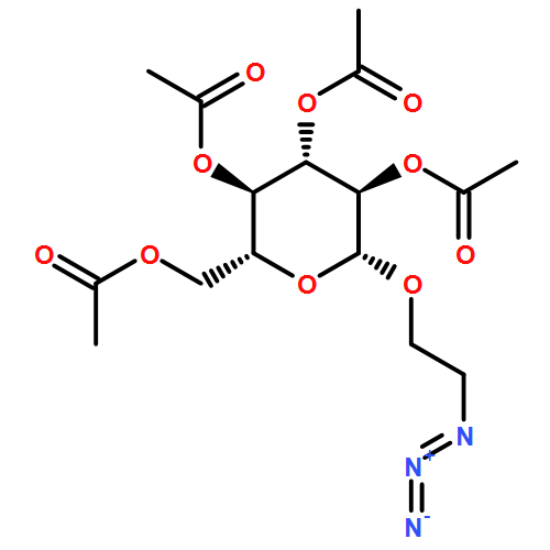


![Benzaldehyde, 2,5-bis[[(1,1-dimethylethyl)dimethylsilyl]oxy]-](http://img.cochemist.com/ccimg/114000/113984-70-6.png)
![Benzaldehyde, 2,5-bis[[(1,1-dimethylethyl)dimethylsilyl]oxy]-](http://img.cochemist.com/ccimg/114000/113984-70-6_b.png)
![5-Oxazolecarboxylicacid,2-[6-[bis(carboxymethyl)amino]-5-[2-[2-[bis(carboxymethyl)amino]-5-methylphenoxy]ethoxy]-2-benzofuranyl]-,potassium salt (1:5)](http://img.cochemist.com/ccimg/113700/113694-64-7.png)
![5-Oxazolecarboxylicacid,2-[6-[bis(carboxymethyl)amino]-5-[2-[2-[bis(carboxymethyl)amino]-5-methylphenoxy]ethoxy]-2-benzofuranyl]-,potassium salt (1:5)](http://img.cochemist.com/ccimg/113700/113694-64-7_b.png)


![Heptacyclo[31.3.1.13,7.19,13.115,19.121,25.127,31]dotetraconta-1(37),3,5,7(42),9,11,13(41),15,17,19(40),21,23,25(39),27,29,31(38),33,35-octadecaene-5,11,17,23,29,35-hexasulfonic acid, 37,38,39,40,41,42-hexahydroxy-](/data/chemimg/103300/102088-39-1.png)
![Heptacyclo[31.3.1.13,7.19,13.115,19.121,25.127,31]dotetraconta-1(37),3,5,7(42),9,11,13(41),15,17,19(40),21,23,25(39),27,29,31(38),33,35-octadecaene-5,11,17,23,29,35-hexasulfonic acid, 37,38,39,40,41,42-hexahydroxy-](/data/chemimg/103300/102088-39-1_b.png)


![ETHANOL, 2-[2-[2-(2-MERCAPTOETHOXY)ETHOXY]ETHOXY]-](http://img.cochemist.com/ccimg/91000/90952-27-5.png)
![ETHANOL, 2-[2-[2-(2-MERCAPTOETHOXY)ETHOXY]ETHOXY]-](http://img.cochemist.com/ccimg/91000/90952-27-5_b.png)
![Piperidine, 1-[(2E,8E)-9-(1,3-benzodioxol-5-yl)-1-oxo-2,8-nonadienyl]-](http://img.cochemist.com/ccimg/88700/88660-10-0.png)
![Piperidine, 1-[(2E,8E)-9-(1,3-benzodioxol-5-yl)-1-oxo-2,8-nonadienyl]-](http://img.cochemist.com/ccimg/88700/88660-10-0_b.png)
![Ethanol, 2-[2-(2-azidoethoxy)ethoxy]-](/data/chemimg/964400/86520-52-7.png)
![Ethanol, 2-[2-(2-azidoethoxy)ethoxy]-](/data/chemimg/964400/86520-52-7_b.png)
![Spiro[1H-isoindole-1,9'-[9H]xanthen]-3(2H)-one, 2-amino-3',6'-bis(diethylamino)-](/data/chemimg/659200/74317-53-6.png)
![Spiro[1H-isoindole-1,9'-[9H]xanthen]-3(2H)-one, 2-amino-3',6'-bis(diethylamino)-](/data/chemimg/659200/74317-53-6_b.png)


![Spiro[2H-1-benzopyran-2,2'-[2H]indole]-1'(3'H)-propanoic acid, 3',3'-dimethyl-6-nitro-](/data/chemimg/583600/55779-26-5.png)
![Spiro[2H-1-benzopyran-2,2'-[2H]indole]-1'(3'H)-propanoic acid, 3',3'-dimethyl-6-nitro-](/data/chemimg/583600/55779-26-5_b.png)


![Acetamide, 2-chloro-N-[2-(3,4-dihydroxyphenyl)ethyl]-](http://img.cochemist.com/ccimg/17700/17639-51-9.png)
![Acetamide, 2-chloro-N-[2-(3,4-dihydroxyphenyl)ethyl]-](http://img.cochemist.com/ccimg/17700/17639-51-9_b.png)


