Co-reporter:Karen Co Tan, Toshiyuki Wakimoto, and Ikuro Abe
Organic Letters 2014 Volume 16(Issue 12) pp:3256-3259
Publication Date(Web):June 6, 2014
DOI:10.1021/ol501271v
Lipodiscamides A–C, three new lipodepsipeptides, were characterized from the marine sponge Discodermia kiiensis. These structurally rare cyclic lipodepsipeptides were found to possess an unprecedented dilactone macrocycle and, thus, represent a new family of lipopeptides. They are the only lipopeptides bearing 4S-hydroxy-trans-2-enoate, and noncanonical amino acids, l-3-ureidoalanine (Uda), E-dehydronorvaline (Denor), and d-citrulline (Cit). MTT assays against P388 and HeLa cells revealed the moderate cytotoxicity of all three compounds.
Co-reporter:Hiroki Tajima, Toshiyuki Wakimoto, Kentaro Takada, Yuji Ise, and Ikuro Abe
Journal of Natural Products 2014 Volume 77(Issue 1) pp:154-158
Publication Date(Web):December 13, 2013
DOI:10.1021/np400668k
A cyclic peptide was isolated from the deep-sea marine sponge Discodermia japonica, and its NMR spectroscopic data were identical to those reported for cyclolithistide A, a known antifungal depsipeptide. However, the interresidue HMBC correlations suggested that the amino acid sequence was different from that of the original structure. Moreover, chiral-phase GC-MS, combined with Marfey’s analysis, indicated that the absolute configurations of three amino acids were also antipodal. Here, we propose the revised structure of cyclolithistide A and address the configuration of the previously unassigned 4-amino-3,5-dihydroxyhexanoic acid (Adha) moiety.
Co-reporter:Yoko Egami, Toshiyuki Wakimoto, Ikuro Abe
Bioorganic & Medicinal Chemistry Letters 2014 Volume 24(Issue 22) pp:5150-5153
Publication Date(Web):15 November 2014
DOI:10.1016/j.bmcl.2014.10.002
Calyculin C, a minor derivative of the calyculins, has an additional methyl group on C32 of calyculin A. A recent biosynthetic study of calyculins revealed that an end product of calyculin biosynthesis is the pyrophosphate form, phosphocalyculin A. However, the pyrophosphate counterpart derived from calyculin C had not been reported. We isolated phosphocalyculin C as a minor pyrophosphate derivative, by a detailed investigation of an extract from the sponge Discodermia calyx. The treatment of phosphocalyculin C with the D. calyx cell-free extract significantly enhanced its cytotoxicity, providing molecular evidence for its role as the protoxin of calyculin C.
Co-reporter:Karen Co Tan, Toshiyuki Wakimoto, Kentaro Takada, Takashi Ohtsuki, Nahoko Uchiyama, Yukihiro Goda, and Ikuro Abe
Journal of Natural Products 2013 Volume 76(Issue 7) pp:1388-1391
Publication Date(Web):July 12, 2013
DOI:10.1021/np400404r
A macrocylic dodecapeptide, cycloforskamide, was isolated from the sea slug Pleurobranchus forskalii, collected off Ishigaki Island, Japan. Its planar structure was deduced by extensive NMR analyses and was further confirmed by MS/MS fragmentation analyses. Finally, the absolute configuration was determined by total hydrolysis and chiral-phase gas chromatographic analysis. This novel dodecapeptide contains three d-amino acids and three thiazoline heterocycles and exhibits cytotoxicity against murine leukemia P388 cells, with an IC50 of 5.8 μM.
Co-reporter:Xiao-Long Yang, Toshiyuki Wakimoto, Yuya Takeshige, Rui He, Yoko Egami, Takayoshi Awakawa, Ikuro Abe
Bioorganic & Medicinal Chemistry Letters 2013 Volume 23(Issue 13) pp:3810-3813
Publication Date(Web):1 July 2013
DOI:10.1016/j.bmcl.2013.04.076
New indole–porphyrin hybrid molecules were isolated from Escherichia coli harboring metagenomic DNA from the Japanese marine sponge Discodermia calyx. The indole was appended to the reactive vinyl substituent of the harderoporphyrin chromophore, encoded by the insert DNA. Thus, the chimeric pathway between the heterologously expressed porphyrins and the endogenous indole generated new indole-conjugated chiral porphyrins in E. coli.
Co-reporter:Miki Kimura, Toshiyuki Wakimoto, Ikuro Abe
Tetrahedron Letters 2013 Volume 54(Issue 1) pp:114-116
Publication Date(Web):2 January 2013
DOI:10.1016/j.tetlet.2012.10.130
Allos-hemicalyculin A (1), a new derivative of calyculin A, was isolated from the marine sponge Discodermia calyx collected off Shikine-jima Island, Japan. The structure of 1, including the absolute configurations, was elucidated by spectroscopic analyses and photochemical degradation experiments. Consequently, its structure was identical to the distal end of the peptide side chain of calyculin A (2), previously isolated from D. calyx and generated by photochemical oxidative cleavage of the oxazole moiety. In stark contrast to the potent cytotoxicity of 2, 1 is no longer cytotoxic, in agreement with the previously reported structure–activity relationship data. Here we describe the isolation and structural elucidation of 1.
Co-reporter:Miki Kimura, Toshiyuki Wakimoto, Yoko Egami, Karen Co Tan, Yuji Ise, and Ikuro Abe
Journal of Natural Products 2012 Volume 75(Issue 2) pp:290-294
Publication Date(Web):January 25, 2012
DOI:10.1021/np2009187
Cyclic peptides containing 5-hydroxytryptophan and thiazole moieties were isolated from the marine sponge Discodermia calyx collected near Shikine-jima Island, Japan. The structures of calyxamides A (1) and B (2), including the absolute configurations of all amino acids, were elucidated by spectroscopic analyses and degradation experiments. The structures are similar to keramamides F and G, previously isolated from Theonella sp. The analysis of the 16S rDNA sequences obtained from the metagenomic DNA of D. calyx revealed the presence of Candidatus Entotheonella sp., an unculturable δ-proteobacterium inhabiting the Theonella genus and implicated in the biosynthesis of bioactive peptides.
Co-reporter:Rui He, Toshiyuki Wakimoto, Yoko Egami, Hiromichi Kenmoku, Takuya Ito, Yoshinori Asakawa, Ikuro Abe
Bioorganic & Medicinal Chemistry Letters 2012 Volume 22(Issue 24) pp:7322-7325
Publication Date(Web):15 December 2012
DOI:10.1016/j.bmcl.2012.10.082
Functional screening based on the antibacterial activity of a metagenomic library of the Japanese marine sponge, Discodermia calyx, afforded three β-hydroxyl fatty acids: 3-hydroxypalmitic acid, 3-hydroxylauric acid and 3-hydroxymyristic acid, heterologously expressed in an antibacterial clone, pDC113. 3-Hydroxypalmitic acid showed moderate antibacterial activity against Bacillus cereus and Candida albicans. A sequence analysis of the insert DNA revealed 23 putative ORFs, with most sharing homology to bacterial fatty acid synthase II and lipid A biosynthesis enzymes. The other ORFs were probably transmembrane proteins involved in lipid A biosynthesis. Although lipid A was not detected under our experimental conditions, the production of β-hydroxyl fatty acids as components of lipid A were enhanced in pDC113.
Co-reporter:Hikaru Kondo;Kaori Kimura;Yusuke Oka;Eri Kida;Masae Yoshida;Yiping Ye;Saeko Akahoshi;Tomohiro Asakawa;Yoko Egami;Hirohiko Nii;Kuniro Tsuji;Hitoshi Ishida;Toshiyuki Kan;Ikuro Abe;Haruo Nukaya;Koichi Matsumura
PNAS 2011 Volume 108 (Issue 42 ) pp:17533-17537
Publication Date(Web):2011-10-18
DOI:10.1073/pnas.1110577108
A lipid extract of Perna canaliculus (New Zealand green-lipped mussel) has reportedly displayed anti-inflammatory effects in animal models and in human controlled
studies. However, the anti-inflammatory lipid components have not been investigated in detail due to the instability of the
lipid extract, which has made the identification of the distinct active components a formidable task. Considering the instability
of the active component, we carefully fractionated a lipid extract of Perna canaliculus (Lyprinol) and detected furan fatty acids (F-acids). These naturally but rarely detected fatty acids show potent radical-scavenging
ability and are essential constituents of plants and algae. Based on these data, it has been proposed that F-acids could be
potential antioxidants, which may contribute to the protective properties of fish and fish oil diets against chronic inflammatory
diseases. However, to date, in vivo data to support the hypothesis have not been obtained, presumably due to the limited availability
of F-acids. To confirm the in vivo anti-inflammatory effect of F-acids in comparison with that of eicosapentaenoic acid (EPA),
we developed a semisynthetic preparation and examined its anti-inflammatory activity in a rat model of adjuvant-induced arthritis.
Indeed, the F-acid ethyl ester exhibited more potent anti-inflammatory activity than that of the EPA ethyl ester. We report
on the in vivo activity of F-acids, confirming that the lipid extract of the green-lipped mussel includes an unstable fatty
acid that is more effective than EPA.
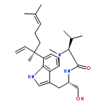
Co-reporter:Toshiyuki Wakimoto, Karen Co Tan, Ikuro Abe
Toxicon (15 September 2013) Volume 72() pp:1-4
Publication Date(Web):15 September 2013
DOI:10.1016/j.toxicon.2013.05.021
•Ergosinine was isolated from the sea slug Pleurobranchus forskalii.•Ergosinine is the first ergot peptide alkaloid (ergopeptine) found in marine life.•This study points to the possible accumulation of ergopeptines in marine macroorganisms particularly around coastal zones.The sea slug Pleurobranchus forskalii is a carnivorous scavenger that is widely distributed in shallow subtidal areas. Very few investigations of the chemical components of this gastropod have been reported. In this study we performed a comprehensive analysis of an extract of the marine mollusc, P. forskalii, collected off Ishigaki Island, Japan. As a result, an alkaloid was isolated from the chloroform extract. Remarkably, the structure elucidation based on the spectral data revealed that it was an ergot alkaloid peptide, ergosinine. Various ergot alkaloids have previously been isolated mainly from terrestrial higher plants or fungi. This is the first report of the isolation of an ergopeptine from marine life, and thus the known geographical extent of ergot alkaloids now includes both terrestrial and aquatic organisms.Download full-size image

![(1s,4s,7z,10s,16e,21r)-7-ethylidene-4,21-di(propan-2-yl)-2-oxa-12,13-dithia-5,8,20,23-tetrazabicyclo[8.7.6]tricos-16-ene-3,6,9,19,22-pentone](http://img.cochemist.com/ccimg/128600/128517-07-7.png)
![(1s,4s,7z,10s,16e,21r)-7-ethylidene-4,21-di(propan-2-yl)-2-oxa-12,13-dithia-5,8,20,23-tetrazabicyclo[8.7.6]tricos-16-ene-3,6,9,19,22-pentone](http://img.cochemist.com/ccimg/128600/128517-07-7_b.png)
![L-Ribonamide,N-[(1R,3S)-3-[4-[(1E)-3-[(2R,3R,5R,7S,8S,9R)-2-[(1S,3S,4S,5R,6R,7E,9E,11E,13Z)-14-cyano-3,5-dihydroxy-1-methoxy-4,6,8,9,13-pentamethyl-7,9,11,13-tetradecatetraen-1-yl]-9-hydroxy-4,4,8-trimethyl-3-(phosphonooxy)-1,6-dioxaspiro[4.5]dec-7-yl]-1-propen-1-yl]-2-oxazolyl]-1-methylbutyl]-4-deoxy-4-(dimethylamino)-5-O-methyl-](http://img.cochemist.com/ccimg/107600/107537-45-1.png)
![L-Ribonamide,N-[(1R,3S)-3-[4-[(1E)-3-[(2R,3R,5R,7S,8S,9R)-2-[(1S,3S,4S,5R,6R,7E,9E,11E,13Z)-14-cyano-3,5-dihydroxy-1-methoxy-4,6,8,9,13-pentamethyl-7,9,11,13-tetradecatetraen-1-yl]-9-hydroxy-4,4,8-trimethyl-3-(phosphonooxy)-1,6-dioxaspiro[4.5]dec-7-yl]-1-propen-1-yl]-2-oxazolyl]-1-methylbutyl]-4-deoxy-4-(dimethylamino)-5-O-methyl-](http://img.cochemist.com/ccimg/107600/107537-45-1_b.png)
![9,15-Methano-3H,11H,13H-furo[3',4':3a,4]indeno[1,7a-g][2]benzoxepin-3,11,16-trione, 5,5a,6,7,8,9,10,10a,14,15,15a,15b-dodecahydro-5,5,9,15b-](/data/chemimg/1334400/79874-93-4.png)
![9,15-Methano-3H,11H,13H-furo[3',4':3a,4]indeno[1,7a-g][2]benzoxepin-3,11,16-trione, 5,5a,6,7,8,9,10,10a,14,15,15a,15b-dodecahydro-5,5,9,15b-](/data/chemimg/1334400/79874-93-4_b.png)


![5H-Indolo[2,3-a]pyrrolo[3,4-c]carbazole-5,7(6H)-dione,12,13-dihydro-](http://img.cochemist.com/ccimg/118500/118458-54-1.png)
![5H-Indolo[2,3-a]pyrrolo[3,4-c]carbazole-5,7(6H)-dione,12,13-dihydro-](http://img.cochemist.com/ccimg/118500/118458-54-1_b.png)


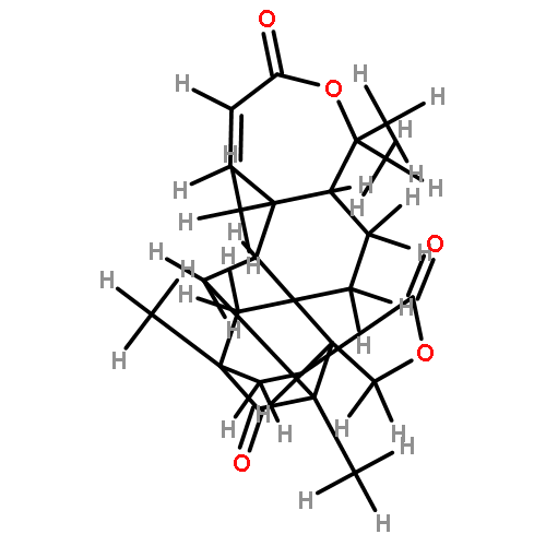
![methyl (2R,4aR,4bS,10aS,12aR)-6,10b-dihydroxy-2,4b,7,7,10a,12a-hexamethyl-12-methylidene-1,4,5,8-tetraoxo-1,4,4a,4b,5,7,8,9,10,10a,10b,11,12,12a-tetradecahydro-2H-naphtho[1,2-h]isochromene-2-carboxylate](http://img.cochemist.com/ccimg/72000/71911-90-5.png)
![methyl (2R,4aR,4bS,10aS,12aR)-6,10b-dihydroxy-2,4b,7,7,10a,12a-hexamethyl-12-methylidene-1,4,5,8-tetraoxo-1,4,4a,4b,5,7,8,9,10,10a,10b,11,12,12a-tetradecahydro-2H-naphtho[1,2-h]isochromene-2-carboxylate](http://img.cochemist.com/ccimg/72000/71911-90-5_b.png)
![Ethanone, 1-(9H-pyrido[3,4-b]indol-1-yl)-](http://img.cochemist.com/ccimg/50900/50892-83-6.png)
![Ethanone, 1-(9H-pyrido[3,4-b]indol-1-yl)-](http://img.cochemist.com/ccimg/50900/50892-83-6_b.png)
![[(2S)-4-butanoyl-5-hydroxy-3-oxo-2,3-dihydrofuran-2-yl]acetic acid](http://img.cochemist.com/ccimg/33500/33404-61-4.png)
![[(2S)-4-butanoyl-5-hydroxy-3-oxo-2,3-dihydrofuran-2-yl]acetic acid](http://img.cochemist.com/ccimg/33500/33404-61-4_b.png)


![Diphosphoric acid,P-[(2E,6E,10E)-3,7,11,15-tetramethyl-2,6,10,14-hexadecatetraen-1-yl] ester](http://img.cochemist.com/ccimg/6700/6699-20-3.png)
![Diphosphoric acid,P-[(2E,6E,10E)-3,7,11,15-tetramethyl-2,6,10,14-hexadecatetraen-1-yl] ester](http://img.cochemist.com/ccimg/6700/6699-20-3_b.png)


![(3AR,4R,5R,6AS)-4-FORMYL-2-OXOHEXAHYDRO-2H-CYCLOPENTA[B]FURAN-5-Y<WBR />L 4-BIPHENYLCARBOXYLATE](http://img.cochemist.com/ccimg/100/72-89-9.png)
![(3AR,4R,5R,6AS)-4-FORMYL-2-OXOHEXAHYDRO-2H-CYCLOPENTA[B]FURAN-5-Y<WBR />L 4-BIPHENYLCARBOXYLATE](http://img.cochemist.com/ccimg/100/72-89-9_b.png)
![7-hydroxy-8,12-dihydro-1H-indolo[2,3-a]pyrrolo[3,4-c]carbazole-1,3,10(2H,9H)-trione](http://img.cochemist.com/ccimg/133900/133805-03-5.png)
![7-hydroxy-8,12-dihydro-1H-indolo[2,3-a]pyrrolo[3,4-c]carbazole-1,3,10(2H,9H)-trione](http://img.cochemist.com/ccimg/133900/133805-03-5_b.png)