Co-reporter:Qiong Hu, Qiangwei Wang, Gengzhi Sun, Jinming Kong, and Xueji Zhang
Analytical Chemistry September 5, 2017 Volume 89(Issue 17) pp:9253-9253
Publication Date(Web):August 15, 2017
DOI:10.1021/acs.analchem.7b02039
The development of convenient and efficient strategies without involving any complex nanomaterials or enzymes for signal amplification is of great importance in bioanalytical applications. In this work, we report the use of electrochemically mediated surface-initiated atom transfer radical polymerization (SI-eATRP) as a novel amplification strategy based on the de novo growth of polymers (dnGOPs) for the electrochemical detection of DNA. Specifically, the capture of target DNA (tDNA) by the immobilized peptide nucleic acid (PNA) probes provides a high density of phosphate groups for the subsequent attachment of ATRP initiators onto the electrode surface by means of the phosphate-Zr4+-carboxylate chemistry, followed by the de novo growth of electroactive polymer via the SI-eATRP. De novo growth of long polymeric chains enables the labeling of numerous electroactive probes, which in turn greatly improves the electrochemical response. Moreover, it circumvents the slow kinetics and poor coupling efficiency encountered when nanomaterials or preformed polymers are used and features sufficient flexibility and simplicity in controlling the degree of signal amplification. Under optimal conditions, it allows a highly sensitive and selective detection of tDNA within a broad linear range from 0.1 fM to 0.1 nM (R2 = 0.996), with the detection limit down to 0.072 fM. Compared with the unamplified method, more than 1.2 × 10⁶-fold sensitivity improvement in DNA detection can be achieved. By virtue of its simplicity, high efficiency, and cost-effectiveness, the proposed dnGOPs-based signal amplification strategy holds great potential in bioanalytical applications for the sensitive detection of biological molecules.
Co-reporter:Qiong Hu;Li Li;Gengzhi Sun;Dali Li;Wei Huang;Xueji Zhang
Journal of Materials Chemistry B 2017 vol. 5(Issue 30) pp:5937-5941
Publication Date(Web):2017/08/02
DOI:10.1039/C7TB01064H
5-Carboxyfluorescein is verified to possess intrinsic peroxidase-like catalytic activity and can serve as a robust artificial peroxidase for the biomimetic synthesis of polyaniline nanoplatelets under harsh conditions. Our work discloses the potential of using this small molecule as a peroxidase surrogate for the biomimetic synthesis of nanostructured polymers.
Co-reporter:Qiong Hu, Minhui He, Yaqi Mei, Wenjie Feng, Su Jing, Jinming Kong, Xueji Zhang
Talanta 2017 Volume 163() pp:146-152
Publication Date(Web):15 January 2017
DOI:10.1016/j.talanta.2016.10.097
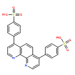
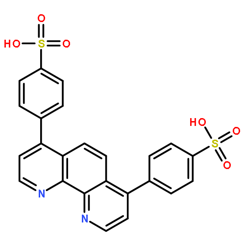
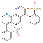
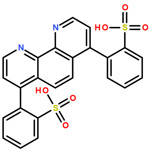
•We report a simple and practical method for the colorimetric assay of ALP activity.•We exploit the Cu(BPDS)23− complex as the reporter for its distinctive MLCT properties.•It allows a naked-eye readout of ALP activity and is highly immune to false positives.•It is selective and holds great potential in ALP inhibitor efficiency evaluation.•It is practical for its satisfactory analytical performance in complex serum samples.Alkaline phosphatase (ALP) plays a vital role in dephosphorylation- and phosphorylation-related cellular regulation and signaling processes. Accordingly, the development of efficient methods for ALP activity assay is of significant importance in clinical diagnosis. In this work, a simple and practical method is reported for the first time for the sensitive and selective colorimetric assay of ALP activity by exploiting a water-soluble Cu(II)-phenanthroline complex as the probe, on the basis of the distinctive metal-to-ligand charge-transfer (MLCT) properties. This method is simply built on a two-step chromogenic reaction: the enzymatic hydrolysis of the substrate ascorbic acid 2-phosphate to ascorbic acid (AA), followed by the reduction of the colorimetric probe Cu(BPDS)22− (BPDS=bathophenanthroline disulfonate) by AA to its cuprous form. The latter process triggers a turn-on spectral absorption at 424 nm and a striking color change of the solution from colorless to blackish-green. Needless of complicated protocols and instrumentation, this method allows a sensitive readout of ALP activity within a wide linear range of 0–200 mU mL−1, with a detection limit down to 1.25 mU mL−1. Results also reveal that it is highly selective and holds great potential in ALP inhibitor efficiency evaluation. In addition, quantitative analysis of ALP activity in spiked serum samples has been realized successfully in the linear range of 0–200 mU mL−1, with a detection limit of 1.75 mU mL−1. Advantages of simplicity, wide linear range, high sensitivity and selectivity, low cost, and little background interference render this method great potential in practical applications.
Co-reporter:Qianrui Liu;Qiong Hu;Lianzhi Li;Xueji Zhang
Analytical Methods (2009-Present) 2017 vol. 9(Issue 25) pp:3825-3830
Publication Date(Web):2017/06/30
DOI:10.1039/C7AY00861A
In this study, a click chemistry-based electrochemical aptasensor is reported for the simple and fast detection of thrombin. Briefly, the thiol-terminated aptamer1 (Apt1) was first self-assembled on a gold electrode for the specific recognition of thrombin in the following step. After this, the azido-labeled aptamer2 (Apt2) was attached to the electrode via the formation of a sandwich structure with Apt1 and thrombin. The azido terminal was designed for the subsequent labeling of Apt2 with ethynylferrocene via electrochemically mediated Cu(I)-catalyzed azide–alkyne cycloaddition (eCuAAC), in which the active Cu(I) was electrochemically generated in situ. Finally, the quantitatively labeled ethynylferrocene probes could be detected on the basis of differential pulse voltammetry (DPV) for the electrochemical detection of thrombin. Under optimal conditions, this aptasensor could detect thrombin down to 84 pM with a good linear response over the range from 0.1 nM to 1000 nM. In addition, it exhibited excellent specificity in the quantitative detection of thrombin and also showed good detection reliability in serum samples.
Co-reporter:Weiwen Hu, Xuehua Yu, Qiong Hu, Jinming Kong, ... Xueji Zhang
Journal of Environmental Sciences 2017 Volume 53(Volume 53) pp:
Publication Date(Web):1 March 2017
DOI:10.1016/j.jes.2016.01.016
A novel poly(ethyleneimine)/Au nanoparticles/hemin nanocomposite (PEI-AuNPs-Hemin) acting for Methyl Orange (MO) removal has been synthesized. PEI-AuNPs was prepared firstly and it was then linked to hemin through the coupling between carboxyl groups in hemin and amino groups in PEI without the activation of carboxyl groups. The high reactivity and stability of AuNPs contributed greatly in the formation of the amido bonds in the nanocomposite. Fourier transform infrared spectroscopy, transmission electron microscopy and UV–visible spectroscopy were used to characterize the PEI-AuNPs-Hemin. Results show that PEI-AuNPs-Hemin has strong adsorption for MO. Adsorption and degradation experiments were carried out at different pHs, nanocomposite concentrations and UV irradiation times. Removal of MO in acidic solutions was more effective than in basic solutions. The real-time study showed that the MO degradation with the nanocomposite under UV irradiation was a fast process. In addition, the photocatalytic degradation mechanism was proposed. The study suggests that the PEI-AuNPs-Hemin may have promising applications in environmental monitoring and protection.Download high-res image (176KB)Download full-size image
Co-reporter:Qiong Hu, Jinming Kong, Yajie Li, Xueji Zhang
Talanta 2016 Volume 147() pp:516-522
Publication Date(Web):15 January 2016
DOI:10.1016/j.talanta.2015.10.039
•This is a signal-on biosensor and it is highly compatible with multiplexed detections.•The φCuAAC is exploited to label hairpins with EFC under mild conditions.•The labeling is rapid, selective and can be realized conveniently and efficiently.•It is highly specific and holds good detection capability in complicated serum sample.•The sensitivity and stability are satisfactory with the protocol has been simplified.A novel electrochemical biosensor was developed for the signal-on detection of sequence-specific DNA by exploiting potential-assisted Cu(I)-catalyzed azide-alkyne cycloaddition (φCuAAC) as an efficient approach for the labeling of hairpin-like oligonucleotide (hairpin) with electroactive probe. The hairpins, dually labeled with thiol and azide at either terminal, were firstly self-assembled on gold electrode and served as the capture probes for the specific recognition of target DNA. Upon hybridization with target DNA, the surface-confined hairpins were unfolded, liberating the azide-containing terminals away from electrode surface. Subsequently, the unfolded hairpins were conveniently and efficiently labeled with ethynylferrocene (EFC) via the φCuAAC. The quantitatively labeled EFC was finally measured via differential pulse voltammetry (DPV) for the signal-on electrochemical detection of sequence-specific DNA. The biosensor presented a good linear response over the range from 1 pM to 1 nM with a detection limit of 0.62 pM. Results also revealed that it was highly specific and held a good detection capability in serum samples. Furthermore, the ability to chemoselectively label hairpin-like oligonucleotide with signal reporter by electrical addressing, together with the simplicity and efficiency of the φCuAAC, makes it compatible with microfluidic devices and microelectrode arrays to achieve the miniaturized and multiplexed detections.
Co-reporter:Weiwen Hu, Yong Ning, Jinming Kong and Xueji Zhang
Analyst 2015 vol. 140(Issue 16) pp:5678-5684
Publication Date(Web):23 Jun 2015
DOI:10.1039/C5AN01109D
Poly(thymine) (polyT) and double-stranded DNA (dsDNA) can act as efficient templates for the formation of copper nanoparticles (CuNPs) at a low concentration of CuSO4, and the formed CuNPs emit excellent fluorescence. In this work, we demonstrated a new and facile strategy for the highly sensitive and selective detection of DNA on streptavidin-functionalized magnetic beads (SA-MB) using DNA-templated CuNPs as the fluorescent probe. Target DNA (tDNA) was hybridized with the capture DNA that was immobilized on the surface of SA-MB. Surface initiated enzymatic polymerization (SIEP) was employed as the signal amplification method to generate the polyT at the 3′ end of tDNA for the formation of CuNPs. The incorporation of polyT by SIEP resulted in ∼35.7 fold signal amplification compared to the dsDNA after hybridization without SIEP. A dose–response curve for detection of DNA was obtained, with a linear dynamic range of 0.1 nM to 10 nM. We showed that this method has a low pM limit of detection (LOD 98.2 pM) and it is also very sensitive to the mismatch type in a specific DNA sequence. In addition, it avoids rigorously controlled temperature, complex synthesis of the fluorescent probe and prelabeling of DNA strands and eliminates the use of sophisticated experimental techniques and equipment. Armed with these intriguing properties, the proposed system could provide an efficient tool for early diagnosis and risk assessment of malignancy.
Co-reporter:Qiong Hu, Xianbao Deng, Jinming Kong, Yuanyuan Dong, Qianrui Liu and Xueji Zhang
Analyst 2015 vol. 140(Issue 12) pp:4154-4161
Publication Date(Web):13 Apr 2015
DOI:10.1039/C5AN00566C
A universal and straightforward electrochemical biosensing strategy for the detection and identification of sequence-specific DNA via click chemistry-mediated labeling of hairpin DNA probes (hairpins) with ethynylferrocene was reported. In the target-unbound form, the immobilized hairpins were kept in the folded stem–loop configuration with their azido terminals held in close proximity of the electrode surface, making them difficult to be labeled with ethynylferrocene due to the remarkable steric hindrance of the densely packed hairpins. Upon hybridization, they were unfolded and underwent a large conformational change, thus enabling the azido terminals to become available for its subsequent conjugation with ethynylferrocene via the Cu(I)-catalyzed azide–alkyne cycloaddition (CuAAC). After that, the quantitatively labeled ethynylferrocene could be exploited as the electroactive probes to monitor the DNA hybridization. As the unfolded hairpins were labeled in a stoichiometric ratio of 1:1, the electrochemical measurement based on differential pulse voltammetry enabled a reliable quantification of sequence-specific DNA. Under optimal conditions, the strategy could detect target single-stranded DNA (ssDNA) down to 0.296 pM with a good linear response over the range from 1 pM to 1 nM, and had excellent specificity in the genotyping of single-nucleotide polymorphisms. Furthermore, it also exhibited good detection reliability in serum samples and required no complicated protocols. More importantly, the simplicity of this strategy together with its compatibility with microfluidic chips makes it show great potential in clinical applications, where simple procedures are generally preferred.
Co-reporter:Weiwen Hu, Yong Ning, Lianzhi Li, Jinming Kong and Xueji Zhang
Analytical Methods 2015 vol. 7(Issue 16) pp:6712-6717
Publication Date(Web):10 Jul 2015
DOI:10.1039/C5AY00628G
In this work, a highly effective biosensor supported on morpholino-functionalized magnetic microspheres was investigated for the sequence-specific detection of DNA. Briefly, morpholino was first modified on the surface of magnetic microspheres through amido bonds, and then hybridized with target DNA in the ensuing step. Biotin-labeled signal probe DNA was designed to hybridize with the DNA nearby the 5′ end of sequence, and the biotin was used to bind with streptavidin-alkaline phosphatase (SA-ALP). L-Ascorbic acid-2-phosphate (AAP) was hydrolyzed by ALP to generate L-ascorbic acid, which could reduce resazurin to resorufin, resulting in a turn-on fluorescence signal. UV-vis absorbance spectroscopy was applied to prove the conjugation of ALP with signal probe DNA. Under optimal conditions, this biosensor displayed a good linear relationship between the fluorescence intensity and logarithm of single-stranded DNA concentrations in the range of 0.1 pM to 0.1 nM with a low detection limit of 9.32 fM. Moreover, fully complementary versus single-base mismatched, three-base mismatched and non-complementary DNA could be effectively distinguished. In addition, this novel approach rendered satisfactory analytical results for the determination of DNA in serum, thus exhibiting practical significance. These results demonstrated that this assay method displays excellent sensitivity and specificity for DNA detection and has great potential for practical applications.
Co-reporter:Weiwen Hu, Qiong Hu, Lianzhi Li, Jinming Kong and Xueji Zhang
Analytical Methods 2015 vol. 7(Issue 6) pp:2406-2412
Publication Date(Web):27 Jan 2015
DOI:10.1039/C4AY02780A
In this work, an efficient method for the sequence-specific detection of DNA based on a morpholino-functionalized silicon chip platform has been proposed. Briefly, morpholino was first immobilized on the surface of a silicon chip using 3-aminopropyltriethoxysilane (APTES) as the silane coupling agent and 1,4-phenylenediisothiocyanate (PDITC) as the cross-linker and then hybridized with DNA in the ensuing step. The fluorescence label was introduced by strongly binding Rhodamine B, which contains a terminal carboxylic group, with DNA by means of phosphate–zirconium–carboxylate coordination reaction. X-ray photoelectron spectroscopy (XPS) was used to characterize the silicon surface. Under optimal conditions, the morpholino-functionalized silicon chip presented a great linear relationship between the fluorescence intensity and the logarithm of target DNA concentrations in the range from 1 pM to 1 nM with a detection limit of 4.52 pM. Furthermore, fully complementary versus single-base mismatched, three-base mismatched and non-complementary DNA could be effectively identified. The chip showed excellent stability because it could be reused for another hybridization experiment after denaturing the morpholino–complementary DNA duplex. In addition, the chip rendered satisfactory analytical performance for the detection of DNA in serum samples, thus exhibiting practical significance. Morpholino-functionalized silicon chips display good sensitivity and selectivity for the detection of DNA and promising applications in single-nucleotide polymorphisms (SNPs).
Co-reporter:Qiong Hu, Xianbao Deng, Xuehua Yu, Jinming Kong, Xueji Zhang
Biosensors and Bioelectronics 2015 Volume 65() pp:71-77
Publication Date(Web):15 March 2015
DOI:10.1016/j.bios.2014.10.015
•We presented a novel and straightforward electrochemical DNA biosensing approach.•AFC was labeled to captured ssDNA as electroactive probe via one-step reaction only.•Imidazole and EDC were exploited as the zero-length cross-linkers in conjugation.•It was highly specific and it also showed acceptable analytical reliability in serum samples.•It could integrate with micro-fabrication techniques to develop microfluidic chips.A straightforward electrochemical DNA biosensing approach based on exploiting organometallic compound, aminoferrocene (AFC), as electroactive probes was firstly demonstrated, where the probes could be directly labeled to the free phosphate groups of the hybridized PNA/DNA heteroduplexes merely through one-step conjugation in the presence of 1-ethyl-3-(3-dimethylaminopropyl) carbodiimide (EDC) and imidazole. Briefly, mercapto-terminated peptide nucleic acid (PNA) was firstly immobilized onto gold electrode and used as the capture probes for the specific recognition of target single-stranded DNA (ssDNA). After hybridization, AFC probes were directly labeled to the free 5′-terminal phosphate groups, which were activated by EDC and imidazole, of the hybridized PNA/DNA heteroduplexes, and then they were exploited as the electroactive probes to monitor the hybridization. As the captured ssDNA was labeled with AFC in the stoichiometric ratio of 1:1, thus the electrochemical analysis of the proportionally labeled AFC based on differential pulse voltammetry (DPV) enabled a quantitative determination of sequence-specific DNA. Under optimal conditions, the approach presented a good linear relationship between the current intensities and logarithm of ssDNA concentrations in the range from 0.1 nM to 100 nM with a detection limit of 93 pM, and it rendered satisfactory analytical performance in serum samples. Furthermore, it exhibited excellent specificity toward single-nucleotide polymorphism (SNP) and precluded complicated protocols. More importantly, the simplicity of this approach together with its compatibility with standard micro-fabrication techniques makes it great potential in practical applications, especially in microarray areas where simple procedures are preferred.
Co-reporter:Qiong Hu, Weiwen Hu, Jinming Kong, Xueji Zhang
Biosensors and Bioelectronics 2015 Volume 63() pp:269-275
Publication Date(Web):15 January 2015
DOI:10.1016/j.bios.2014.07.034
•We presented a novel electrochemical DNA biosensor for the detection of target ssDNA.•Hematin was exploited as efficient biomimetic catalyst toward in situ metallization.•Hematin-based signal amplification and SWV synergistically improved the sensitivity.•It exhibited a wide linearity from 0.1 fM to 0.1 nM and a detection limit of 62.41 aM.•It was highly specific and showed acceptable analytical reliability in serum samples.In this work, we presented a novel signal amplification approach to construct an electrochemical DNA biosensor for the ultrasensitive determination of sequence-specific DNA by exploiting hematin as biomimetic catalyst toward in situ metallization. Briefly, thiolated peptide nucleic acid (PNA) probes were firstly immobilized onto gold electrode through the formation of self-assembled monolayer (SAM) and then hybridization was accomplished in the ensuing step. After that, hematin molecules were introduced to the hybridized PNA/DNA heteroduplexes by employing phosphate–zirconium–carboxylate coordination chemistry. Next, the attached hematin molecules acted as catalyst in accelerating the reduction of silver ions in the presence of catechol, leading to the in situ deposition of silver particles onto the electrode. Finally, the deposited silver particles were electrochemically stripped into KCl solution and measured by square wave voltammetry (SWV). Under optimal conditions, the hematin-based electrochemical DNA biosensor presented a good linear relationship between the stripping peak currents and logarithm of single-stranded DNA (ssDNA) concentrations in the range from 0.1 fM to 0.1 nM with a low detection limit of 62.41 aM, and it rendered satisfactory analytical performance for the determination of ssDNA in serum samples. Furthermore, it exhibited good reproducibility and stability, meanwhile, it also showed excellent specificity toward single-nucleotide polymorphism (SNP). Therefore, the hematin-based signal amplification approach has great potential in clinical applications and is also suitable for quantification of biomarkers at ultralow level.
Co-reporter:X. H. Yu, J. M. Kong, X. J. Han and X. J. Zhang
RSC Advances 2014 vol. 4(Issue 87) pp:46980-46986
Publication Date(Web):05 Sep 2014
DOI:10.1039/C4RA05886K
In this work, we prepare a novel platform based on poly(3,4-ethylenedioxythiophene) (PEDOT) and 1-pyrenebutanoic acid (PBA). PEDOT is a conductive material of heteroatom doping, which can connect with PBA through π–π stacking. The feasibility of the film is verified via fabricating it on a glassy carbon electrode (GCE); then, hematin is linked with PBA via carboxylate–zirconium–carboxylate coordination bond to prepare a GCE/PEDOT–PBA–hematin biosensor. The electrochemical performance of the biosensor has been tested by electrochemical impedance spectroscopy (EIS), cyclic voltammetry (CV) and current-time curve method (I–T). From CV, a pair of well-defined and quasi-reversible redox peaks, corresponding to the hematin Fe(III)/Fe(II) redox couple, is observed, and the surface coverage (Γ*) of hematin on GCE has been calculated to be 1.2 × 10⁻⁹ mol cm−2, which is almost 20 times larger than the monolayer coverage of hemin. This value shows that the PEDOT and PBA composite results in a better loading of hematin on the surface of the GCE. In addition, the GCE/PEDOT–PBA–hematin biosensor exhibits strong electro-catalysis activity for H2O2 and displays a linear response for the reduction of H2O2 in the range of 0.005 to 1.322 mmol L−1 with a detection limit of 0.03 μmol L−1 and a high sensitivity of 2.83 μA mM−1 cm−2. In addition, the sensor has been applied to the determination of H2O2 in real samples, and the response is in the ideal range, which implies that the GCE/PEDOT–PBA–hematin biosensor has promising future applications.
Co-reporter:Lin Bian;Lianzhi Li;Qingfu Zhang;Jianfang Dong;Tao Xu
Transition Metal Chemistry 2012 Volume 37( Issue 8) pp:783-790
Publication Date(Web):2012 November
DOI:10.1007/s11243-012-9653-9
Two new V(IV) complexes, [VO(Naph–trp)(phen)]·CH3OH (1) and [VO(o-Van–trp)(phen)]·CH3OH·H2O (2) (Naph–Trp = Schiff base derived from 2-hydroxy-1-naphthaldehyde and l-tryptophan, o-Van–trp = Schiff base derived from o-vanillin and l-tryptophan, phen = 1,10-phenanthroline), have been synthesized and characterized by physicochemical methods. The V(IV) atoms in both complexes are six-coordinated in a distorted octahedral environment. In the crystals of complex 1, the C–H···π and π–π stacking interactions form a 1D chain structure, whereas for complex 2, hydrogen bonds connect the molecular units into a 2D plane structure. The DNA binding properties and cleavage efficiencies of the complexes have been investigated by spectroscopic methods, viscosity measurements and agarose gel electrophoresis. The results suggest that both complexes can bind to CT-DNA in an intercalative mode and can also cleave pBR322 DNA.
Co-reporter:Qiong Hu, Kefeng Ma, Yaqi Mei, Minhui He, Jinming Kong, Xueji Zhang
Talanta (15 May 2017) Volume 167() pp:253-259
Publication Date(Web):15 May 2017
DOI:10.1016/j.talanta.2017.02.027
Co-reporter:Qiong Hu, Baojing Zhou, Pengyun Dang, Lianzhi Li, Jinming Kong, Xueji Zhang
Analytica Chimica Acta (15 January 2017) Volume 950() pp:
Publication Date(Web):15 January 2017
DOI:10.1016/j.aca.2016.11.012
•A Fe(II)-phenanthroline reporter was used for the colorimetric assay of ALP activity.•The approach is based on the distinctive MLCT absorption properties of the reporter.•It is highly selective and holds great potential in ALP inhibitor screening.•It holds considerable simplicity and flexibility with respect to reporter design.•It was successfully employed to detect the endogenous ALP level of serum samples.We report a versatile approach for the colorimetric assay of alkaline phosphatase (ALP) activity based on the distinctive metal-to-ligand charge-transfer (MLCT) absorption properties of Fe(II)-phenanthroline reporter. In the presence of ALP, the applied substrate ascorbic acid 2-phosphate is enzymatically hydrolyzed to produce ascorbic acid, which then reduces Fe3+ to Fe2+. The complexation of Fe2+ with the bathophenanthroline disulfonate (BPS) ligand generates a blood-red Fe(BPS)34− reporter, which is characterized by an intense MLCT absorption band at 535 nm in the visible range. Under optimal conditions, the spectral output exhibits a good quantitative relationship with ALP activity over the range of 0–220 mU mL−1 with a detection limit of 0.94 mU mL−1. Moreover, the activity of ALP can also be conveniently judged through naked-eye observations. Results indicate that it is highly selective and can be applied to the screening of ALP inhibitors. In addition, it has been successfully employed to detect the endogenous ALP level of undiluted human serum samples, with a detection limit of 1.05 mU mL−1 being achieved. This approach avoids any elaborately designed substrates and holds considerable simplicity and flexibility for reporter design. This study broadens the horizon of the applications of phenanthroline-based transition metal complexes. Furthermore, an efficient and practical method like this has the potential to be widely used in clinical applications and in the point-of-care testing.In this work, a versatile approach is reported for the colorimetric assay of alkaline phosphatase (ALP) activity on the basis of the distinctive metal-to-ligand charge-transfer (MLCT) absorption properties of Fe(II)-phenanthroline complex. The limit of detection is lower than that of most of the reported colorimetric methods. It is highly selective and can be applied to the screening of ALP inhibitors. Our approach features high interference immunity and reproducibility, and is highly immune to false-positive results. In addition, it has been successfully employed to detect the endogenous ALP level of human serum samples, and a detection limit of 1.05 mU mL−1 was achieved. This approach is highly efficient and involves only a single step. It avoids elaborately designed substrates, and in particular all chemicals used in this approach are commercially available and inexpensive.
Co-reporter:Qiong Hu, Li Li, Gengzhi Sun, Dali Li, Jinming Kong, Wei Huang and Xueji Zhang
Journal of Materials Chemistry A 2017 - vol. 5(Issue 30) pp:NaN5941-5941
Publication Date(Web):2017/07/06
DOI:10.1039/C7TB01064H
5-Carboxyfluorescein is verified to possess intrinsic peroxidase-like catalytic activity and can serve as a robust artificial peroxidase for the biomimetic synthesis of polyaniline nanoplatelets under harsh conditions. Our work discloses the potential of using this small molecule as a peroxidase surrogate for the biomimetic synthesis of nanostructured polymers.