Co-reporter:Yanfang Wang, Pengfei Li, Ping Xiang, Jueting Lu, Jiang Yuan and Jian Shen
Journal of Materials Chemistry A 2016 vol. 4(Issue 4) pp:635-648
Publication Date(Web):21 Dec 2015
DOI:10.1039/C5TB02358K
Keratin based biomaterials have emerged as potential candidates for various biomedical and biotechnological applications due to their intrinsic biocompatibility, biodegradability, mechanical durability, and natural abundance. The objective of this study is to combine the merits of polyurethane, keratin, and silver nanoparticles (AgNPs) together and develop a novel nanofibrous mat for wound dressing. Herein, keratin was first extracted from human hair and chemically modified with iodoacetic acid to afford S-(carboxymethyl) keratin. The modified keratin was examined using Raman spectroscopy, infrared spectroscopy, and SDS-PAGE. The keratin was then blended with polyurethane (PU) and electrospun. Subsequently, AgNPs were formed in situ to afford antibacterial PU/keratin/AgNP mats. These mats were characterized using field emission scanning electron microscopy (FE-SEM), attenuated total reflection Fourier transform infrared spectroscopy (ATR-FTIR), water contact angle measurements, and X-ray photoelectron spectroscopy (XPS). MTT results indicated that the introduction of keratin could accelerate fibroblast cell proliferation, while the loaded AgNPs did not weaken cytocompatibility. Antibacterial test results showed that PU/keratin/AgNP mats exerted good antibacterial property. The results from a wound healing test and a histological examination suggested that these biocomposite mats could remarkably accelerate wound recovery as compared to the conventional gauze sponge dressing. Given their excellent biocompatibility, antibacterial properties, and very mild inflammatory responses, PU/keratin/AgNP mats have great potential for wound dressing applications.
Co-reporter:Xingxing Jin, Yanfang Wang, Jiang Yuan, Jian Shen
Materials Letters 2016 Volume 175() pp:188-190
Publication Date(Web):15 July 2016
DOI:10.1016/j.matlet.2016.04.036
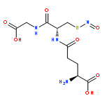
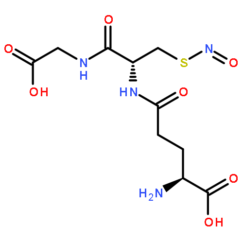
•Keratin is extracted and modified to afford S-carboxymethyl keratin.•The amino acid residues, isoelectric point, and molecular weight are quantified.•Keratin is capable of promoting NO release from GSNO in the presence of glutathione.Keratin-based biomaterials have emerged as potential candidates for various biomedical and biotechnological applications due to their intrinsic biocompatibility, biodegradability, mechanical durability, and natural abundance. The objective of our study is to explore the feasibility and potential applications of keratin for promoting nitric oxide (NO) release. Herein, keratin was first extracted from human hair and then modified with iodoacetic acid to afford S-carboxymethyl keratin. The amino acid residues, isoelectric point, and molecular weight of keratin were quantified with amino acid analyzer, zeta potential measurement, and SDS-PAGE method. Interestingly, human hair keratin was proved to be capable of promoting NO release from endogenous donor of S-nitrosoglutathione (GSNO) in the presence of glutathione.Keratin was extracted and modified to afford S-carboxymethyl keratin. The amino acid residues, isoelectric point, and molecular weight of keratin were quantified with amino acid analyzer, zeta potential measurement, and SDS-PAGE method. Human hair keratin was proved to be capable of promoting NO release from endogenous donor of S-nitrosoglutathione (GSNO) in the presence of glutathione (GSH).
Co-reporter:Yanfang Wang, Weiwei Zhang, Jiang Yuan, Jian Shen
Materials Science and Engineering: C 2016 Volume 59() pp:30-34
Publication Date(Web):1 February 2016
DOI:10.1016/j.msec.2015.09.093
•Collagen, gelatin and keratin were coelectrospun with PHBV to afford nanofibrous mats.•Cytocompatibility was evaluated with cell adhesion, cell viability and cell proliferation.•Collagen had significantly superior cytocompatibility as compared to gelatin and keratin.Keratins are cysteine-rich intermediate filament proteins found in the cytoskeleton of the epithelial cells and in the matrix of hair, feathers, wool, nails and horns. The natural abundance of cell adhesion sequences, RGD (Arg-Gly-Asp) and LDV (Leu-Asp-Val), makes them suitable for tissue engineering applications. The purpose of our study is to evaluate their cytocompatibility as compared to well-known collagen and gelatin proteins. Herein, collagen, gelatin and keratin were blended with poly(hydroxybutyrate-co-hydroxyvalerate) (PHBV) and electrospun to afford nanofibrous mats, respectively. These PHBV/protein composite mats were characterized by field emission scanning electron microscopy (FE-SEM), attenuated total reflection Fourier transform infrared spectroscopy (ATR-FTIR), X-ray photoelectron spectroscopy (XPS), and dynamic mechanical analysis (DMA). The cytocompatibility was evaluated with cell adhesion, cell viability and cell proliferation. The data from MTT and BrDU revealed that collagen had significantly superior cytocompatibility as compared to gelatin and keratin. Gelatin showed a better cytocompatibility than keratin without statistical significance difference. Finally, we gave the reasons to account for the above conclusions.
Co-reporter:Yanmei Li, Yanfang Wang, Jingjie Ye, Jiang Yuan, Yinghong Xiao
Materials Science and Engineering: C 2016 Volume 68() pp:177-183
Publication Date(Web):1 November 2016
DOI:10.1016/j.msec.2016.05.117
•Keratins are coelectrospun with PCL to afford nanofibrous mats.•PCL/keratin mats show good cytocomatibility and blood compatibility.•PCL/keratin mats have great potential as scaffold for vascular tissue engineering.The natural abundance of cell adhesion sequences, RGD (Arg-Gly-Asp) and LDV (Leu-Asp-Val) in the keratins make them suitable as biomaterials for tissue engineering applications. Herein, keratins were coelectrospun with poly(ε-caprolactone)(PCL) at the ratio of 9/1, 8/2, and 7/3 to afford nanofibrous mats. The resulting mats were surface-characterized by ATR-FTIR, SEM, WCA, and XPS. Cell attachment data showed that NIH 3T3 cells adhered more to the PCL/keratin nanofibrous mats than the pristine PCL mats. The MTT assay revealed that the PCL/keratin mats had improved cell viability. The blood clotting time test (APTT, PT, and TT) indicated the PCL/keratin mats exerted good blood compatibility. These mats would be a good candidate as a scaffold for vascular tissue engineering.
Co-reporter:Miao Wang, Jiang Yuan, Xiaobo Huang, Xianmei Cai, Li Li, Jian Shen
Colloids and Surfaces B: Biointerfaces 2013 Volume 103() pp:52-58
Publication Date(Web):1 March 2013
DOI:10.1016/j.colsurfb.2012.10.025
Grafting-from has proven to be a very effective way to create high grafting densities and well-controlled polymer chains on different kinds of surfaces. In this work, we aim to graft zwitterionic brush from cellulose membrane (CM) via ARGET-ATRP (Activator Regenerated by Electron Transfer ATRP) method indirectly for blood compatibility improvement. Characterization of the CM substrates before and after modification was carried out by attenuated total reflection Fourier transform infrared spectroscopy (ATR-FTIR), water contact angle measurements, X-ray photoelectron spectroscopy analysis, and atomic force microscopy, respectively. The results demonstrated zwitterionic brushes were successfully grafted on the CM surfaces, and the content of the grafted layer increased gradually with the polymerization time. The platelet adhesion, hemolytic test and plasma protein adsorption results indicated the cellulose membrane had significantly excellent blood compatibility featured on lower platelet adhesion and protein adsorption without causing hemolysis. The functionalized cellulose substrate could have a great potential usage for biomedical applications.Graphical abstractIn this work, we aim to graft zwitterionic brush from cellulose membrane (CM) via ARGET-ATRP (Activator Regenerated by Electron Transfer ATRP) method indirectly for blood compatibility improvement. Characterization of the CM substrates before and after modification was carried out by attenuated total reflection Fourier transform infrared spectroscopy (ATR-FTIR), water contact angle measurements, X-ray photoelectron spectroscopy analysis, and atomic force microscopy, respectively. The platelet adhesion, hemolytic test and plasma protein adsorption results indicated the cellulose membrane had significantly excellent blood compatibility featured on lower platelet adhesion and protein adsorption without causing hemolysis.Highlights► Carboxybetaine brush was grafted onto cellulose surface via surface-initiated ARGET ATRP for improving blood compatibility ► Cellulose membrane had significantly excellent blood compatibility featured on lower platelet adhesion and protein adsorption without causing hemolysis. ► Cellulose membrane could have a great potential usage for biomedical applications.
Co-reporter:Ninglin Zhou;Dong Xu;Jun Zhang;Yinchen Ma;Jian Shen
Journal of Biomedical Materials Research Part A 2012 Volume 100A( Issue 6) pp:1623-1627
Publication Date(Web):
DOI:10.1002/jbm.a.34110
Abstract
A heparin (Hep)—benzalkonium chloride (C12)-graphite oxide (GO)/polymethylvinyl siloxane (PMVS) nancomposite was prepared via melting intercalation at different temperatures. Scanning electron microscopy images showed the Hep-C12-GO was well dispersed into PMVS processed at 100°C. Mechanical properties measurement demonstrated that the addition of Hep-C12-GO maintained its strength. XRD data indicated that Hep-C12-GO lost its layer structure completely. FTIR results suggested that Hep-C12-GO interacted with PMVS strongly. Antibacterial activity of resulting nanocomposite was evaluated using zone of inhibition and bacteria adhesion methods. The results demonstrated that Hep-C12-GO/PMVS had a good capability against Escherichia coli, Staphylococcus aureus, and Pseudomonas aeruginosa. Antithrombogenic properties were assessed using platelet adhesion experiment and the results showed that Hep-C12-GO/PMVS was blood-compatible. © 2012 Wiley Periodicals, Inc. J Biomed Mater Res Part A: 2012.
Co-reporter:S. C. Chen;X. B. Huang;X. M. Cai;J. Lu;J. Yuan;J. Shen
Fibers and Polymers 2012 Volume 13( Issue 9) pp:1120-1125
Publication Date(Web):2012 November
DOI:10.1007/s12221-012-1120-x
Electrospinning is a simple process for the production of fibers with diameters in the range from submicron to micron. Herein we aim to explore the influence of fibrous diameter on the drug delivery. The feasible methods by making choice of solvents and changing flow rate were used to prepare 5-fluorouracil-loaded polylactide (PLA) fibers with a large diameter gap. The drug release behavior in vitro was investigated and analyzed in phosphate buffer solution. The drug distribution and fiber diameter both affected the initial burst release. The results showed that all the asspun fibers could not avoid of burst release. The coarse fibers exhibited slight burst release as compared to fine fibers. During the second stage, the fine fibers released faster than that of the coarse fibers. For the whole release stage, the large-diameter fibers seemed to be beneficial for drug release in the long term and smoothly. The MTT results showed that the cytotoxicity of drugs was maintained.
Co-reporter:Chunli He, Miao Wang, Xianmei Cai, Xiaobo Huang, Li Li, Haomiao Zhu, Jian Shen, Jiang Yuan
Applied Surface Science 2011 Volume 258(Issue 2) pp:755-760
Publication Date(Web):1 November 2011
DOI:10.1016/j.apsusc.2011.08.074
Abstract
To improve hydrophilicity and blood compatibility properties of polyurethane (PU) film, we chemically induced graft copolymerization of 2-hydroxyethyl methacrylate (HEMA) onto the surface of polyurethane film using benzoyl peroxide as an initiator. The effects of grafting temperature, grafting time, monomer and initiator concentrations on the grafting yields were studied. The maximum grafting yield value was obtained 0.0275 g/cm2 for HEMA. Characterization of the films was carried out by attenuated total reflection Fourier transform infrared spectroscopy (ATR-FTIR), water contact angle measurements. ATR-FTIR data showed that HEMA was successfully grafted onto the PU films surface. Water contact angle measurement demonstrated the grafted films possessed a relatively hydrophilic surface. The blood compatibility of the grafted films was preliminarily evaluated by a platelet-rich plasma adhesion test and hemolysis test. The results of platelet adhesion experiment showed that polyurethane grafted polymerization with monomer of 2-hydroxyethyl methacrylate had good blood compatibility featured by the low platelet adhesion. Hemolysis rate of the PU-g-PHEMA films was dramatically decreased than the ungrafted PU films. This kind of new biomaterials grafted with HEMA monomers might have a potential usage for biomedical applications.
Co-reporter:Yanmei Li, Xuelian Zhi, Jiantao Lin, Xin You, Jiang Yuan
Materials Science and Engineering: C (1 April 2017) Volume 73() pp:
Publication Date(Web):1 April 2017
DOI:10.1016/j.msec.2016.12.067
•KDNPs exhibit pH and GSH dual-responsive characters.•KDNPs perform surface negative-to-positive charge conversion and accumulation at the tumor region through EPR effect.•KDNPs promote NO release from endogenous donor of S-nitrosoglutathione in the presence of GSHSmart drug carriers are the current need of the hour in controlled drug delivery applications. In this work, pH and redox dual responsive keratin based drug-loaded nanoparticles (KDNPs) were fabricated through two-step strategies. Keratin nanoparticles were first prepared by desolvation method and chemical crosslinking, followed by electrostatic adsorbing doxorubicin (DOX) to afford drug loaded keratin nanoparticles (KDNPs). The size, size distribution, and morphology of the KDNPs were characterized with dynamic light scattering (DLS) and Scan electronic microscope (SEM). Drug delivery profiles showed that KDNPs exhibited pH and glutathione (GSH) dual-responsive characters. Under tumor tissue/cell microenvironments (more acidic and high GSH level), KDNPs tended to accumulate at the tumor region through a potential enhanced permeability and retention (EPR) effect and perform surface negative-to-positive charge conversion. Hemolysis assay indicated that KDNPs had good blood compatibility. Cellular uptake assay demonstrated that KDNPs could be internalized by A 549 cells through endocytosis. Intriguingly, KDNPs were capable of promoting nitric oxide (NO) release from endogenous donor of S-nitrosoglutathione in the presence of GSH. All of these results demonstrated that keratin based drug carriers had potential for drug/NO delivery and cancer therapy in clinical medicine.pH and redox dual responsive keratin based drug-loaded nanoparticles (KDNPs) were fabricated by desolvation with chemical crosslinking, followed by electrostatic adsorbing DOX to afford DOX loaded keratin nanoparticles (KDNPs). Drug delivery profiles showed that KDNPs exhibited pH and GSH dual-responsive characters. Under tumor tissue/cell microenvironments (more acidic and high GSH level), KDNPs tended to accumulate at the tumor region through a potential enhanced permeability and retention (EPR) effect and perform surface negative-to-positive charge conversion. Hemolysis assay indicated that KDNPs had good blood compatibility. Cellular uptake assay demonstrated that KDNPs could be internalized by A 549 cells through endocytosis. Intriguingly, KDNPs were capable of promoting NO release from endogenous donor of S-nitrosoglutathione in the presence of GSH. All of these results demonstrated that keratin based drug carriers had potential for drug/NO delivery and cancer therapy in clinical medicine.
Co-reporter:Yanfang Wang, Pengfei Li, Ping Xiang, Jueting Lu, Jiang Yuan and Jian Shen
Journal of Materials Chemistry A 2016 - vol. 4(Issue 4) pp:NaN648-648
Publication Date(Web):2015/12/21
DOI:10.1039/C5TB02358K
Keratin based biomaterials have emerged as potential candidates for various biomedical and biotechnological applications due to their intrinsic biocompatibility, biodegradability, mechanical durability, and natural abundance. The objective of this study is to combine the merits of polyurethane, keratin, and silver nanoparticles (AgNPs) together and develop a novel nanofibrous mat for wound dressing. Herein, keratin was first extracted from human hair and chemically modified with iodoacetic acid to afford S-(carboxymethyl) keratin. The modified keratin was examined using Raman spectroscopy, infrared spectroscopy, and SDS-PAGE. The keratin was then blended with polyurethane (PU) and electrospun. Subsequently, AgNPs were formed in situ to afford antibacterial PU/keratin/AgNP mats. These mats were characterized using field emission scanning electron microscopy (FE-SEM), attenuated total reflection Fourier transform infrared spectroscopy (ATR-FTIR), water contact angle measurements, and X-ray photoelectron spectroscopy (XPS). MTT results indicated that the introduction of keratin could accelerate fibroblast cell proliferation, while the loaded AgNPs did not weaken cytocompatibility. Antibacterial test results showed that PU/keratin/AgNP mats exerted good antibacterial property. The results from a wound healing test and a histological examination suggested that these biocomposite mats could remarkably accelerate wound recovery as compared to the conventional gauze sponge dressing. Given their excellent biocompatibility, antibacterial properties, and very mild inflammatory responses, PU/keratin/AgNP mats have great potential for wound dressing applications.

![Poly[oxy(ethenylmethylsilylene)]](http://img.cochemist.com/ccimg/28400/28323-46-8.png)
![Poly[oxy(ethenylmethylsilylene)]](http://img.cochemist.com/ccimg/28400/28323-46-8_b.png)