Co-reporter:Ariana B. Jones, Guy C. Lloyd-Jones, and Dušan Uhrín
Analytical Chemistry September 19, 2017 Volume 89(Issue 18) pp:10013-10013
Publication Date(Web):August 7, 2017
DOI:10.1021/acs.analchem.7b02437
We report a new pure-shift method, termed SHARPER (Sensitive, Homogeneous, And Resolved PEaks in Real time) designed for the analysis of reactions and equilibria by NMR. By focusing on a single selected signal, SHARPER removes all heteronuclear couplings of a selected nucleus without the need to pulse on X channels, thus overcoming hardware limitations of conventional spectrometers. A more versatile decoupling scheme, termed sel-SHARPER, removes all heteronuclear and homonuclear couplings of the selected signal. Both methods are characterized by a periodic inversion of the active spin during the real-time acquisition. In addition to decoupling, they also compensate for pulse imperfections and magnetic field inhomogeneity, generating an extremely narrow singlet with a linewidth approaching limits dictated by the spin–spin relaxation. The decoupling and line narrowing effected by (sel)-SHARPER provide significant increases in the signal-to-noise (S/N) ratio. Increases of 20-fold were routinely achieved for 19F detection. sel-SHARPER is also applicable to first- and higher-order 1H spectra. The sensitivity gains are substantially greater for inhomogeneous magnetic fields, including dynamic inhomogeneity caused by gas sparging. The parameters of the pulse sequences have been analyzed in detail to provide guidelines for their most effective application. The considerable reduction in the detection threshold induced by (sel)-SHARPER make the technique particularly suited for in situ monitoring of reaction kinetics. The approach is illustrated by a 19F NMR study of the protodeboronation of an aryl boronic acid. Here, the high S/N allowed reliable determination of the net protodeoboronation kinetics, and the excess line broadening of 19F singlets was utilized to characterize the boronic acid/boronate equilibrium kinetics. Oxidation of diphenylphosphine, monitored by 31P NMR under optimized gas-flow conditions, demonstrated the high tolerance of SHARPER to dynamic inhomogeneity. The principles of the (sel)-SHARPER sequences are expected to find numerous applications in the design of new NMR experiments.
Co-reporter:John W. T. Blackburn, Will Kew, Margaret C. Graham, and Dušan Uhrín
Analytical Chemistry April 18, 2017 Volume 89(Issue 8) pp:4382-4382
Publication Date(Web):March 23, 2017
DOI:10.1021/acs.analchem.6b04817
Laser desorption/ionization (LDI) was investigated as an ionization method for Fourier transform ion cyclotron resonance mass spectrometry (FTICR MS) studies of natural organic matter (NOM). Using International Humic Substances Society standards, Suwannee River fulvic acid (SRFA) and Suwannee River natural organic matter (SRNOM), LDI was found to ionize a very similar set of compounds (>90% of molecular formulas identity) to the matrix assisted laser desorption/ionization (MALDI), while producing higher quality spectra. A comparison of electrospray ionization (ESI) and LDI spectra showed that different types of compounds are ionized by these methods with only 9.9% of molecular formulas common to both. The compounds ionized by LDI/MALDI belong to low oxygen classes (maximum number of species for O7–O9), while ESI compounds belong to higher oxygen classes (maximum number of species for O14–O16). Compounds ionized by LDI can be classified as aliphatic, aromatic, and condensed aromatics in approximately equal measure, while aliphatic compounds dominated the ESI spectra of SRFA. In order to maximize the coverage of molecular species, LDI, as a particularly convenient and readily deployable ionization method, should be used routinely in combination with other ionization methods, such as ESI, for FTICR MS studies of NOM.
Co-reporter:Will Kew;Ian Goodall;David Clarke
Journal of The American Society for Mass Spectrometry 2017 Volume 28( Issue 1) pp:200-213
Publication Date(Web):17 October 2016
DOI:10.1007/s13361-016-1513-y
Scotch Whisky is an important product, both culturally and economically. Chemically, Scotch Whisky is a complex mixture, which comprises thousands of compounds, the nature of which are largely unknown. Here, we present a thorough overview of the chemistry of Scotch Whisky as observed by Fourier transform ion cyclotron resonance mass spectrometry (FT-ICR MS). Eighty-five whiskies, representing the majority of Scotch Whisky produced and sold, were analyzed by untargeted high-resolution mass spectrometry. Thousands of chemical formulae were assigned for each sample based on parts-per-billion mass accuracy of FT-ICR MS spectra. For the first time, isotopic fine structure analysis was used to confirm the assignment of high molecular weight CHOS species in Scotch Whisky. The assigned spectra were compared using a number of visualization techniques, including van Krevelen diagrams, double bond equivalence (DBE) plots, as well as heteroatomic compound class distributions. Additionally, multivariate analysis, including PCA and OPLS-DA, was used to interpret the data, with key compounds identified for discriminating between types of whisky (blend or malt) or maturation wood type. FT-ICR MS analysis of Scotch Whisky was shown to be of significant potential in further understanding of the complexity of mature spirit drinks and as a tool for investigating the chemistry of the maturation processes.
Co-reporter:N. G. A. Bell, M. C. Graham and D. Uhrín
Analyst 2016 vol. 141(Issue 15) pp:4614-4624
Publication Date(Web):01 Jun 2016
DOI:10.1039/C6AN00999A
Unravelling structures of molecules contained in complex, chromatographically inseparable mixtures is a challenging task. Due to the number of overlapping resonances in NMR spectra of these mixtures, unambiguous chemical shift correlations attributable to individual molecules cannot be achieved and thus their structure determination is elusive by this technique. Placing a tag carrying an NMR active nucleus onto a subset of molecules enables (i) to eliminate signals from the non-tagged molecules, and (ii) to obtain a set of correlated chemical shifts and coupling constants belonging to a single molecular type. This approach provides an opportunity for structure determination without the need for compound separation. Focusing on the most abundant functional groups of natural organic matter molecules, the carboxyl and hydroxyl groups were converted into esters and ethers, respectively by introducing 13CH3O groups. A set of 13C-filtered nD NMR experiments was designed yielding structures/structural motives of tagged molecules. The relative sensitivity of these experiments was compared and a step-by-step guide how to use these experiments to analyse the structures of methylated phenolics is provided. The methods are illustrated using an operational fraction of soil organic matter, fulvic acid isolated from a Scottish peat bog. Analysis of 33 structures identified in this sample revealed a correlation between the position of the methoxy cross-peaks in the 1H, 13C HSQC spectra and the compound type. This information enables profiling of phenolic compounds in natural organic matter without the need to acquire a full set of experiments described here or access to high field cryoprobe NMR spectrometers.
Co-reporter:Nicholle G. A. Bell;Adam A. L. Michalchuk;John W. T. Blackburn;Dr. Margaret C. Graham;Dr. Du&x161;an Uhrín
Angewandte Chemie International Edition 2015 Volume 54( Issue 29) pp:8382-8385
Publication Date(Web):
DOI:10.1002/anie.201503321
Abstract
Humic substances, the main component of soil organic matter, could form an integral part of green and sustainable solutions to the soil fertility problem. However, their global-scale application is hindered from both scientific and regulatory perspectives by the lack of understanding of the molecular make-up of these chromatographically inseparable mixtures containing thousands of molecules. Here we show how multidimensional NMR spectroscopy of isotopically tagged molecules enables structure characterization of humic compounds. We illustrate this approach by identifying major substitution patterns of phenolic aromatic moieties of a peat soil fulvic acid, an operational fraction of humic substances. Our methodology represents a paradigm shift in the use of NMR active tags in structure determination of small molecules in complex mixtures. Unlike previous tagging methodologies that focused on the signals of the tags, we utilize tags to directly probe the identity of the molecules they are attached to.
Co-reporter:Nicholle G. A. Bell;Adam A. L. Michalchuk;John W. T. Blackburn;Dr. Margaret C. Graham;Dr. Du&x161;an Uhrín
Angewandte Chemie 2015 Volume 127( Issue 29) pp:8502-8505
Publication Date(Web):
DOI:10.1002/ange.201503321
Abstract
Humic substances, the main component of soil organic matter, could form an integral part of green and sustainable solutions to the soil fertility problem. However, their global-scale application is hindered from both scientific and regulatory perspectives by the lack of understanding of the molecular make-up of these chromatographically inseparable mixtures containing thousands of molecules. Here we show how multidimensional NMR spectroscopy of isotopically tagged molecules enables structure characterization of humic compounds. We illustrate this approach by identifying major substitution patterns of phenolic aromatic moieties of a peat soil fulvic acid, an operational fraction of humic substances. Our methodology represents a paradigm shift in the use of NMR active tags in structure determination of small molecules in complex mixtures. Unlike previous tagging methodologies that focused on the signals of the tags, we utilize tags to directly probe the identity of the molecules they are attached to.
Co-reporter:Nicholle G. A. Bell, Lorna Murray, Margaret C. Graham and Dušan Uhrín
Chemical Communications 2014 vol. 50(Issue 14) pp:1694-1697
Publication Date(Web):02 Jan 2014
DOI:10.1039/C3CC48907H
Mixture ‘separation’ by NMR is demonstrated through the development of a pseudo 4D NMR experiment, 3D IPAP INEPT-INADEQUATE-HSQC, designed for the structural elucidation of 13C tagged compounds.
Co-reporter:Charalampos G. Panagos, Derek Thomson, Claire Moss, Charles D. Bavington, Halldór G. Ólafsson, Dušan Uhrín
Carbohydrate Polymers 2014 Volume 106() pp:25-33
Publication Date(Web):15 June 2014
DOI:10.1016/j.carbpol.2014.01.090
•Glycosaminoglycans (GAGs) were isolated from the lumpsucker fish, Cyclopterus lumpus.•Two key GAG components were isolated by ion-exchange and preparative chromatography.•The structures were characterised by analytical techniques and confirmed by NMR as HA and CD/DS.•Biological activity of the polymers was determined and is in keeping with the low sulfate content.•The potential commercial use of lumpsucker by-catch, as a new source of HA and CS, will be explored.The lumpsucker, Cyclopterus lumpus, a cottoid teleost fish found in the cold waters of the North Atlantic, and North Pacific, was identified as a possible source of GAGs. The GAGs present in the C. lumpus dorsal hump and body wall tissue were isolated and purified. Two fractions were analysed by NMR and their GAG structures determined as hyaluronic acid and CS/DS chains. The latter fraction contained GlcA (65% of the total uronic acids) and IdoA (the remaining 35%). All uronic acid residues were unsulfated, whilst 86% of the GalNAc was 4-sulfated and 14% was 6-sulfated. The presence of GlcA-GalNAc4S, IdoA-GalNAc4S and GlcA-GalNAc6S disaccharide fragments was confirmed. The isolated GAGs obtained from each tissue were biochemically characterised. The lumpsucker offers a high yield source of GAGs, which compares favourably with other sources such as shark cartilage.
Co-reporter:Nicholle G. A. Bell, Graeme Rigg, Sarah Masters, Juraj Bella and Dušan Uhrín
Physical Chemistry Chemical Physics 2013 vol. 15(Issue 41) pp:18223-18234
Publication Date(Web):05 Sep 2013
DOI:10.1039/C3CP52987H
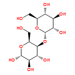
We have developed novel NMR methods for the measurement of heteronuclear residual dipolar couplings (RDCs) in molecules with severely overlapping NMR resonances. These and other methods enabled us to obtain 31 RDCs for α-D-cellobiose and 24 RDCs for β-D-cellobiose. The interpretation of the data in the approximation of a rigid disaccharide structure, using RDCs and interglycosidic 3J coupling constants, yielded conformation that is very close to that determined using X-ray crystallography. However, depending on which ring was used to calculate the order parameters, the dihedral angle ψH varied up to 30° or 40°, while the ϕH angle was always the same. This indicates residual flexibility of the glycosidic linkage between the two monosaccharide rings and was observed for both α- and β-D-cellobiose. The RDC analysis using rigid fragments rather than a complete molecule has thus shown that the glycosidic bond of cellobiose is not completely rigid and exhibits low-level flexibility. The sources of this flexibility are discussed and evidence presented to support a hypothesis that it is associated with the ψ more than the ϕ angle.
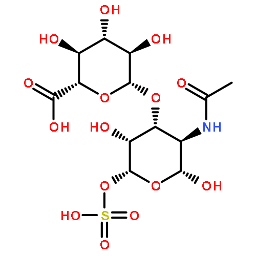
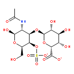
Co-reporter:Charalambos Panagos, Derek Thomson, Charles D. Bavington, Dušan Uhrín
Carbohydrate Polymers 2012 Volume 87(Issue 3) pp:2086-2092
Publication Date(Web):14 February 2012
DOI:10.1016/j.carbpol.2011.10.031
The contamination of heparin in 2008 brought to the attention of health authorities an urgent need for structural characterisation of low molecular weight heparins and other glycosaminoglycans (GAGs) intended for clinical applications. Potentially harmful compounds can be introduced into these preparations as contaminants of the original material or as by-products of the depolymerisation process. Radical depolymerisation is one of the methods used for fractionations of GAGs. We report here on the results of the Fenton-type radical depolymerisation of dermatan sulfate (DS) by hydrogen peroxide in the presence of Cu2+ cations. A low molecular fraction of the reaction mixture was investigated by a combination of 2D 1H,13C HSQC, 2D HSQC-TOCSY and 2D HMBC experiments at 800 MHz. The analysis of the spectra revealed the formation of oligosaccharides with structures corresponding to the native DS sequence and containing almost exclusively GalNAc4S as the reducing end monosaccharide. In addition, oligosaccharides containing a C-4 sulfated N-acetylgalactosaminic acid in place of the reducing end GalNAc4S were identified. This open chain monosaccharide represents a non-native DS structure.Highlights► All structures in low molecular weight heparins and other GAGs must be characterised. ► NMR analysis of the Fenton-type radical depolymerisation of dermatansulfate reported. ► Native DS sequences containing almost exclusively GalNAc as the reducing sugar observed. ► Non-native C-4 sulfated N-acetylgalactosaminic acid in place of the reducing GalNAc4S present. ► Hetercorrelated NMR methods at 800 MHz capable of analysing mixtures of related oligosaccharides.
Co-reporter:Andrew P. Herbert, David Kavanagh, Conny Johansson, Hugh P. Morgan, Bärbel S. Blaum, Jonathan P. Hannan, Paul N. Barlow, and Dušan Uhrín
Biochemistry 2012 Volume 51(Issue 9) pp:1874
Publication Date(Web):February 9, 2012
DOI:10.1021/bi201689j
Numerous complement factor H (FH) mutations predispose patients to atypical hemolytic uremic syndrome (aHUS) and other disorders arising from inadequately regulated complement activation. No unifying structural or mechanistic consequences have been ascribed to these mutants beyond impaired self-cell protection. The S1191L and V1197A mutations toward the C-terminus of FH, which occur in patients singly or together, arose from gene conversion between CFH encoding FH and CFHR1 encoding FH-related 1. We show that neither single nor double mutations structurally perturbed recombinant proteins consisting of the FH C-terminal modules, 19 and 20 (FH19-20), although all three FH19-20 mutants were poor, compared to wild-type FH19-20, at promoting hemolysis of C3b-coated erythrocytes through competition with full-length FH. Indeed, our new crystal structure of the S1191L mutant of FH19-20 complexed with an activation-specific complement fragment, C3d, was nearly identical to that of the wild-type FH19-20:C3d complex, consistent with mutants binding to C3b with wild-type-like affinity. The S1191L mutation enhanced thermal stability of module 20, whereas the V1197A mutation dramatically decreased it. Thus, although mutant proteins were folded at 37 °C, they differ in conformational rigidity. Neither single substitutions nor double substitutions increased measurably the extent of FH19-20 self-association, nor did these mutations significantly affect the affinity of FH19-20 for three glycosaminoglycans, despite critical roles of module 20 in recognizing polyanionic self-surface markers. Unexpectedly, FH19-20 mutants containing Leu1191 self-associated on a heparin-coated surface to a higher degree than on surfaces coated with dermatan or chondroitin sulfates. Thus, potentially disease-related functional distinctions between mutants, and between FH and FH-related 1, may manifest in the presence of specific glycosaminoglycans.
Co-reporter:Bärbel S. Blaum ; Jon A. Deakin ; Conny M. Johansson ; Andrew P. Herbert ; Paul N. Barlow ; Malcolm Lyon ;Dušan Uhrín
Journal of the American Chemical Society 2010 Volume 132(Issue 18) pp:6374-6381
Publication Date(Web):April 15, 2010
DOI:10.1021/ja1000517
We have used the interaction between module 7 of complement factor H (CFH∼7) and a fully sulfated heparin tetrasaccharide to exemplify a new approach for studying contributions of basic side chains to the formation of glycosaminoglycan (GAG)−protein complexes. We first employed HISQC and H2CN NMR experiments to monitor the side-chain resonances of lysines and arginines in 15N, 13C-labeled protein during titrations with a fully sulfated heparin tetrasaccharide under physiological conditions. Under identical conditions and using 15N-labeled protein, we then cross-linked tetrasaccharide to CFH∼7 and confirmed the 1:1 stoichiometry by FT-ICR-MS. We subsequently characterized this covalent protein−GAG conjugate by NMR and further MS techniques. MALDI-TOF MS identified protein fragments obtained via trypsin digestion or chemical fragmentation, yielding information concerning the site of GAG attachment. Combining MS and NMR data allowed us to identify the side chain of K405 as the point of attachment of the cross-linked heparin oligosaccharide to CFH∼7. On the basis of the analysis of NMR and MS data of the noncovalent and cross-linked CFH∼7−tetrasaccharide complexes, we conclude that the K446 side chain is not essential for binding the tetrasaccharide, despite the large chemical shift perturbations of its backbone amide 15N and 1H resonances during titrations. We show that R444 provides the most important charge−charge interaction within a C-terminal heparin-binding subsite of CFH∼7 whereas side chains of R404, K405, and K388 are the predominant contributors to an N-terminal binding subsite located in the immediate vicinity of residue 402, which is implicated in age-related macular degeneration (AMD).
Co-reporter:Emily S. Seo, Bärbel S. Blaum, Thomas Vargues, Martin De Cecco, Jon A. Deakin, Malcolm Lyon, Perdita E. Barran, Dominic J. Campopiano, and Dušan Uhrín
Biochemistry 2010 Volume 49(Issue 49) pp:
Publication Date(Web):November 9, 2010
DOI:10.1021/bi1011749
Human β-defensin 2 (HBD2) is a member of the defensin family of antimicrobial peptides that plays important roles in the innate and adaptive immune system of both vertebrates and invertebrates. In addition to their direct bactericidal action, defensins are also involved in chemotaxis and Toll-like receptor activation. In analogy to chemokine/glycosaminoglycan (GAG) interactions, GAG−defensin complexes are likely to play an important role in chemotaxis and in presenting defensins to their receptors. Using a gel mobility shift assay, we found that HBD2 bound to a range of GAGs including heparin/heparan sulfate (HS), dermatan sulfate (DS), and chondroitin sulfate. We used NMR spectroscopy of 15N-labeled HBD2 to map the binding sites for two GAG model compounds, a heparin/HS pentasaccharide (fondaparinux sodium; FX) and enzymatically prepared DS hexasaccharide (DSdp6). We identified a number of basic amino acids that form a common ligand binding site, which indicated that these interactions are predominantly electrostatic. The dissociation constant of the [DSdp6−HBD2] complex was determined by NMR spectroscopy to be 5 ± 5 μM. Binding of FX could not be quantified because of slow exchange on the NMR chemical shift time scale. FX was found to induce HBD2 dimerization as evidenced by the analysis of diffusion coefficients, 15N relaxation, and nESI-MS measurements. The formation of FX-bridged HBD2 dimers exhibited features of a cooperative binding mechanism. In contrast, the complex with DSdp6 was found to be mostly monomeric.
Co-reporter:Emiliano Gemma, Alison N. Hulme, Astrid Jahnke, Lan Jin, Malcolm Lyon, Ralf M. Müller and Dušan Uhrín
Chemical Communications 2007 (Issue 26) pp:2686-2688
Publication Date(Web):16 Apr 2007
DOI:10.1039/B617038B
Efficient functionalisation of the non-reducing end of uronic acid derivatives and glycosaminoglycan-derived disaccharides using peptide coupling has been achieved, mediated by the water-soluble agent DMT-MM.
Co-reporter:Lan Jin;Dušan Uhrín
Magnetic Resonance in Chemistry 2007 Volume 45(Issue 8) pp:628-633
Publication Date(Web):11 JUN 2007
DOI:10.1002/mrc.2031
The sensitivity of cryoprobes, which are rapidly becoming available, means that the measurement of coupling constants involving 13C, 13C pairs at the natural abundance of 13C can now, in principle, be done by using tens rather then hundreds of milligrams of compounds. However, a robust method that would yield reliable values of small long-range carbon--carbon coupling constants is still missing. In this Communication, we describe a novel 13C–detected incredible natural-abundance double-quantum transfer experiment (INADEQUATE) experiment for simultaneous correlation of one-bond and long-range 13C13C pairs and the measurement of both types of coupling constants in 13C natural abundance samples. This method yields accurate values of one-bond and long-range coupling constants by manipulation of pure phase in-phase (IP) and antiphase (AP) doublets, and is referred to as 13C-detected IPAP-INADEQUATE. It is illustrated by the measurement of interglycosidic 3JCCOC coupling constants in a disaccharide molecule providing important information about the conformation of the glycosidic linkage. Owing to the simplicity of INADEQUATE spectra the carbon–carbon coupling constants are particularly suitable for studies of partially oriented molecules through the measurement of carbon–carbon residual dipolar couplings (RDCs). An example of this approach is presented. We expect the method to find a variety of applications in the conformational analysis of small molecules, determination of diastereoisomers and enantiomers, and studies of molecules in aligned media. Copyright © 2007 John Wiley & Sons, Ltd.
Co-reporter:Lan Jin;Tran N. Pham Dr.;Dušan Uhrín Dr.
ChemPhysChem 2007 Volume 8(Issue 8) pp:1228-1235
Publication Date(Web):24 APR 2007
DOI:10.1002/cphc.200700071
Residual dipolar coupling constants (RDCs) are being increasingly applied to elucidate the configuration and conformation of small organic molecules, peptides and oligosaccharides. In this paper we describe a set of robust 1D NMR methods for accurate and precise measurement of proton–proton RDCs of small and medium size molecules. The performance of these techniques is not impeded by the presence of overlapping and broad 1H multiplets that are typically observed for such molecules in weakly aligned media. The use of these techniques provides access to a large pool of proton–proton RDCs opening new avenues for the solution structure elucidation of medium size molecules by NMR. The techniques are illustrated on the determination of the alignment tensor of the reducing monosaccharide ring of cellobiose and the determination of the relative configuration of sodium cholate.
Co-reporter:Graeme Ball, Nicola Meenan, Krystyna Bromek, Brian O. Smith, Juraj Bella, Dušan Uhrín
Journal of Magnetic Resonance 2006 Volume 180(Issue 1) pp:127-136
Publication Date(Web):May 2006
DOI:10.1016/j.jmr.2006.01.017
We have developed new 2D and 3D experiments for the measurement of Cα–Hα residual dipolar coupling constants in 13C and 15N labelled proteins. Two experiments, 2D (HNCO)-(J-CA)NH and 3D (HN)CO-(J-CA)NH, sample the Cα–Hα splitting by means of Cα magnetization, while 2D (J-HACACO)NH and 3D J-HA(CACO)NH use Hα magnetization to achieve a similar result. In the 2D experiments the coupling evolution is superimposed on the evolution of the 15N chemical shifts and the IPAP principle is used to obtain 1H–15N HSQC-like spectra from which the splitting is determined. The use of a third dimension in 3D experiments reduces spectral overlap to the point where use of an IPAP scheme may not be necessary. The length of the sampling interval in the J-dimension of these experiments is dictated solely by the relaxation properties of Cα or Hα nuclei. This was made possible by the use of Cα selective pulses in combination with either a DPFGSE or modified BIRD pulses. Inclusion of these pulse sequence elements in the J-evolution periods removes unwanted spin–spin interactions. This allows prolonged sampling periods (∼25 ms) yielding higher precision Cα–Hα splitting determination than is achievable with existing frequency based methods.
Co-reporter:Tran N. Pham, Katalin E. Kövér, Lan Jin, Dušan Uhrín
Journal of Magnetic Resonance 2005 Volume 176(Issue 2) pp:199-206
Publication Date(Web):October 2005
DOI:10.1016/j.jmr.2005.06.010
We describe a new NMR experiment, 1H-detected double-J-modulated (DJM)-INEPT-INADEQUATE, for tracing out the carbon skeleton of molecules. This experiment allows simultaneous correlation of directly bonded carbon atoms and those separated by multiple bonds, while at the same time also providing the values of all JCC coupling constants. This is achieved by replacing both fixed carbon–carbon coupling evolution intervals of the INEPT-INADEQUATE experiment by variable time intervals, which are incremented in concert with the DQ evolution period (t1). We show that the analysis of the fine structure of cross-peaks in DJM-INEPT-INADEQUATE spectra leads to accurate values of coupling constants and give guidelines for the proper usage of the method. The proposed experiment is two times less sensitive that the original INEPT-INADEQUATE experiment. We show that, using a 600-MHz cryoprobe and 20 mg of a monosaccharide, spectra that are suitable for the analysis of coupling constants as small as 2 Hz can be obtained within 24 h. Instead of performing multiple experiments, a single DJM-INEPT-INADEQUATE experiment can thus provide a wealth of information for the structural analysis of small molecules.
Co-reporter:Emiliano Gemma, Alison N. Hulme, Astrid Jahnke, Lan Jin, Malcolm Lyon, Ralf M. Müller and Dušan Uhrín
Chemical Communications 2007(Issue 26) pp:
Publication Date(Web):
DOI:10.1039/B617038B
Co-reporter:Nicholle G. A. Bell, Lorna Murray, Margaret C. Graham and Dušan Uhrín
Chemical Communications 2014 - vol. 50(Issue 14) pp:NaN1697-1697
Publication Date(Web):2014/01/02
DOI:10.1039/C3CC48907H
Mixture ‘separation’ by NMR is demonstrated through the development of a pseudo 4D NMR experiment, 3D IPAP INEPT-INADEQUATE-HSQC, designed for the structural elucidation of 13C tagged compounds.
Co-reporter:Nicholle G. A. Bell, Graeme Rigg, Sarah Masters, Juraj Bella and Dušan Uhrín
Physical Chemistry Chemical Physics 2013 - vol. 15(Issue 41) pp:NaN18234-18234
Publication Date(Web):2013/09/05
DOI:10.1039/C3CP52987H
We have developed novel NMR methods for the measurement of heteronuclear residual dipolar couplings (RDCs) in molecules with severely overlapping NMR resonances. These and other methods enabled us to obtain 31 RDCs for α-D-cellobiose and 24 RDCs for β-D-cellobiose. The interpretation of the data in the approximation of a rigid disaccharide structure, using RDCs and interglycosidic 3J coupling constants, yielded conformation that is very close to that determined using X-ray crystallography. However, depending on which ring was used to calculate the order parameters, the dihedral angle ψH varied up to 30° or 40°, while the ϕH angle was always the same. This indicates residual flexibility of the glycosidic linkage between the two monosaccharide rings and was observed for both α- and β-D-cellobiose. The RDC analysis using rigid fragments rather than a complete molecule has thus shown that the glycosidic bond of cellobiose is not completely rigid and exhibits low-level flexibility. The sources of this flexibility are discussed and evidence presented to support a hypothesis that it is associated with the ψ more than the ϕ angle.


![BETA-D-GAL-[1->4]-BETA-D-GLC-1->OME](http://img.cochemist.com/ccimg/7300/7216-69-5.png)
![BETA-D-GAL-[1->4]-BETA-D-GLC-1->OME](http://img.cochemist.com/ccimg/7300/7216-69-5_b.png)


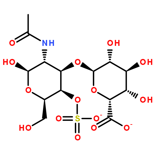
![ALPHA-DELTA-UA-2S-[1->4]-GLCNS-6S SODIUM SALT](http://img.cochemist.com/ccimg/136100/136098-10-7.png)
![ALPHA-DELTA-UA-2S-[1->4]-GLCNS-6S SODIUM SALT](http://img.cochemist.com/ccimg/136100/136098-10-7_b.png)