Co-reporter:Sida Shen, Dawei Jiang, Liang Cheng, Yu Chao, Kaiqi Nie, Ziliang Dong, Christopher J. Kutyreff, Jonathan W. Engle, Peng Huang, Weibo Cai, and Zhuang Liu
ACS Nano September 26, 2017 Volume 11(Issue 9) pp:9103-9103
Publication Date(Web):August 30, 2017
DOI:10.1021/acsnano.7b03857
Developing tumor-homing nanoparticles with integrated diagnostic and therapeutic functions, and meanwhile could be rapidly excreted from the body, would be of great interest to realize imaging-guided precision treatment of cancer. In this study, an ultrasmall coordination polymer nanodot (CPN) based on the coordination between tungsten ions (WVI) and gallic acid (W-GA) was developed via a simple method. After polyethylene glycol (PEG) modification, PEGylated W-GA (W-GA-PEG) CPNs with an ultrasmall hydrodynamic diameter of 5 nm were rather stable in various physiological solutions. Without the need of chelator molecules, W-GA-PEG CPNs could be efficiently labeled with radioisotope 64Cu2+, enabling positron emission tomography (PET) imaging, which reveals efficient tumor accumulation and rapid renal clearance of W-GA-PEG CPNs upon intravenous injection. Utilizing the radio-sensitizing function of tungsten with strong X-ray absorption, such W-GA-PEG CPNs were able to greatly enhance the efficacy of cancer radiotherapy in inhibiting the tumor growth. With fast clearance and little long-term body retention, those W-GA-PEG CPNs exhibited no appreciable in vivo toxicity. This study presents a type of CPNs with excellent imaging and therapeutic abilities as well as rapid renal clearance behavior, promising for further clinic translation.Keywords: chelator-free 64Cu-labeling; coordination polymer nanodots; positron emission tomography imaging; radiotherapy; rapid renal clearance;
Co-reporter:Qiutong Jin;Wenjun Zhu;Dawei Jiang;Rui Zhang;Christopher J. Kutyreff;Jonathan W. Engle;Peng Huang;Weibo Cai;Zhuang Liu
Nanoscale (2009-Present) 2017 vol. 9(Issue 34) pp:12609-12617
Publication Date(Web):2017/08/31
DOI:10.1039/C7NR03086J
Cancer nanotechnology has become the hot topic nowadays. While various kinds of nanomaterials have been widely explored for innovative cancer imaging and therapy applications, safe multifunctional nano-agents without long-term retention and toxicity are still demanded. Herein, iron-gallic acid coordination nanoparticles (Fe-GA CPNs) with ultra-small sizes are successfully synthesized by a simple method for multimodal imaging-guided cancer therapy. After surface modification with polyethylene glycol (PEG), the synthesized Fe-GA-PEG CPNs show high stability in various physiological solutions. Taking advantage of high near-infrared (NIR) absorbance as well as the T1-MR contrasting ability of Fe-GA-PEG CPNs, in vivo photoacoustic tomography (PAT) and magnetic resonance (MR) bimodal imaging are carried out, revealing the efficient passive tumor targeting of these ultra-small CPNs after intravenous (i.v.) injection. Interestingly, such Fe-GA-PEG CPNs could be labeled with the 64Cu isotope via a chelator-free method for in vivo PET imaging, which also illustrates the high tumor uptake of Fe-GA CPNs. We further utilize Fe-GA-PEG CPNs for in vivo photothermal therapy and achieve highly effective tumor destruction after i.v. injection of Fe-GA-PEG CPNs and the following NIR laser irradiation of the tumors, without observing any apparent toxicity of such CPNs to the treated animals. Our work highlights the promise of ultra-small iron coordination nanoparticles for imaging-guided cancer therapy.
Co-reporter:Yuyan Chen;Dr. Liang Cheng;Ziliang Dong;Yu Chao;Huali Lei;He Zhao; Jian Wang; Zhuang Liu
Angewandte Chemie 2017 Volume 129(Issue 42) pp:13171-13176
Publication Date(Web):2017/10/09
DOI:10.1002/ange.201707128
AbstractMultifunctional biodegradable inorganic theranostic nano-agents are of great interest to the field of nanomedicine. Upon lipid modification, VS2 nanosheets could be converted into ultra-small VS2 nanodots encapsulated inside polyethylene glycol (PEG) modified lipid micelles. Owing to paramagnetism, high near-infrared (NIR) absorbance, and chelator-free 99mTc4+ labeling of VS2, such VS2@lipid-PEG nanoparticles could be used for T1-weighted magnetic resonance (MR), photoacoustic (PA),and single photon emission computed tomography (SPECT) tri-modal imaging guided photothermal ablation of tumors. Importantly, along with the gradual degradation of VS2, our VS2@lipid-PEG nanoparticles exhibit effective body excretion without appreciable toxicity. The unique advantages of VS2 nanostructures with highly integrated functionalities and biodegradable behaviors mean they are promising for applications in cancer theranostics.
Co-reporter:Yuyan Chen;Dr. Liang Cheng;Ziliang Dong;Yu Chao;Huali Lei;He Zhao; Jian Wang; Zhuang Liu
Angewandte Chemie International Edition 2017 Volume 56(Issue 42) pp:12991-12996
Publication Date(Web):2017/10/09
DOI:10.1002/anie.201707128
AbstractMultifunctional biodegradable inorganic theranostic nano-agents are of great interest to the field of nanomedicine. Upon lipid modification, VS2 nanosheets could be converted into ultra-small VS2 nanodots encapsulated inside polyethylene glycol (PEG) modified lipid micelles. Owing to paramagnetism, high near-infrared (NIR) absorbance, and chelator-free 99mTc4+ labeling of VS2, such VS2@lipid-PEG nanoparticles could be used for T1-weighted magnetic resonance (MR), photoacoustic (PA),and single photon emission computed tomography (SPECT) tri-modal imaging guided photothermal ablation of tumors. Importantly, along with the gradual degradation of VS2, our VS2@lipid-PEG nanoparticles exhibit effective body excretion without appreciable toxicity. The unique advantages of VS2 nanostructures with highly integrated functionalities and biodegradable behaviors mean they are promising for applications in cancer theranostics.
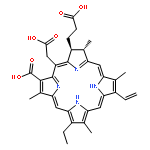
Co-reporter:Liang Cheng;Dawei Jiang;Anyanee Kamkaew;Hector F. Valdovinos;Hyung-Jun Im;Liangzhu Feng;Christopher G. Engl;Shreya Goel;Todd E. Barnhart;Zhuang Liu;Weibo Cai
Advanced Functional Materials 2017 Volume 27(Issue 34) pp:
Publication Date(Web):2017/09/01
DOI:10.1002/adfm.201702928
Nanomaterials with renal clearance from the body within a reasonable timescale have shown great promises in the area of nanomedicine recently. However, the integration of theranostic and renal clearance properties into a single ultrasmall nanostructure remains a great challenge. Herein, meso-tetra(4-carboxyphenyl)porphyrin (TCPP) structure is utilized as a model, for the first time using noninvasive dynamic positron emission tomography (PET) imaging to investigate the balance of the renal clearance and tumor uptake behaviors of polyethylene glycol (PEG)-modified porphyrin nanoparticles (TCPP-PEG) with various molecular weights. This study finds that TCPP-PEG nanoparticles with larger molecular weight show higher tumor uptake due to the enhanced permeability and retention effect, while the lower ones tend to be better for renal clearance. Based on dynamic PET and fluorescence dual-modal imaging modalities, the TCPP-PEG10K nanoparticles seem to be an excellent choice for the balance of renal clearance and tumor retention. In vitro and in vivo photodynamic therapy confirms an excellent therapeutic efficacy. Therefore, this work presents a simplified approach to fabricate and select biocompatible multifunctional TCPP-PEG-based theranostic agents with renal clearance behavior, which highlights the clinical application potential of TCPP-PEG nanoparticles as theranostic probes for imaging-guided cancer therapy.
Co-reporter:Sida Shen;Yu Chao;Ziliang Dong;Guanglin Wang;Xuan Yi;Guosheng Song;Kai Yang;Zhuang Liu
Advanced Functional Materials 2017 Volume 27(Issue 28) pp:
Publication Date(Web):2017/07/01
DOI:10.1002/adfm.201700250
Facile preparation of multifunctional theranostic nanoplatforms with well-controlled morphology and sizes remains an attractive in the area of nanomedicine. Here, a new kind of 2D transition metal dichalcogenide, rhenium disulfide (ReS2) nanosheets, with uniform sizes, strong near-infrared (NIR) light, and strong X-ray attenuation, is successfully synthesized. After surface modification with poly(ethylene glycol) (PEG), the synthesized ReS2-PEG nanosheets are stable in various physiological solutions. In addition to their contrasts in photoacoustic imaging and X-ray computed tomography imaging because of their strong NIR light and X-ray absorptions, respectively, such ReS2-PEG nanosheets can also be tracked under nuclear imaging after chelator-free labeling with radioisotope ions, 99mTc4+. Efficient tumor accumulation of ReS2-PEG nanosheets is then observed after intravenous injection into tumor-bearing mice under triple-modal imaging. The combined in vivo photothermal radiotherapy is further conducted, achieving a remarkable synergistic tumor destruction effect. Finally, no obvious toxicity of ReS2-PEG nanosheets is observed from the treated mice within 30 d. This work suggests that such ultrathin ReS2 nanosheets with well-controlled morphology and uniform sizes may be a promising type of multifunctional theranostic agent for remotely triggered cancer combination therapy.
Co-reporter:Liang Cheng;Sida Shen;Sixiang Shi;Yuan Yi;Xiaoyong Wang;Guosheng Song;Kai Yang;Gang Liu;Todd E. Barnhart;Weibo Cai;Zhuang Liu
Advanced Functional Materials 2016 Volume 26( Issue 13) pp:2185-2197
Publication Date(Web):
DOI:10.1002/adfm.201504810
Multifunctional theranostic agents have become rather attractive to realize image-guided combination cancer therapy. Herein, a novel method is developed to synthesize Bi2Se3 nanosheets decorated with mono-dispersed FeSe2 nanoparticles (FeSe2/Bi2Se3) for tetra-modal image-guided combined photothermal and radiation tumor therapy. Interestingly, upon addition of Bi(NO3)3, pre-made FeSe2 nanoparticles via cation exchange would be gradually converted into Bi2Se3 nanosheets, on which remaining FeSe2 nanoparticles are decorated. The yielded FeSe2/Bi2Se3 composite-nanostructures are then modified with polyethylene glycol (PEG). Taking advantages of the high r 2 relaxivity of FeSe2, the X-ray attenuation ability of Bi2Se3, the strong near-infrared optical absorbance of the whole nanostructure, as well as the chelate-free radiolabeling of 64Cu on FeSe2/Bi2Se3-PEG, in vivo magnetic resonance/computer tomography/photoacoustic/position emission tomography multimodal imaging is carried out, revealing efficient tumor homing of FeSe2/Bi2Se3-PEG after intravenous injection. Utilizing the intrinsic physical properties of FeSe2/Bi2Se3-PEG, in vivo photothermal and radiation therapy to achieve synergistic tumor destruction is then realized, without causing obvious toxicity to the treated animals. This work presents a unique method to synthesize composite-nanostructures with highly integrated functionalities, promising not only for nano-biomedicine but also potentially for other different nanotechnology fields.
Co-reporter:Liang Cheng, Anyanee Kamkaew, Haiyan Sun, Dawei Jiang, Hector F. Valdovinos, Hua Gong, Christopher G. England, Shreya Goel, Todd E. Barnhart, and Weibo Cai
ACS Nano 2016 Volume 10(Issue 8) pp:7721
Publication Date(Web):July 26, 2016
DOI:10.1021/acsnano.6b03074
Multifunctional nanoparticles with combined diagnostic and therapeutic functions show great promise in nanomedicine. Herein, we develop an organic photodynamic therapy (PDT) system based on polyethylene glycol (PEG)-coated nanomicelles conjugated with ∼20% chlorin e6 (PEG-Ce 6 nanomicelles), which functions as an optical imaging agent, as well as a PDT agent. The formed PEG-Ce 6 nanomicelles with the size of ∼20 nm were highly stable in various physiological solutions for a long time. Moreover, Ce 6 can also be a 64Cu chelating agent for in vivo positron emission tomography (PET). By simply mixing, more than 90% of 64Cu was chelator-free labeled on PEG-Ce 6 nanomicelles, and they also showed high stability in serum conditions. Both fluorescence imaging and PET imaging revealed that PEG-Ce 6 nanomicelles displayed high tumor uptake (13.7 ± 2.2%ID/g) after intravenous injection into tumor-bearing mice at the 48 h time point. In addition, PEG-Ce 6 nanomicelles exhibited excellent PDT properties upon laser irradiation, confirming the theranostic properties of PEG-Ce 6 nanomicelles for imaging and treatment of cancer. In addition, PDT was not shown to render any appreciable toxicity. This work presents a theranostic platform based on polymer nanomicelles with great potential in multimodality imaging-guided photodynamic cancer therapy.Keywords: EPR effect; fluorescence imaging; PEG-Ce 6 nanomicelles; PET imaging; photodynamic therapy
Co-reporter:Teng Liu;Yu Chao;Min Gao;Chao Liang;Qian Chen;Guosheng Song
Nano Research 2016 Volume 9( Issue 10) pp:3003-3017
Publication Date(Web):2016 October
DOI:10.1007/s12274-016-1183-x
The clinical translation of many inorganic nanomaterials is severely hampered by toxicity issues because of the long-term retention of these nanomaterials in the body. In this study, we developed a bio-clearable theranostic agent based on ultra-small MoS2 nanodots, which were synthesized by a facile bottom-up approach through one-step solvothermal decomposition of ammonium tetrathiomolybdate. After modification by glutathione (GSH), the obtained MoS2-GSH nanodots exhibited sub-10-nm hydrodynamic diameters without aggregation in various physiological buffers. Without showing appreciable in vitro toxicity, such MoS2-GSH nanodots with strong near-infrared (NIR) absorbance could induce remarkable photothermal ablation of cancer cells. Upon intravenous (i.v.) injection, efficient tumor accumulation of MoS2-GSH nanodots was observed by photoacoustic imaging, and further confirmed by analysis of the biodistribution of Mo. Notably, the MoS2-GSH nanodots, in contrast to conventional MoS2 nanoflakes with larger sizes, showed rather efficient body clearance via urine, where the majority of the injected dose was cleared within just seven days. Photothermal ablation of tumors on mice was then realized with the MoS2-GSH nanodots, achieving excellent therapeutic efficacy. This study presents a new type of ultra-small nanoparticle with efficient tumor homing/treatment abilities, as well as rapid body clearance behavior, making it promising for cancer theranostics without long-term toxicity concerns.
Co-reporter:Tingting Fu, Yuyan Chen, Jiali Hao, Xiaoyong Wang, Gang Liu, Yonggang Li, Zhuang Liu and Liang Cheng
Nanoscale 2015 vol. 7(Issue 48) pp:20757-20768
Publication Date(Web):12 Nov 2015
DOI:10.1039/C5NR06840A
Recently, magnetic photothermal nanomaterials have emerged as a new class of bio-nanomaterials for application in cancer diagnosis and therapy. Hence, we developed a new kind of magnetic nanomaterials, iron diselenide (FeSe2) nanoparticles, for multimodal imaging-guided photothermal therapy (PTT) to improve the efficacy of cancer treatment. By controlling the reaction time and temperature, FeSe2 nanoparticles were synthesized by a simple solution-phase method. After modification with polyethylene glycol (PEG), the obtained FeSe2-PEG nanoparticles showed high stability under various physiological conditions. FeSe2-PEG could serve as a T2-weighted magnetic resonance (MR) imaging contrast agent because of its strong superparamagnetic properties, with its r2 relaxivity determined to be 133.38 mM−1 S−1, a value higher than that of the clinically used Feridex. On the other hand, with high absorbance in the near-infrared (NIR) region, FeSe2-PEG also appeared to be a useful contrast agent for photoacoustic imaging (PA) as well as an effective photothermal agent for PTT cancer treatment, as demonstrated in our animal tumor model experiments. Moreover, long-term toxicity tests have proven that FeSe2-PEG nanoparticles after systematic administration rendered no appreciable toxicity to the treated animals, and could be gradually excreted from the major organs of mice. Our work indicates that FeSe2-PEG nanoparticles would be a new class of theranostic agents promising for application in bioimaging and cancer therapy.
Co-reporter:Xiaoxin Qian, Sida Shen, Teng Liu, Liang Cheng and Zhuang Liu
Nanoscale 2015 vol. 7(Issue 14) pp:6380-6387
Publication Date(Web):09 Mar 2015
DOI:10.1039/C5NR00893J
Recently, transition metal dichalcogenides (TMDCs) have attracted significant attention in nanomedicine owing to their intriguing properties. In this study, TiS2 nanosheets, a new TMDC nanomaterial, are synthesized by a bottom-up solution-phase method and then modified with polyethylene glycol (PEG), obtaining TiS2-PEG with high stability in physiological solutions and no appreciable in vitro toxicity. Due to their high absorbance in the near-infrared (NIR) region, TiS2-PEG nanosheets could offer a strong contrast in photoacoustic imaging, which uncovers the high tumor uptake and retention of these nanosheets after systemic administration into tumor-bearing mice. We further apply TiS2-PEG nanosheets for in vivo photothermal therapy, which are able to completely eradicate the tumors in mice upon intravenous injection of TiS2-PEG followed by NIR laser irradiation. Our work indicates that TiS2 nanosheets with appropriate surface coating (e.g. PEGylation) would be a promising new class of photothermal agents for imaging-guided cancer therapy.
Co-reporter:Jiaxiang Juan, Liang Cheng, Min Shi, Zhuang Liu and Xinliang Mao
Journal of Materials Chemistry A 2015 vol. 3(Issue 28) pp:5769-5776
Publication Date(Web):02 Jun 2015
DOI:10.1039/C5TB00646E
Upconversion nanoparticles (UCNPs) have gained increased attention due to their various medical applications such as drug delivery, detection, imaging, and photodynamic therapy. But little is known about their direct biological activity. In the present study, we synthesized NaYF4:Yb,Er UCNPs coated with poly-(allylamine hydrochloride) (PAH) or poly(acrylic acid) (PAA) and investigated their effects in human myeloma and leukemia cells. When these cells were incubated with both types of UCNPs, we found that PAH-UCNPs but not PAA-UCNPs increased the expression of LC3-II, a hallmark of autophagy. This effect was confirmed by the accumulation of LC3 puncta as analyzed by immunofluorescence microscopy. Induction of LC3-II could be blocked by 3-methyl adenosine (3-MA), an autophagy inhibitor. Consistent with this observation, PAH-UCNPs also inhibited both AKT and mTOR activation, the key step in autophagy activation. Further studies demonstrated that PAH-UCNPs also decreased Bcl-2 but increased Beclin1 and Atg14 expression, suggesting that PAH-UCNPs-induced autophagy was associated with increased PI3KC3–Beclin1 activity. Moreover, PAH-UCNPs induced apoptosis in myeloma cells and enhanced apoptosis induced by bortezomib and doxorubicin, two major anti-myeloma drugs. Therefore, our study suggested that PAH-UCNPs alone can induce both apoptosis and autophagy in human blood cancer cells by modulating the AKT–mTOR and PI3KC3–Beclin1–Atg14 signaling pathways. This study implies the potential application of PAH-UCNPs in blood cancer-cell killing.
Co-reporter:Wenwen Zhu, Kai Liu, Xiaoqi Sun, Xin Wang, Yonggang Li, Liang Cheng, and Zhuang Liu
ACS Applied Materials & Interfaces 2015 Volume 7(Issue 21) pp:11575
Publication Date(Web):May 12, 2015
DOI:10.1021/acsami.5b02510
Prussian blue (PB) as a clinically adapted agent recently has drawn much attention in cancer theranostics for potential applications in magnetic resonance (MR) imaging as well as photothermal cancer treatment. In this work, we take a closer look at the imaging and therapy performance of PB agents once they are doped with Mn2+. It is found that Mn2+-doped PB nanocubes exhibit increased longitudinal relaxivity along with enhanced optical absorption red-shifted to the near-infrared (NIR) region. Those properties make PB:Mn nanocubes with appropriate surface coatings rather attractive agents for biomedical imaging and cancer therapy, which have been successfully demonstrated in our in vivo experiments for effectively tumor ablation.Keywords: Mn doping; MR imaging; photoacoustic imaging; photothermal therapy; Prussian blue;
Co-reporter:Liang Cheng, Chao Yuan, Sida Shen, Xuan Yi, Hua Gong, Kai Yang, and Zhuang Liu
ACS Nano 2015 Volume 9(Issue 11) pp:11090
Publication Date(Web):October 7, 2015
DOI:10.1021/acsnano.5b04606
Recently, two-dimensional transition metal dichalcogenides (TMDCs) have received tremendous attention in many fields including biomedicine. Herein, we develop a general method to dope different types of metal ions into WS2 nanoflakes, a typical class of TMDCs, and choose Gd3+-doped WS2 (WS2:Gd3+) with polyethylene glycol (PEG) modification as a multifunctional agent for imaging-guided combination cancer treatment. While WS2 with strong near-infrared (NIR) absorbance and X-ray attenuation ability enables contrasts in photoacoustic (PA) imaging and computed tomography (CT), Gd3+ doping offers the nanostructure a paramagnetic property for magnetic resonance (MR) imaging. As revealed by trimodal PA/CT/MR imaging, WS2:Gd3+-PEG nanoflakes showed efficient tumor homing after intravenous injection. In vivo cancer treatment study further uncovered that WS2:Gd3+-PEG could not only convert NIR light into heat for photothermal therapy (PTT) but also enhance the ionizing irradiation-induced tumor damage to boost radiation therapy (RT). Owing to the improved tumor oxygenation after the mild PTT, the combination of PTT and RT induced by WS2:Gd3+-PEG resulted in a remarkable synergistic effect to destroy cancer. Our work highlights the promise of utilizing inherent physical properties of TMDC-based nanostructures, whose functions could be further enriched by elementary doping, for applications in multimodal bioimaging and synergistic cancer therapy.Keywords: cancer theranostics; metal-ion-doped WS2 nanoflakes; multimodal imaging; photothermal therapy; radiation therapy;
Co-reporter:Jianwei Shen, Kunyang Li, Liang Cheng, Zhuang Liu, Shuit-Tong Lee, and Jian Liu
ACS Applied Materials & Interfaces 2014 Volume 6(Issue 9) pp:6443
Publication Date(Web):April 2, 2014
DOI:10.1021/am405924g
There is a great need to develop multifunctional nanoparticles (MFNPs) for cancer biomarker-based detection and highly selective therapeutic treatment simultaneously. Here we describe a facile approach of layer-by-layer-assembled MFNPs conjugated with monoclonal antibody anti-HER2, demonstrating the specific detection of breast cancer BT474 cells (biomarker HER2 positive) with a high signal-to-noise ratio. The MFNPs contain a well-defined core–shell structure of UCNP@Fe3O4@Au coated by poly(ethylene glycol) (PEG) and anti-HER2 antibody, displaying excellent dispersity in various aqueous solutions. This unique combination of nanoparticles and ligand molecules allows us to perform photothermal treatment (PTT) of the cancer cells, while simultaneously quantifying the distribution of MFNPs on a cancer cell surface induced by antigen–antibody binding events. An important finding is that cancer cells adjacent to each other or in physical proximity within micrometers may end up with different fates of survival or death in PTT. This dramatic difference is determined by the antigen–antibody binding events at the interface of MFNPs and cells because of tumor cell heterogeneity. Therefore, our experiments reveal a new scale of the highly localized feature of the photothermal effect at the single−cell level illuminated by a continuous−wave near−IR laser.Keywords: detection of cancer cells; multifunctional nanoparticles; nano−bio interface; photothermal treatment; tumor heterogeneity;
Co-reporter:Liang Cheng, Hua Gong, Wenwen Zhu, Jingjing Liu, Xiaoyong Wang, Gang Liu, Zhuang Liu
Biomaterials 2014 35(37) pp: 9844-9852
Publication Date(Web):
DOI:10.1016/j.biomaterials.2014.09.004
Co-reporter:Zhen Liu, Liang Cheng, Lei Zhang, Zhongbo Yang, Zhuang Liu, Jixiang Fang
Biomaterials 2014 35(13) pp: 4099-4107
Publication Date(Web):
DOI:10.1016/j.biomaterials.2014.01.053
Co-reporter:Shengnan Yin, Zhiwei Li, Liang Cheng, Chao Wang, Yumeng Liu, Qian Chen, Hua Gong, Liang Guo, Yonggang Li and Zhuang Liu
Nanoscale 2013 vol. 5(Issue 24) pp:12464-12473
Publication Date(Web):10 Oct 2013
DOI:10.1039/C3NR04212J
Magnetic resonance (MR) imaging using magnetic nanoparticles as the contrast agent has been extensively explored in biomedical imaging and disease diagnosis. Herein, we develop biocompatible polymer coated ultra-small Pt3Co magnetic nanoparticles as a new T2-weighted MR imaging contrast agent. A unique class of alloy Pt3Co nanoparticles is synthesized through a thermal decomposition method. After being modified with polyethylene glycol (PEG), the obtained Pt3Co–PEG nanoparticles exhibit an extremely high T2-weighted relaxivity rate (r2) up to 451.2 mM s−1, which is much higher than that of Resovist®, a commercial T2-MR contrast agent used in the clinic. In vitro experiments indicate no obvious cytotoxicity of Pt3Co–PEG nanoparticles to various cell lines. After intravenous injection of Pt3Co–PEG nanoparticles, in vivo T2-weighted MR imaging of tumor-bearing mice reveals strong tumor contrast, which is much higher than that offered by injecting Resovist®. We further study the long-term biodistribution and toxicology of this new type of MR contrast nanoparticles after intravenous injection into healthy mice. Despite the significant retention of Pt3Co–PEG nanoparticles in the mouse liver and spleen, no appreciable toxicity of these nanoparticles to the treated animals has been noted in our detailed histological and hematological analysis over a course of 60 days. Our work demonstrates that functionalized Pt3Co nanoparticles may be a promising new type of T2-weighted MR contrast agent potentially useful in biomedical imaging and diagnosis.
Co-reporter:Weiwei He, Lifen Zhang, Bing Han, Liang Cheng, Nianchen Zhou, Zhuang Liu and Zhenping Cheng
Journal of Materials Chemistry A 2013 vol. 1(Issue 26) pp:3257-3266
Publication Date(Web):24 May 2013
DOI:10.1039/C3TB20262C
We, the named authors, hereby wholly retract this Journal of Materials Chemistry B article due to the subsequent realisation that fluorescence measurements reported in the article should have been measured through a long-pass filter. The emission at 750 nm reported from the excitation of the monomer 1-(4-vinyl benzyl)-2-naphthyl-benzimidazole by UV light, is actually a second order diffraction artefact of the 380 nm emission. The claim that the monomer is capable of emitting near infrared fluorescence under UV light excitation in the article is false. Signed: Weiwei He, Lifen Zhang, Bing Han, Liang Cheng, Nianchen Zhou, Zhuang Liu and Zhenping Cheng, September 2013. Retraction endorsed by Liz Dunn, Managing Editor, Journal of Materials Chemistry B. Retraction published 24 September 2013
Co-reporter:Yumeng Liu, Kai Yang, Liang Cheng, Jing Zhu, Xinxing Ma, Huan Xu, Yonggang Li, Liang Guo, Hongwei Gu, Zhuang Liu
Nanomedicine: Nanotechnology, Biology and Medicine 2013 Volume 9(Issue 7) pp:1077-1088
Publication Date(Web):October 2013
DOI:10.1016/j.nano.2013.02.010
Herein, we develop FePt@Fe2O3 core-shell magnetic nanoparticles as a T2 magnetic resonance (MR) imaging contrast agent as well as a drug carrier for potential cancer theranostic applications. The FePt@Fe2O3 core-shell nanoparticles are synthesized and then functionalized with polyethylene glycol (PEG). Folic acid (FA) is conjugated on the surface of FePt@Fe2O3-PEG nanoparticles for effective targeting of folate receptor (FR)-positive tumor cells. A chemotherapy drug, doxorubicin (DOX), is then loaded onto those nanoparticles via hydrophobic physical adsorption, for targeted intracellular drug delivery and selective cancer cell killing. We then use those FePt@Fe2O3-PEG nanoparticles for in vivo MR imaging, observing obvious tumor MR contrasts, which resulted from both passive tumor accumulation and active tumor targeting of nanoparticles. Moreover, both in vitro and in vivo studies uncover no obvious toxicity for FePt@Fe2O3-PEG nanoparticles. Therefore, our PEGylated FePt@Fe2O3 core-shell nanoparticles could serve as a promising multifunctional theranostic nano-platform in imaging guided cancer therapy.From the Clinical EditorIn this study of PEGylated FePt@Fe2O3 core-shell magnetic nanoparticles, both therapeutic and diagnostic applications are demonstrated. Folic acid surface-conjugation resulted in uptake by folate receptor positive cancer cells, the iron oxide particles enabled MRI imaging using T2* weighted sequences, and the absorbed doxorubicin provided treatment effects in this model. Similar multi-modality approaches will hopefully find their way to clinical applications in the near future.Herein, we develop FePt@Fe2O3 core-shell magnetic nanoparticles functionalized with polyethylene glycol (PEG) as a T2 magnetic resonance (MR) imaging contrast agent as well as a drug carrier. In vitro targeted drug delivery and in vivo tumor-targeted MR imaging, have both been realized using this novel type of magnetic nanoparticles. No noticeable short-term toxicity of those nanoparticles is found in our careful in vivo toxicology examinations. Our results suggest that FePt@Fe2O3 core-shell nanoparticles with appropriate surface engineering may be a promising T2 MR contrast agent potentially useful for in vivo molecular imaging as well as cancer theranostics.
Co-reporter:Weiwei He, Lifen Zhang, Bing Han, Liang Cheng, Nianchen Zhou, Zhuang Liu and Zhenping Cheng
Journal of Materials Chemistry A 2013 - vol. 1(Issue 26) pp:NaN3266-3266
Publication Date(Web):2013/05/24
DOI:10.1039/C3TB20262C
We, the named authors, hereby wholly retract this Journal of Materials Chemistry B article due to the subsequent realisation that fluorescence measurements reported in the article should have been measured through a long-pass filter. The emission at 750 nm reported from the excitation of the monomer 1-(4-vinyl benzyl)-2-naphthyl-benzimidazole by UV light, is actually a second order diffraction artefact of the 380 nm emission. The claim that the monomer is capable of emitting near infrared fluorescence under UV light excitation in the article is false. Signed: Weiwei He, Lifen Zhang, Bing Han, Liang Cheng, Nianchen Zhou, Zhuang Liu and Zhenping Cheng, September 2013. Retraction endorsed by Liz Dunn, Managing Editor, Journal of Materials Chemistry B. Retraction published 24 September 2013
Co-reporter:Jiaxiang Juan, Liang Cheng, Min Shi, Zhuang Liu and Xinliang Mao
Journal of Materials Chemistry A 2015 - vol. 3(Issue 28) pp:NaN5776-5776
Publication Date(Web):2015/06/02
DOI:10.1039/C5TB00646E
Upconversion nanoparticles (UCNPs) have gained increased attention due to their various medical applications such as drug delivery, detection, imaging, and photodynamic therapy. But little is known about their direct biological activity. In the present study, we synthesized NaYF4:Yb,Er UCNPs coated with poly-(allylamine hydrochloride) (PAH) or poly(acrylic acid) (PAA) and investigated their effects in human myeloma and leukemia cells. When these cells were incubated with both types of UCNPs, we found that PAH-UCNPs but not PAA-UCNPs increased the expression of LC3-II, a hallmark of autophagy. This effect was confirmed by the accumulation of LC3 puncta as analyzed by immunofluorescence microscopy. Induction of LC3-II could be blocked by 3-methyl adenosine (3-MA), an autophagy inhibitor. Consistent with this observation, PAH-UCNPs also inhibited both AKT and mTOR activation, the key step in autophagy activation. Further studies demonstrated that PAH-UCNPs also decreased Bcl-2 but increased Beclin1 and Atg14 expression, suggesting that PAH-UCNPs-induced autophagy was associated with increased PI3KC3–Beclin1 activity. Moreover, PAH-UCNPs induced apoptosis in myeloma cells and enhanced apoptosis induced by bortezomib and doxorubicin, two major anti-myeloma drugs. Therefore, our study suggested that PAH-UCNPs alone can induce both apoptosis and autophagy in human blood cancer cells by modulating the AKT–mTOR and PI3KC3–Beclin1–Atg14 signaling pathways. This study implies the potential application of PAH-UCNPs in blood cancer-cell killing.

![N-[4-[[(2-amino-3,4-dihydro-4-oxo-6-pteridinyl)methyl]amino]benzoyl]-L-Glutamic acid 5-(2,5-dioxo-1-pyrrolidinyl) ester](http://img.cochemist.com/ccimg/153500/153445-05-7.png)
![N-[4-[[(2-amino-3,4-dihydro-4-oxo-6-pteridinyl)methyl]amino]benzoyl]-L-Glutamic acid 5-(2,5-dioxo-1-pyrrolidinyl) ester](http://img.cochemist.com/ccimg/153500/153445-05-7_b.png)


![1,4-Bis[2-(dimethylamino-N-oxide)ethylamino]-5,8-dihydroxyanthracene-9,10-dione](/data/chemimg/501200/136470-65-0.png)
![1,4-Bis[2-(dimethylamino-N-oxide)ethylamino]-5,8-dihydroxyanthracene-9,10-dione](/data/chemimg/501200/136470-65-0_b.png)


![Benzoic acid,4-(decyloxy)-, 4-[(2S)-2-methylbutyl]phenyl ester](http://img.cochemist.com/ccimg/69800/69777-63-5.png)
![Benzoic acid,4-(decyloxy)-, 4-[(2S)-2-methylbutyl]phenyl ester](http://img.cochemist.com/ccimg/69800/69777-63-5_b.png)