Co-reporter:Li Wu, I-Che Wu, Christopher C. DuFort, Markus A. Carlson, Xu Wu, Lei Chen, Chun-Ting Kuo, Yuling Qin, Jiangbo Yu, Sunil R. Hingorani, and Daniel T. Chiu
Journal of the American Chemical Society May 24, 2017 Volume 139(Issue 20) pp:6911-6911
Publication Date(Web):May 1, 2017
DOI:10.1021/jacs.7b01545
Developing probes for the detection of reactive oxygen species (ROS), a hallmark of many pathophysiological process, is imperative to both understanding the precise roles of ROS in many life-threatening diseases and optimizing therapeutic interventions. We herein report an all-in-one fluorescent semiconducting polymer based far-red to near-infrared (NIR) Pdot nanoprobe for the ratiometric detection of hypochlorous acid (HOCl). The fabrication takes the advantage of flexible polymer design by incorporating target-sensitive and target-inert fluorophores into a single conjugated polymer to avoid leakage or differential photobleaching problems existed in other nanoprobes. The obtained nanoprobe has improved performance in HOCl sensing, such as high brightness, ideal far-red to NIR optical window, excellent photostability, self-referenced ratiometric response, fast response, and high selectivity. The dual-emission property allows the sensitive imaging of HOCl fluctuations produced in living macrophage cells and peritonitis of living mice with high contrast. This study not only provides a powerful and promising nanoprobe to be potentially used in the investigations of in situ HOCl status of diseases in living systems but also puts forward the design strategy of a new category of ratiometric fluorescent probes facilitating precise and reliable measurement in biological systems.
Co-reporter:Chun-Ting Kuo, Hong-Shang Peng, Yu Rong, Jiangbo Yu, Wei Sun, Bryant Fujimoto, and Daniel T. Chiu
Analytical Chemistry June 6, 2017 Volume 89(Issue 11) pp:6232-6232
Publication Date(Web):May 12, 2017
DOI:10.1021/acs.analchem.7b01214
Multiplexed optical encoding is emerging as a powerful technique for high-throughput cellular analysis and molecular assays. Most of the developed optical barcodes, however, either suffer from large particle size or are incompatible with most commercial optical instruments. Here, a new type of nanoscale fluorescent barcode (Pdot barcodes) was prepared from semiconducting polymers. The Pdot barcodes possess the merits of small size (∼20 nm in diameter), narrow emission bands (full-width-at-half-maximum (fwhm) of 30–40 nm), three-color emissions (blue, green, and red) under single-wavelength excitation, a high brightness, good pH and thermal stability, and efficient cellular uptake. The Pdot barcodes were prepared using a three-color and six-intensity encoding strategy; for ratiometric readout of the barcodes, one of the colors might be used as an internal reference. We used the Pdot barcodes to label 20 sets of cancer cells and then distinguished and identified each set based on the Pdot barcodes using flow cytometry. We also monitored and tracked single cells labeled with different Pdot barcodes, even through rounds of cell division. These results suggest Pdot barcodes are strong candidates for discriminating different labeled cell and for long-term cell tracking.
Co-reporter:Dr. Xu Wu;Dr. Quinn DeGottardi;I-Che Wu;Jiangbo Yu;Li Wu;Fangmao Ye;Chun-Ting Kuo; William W. Kwok; Daniel T. Chiu
Angewandte Chemie 2017 Volume 129(Issue 47) pp:15104-15108
Publication Date(Web):2017/11/20
DOI:10.1002/ange.201708463
AbstractSimultaneous monitoring of biomarkers as well as single-cell analyses based on flow cytometry and mass cytometry are important for investigations of disease mechanisms, drug discovery, and signaling-network studies. Flow cytometry and mass cytometry are complementary to each other; however, probes that can satisfy all the requirements for these two advanced technologies are limited. In this study, we report a probe of lanthanide-coordinated semiconducting polymer dots (Pdots), which possess fluorescence and mass signals. We demonstrated the usage of this dual-functionality probe for both flow cytometry and mass cytometry in a mimetic cell mixture and human peripheral blood mononuclear cells as model systems. The probes not only offer high fluorescence signal for use in flow cytometry, but also show better performance in mass cytometry than the commercially available counterparts.
Co-reporter:Dr. Xu Wu;Dr. Quinn DeGottardi;I-Che Wu;Jiangbo Yu;Li Wu;Fangmao Ye;Chun-Ting Kuo; William W. Kwok; Daniel T. Chiu
Angewandte Chemie International Edition 2017 Volume 56(Issue 47) pp:14908-14912
Publication Date(Web):2017/11/20
DOI:10.1002/anie.201708463
AbstractSimultaneous monitoring of biomarkers as well as single-cell analyses based on flow cytometry and mass cytometry are important for investigations of disease mechanisms, drug discovery, and signaling-network studies. Flow cytometry and mass cytometry are complementary to each other; however, probes that can satisfy all the requirements for these two advanced technologies are limited. In this study, we report a probe of lanthanide-coordinated semiconducting polymer dots (Pdots), which possess fluorescence and mass signals. We demonstrated the usage of this dual-functionality probe for both flow cytometry and mass cytometry in a mimetic cell mixture and human peripheral blood mononuclear cells as model systems. The probes not only offer high fluorescence signal for use in flow cytometry, but also show better performance in mass cytometry than the commercially available counterparts.
Co-reporter:Dandan Chen;I-Che Wu;Zhihe Liu;Ying Tang;Haobin Chen;Jiangbo Yu;Changfeng Wu
Chemical Science (2010-Present) 2017 vol. 8(Issue 5) pp:3390-3398
Publication Date(Web):2017/05/03
DOI:10.1039/C7SC00441A
The development of near-infrared (NIR) fluorescent probes is critical for in vivo exploration of the fundamental and complex processes in living systems by noninvasive fluorescence imaging techniques. Semiconducting polymer dots (Pdots) are emerging as important probes that exhibit several advantages, such as high brightness and biocompatibility. However, NIR-emitting Pdots are very rare, particularly at the center (∼800 nm) of the first optical window of biological tissues (between 650 nm and 950 nm). In this paper, we describe the synthesis of a semiconducting polymer with bright and narrow-band emission at 800 nm. The polymer was designed by incorporating a NIR porphyrin unit into a conjugated backbone; the polymer used a cascade energy transfer to produce the signal. The resulting Pdots possessed a narrow emission bandwidth (FWHM ∼ 23 nm) and good fluorescence quantum yield (QY = 8%), which is high for a near-IR emitter. The Pdots were bioconjugated with streptavidin for specific labeling of cellular targets, yielding higher staining index when compared with commercial NIR probes, such as PE-Cy7. Moreover, the NIR polymer was combined with a long-wavelength absorbing polymer to make bright Pdots (QY = 15%) for in vivo noninvasive imaging. These NIR Pdots with surface PEGylation led to high-contrast imaging of lymph nodes and tumors in a mouse model. This work highlights the great potential of the NIR Pdots for cellular and in vivo imaging applications.
Co-reporter:Hong-Shang Peng and Daniel T. Chiu
Chemical Society Reviews 2015 vol. 44(Issue 14) pp:4699-4722
Publication Date(Web):22 Dec 2014
DOI:10.1039/C4CS00294F
Soft fluorescent nanomaterials have attracted recent attention as imaging agents for biological applications, because they provide the advantages of good biocompatibility, high brightness, and easy biofunctionalization. Here, we provide a survey of recent developments in fluorescent soft nano-sized biological imaging agents. Various soft fluorescent nanoparticles (NPs) (including dye-doped polymer NPs, semiconducting polymer NPs, small-molecule organic NPs, nanogels, micelles, vesicles, and biomaterial-based NPs) are summarized from the perspectives of preparation methods, structure, optical properties, and surface functionalization. Based on both optical and functional properties of the nano-sized imaging agents, their applications are then reviewed in terms of in vitro imaging, in vivo imaging, and cellular-process imaging, by means of specific or nonspecific targeting.
Co-reporter:Robbyn K. Anand; Eleanor S. Johnson
Journal of the American Chemical Society 2015 Volume 137(Issue 2) pp:776-783
Publication Date(Web):January 6, 2015
DOI:10.1021/ja5102689
This paper describes the dielectrophoretic (DEP) forces generated by a bipolar electrode (BPE) in a microfluidic device and elucidates the impact of faradaic ion enrichment and depletion (FIE and FID) on electric field gradients. DEP technologies for manipulating biological cells provide several distinct advantages over other cell-handling techniques including label-free selectivity, inexpensive device components, and amenability to single-cell and array-based applications. However, extension to the array format is nontrivial, and DEP forces are notoriously short-range, limiting device dimensions and throughput. BPEs present an attractive option for DEP because of the ease with which they can be arrayed. Here, we present experimental results demonstrating both negative DEP (nDEP) attraction and repulsion of B-cells from each a BPE cathode and anode. The direction of nDEP force in each case was determined by whether the conditions for FIE or FID were chosen in the experimental design. We conclude that FIE and FID zones generated by BPEs can be exploited to shape and extend the electric field gradients that are responsible for DEP force.
Co-reporter:Yue Zhang, Fangmao Ye, Wei Sun, Jiangbo Yu, I-Che Wu, Yu Rong, Yong Zhang and Daniel T. Chiu
Chemical Science 2015 vol. 6(Issue 3) pp:2102-2109
Publication Date(Web):23 Jan 2015
DOI:10.1039/C4SC03959A
This paper describes a synthetic approach for photocrosslinkable polyfluorene (pc-PFO) semiconducting polymer dots, and demonstrates their superior ability to crosslink and form 3-D intermolecular polymer networks. The crosslinked pc-PFO Pdots are equipped with excellent encapsulating ability of functional small molecules. Optimum conditions of light irradiation on pc-PFO Pdots were investigated and clarified by using polymer thin films as a model. By employing the optimal light irradiation conditions, we successfully crosslinked pc-PFO Pdots and studied their particle sizes, photophysical, and colloidal properties. Single-particle imaging and dynamic-light-scattering measurements were conducted to understand the behaviors of photocrosslinked Pdots. Our results indicate pc-PFO Pdots can be easily photocrosslinked and the crosslinked species have excellent colloidal stability, physical and chemical stability, fluorescence brightness, and specific binding properties for cellular labeling. Considering that optical stimulus can work remotely, cleanly, and non-invasively, this study should pave the way for a promising approach to further develop stimuli-responsive ultrabright and versatile Pdot probes for biomedical imaging.
Co-reporter:Fangmao Ye, Collin C. White, Yuhui Jin, Xiaoge Hu, Sarah Hayden, Xuanjun Zhang, Xiaohu Gao, Terrance J. Kavanagh and Daniel T. Chiu
Nanoscale 2015 vol. 7(Issue 22) pp:10085-10093
Publication Date(Web):15 May 2015
DOI:10.1039/C5NR01857A
The rapid development and acceptance of PDots for biological applications depends on an in depth understanding of their cytotoxicity. In this paper, we performed a comprehensive study of PDot cytotoxicity at both the gross cell effect level (such as cell viability, proliferation and necrosis) and more subtle effects (such as redox stress) on RAW264.7 cells, a murine macrophage cell line with high relevance to in vivo nanoparticle disposition. The redox stress measurements assessed were inner mitochondrial membrane lipid peroxidation (nonyl-acridine orange, NAO), total thiol level (monobromobimane, MBB), and pyridine nucleotide redox status (NAD(P)H autofluorescence). Because of the extensive work already performed with QDots on nanotoxicity and also because of their comparable size, QDots were chosen as a comparison/reference nanoparticle for this study. The results showed that PDots exhibit cytotoxic effects to a much lesser degree than their inorganic analogue (QDots) and are much brighter, allowing for much lower concentrations to be used in various biological applications. In addition, at lower dose levels (2.5 nM to 10 nM) PDot treatment resulted in higher total thiol level than those found with QDots. At higher dose levels (20 nM to 40 nM) QDots caused significantly higher thiol levels in RAW264.7 cells, than was seen with PDots, suggesting that QDots elicit compensation to oxidative stress by upregulating GSH synthesis. At the higher concentrations of QDots, NAD(P)H levels showed an initial depletion, then repletion to a level that was greater than vehicle controls. PDots showed a similar trend but this was not statistically significant. Because PDots elicit less oxidative stress and cytotoxicity at low concentrations than QDots, and because they exhibit superior fluorescence at these low concentrations, PDots are predicted to have enhanced utility in biomedical applications.
Co-reporter:Eleanor S. Johnson, Robbyn K. Anand, and Daniel T. Chiu,
Analytical Chemistry 2015 Volume 87(Issue 18) pp:9389
Publication Date(Web):August 24, 2015
DOI:10.1021/acs.analchem.5b02241
Circulating tumor cells (CTCs) are shed from a solid tumor into the bloodstream and can seed new metastases. CTCs hold promise for cancer diagnosis and prognosis and to increase our understanding of the metastatic process. However, their low numbers in blood and varied phenotypic characteristics make their detection and isolation difficult. One source of heterogeneity among CTCs is molecular: When they leave the primary tumor, these cells must undergo a molecular transition, which increases their mobility and chance of survival in the blood. During this molecular transition, the cells lose some of their epithelial character, which is manifested by the expression of the cell surface antigen known as epithelial cell adhesion molecule (EpCAM). Some tumors shed CTCs that express high levels of EpCAM; others release cells that have a low level of the antigen. Nevertheless, many CTC isolation techniques rely on the detection of EpCAM to discriminate CTCs from other cells in the blood. We previously reported a high-throughput immunofluorescence-based technology that targets EpCAM to rank aliquots of blood for the presence or absence of a CTC. This technology, termed ensemble decision aliquot ranking (eDAR), recovered spiked-in cancer cells (taken from a model EpCAMhigh cell line) from blood at an efficiency of 95%. In this paper, we evaluated eDAR for recovery of cells that have low EpCAM expression and developed an immunofluorescence labeling strategy that significantly enhances the method’s performance. Specifically, we used a cocktail of primary antibodies for both epithelial and mesenchymal antigens as well as a dye-linked secondary antibody. The cocktail allowed us to reliably detect a model EpCAMlow cell line for triple negative breast cancer, MDA-MB-231, with a recovery efficiency of 86%. Most significantly, we observed an average of 6-fold increase in the number of CTCs isolated from blood samples from breast cancer patients. These findings underscore the importance of benchmarking CTC technologies with model cell lines that express both high and low levels of EpCAM.
Co-reporter:Mengxia Zhao, Bingchuan Wei, Wyatt C. Nelson, Perry G. Schiro and Daniel T. Chiu
Lab on a Chip 2015 vol. 15(Issue 16) pp:3391-3396
Publication Date(Web):10 Jul 2015
DOI:10.1039/C5LC00384A
Rare cells, such as circulating tumor cells (CTCs), can be heterogeneous. The isolation and identification of rare cells with different phenotypes is desirable, for clinical and biological applications. However, CTCs exist in a complex biological environment, which complicates the isolation and identification of particular subtypes. To address this need, we re-designed our ensemble-decision aliquot ranking (eDAR) system to detect, isolate, and study two subpopulations of rare cells in the same microchip. With this dual-capture eDAR device, we simultaneously and selectively isolated two subsets of CTCs from the same blood sample: One set expressed epithelial markers and the other had mesenchymal characteristics. We could apply other selection schemes with different sorting logics to isolate the two subpopulations on demand. The average recovery rate for each subpopulation was higher than 88% with a nearly 100% selectivity of the targeted cells; the throughput was 50 μL min−1.
Co-reporter:Fangmao Ye, Wei Sun, Yue Zhang, Changfeng Wu, Xuanjun Zhang, Jiangbo Yu, Yu Rong, Miqin Zhang, and Daniel T. Chiu
Langmuir 2015 Volume 31(Issue 1) pp:499-505
Publication Date(Web):December 18, 2014
DOI:10.1021/la5038684
This work describes the preparation and validation of single-chain semiconducting polymer dots (sPdots), which were generated using a method based on surface immobilization, washing, and cleavage. The sPdots have an ultrasmall size of ∼3.0 nm as determined by atomic force microscopy, a size that is consistent with the anticipated diameter calculated from the molecular weight of the single-chain semiconducting polymer. sPdots should find use in biology and medicine as a new class of fluorescent probes. The FRET assay this work presents is a simple and rapid test to ensure methods developed for preparing sPdot indeed produced single-chain Pdots as designed.
Co-reporter:I-Che Wu; Jiangbo Yu; Fangmao Ye; Yu Rong; Maria Elena Gallina; Bryant S. Fujimoto; Yong Zhang; Yang-Hsiang Chan; Wei Sun; Xing-Hua Zhou; Changfeng Wu
Journal of the American Chemical Society 2014 Volume 137(Issue 1) pp:173-178
Publication Date(Web):December 10, 2014
DOI:10.1021/ja5123045
This article describes the design and development of squaraine-based semiconducting polymer dots (Pdots) that show large Stokes shifts and narrow-band emissions in the near-infrared (NIR) region. Fluorescent copolymers containing fluorene and squaraine units were synthesized and used as precursors for preparing the Pdots, where exciton diffusion and likely through-bond energy transfer led to highly bright and narrow-band NIR emissions. The resulting Pdots exhibit the emission full width at half-maximum of ∼36 nm, which is ∼2 times narrower than those of inorganic quantum dots in the same wavelength region (∼66 nm for Qdot705). The squaraine-based Pdots show a high fluorescence quantum yield (QY) of 0.30 and a large Stokes shift of ∼340 nm. Single-particle analysis indicates that the average per-particle brightness of the Pdots is ∼6 times higher than that of Qdot705. We demonstrate bioconjugation of the squaraine Pdots and employ the Pdot bioconjugates in flow cytometry and cellular imaging applications. Our results suggest that the narrow bandwidth, high QY, and large Stokes shift are promising for multiplexed biological detections.
Co-reporter:Fangmao Ye, Changfeng Wu, Wei Sun, Jiangbo Yu, Xuanjun Zhang, Yu Rong, Yue Zhang, I-Che Wu, Yang-Hsiang Chan and Daniel T. Chiu
Chemical Communications 2014 vol. 50(Issue 42) pp:5604-5607
Publication Date(Web):14 Apr 2014
DOI:10.1039/C4CC01689K
This communication describes an approach for preparing monovalent semiconducting polymer dots (mPdots) with a size of 5 nm where each mPdot was composed of precisely a single active functional group.
Co-reporter:Alison M. Thompson, Alexander Gansen, Amy L. Paguirigan, Jason E. Kreutz, Jerald P. Radich, and Daniel T. Chiu
Analytical Chemistry 2014 Volume 86(Issue 24) pp:12308
Publication Date(Web):November 12, 2014
DOI:10.1021/ac5035924
Quantification of mRNA in single cells provides direct insight into how intercellular heterogeneity plays a role in disease progression and outcomes. Quantitative polymerase chain reaction (qPCR), the current gold standard for evaluating gene expression, is insufficient for providing absolute measurement of single-cell mRNA transcript abundance. Challenges include difficulties in handling small sample volumes and the high variability in measurements. Microfluidic digital PCR provides far better sensitivity for minute quantities of genetic material, but the typical format of this assay does not allow for counting of the absolute number of mRNA transcripts samples taken from single cells. Furthermore, a large fraction of the sample is often lost during sample handling in microfluidic digital PCR. Here, we report the absolute quantification of single-cell mRNA transcripts by digital, one-step reverse transcription PCR in a simple microfluidic array device called the self-digitization (SD) chip. By performing the reverse transcription step in digitized volumes, we find that the assay exhibits a linear signal across a wide range of total RNA concentrations and agrees well with standard curve qPCR. The SD chip is found to digitize a high percentage (86.7%) of the sample for single-cell experiments. Moreover, quantification of transferrin receptor mRNA in single cells agrees well with single-molecule fluorescence in situ hybridization experiments. The SD platform for absolute quantification of single-cell mRNA can be optimized for other genes and may be useful as an independent control method for the validation of mRNA quantification techniques.
Co-reporter:Yu Rong, Jiangbo Yu, Xuanjun Zhang, Wei Sun, Fangmao Ye, I-Che Wu, Yong Zhang, Sarah Hayden, Yue Zhang, Changfeng Wu, and Daniel T. Chiu
ACS Macro Letters 2014 Volume 3(Issue 10) pp:1051
Publication Date(Web):October 3, 2014
DOI:10.1021/mz500383c
Cross-linked polymer dots with intense and narrow yellow emission were designed using boron-dipyrromethene (BODIPY) polymer as the acceptor and poly[9,9-dioctylfluorenyl-2,7-diyl-co-1,4-benzo-{2,1′-3}-thiadiazole] (PFBT) polymer as the donor. The emission fwhm’s of the polymer dots (Pdots) were 37 nm. CL-BODIPY 565 Pdots were about 5 times brighter than commercial quantum dots (Qdots) 565 under identical experimental conditions. Specific cellular targeting indicated that the small, bright, and narrow emissive CL-BODIPY 565 Pdots are promising probes for biological applications.
Co-reporter:Michael Volný, Joelle Rolfs, Bejan Hakimi, Petr Fryčák, Thomas Schneider, Dingsheng Liu, Gloria Yen, Daniel T. Chiu, and František Tureček
Analytical Chemistry 2014 Volume 86(Issue 7) pp:3647
Publication Date(Web):March 13, 2014
DOI:10.1021/ac500365r
We report a microfluidic device, using segmented flow in a two-phase system of immiscible liquids, which delivers aqueous droplets into a modified commercial mass spectrometer. The interface coupling the microfluidics to the mass spectrometer achieves up to 96% sample transfer efficiency to the vacuum chamber. Sample ionization is assisted by multipass infrared laser beam in the interface. The system achieves low femtomole detection limits of several analytes ranging from drugs to proteins. Sample ionization in this segmented-flow sampling was found to be remarkably insensitive to the presence of buffer salts and other matrices.
Co-reporter:A. M. Thompson, A. L. Paguirigan, J. E. Kreutz, J. P. Radich and D. T. Chiu
Lab on a Chip 2014 vol. 14(Issue 17) pp:3135-3142
Publication Date(Web):31 Mar 2014
DOI:10.1039/C4LC00175C
The ability to correlate single-cell genetic information to cellular phenotypes will provide the kind of detailed insight into human physiology and disease pathways that is not possible to infer from bulk cell analysis. Microfluidic technologies are attractive for single-cell manipulation due to precise handling and low risk of contamination. Additionally, microfluidic single-cell techniques can allow for high-throughput and detailed genetic analyses that increase accuracy and decrease reagent cost compared to bulk techniques. Incorporating these microfluidic platforms into research and clinical laboratory workflows can fill an unmet need in biology, delivering the highly accurate, highly informative data necessary to develop new therapies and monitor patient outcomes. In this perspective, we describe the current and potential future uses of microfluidics at all stages of single-cell genetic analysis, including cell enrichment and capture, single-cell compartmentalization and manipulation, and detection and analyses.
Co-reporter:Qiong Li, Jianan Zhang, Wei Sun, Jiangbo Yu, Changfeng Wu, Weiping Qin, and Daniel T. Chiu
Langmuir 2014 Volume 30(Issue 28) pp:8607-8614
Publication Date(Web):2017-2-22
DOI:10.1021/la501876m
We report on a europium-complex-grafted polymer for preparing stable nanoparticle probes with high luminescence brightness, narrow emission bandwidth, and long luminescence lifetimes. A Eu complex bearing an amino group was used to react with a functional copolymer poly(styrene-co-maleic anhydride) by the spontaneous amidation reaction, producing the polymer grafted with Eu complexes in the side chains. The Eu-complex-grafted polymer was further used to prepare Eu-complex-grafted polymer dots (Pdots) and Eu-complex-blended poly(9-vinylcarbazole) composite Pdots, which showed improved colloidal stability as compared to those directly doped with Eu-complex molecules. Both types of Pdots can be efficiently quenched by a nile blue dye, exhibiting much lower detection limit and higher quenching sensitivity as compared to free Eu-complex molecules. Steady-state spectroscopy and time-resolved decay dynamics suggest the quenching mechanism is via efficient fluorescence resonance energy transfer from the Eu complex inside a Pdot to surface dye molecules. The amplified quenching in Eu-complex Pdots, together with efficient cell uptake and specific cell surface labeling observed in mammalian cells, suggests their potential applications in time-resolved bioassays and cellular imaging.
Co-reporter:Maxwell B. Zeigler ; Wei Sun ; Yu Rong
Journal of the American Chemical Society 2013 Volume 135(Issue 31) pp:11453-11456
Publication Date(Web):July 29, 2013
DOI:10.1021/ja404719f
Much work has been done on collapsed chains of conjugated semiconducting polymers and their applications as fluorescent probes or sensors. On surfaces spin-coated with semiconducting polymers, excitation energy transfer along the polymer backbone can be used to quickly and efficiently funnel energy to chromophores with localized energy minima. If each chromophore is immobilized within its matrix, this can result in a large fluorescence anisotropy. Through nanoprecipitation of a matrix polymer blended at low mass ratios with short-chain, hydrophobic, fluorescent semiconducting polymers, we took advantage of this large fluorescence anisotropy to make polarization-sensitive nanoparticles (NPs). These NPs are small (∼7 nm in diameter), exhibit a high quantum yield of 0.75, and are easily functionalized to bind to protein targets. Excitation of the NPs with polarized light on a wide-field fluorescence microscope enabled monitoring of both protein location and changes in protein orientation.
Co-reporter:Xuanjun Zhang, Jiangbo Yu, Yu Rong, Fangmao Ye, Daniel T. Chiu and Kajsa Uvdal
Chemical Science 2013 vol. 4(Issue 5) pp:2143-2151
Publication Date(Web):27 Feb 2013
DOI:10.1039/C3SC50222H
Near-IR (NIR) emitting semiconducting polymer dots (Pdots) with ultrabright fluorescence have been prepared for specific cellular targeting. A series of π-conjugated polymers were synthesized to form water dispersible multicomponent Pdots by an ultrasonication-assisted co-precipitation method. By optimizing cascade energy transfer in Pdots, high-intensity NIR fluorescence (Φ = 0.32) with tunable excitations, large absorption–emission separation (up to 330 nm), and narrow emission bands (FWHM = 44 nm) have been achieved. Single-particle fluorescence imaging show that the as-prepared NIR Pdots were more than three times brighter than the commercially available Qdot705 with comparable sizes under identical conditions of excitation and detection. Because of the covalent introduction of carboxylic acid groups into polymer side chains, the bioconjugation between NIR-emitting Pdots and streptavidins can be readily completed via these functional groups on the surface of Pdots. Furthermore, through flow cytometry and confocal fluorescence microscopy the NIR-emitting Pdot–streptavidin conjugates proved that they could effectively label EpCAM receptors on the surface of MCF-7 cells, via specific binding between streptavidin and biotin.
Co-reporter:Yong Zhang, Jiangbo Yu, Maria Elena Gallina, Wei Sun, Yu Rong and Daniel T. Chiu
Chemical Communications 2013 vol. 49(Issue 74) pp:8256-8258
Publication Date(Web):08 Aug 2013
DOI:10.1039/C3CC44048F
A highly fluorescent fluorinated semiconducting polymer dot (Pdot) with a quantum yield of up to 49% was developed. The fluorinated Pdot was eight times brighter in cell-labeling applications than its non-fluorinated counterpart, and was rod shaped rather than spherical.
Co-reporter:Thomas Schneider, Jason Kreutz, and Daniel T. Chiu
Analytical Chemistry 2013 Volume 85(Issue 7) pp:3476
Publication Date(Web):March 15, 2013
DOI:10.1021/ac400257c
Droplet microfluidics, which involves micrometer-sized emulsion droplets on a microfabricated platform, is an active research endeavor that evolved out of the larger field of microfluidics. Recently, this subfield of microfluidics has started to attract greater interest because researchers have been able to demonstrate applications of droplets as miniaturized laboratories for biological measurements. This perspective explores the recent developments and the potential future biological applications of droplet microfluidics.
Co-reporter:Mengxia Zhao, Perry G. Schiro, Jason S. Kuo, Karen M. Koehler, Daniel E. Sabath, Viorica Popov, Qinghua Feng, and Daniel T. Chiu
Analytical Chemistry 2013 Volume 85(Issue 4) pp:2465
Publication Date(Web):February 6, 2013
DOI:10.1021/ac400193b
Enumeration of circulating tumor cells (CTCs) has proved valuable for early detection and prognosis in cancer treatment. This paper describes an automated high-throughput counting method for CTCs based on microfluidics and line-confocal microscopy. Peripheral blood was directly labeled with multiple antibodies, each conjugated with a different fluorophore, pneumatically pumped through a microfluidic channel, and interrogated by a line-confocal microscope. On the basis of the fluorescence signals and labeling schemes, the count of CTCs was automatically reported. Due to the high flow rate, 1 mL of whole blood can be analyzed in less than 30 min. We applied this method in analyzing CTCs from 90 stage IV breast cancer patient samples and performed a side-by-side comparison with the results of the CellSearch assay, which is the only method approved by the U.S. Food and Drug Administration at present for enumeration of CTCs. This method has a recovery rate for cultured breast cancer cells of 94% (n = 9), with an average of 1.2 counts/mL of background level of detected CTCs from healthy donors. It detected CTCs from breast cancer patients ranging from 15 to 3375 counts/7.5 mL. Using this method, we also demonstrate the ability to enumerate CTCs from breast cancer patients that were positive for Her2 or CD44+/CD24–, which is a putative cancer stem cell marker. This automated method can enumerate CTCs from peripheral blood with high throughput and sensitivity. It could potentially benefit the clinical diagnosis and prognosis of cancer.
Co-reporter:Wei Sun, Fangmao Ye, Maria E. Gallina, Jiangbo Yu, Changfeng Wu, and Daniel T. Chiu
Analytical Chemistry 2013 Volume 85(Issue 9) pp:4316
Publication Date(Web):April 19, 2013
DOI:10.1021/ac4007123
Semiconducting polymer dot (Pdot) bioconjugates are a new class of ultrabright fluorescent probes. Here, we report a procedure for lyophilizing Pdot bioconjugates so that they successfully retain their optical properties, colloidal stability, and cell-targeting capability during storage. We found that, when Pdot bioconjugates were lyophilized in the presence of 10% sucrose, the rehydrated Pdot bioconjugates did not show any signs of aggregation and exhibited the same hydrodynamic diameters as before lyophilization. The brightness of the lyophilized Pdots was at least as good as before lyophilization, but in some cases, the quantum yield of lyophilized Pdots curiously showed an improvement. Finally, using flow cytometry, we demonstrated that lyophilized Pdot bioconjugates retained their biological targeting properties and were able to effectively label cells; in fact, cells labeled with lyophilized Pdot bioconjugates composed of PFBT, which were stored for 6 months at −80 °C, were ∼22% brighter than those labeled with identical but unlyophilized Pdot bioconjugates. These results indicate lyophilization may be a preferred approach for storing and shipping Pdot bioconjugates, which is an important practical consideration for ensuring Pdots are widely adopted in biomedical research.
Co-reporter:Dingsheng Liu, Bejan Hakimi, Michael Volny, Joelle Rolfs, Xudong Chen, Frantisek Turecek, and Daniel T. Chiu
Analytical Chemistry 2013 Volume 85(Issue 13) pp:6190
Publication Date(Web):June 14, 2013
DOI:10.1021/ac400844p
This Letter describes the controlled generation of double emulsions in the gas phase, which was carried out using an integrated emitter in a poly(dimethylsiloxane) (PDMS) microfluidic chip. The integrated emitter was formed using a molding approach, in which metal wires with desirable diameters were used as emitter molds. The generation of double emulsions in air was achieved with electrohydrodynamics actuation, which offers controllable force exerting on the double emulsions. We developed this capability for future integration of droplet microfluidics with mass spectrometry (MS), where each aqueous droplet in the microchannel is introduced into the gas phase as a double emulsion for subsequent ionization and MS analysis.
Co-reporter:Mengxia Zhao, Wyatt C. Nelson, Bingchuan Wei, Perry G. Schiro, Bejan M. Hakimi, Eleanor S. Johnson, Robbyn K. Anand, Grace S. Gyurkey, Lisa M. White, Samuel H. Whiting, Andrew L. Coveler, and Daniel T. Chiu
Analytical Chemistry 2013 Volume 85(Issue 20) pp:9671
Publication Date(Web):October 2, 2013
DOI:10.1021/ac401985r
Ensemble-decision aliquot ranking (eDAR) is a sensitive and high-throughput method to analyze circulating tumor cells (CTCs) from peripheral blood. Here, we report the next generation of eDAR, where we designed and optimized a new hydrodynamic switching scheme for the active sorting step in eDAR, which provided fast cell sorting with an improved reproducibility and stability. The microfluidic chip was also simplified by incorporating a functional area for subsequent purification using microslits fabricated by standard lithography method. Using the reported second generation of eDAR, we were able to analyze 1 mL of whole-blood samples in 12.5 min, with a 95% recovery and a zero false positive rate (n = 15).
Co-reporter:Thomas Schneider, Gloria S. Yen, Alison M. Thompson, Daniel R. Burnham, and Daniel T. Chiu
Analytical Chemistry 2013 Volume 85(Issue 21) pp:10417
Publication Date(Web):October 7, 2013
DOI:10.1021/ac402383n
This paper describes a sample digitization method that generates tens of thousands of nanoliter-sized droplets in a high-density array in a matter of minutes. We show that the sample digitization depends on both the geometric design of the microfluidic device and the viscoelastic forces between the aqueous sample and a continuous oil phase. Our design avoids sample loss: Samples are split into tens of thousands of discrete volumes with close to 100% efficiency without the need for any expensive valving or pumping systems. We envision this technology will have broad applications that require simple sample digitization within minutes, such as digital polymerase chain reactions and single-cell studies.
Co-reporter:Fangmao Ye;Polina B. Smith;Changfeng Wu
Macromolecular Rapid Communications 2013 Volume 34( Issue 9) pp:785-790
Publication Date(Web):
DOI:10.1002/marc.201200809
Co-reporter:Allyson E. Sgro, Sandra M. Bajjalieh, and Daniel T. Chiu
ACS Chemical Neuroscience 2013 Volume 4(Issue 2) pp:277
Publication Date(Web):November 30, 2012
DOI:10.1021/cn300136y
Axonal transport of synaptic vesicle proteins is required to maintain neurons’ ability to communicate via synaptic transmission. Neurotransmitter-containing synaptic vesicles are assembled at synaptic terminals via highly regulated endocytosis of membrane proteins. These synaptic vesicle membrane proteins are synthesized in the cell body and transported to synapses in carrier vesicles that make their way down axons via microtubule-based transport utilizing kinesin molecular motors. Identifying the cargos that each kinesin motor protein carries from the cell bodies to the synapse is key to understanding both diseases caused by motor protein dysfunction and how synaptic vesicles are assembled. However, obtaining a bulk sample of axonal transport complexes from central nervous system (CNS) neurons to use for identification of their contents has posed a challenge to researchers. To obtain axonal carrier vesicles from primary cultured neurons, we fabricated a microfluidic chip designed to physically isolate axons from dendrites and cell bodies and developed a method to remove bulk axonal samples and label their contents. Synaptic vesicle protein carrier vesicles in these samples were labeled with antibodies to the synaptic vesicle proteins p38, SV2A, and VAMP2, and the anterograde axonal transport motor KIF1A, after which antibody overlap was evaluated using single-organelle TIRF microscopy. This work confirms a previously discovered association between KIF1A and p38 and shows that KIF1A also transports SV2A- and VAMP2-containing carrier vesicles.Keywords: axonal transport; kinesin motor proteins; microfluidics; single organelles; synaptic vesicle proteins; TIRF microscopy
Co-reporter:Yu Rong, Changfeng Wu, Jiangbo Yu, Xuanjun Zhang, Fangmao Ye, Maxwell Zeigler, Maria Elena Gallina, I-Che Wu, Yong Zhang, Yang-Hsiang Chan, Wei Sun, Kajsa Uvdal, and Daniel T. Chiu
ACS Nano 2013 Volume 7(Issue 1) pp:376
Publication Date(Web):January 2, 2013
DOI:10.1021/nn304376z
Fluorescent semiconducting polymer dots (Pdots) have attracted great interest because of their superior characteristics as fluorescent probes, such as high fluorescence brightness, fast radiative rates, and excellent photostability. However, currently available Pdots generally exhibit broad emission spectra, which significantly limit their usefulness in many biological applications involving multiplex detections. Here, we describe the design and development of multicolor narrow emissive Pdots based on different boron dipyrromethene (BODIPY) units. BODIPY-containing semiconducting polymers emitting at multiple wavelengths were synthesized and used as precursors for preparing the Pdots, where intraparticle energy transfer led to highly bright, narrow emissions. The emission full width at half-maximum of the resulting Pdots varies from 40 to 55 nm, which is 1.5–2 times narrower than those of conventional semiconducting polymer dots. BODIPY 520 Pdots were about an order of magnitude brighter than commercial Qdot 525 under identical laser excitation conditions. Fluorescence imaging and flow cytometry experiments indicate that the narrow emissions from these bright Pdots are promising for multiplexed biological detections.Keywords: bioimaging; fluorescence; narrow emission; polymer dots; semiconducting polymer
Co-reporter: Changfeng Wu; Daniel T. Chiu
Angewandte Chemie International Edition 2013 Volume 52( Issue 11) pp:3086-3109
Publication Date(Web):
DOI:10.1002/anie.201205133
Abstract
In recent years, semiconducting polymer nanoparticles have attracted considerable attention because of their outstanding characteristics as fluorescent probes. These nanoparticles, which primarily consist of π-conjugated polymers and are called polymer dots (Pdots) when they exhibit small particle size and high brightness, have demonstrated utility in a wide range of applications such as fluorescence imaging and biosensing. In this review, we summarize recent findings of the photophysical properties of Pdots which speak to the merits of these entities as fluorescent labels. This review also highlights the surface functionalization and biomolecular conjugation of Pdots, and their applications in cellular labeling, in vivo imaging, single-particle tracking, biosensing, and drug delivery. We discuss the relationship between the physical properties and performance, and evaluate the merits and limitations of the Pdot probes for certain imaging tasks and fluorescence assays. We also tackle the current challenges of Pdots and share our perspective on the future directions of the field.
Co-reporter:Dr. Wei Sun;Dr. Jiangbo Yu;Dr. Ruiping Deng;Dr. Yu Rong;Dr. Bryant Fujimoto; Changfeng Wu; Hongjie Zhang; Daniel T. Chiu
Angewandte Chemie International Edition 2013 Volume 52( Issue 43) pp:11294-11297
Publication Date(Web):
DOI:10.1002/anie.201304822
Co-reporter: Changfeng Wu; Daniel T. Chiu
Angewandte Chemie 2013 Volume 125( Issue 11) pp:3164-3190
Publication Date(Web):
DOI:10.1002/ange.201205133
Abstract
In den letzten Jahren haben halbleitende Polymernanopartikel beträchtliches Interesse geweckt, und zwar aufgrund ihrer herausragenden Eigenschaften als Fluoreszenzsonden. Diese Nanopartikel, die vorwiegend aus π-konjugierten Polymeren bestehen und Polymerpunkte (P-Punkte) genannt werden, wenn sie eine kleine Partikelgröße und eine hohe Helligkeit aufweisen, wurden für eine ganze Reihe verschiedener Anwendungen vorgestellt, darunter auch Fluoreszenzbildgebung und Biosensorik. Dieser Aufsatz gibt einen Überblick über kürzlich erzielte Ergebnisse hinsichtlich der photophysikalischen Eigenschaften von P-Punkten, die die Vorzüge dieser Substanzklasse als Fluoreszenzmarker unterstreichen. Ein weiterer Schwerpunkt liegt auf der Oberflächenfunktionalisierung und der biomolekularen Konjugation der P-Punkte sowie deren Anwendungen für die Zellmarkierung, In-vivo-Bildgebung, Einzelpartikelverfolgung, Biosensorik und den Wirkstofftransport.
Co-reporter:Dr. Wei Sun;Dr. Jiangbo Yu;Dr. Ruiping Deng;Dr. Yu Rong;Dr. Bryant Fujimoto; Changfeng Wu; Hongjie Zhang; Daniel T. Chiu
Angewandte Chemie 2013 Volume 125( Issue 43) pp:11504-11507
Publication Date(Web):
DOI:10.1002/ange.201304822
Co-reporter:Jiangbo Yu;Changfeng Wu;Xuanjun Zhang;Fangmao Ye;Maria Elena Gallina;Yu Rong;I-Che Wu;Wei Sun;Yang-Hsiang Chan
Advanced Materials 2012 Volume 24( Issue 26) pp:3498-3504
Publication Date(Web):
DOI:10.1002/adma.201201245
Co-reporter:Jiangbo Yu;Changfeng Wu;Xuanjun Zhang;Fangmao Ye;Maria Elena Gallina;Yu Rong;I-Che Wu;Wei Sun;Yang-Hsiang Chan
Advanced Materials 2012 Volume 24( Issue 26) pp:
Publication Date(Web):
DOI:10.1002/adma.201290160
Co-reporter:Yang-Hsiang Chan ; Fangmao Ye ; Maria Elena Gallina ; Xuanjun Zhang ; Yuhui Jin ; I-Che Wu
Journal of the American Chemical Society 2012 Volume 134(Issue 17) pp:7309-7312
Publication Date(Web):April 19, 2012
DOI:10.1021/ja3022973
This communication describes a new class of semiconducting polymer nanoparticle–quantum dot hybrid with high brightness, narrow emission, near-IR fluorescence, and excellent cellular targeting capability. Using this approach, we circumvented the current difficulty with obtaining narrow-band-emitting and near-IR-fluorescing semiconducting polymer nanoparticles while combining the advantages of both semiconducting polymer nanoparticles and quantum dots. We further demonstrated the use of this new class of hybrid nanomaterial for effective and specific cellular and subcellular labeling without any noticeable nonspecific binding. This hybrid nanomaterial is anticipated to find use in a variety of in vitro and in vivo biological applications.
Co-reporter:Wei Sun, Sarah Hayden, Yuhui Jin, Yu Rong, Jiangbo Yu, Fangmao Ye, Yang-Hsiang Chan, Max Zeigler, Changfeng Wu and Daniel T. Chiu
Nanoscale 2012 vol. 4(Issue 22) pp:7246-7249
Publication Date(Web):16 Oct 2012
DOI:10.1039/C2NR32055J
This paper describes a method, based on co-precipitation, for generating small semiconducting polymer dot (Pdot) nanocomposites, which contain either gold or iron oxide nanoparticles within the Pdot matrix. We demonstrate the utility of Pdot–Au nanoparticles (Au-NP–Pdots) in dual-modality imaging in which co-localization of fluorescence from Pdot and scattering from Au was used to identify Au-NP–Pdot probes for downstream single-particle tracking and cellular imaging. We also demonstrate the potential of employing Pdot–FeOx nanoparticles (FeOx-NP–Pdots) for both sample preparation, where cells tagged with FeOx-NP–Pdots were isolated using an external magnet, and cellular imaging and detection, owing to the intense fluorescence from Pdots. The method we present here should be generalizable to the formation of other Pdot nanocomposites for creating the next generation of multi-functional Pdot probes.
Co-reporter:Yuhui Jin, Fangmao Ye, Changfeng Wu, Yang-Hsiang Chan and Daniel T. Chiu
Chemical Communications 2012 vol. 48(Issue 26) pp:3161-3163
Publication Date(Web):03 Feb 2012
DOI:10.1039/C2CC17703J
We describe a facile method to functionalize semiconducting polymer dots (Pdots) with polyelectrolytes. The polyelectrolyte coating dramatically improves the colloidal stability of the Pdots in solutions which are either of high ionic strength or contain bivalent metal ions: this feature allows Pdots to be used under physiologically relevant environments without losing their functionality. We conjugated the polyelectrolyte-coated Pdots with streptavidin to demonstrate their application in specific cell labeling.
Co-reporter:Fangmao Ye, Changfeng Wu, Yuhui Jin, Meng Wang, Yang-Hsiang Chan, Jiangbo Yu, Wei Sun, Sarah Hayden and Daniel T. Chiu
Chemical Communications 2012 vol. 48(Issue 12) pp:1778-1780
Publication Date(Web):07 Dec 2011
DOI:10.1039/C2CC16486H
We demonstrate a new compact CN-PPV dot, which emits in the orange wavelength range with high brightness. The small particle size, high brightness, and the ability to highly specifically target subcellular structures make the CN-PPV dots promising probes for biological imaging and bioanalytical applications.
Co-reporter:Alexander Gansen, Alison M. Herrick, Ivan K. Dimov, Luke P. Lee and Daniel T. Chiu
Lab on a Chip 2012 vol. 12(Issue 12) pp:2247-2254
Publication Date(Web):08 Feb 2012
DOI:10.1039/C2LC21247A
This paper describes the realization of digital loop-mediated DNA amplification (dLAMP) in a sample self-digitization (SD) chip. Digital DNA amplification has become an attractive technique to quantify absolute concentrations of DNA in a sample. While digital polymerase chain reaction is still the most widespread implementation, its use in resource-limited settings is impeded by the need for thermal cycling and robust temperature control. In such situations, isothermal protocols that can amplify DNA or RNA without thermal cycling are of great interest. Here, we accomplished the successful amplification of single DNA molecules in a stationary droplet array using isothermal digital loop-mediated DNA amplification. Unlike most (if not all) existing methods for sample discretization, our design allows for automated, loss-less digitization of sample volumes on-chip. We demonstrated accurate quantification of relative and absolute DNA concentrations with sample volumes of less than 2 μl. We assessed the homogeneity of droplet size during sample self-digitization in our device, and verified that the size variation was small enough such that straightforward counting of LAMP-active droplets sufficed for data analysis. We anticipate that the simplicity and robustness of our SD chip make it attractive as an inexpensive and easy-to-operate device for DNA amplification, for example in point-of-care settings.
Co-reporter:Yang-Hsiang Chan, Maria Elena Gallina, Xuanjun Zhang, I-Che Wu, Yuhui Jin, Wei Sun, and Daniel T. Chiu
Analytical Chemistry 2012 Volume 84(Issue 21) pp:9431
Publication Date(Web):October 4, 2012
DOI:10.1021/ac302245t
Semiconducting polymer dots (Pdots) recently have emerged as a new class of ultrabright fluorescent probes with promising applications in biological detection and imaging. We developed photoswitchable Pdots by conjugating photochromic spiropyran molecules onto poly[9,9-dioctylfluorenyl-2,7-diyl)-co-1,4-benzo-{2,1′-3}-thiadiazole)] (PFBT). The modulation of fluorescence was achieved by ultraviolet irradiation, which converted spiropyran into its visible-absorbing merocyanine form. The merocyanine efficiently quenched the fluorescence of PFBT via Förster resonance energy transfer (FRET). We then reversed the quenching by subsequent irradiation with visible light to get back the fluorescence of PFBT. This FRET-based photomodulation of Pdot fluorescence could be repeated multiple times. We next conjugated biomolecules onto the surface of these photoswitchable Pdots and demonstrated their specific cellular and subcellular labeling to different types of cells without any noticeable nonspecific binding. We anticipate these photoswitchable and biocompatible Pdots will be useful in developing bioimaging techniques in the future.
Co-reporter:Jennifer C. Gadd, Bryant S. Fujimoto, Sandra M. Bajjalieh, and Daniel T. Chiu
Analytical Chemistry 2012 Volume 84(Issue 24) pp:10522
Publication Date(Web):December 4, 2012
DOI:10.1021/ac303032m
In cellular and molecular biology, fluorophores are employed to aid in tracking and quantifying molecules involved in cellular function. We previously developed a sensitive single-molecule quantification technique to count the number of proteins and the variation of the protein number over the population of individual subcellular organelles. However, environmental effects on the fluorescent intensity of fluorophores can make it difficult to accurately quantify proteins using these sensitive techniques. In this letter, we demonstrate the use of photobleaching to extract an accurate single-molecule calibration intensity distribution from the sample directly to avoid any differences in environment that may alter the count. Using this technique, we were able to show that goat antimouse IgG antibody labeled with Alexa Fluor 488, an environmentally insensitive fluorophore, exhibited an average fluorescence equivalent to 4.6 single fluorophores. SynaptopHluorin vesicles, which contain the environmentally sensitive green fluorescent protein, exhibited an average of 4.4 single green fluorescent proteins per vesicle.
Co-reporter:Perry G. Schiro, Jennifer C. Gadd, Gloria S. Yen, and Daniel T. Chiu
The Journal of Physical Chemistry B 2012 Volume 116(Issue 35) pp:10490-10495
Publication Date(Web):May 10, 2012
DOI:10.1021/jp3019233
Recent single-cell and single-molecule studies have shown that a variety of subpopulations exist within biological systems, such as synaptic vesicles, that have previously been overlooked in common bulk studies. By isolating and enriching these various subpopulations, detailed analysis with a variety of analytical techniques can be done to further understand the role that various subpopulations play in cellular dynamics and how alterations to these subpopulations affect the overall function of the biological system. Previous sorters lack the sensitivity, sorting speed, and efficiency to isolate synaptic vesicles and other nanoscale systems. This paper describes the development of a fluorescence-activated nanoscale subcellular sorter that can sort nearly 10 million objects per hour with single-molecule sensitivity. Utilizing a near-nanoscale channel system, we were able to achieve upward of 91% recovery of desired objects with a 99.7% purity.
Co-reporter:Xuanjun Zhang, Jiangbo Yu, Changfeng Wu, Yuhui Jin, Yu Rong, Fangmao Ye, and Daniel T. Chiu
ACS Nano 2012 Volume 6(Issue 6) pp:5429
Publication Date(Web):May 20, 2012
DOI:10.1021/nn301308w
Semiconducting polymers with low-density side-chain carboxylic acid groups were synthesized to form stable, functionalized, and highly fluorescent polymer dots (Pdots). The influence of the molar fraction of hydrophilic side-chains on Pdot properties and performance was systematically investigated. Our results show that the density of side-chain carboxylic acid groups significantly affects Pdot stability, internal structure, fluorescence brightness, and nonspecific binding in cellular labeling. Fluorescence spectroscopy, single-particle imaging, and a dye-doping method were employed to investigate the fluorescence brightness and the internal structure of the Pdots. The results of these experiments indicate that semiconducting polymers with low density of side-chain functional groups can form stable, compact, and highly bright Pdots as compared to those with high density of hydrophilic side-chains. The functionalized polymer dots were conjugated to streptavidin (SA) by carbodiimide-catalyzed coupling and the Pdot-SA probes effectively and specifically labeled the cancer cell-surface marker Her2 in human breast cancer cells. The carboxylate-functionalized polymer could also be covalently modified with small functional molecules to generate Pdot probes for click chemistry-based bio-orthogonal labeling. This study presents a promising approach for further developing functional Pdot probes for biological applications.Keywords: brightness; cellular targeting; low-density functionalization; nanoparticles; semiconducting polymer
Co-reporter:Perry G. Schiro;Mengxia Zhao;Jason S. Kuo;Karen M. Koehler; Daniel E. Sabath; Daniel T. Chiu
Angewandte Chemie 2012 Volume 124( Issue 19) pp:4696-4700
Publication Date(Web):
DOI:10.1002/ange.201108695
Co-reporter:Perry G. Schiro;Mengxia Zhao;Jason S. Kuo;Karen M. Koehler; Daniel E. Sabath; Daniel T. Chiu
Angewandte Chemie 2012 Volume 124( Issue 19) pp:
Publication Date(Web):
DOI:10.1002/ange.201201432
Co-reporter:Perry G. Schiro;Mengxia Zhao;Jason S. Kuo;Karen M. Koehler; Daniel E. Sabath; Daniel T. Chiu
Angewandte Chemie International Edition 2012 Volume 51( Issue 19) pp:4618-4622
Publication Date(Web):
DOI:10.1002/anie.201108695
Co-reporter:Perry G. Schiro;Mengxia Zhao;Jason S. Kuo;Karen M. Koehler; Daniel E. Sabath; Daniel T. Chiu
Angewandte Chemie International Edition 2012 Volume 51( Issue 19) pp:
Publication Date(Web):
DOI:10.1002/anie.201201432
Co-reporter:Yang-Hsiang Chan, Yuhui Jin, Changfeng Wu and Daniel T. Chiu
Chemical Communications 2011 vol. 47(Issue 10) pp:2820-2822
Publication Date(Web):14 Jan 2011
DOI:10.1039/C0CC04929H
This communication describes a simple platform that employs carboxyl functionalized semiconducting polymer dots as a fluorescent probe for sensitive ratiometric Cu2+ and Fe2+ detection, in which the sensing mechanism is based on aggregation-induced fluorescence quenching.
Co-reporter:Jason S. Kuo and Daniel T. Chiu
Lab on a Chip 2011 vol. 11(Issue 16) pp:2656-2665
Publication Date(Web):05 Jul 2011
DOI:10.1039/C1LC20125E
As more microfluidic applications emerge for clinical diagnostics, the choice of substrate and production method must be considered for eventual regulatory approval. In this review, we survey recent developments in disposable microfluidic substrates and their fabrication methods. We note regulatory approval for disposable microfluidic substrates will be more forthcoming if the substrates are developed with the United States Pharmacopeia's biocompatibility compliance guidelines in mind. We also review the recent trend in microfluidic devices constructed from a hybrid of substrates that takes advantage of each material's attributes.
Co-reporter:Thomas Schneider, Daniel R. Burnham, Jaylen VanOrden and Daniel T. Chiu
Lab on a Chip 2011 vol. 11(Issue 12) pp:2055-2059
Publication Date(Web):17 May 2011
DOI:10.1039/C1LC20259F
Droplet microfluidics has attracted much attention in recent years. For many droplet-based applications, researchers want to predict the size of the droplets in a certain experimental condition. To meet this need, van Steijn and colleagues proposed an elegant theoretical model that predicts the volume of droplets generated in a common channel configuration for forming a steady-state, continuous stream of droplets, the T-junction geometry. To determine the accuracy of this model in predicting droplet volume, we performed a systematic experimental study over two orders of magnitude in capillary number. We found that this model, albeit elegant, has a limited range of interfacial tension over which it can predict accurately the droplet volume. Our experimental results, together with fluid dynamic simulations, allowed us to highlight the importance of physical fluid properties when employing theoretical models.
Co-reporter:Gloria S. Yen, Bryant S. Fujimoto, Thomas Schneider, Desmond T.K. Huynh, Gavin D.M. Jeffries and Daniel T. Chiu
Lab on a Chip 2011 vol. 11(Issue 5) pp:974-977
Publication Date(Web):13 Jan 2011
DOI:10.1039/C0LC00470G
Quality control is an important and integral part to any microfabrication process. While the widths of features often can be easily assessed by light microscopy, the heights of the fabricated structures are more difficult to determine. Here, we present a rapid, accurate, and low-cost method to measure the heights of microfabricated structures during and after the fabrication process. This technique is based on white-light interferometry, which offers accuracy on the submicrometre scale.
Co-reporter:Yang-Hsiang Chan, Changfeng Wu, Fangmao Ye, Yuhui Jin, Polina B. Smith, and Daniel T. Chiu
Analytical Chemistry 2011 Volume 83(Issue 4) pp:1448
Publication Date(Web):January 18, 2011
DOI:10.1021/ac103140x
Semiconducting polymer-based nanoparticles (Pdots) have recently emerged as a new class of ultrabright probes for biological detection and imaging. This paper describes the development of poly(2,5-di(3′,7′-dimethyloctyl)phenylene-1,4-ethynylene) (PPE) Pdots as a platform for designing Förster resonance energy transfer (FRET)-based ratiometric pH nanoprobes. We describe and compare three routes for coupling the pH-sensitive dye, fluorescein, to PPE Pdots, which is a pH-insensitive semiconducting polymer. This approach offers a rapid and robust sensor for pH determination using the ratiometric methodology where excitation at a single wavelength results in two emission peaks, one that is pH sensitive and the other one that is pH insensitive for use as an internal reference. The linear range for pH sensing of the fluorescein-coupled Pdots is between pH 5.0 and 8.0, which is suitable for most cellular studies. The pH-sensitive Pdots show excellent reversibility and stability in pH measurements. In this paper, we use them to measure the intracellular pH in HeLa cells following their uptake by endocytosis, thus demonstrating their utility for use in cellular and imaging experiments.
Co-reporter:Kristi L. Budzinski, Allyson E. Sgro, Bryant S. Fujimoto, Jennifer C. Gadd, Noah G. Shuart, Tamir Gonen, Sandra M. Bajjaleih, and Daniel T. Chiu
ACS Chemical Neuroscience 2011 Volume 2(Issue 5) pp:236
Publication Date(Web):March 8, 2011
DOI:10.1021/cn200009n
Synaptosomes are intact, isolated nerve terminals that contain the necessary machinery to recycle synaptic vesicles via endocytosis and exocytosis upon stimulation. Here we use this property of synaptosomes to load quantum dots into synaptic vesicles. Vesicles are then isolated from the synaptosomes, providing a method to probe isolated, individual synaptic vesicles where each vesicle contains a single, encapsulated nanoparticle. This technique provided an encapsulation efficiency of ∼16%; that is, ∼16% of the vesicles contained a single quantum dot while the remaining vesicles were empty. The ability to load single nanoparticles into synaptic vesicles opens new opportunity for employing various nanoparticle-based sensors to study the dynamics of vesicular transporters.Keywords: nanoparticles; quantum dots; single molecule; synaptic vesicles; Synaptosome
Co-reporter:Yuhui Jin, Fangmao Ye, Maxwell Zeigler, Changfeng Wu, and Daniel T. Chiu
ACS Nano 2011 Volume 5(Issue 2) pp:1468
Publication Date(Web):January 31, 2011
DOI:10.1021/nn103304m
Near-infrared (NIR) fluorescence sensing is desirable for in vivo biological measurements, but the method is currently limited by the availability of NIR fluorescent markers as well as by their poor performance, such as self-aggregation and dim fluorescence, in a physiological environment. To address this issue, this paper describes a NIR fluorescent polymer dot (Pdot) that emits at 777 nm. This Pdot was comparable in size to a water-soluble NIR quantum dot that emits at 800 nm (ITK Qdot800) but was about four times brighter and with a narrower emission peak. We formed the NIR Pdot by doping the NIR dye, silicon 2,3-naphthalocyanine bis(trihexylsilyloxide) (NIR775), into the matrix of poly (9,9-dioctylfluorene-co-benzothiadiazole) (PFBT) as the Pdot formed using a nanoscale precipitation technique. Free molecules of NIR775 aggregate in aqueous solution, but encapsulating them into the hydrophobic Pdot matrix effectively introduced them into aqueous solution for use in biological studies. Most importantly, the brightness of NIR775 was dramatically enhanced because of the excellent light-harvesting ability of PFBT and the very efficient energy transfer from PFBT to NIR775. We anticipate this bright NIR Pdot will be useful in biological measurements and cellular imaging where strong NIR emission is beneficial.Keywords: energy transfer; in vivo imaging; large stokes shift; near-infrared fluorescence; Pdots; semiconducting polymer nanoparticles
Co-reporter:Changfeng Wu;Stacey J. Hansen; Qiong Hou;Jiangbo Yu;Maxwell Zeigler;Yuhui Jin;Daniel R. Burnham; Jason D. McNeill; James M. Olson; Daniel T. Chiu
Angewandte Chemie International Edition 2011 Volume 50( Issue 15) pp:3430-3434
Publication Date(Web):
DOI:10.1002/anie.201007461
Co-reporter:Changfeng Wu;Stacey J. Hansen; Qiong Hou;Jiangbo Yu;Maxwell Zeigler;Yuhui Jin;Daniel R. Burnham; Jason D. McNeill; James M. Olson; Daniel T. Chiu
Angewandte Chemie 2011 Volume 123( Issue 15) pp:3492-3496
Publication Date(Web):
DOI:10.1002/ange.201007461
Co-reporter:Changfeng Wu ; Thomas Schneider ; Maxwell Zeigler ; Jiangbo Yu ; Perry G. Schiro ; Daniel R. Burnham ; Jason D. McNeill
Journal of the American Chemical Society 2010 Volume 132(Issue 43) pp:15410-15417
Publication Date(Web):October 7, 2010
DOI:10.1021/ja107196s
Semiconducting polymer dots (Pdots) represent a new class of ultrabright fluorescent probes for biological imaging. They exhibit several important characteristics for experimentally demanding in vitro and in vivo fluorescence studies, such as their high brightness, fast emission rate, excellent photostability, nonblinking, and nontoxic feature. However, controlling the surface chemistry and bioconjugation of Pdots has been a challenging problem that prevented their widespread applications in biological studies. Here, we report a facile yet powerful conjugation method that overcomes this challenge. Our strategy for Pdot functionalization is based on entrapping heterogeneous polymer chains into a single dot, driven by hydrophobic interactions during nanoparticle formation. A small amount of amphiphilic polymer bearing functional groups is co-condensed with the majority of semiconducting polymers to modify and functionalize the nanoparticle surface for subsequent covalent conjugation to biomolecules, such as streptavidin and immunoglobulin G (IgG). The Pdot bioconjugates can effectively and specifically label cellular targets, such as cell surface marker in human breast cancer cells, without any detectable nonspecific binding. Single-particle imaging, cellular imaging, and flow cytometry experiments indicate a much higher fluorescence brightness of Pdots compared to those of Alexa dye and quantum dot probes. The successful bioconjugation of these ultrabright nanoparticles presents a novel opportunity to apply versatile semiconducting polymers to various fluorescence measurements in modern biology and biomedicine.
Co-reporter:Jason S. Kuo, Yongxi Zhao, Perry G. Schiro, Laiying Ng, David S. W. Lim, J. Patrick Shelby and Daniel T. Chiu
Lab on a Chip 2010 vol. 10(Issue 7) pp:837-842
Publication Date(Web):19 Jan 2010
DOI:10.1039/B922301K
Biological cells are highly sensitive to variation in local pressure because cellular membranes are not rigid. Unlike microbeads, cells deform under pressure or even lyse. In isolating or enriching cells by mechanical filtration, pressure-induced lysis is exacerbated when high local fluidic velocity is present or when a filter reaches its intended capacity. Microfabrication offers new possibilities to design fluidic environments to reduce cellular stress during the filtration process. We describe the underlying biophysics of cellular stress and general solutions to scale up filtration processes for biological cells.
Co-reporter:Peter B. Allen, Graham Milne, Byron R. Doepker and Daniel T. Chiu
Lab on a Chip 2010 vol. 10(Issue 6) pp:727-733
Publication Date(Web):04 Jan 2010
DOI:10.1039/B919639K
This paper describes a technique for rapidly exchanging the solution environment near a surface by displacing laminar flow fluid streams using sudden changes in applied pressure. The method employs off-chip solenoid valves to induce pressure changes, which is important in keeping the microfluidic design simple and the operation of the system robust. The performance of this technique is characterized using simulation and validated with experiments. This technique adds to the microfluidic tool box that is currently available for manipulating the solution environment around biological particles and molecules.
Co-reporter:Graham Milne, Yongxi Zhao and Daniel T. Chiu
Analytical Chemistry 2010 Volume 82(Issue 9) pp:3943
Publication Date(Web):March 30, 2010
DOI:10.1021/ac902722d
This technical note describes an imaging algorithm for analyzing colloidal monolayers, including the measurement of particle-to-particle distances with nanometer-scale resolution and the automated detection of defects and edges, as well as determining the uniformity of the colloid size distribution. The algorithm also allows for the automatic detection and measurement of scaling introduced by nonsquare detector pixels, a common problem in imaging. As an application, we demonstrate the use of this method for spatially calibrating digital video microscopy systems that can be applied in situations where conventional methods may be inappropriate. Here, we provide an overview of the workings of the algorithm, which we have made freely available.
Co-reporter:Dawn E. Cohen, Thomas Schneider, Michelle Wang and Daniel T. Chiu
Analytical Chemistry 2010 Volume 82(Issue 13) pp:5707
Publication Date(Web):June 15, 2010
DOI:10.1021/ac100713u
This paper describes a very simple and robust microfluidic device for digitizing samples into an array of discrete volumes. The device is based on an inherent fluidic phenomenon, where an incoming aqueous sample divides itself into an array of chambers that have been primed with an immiscible phase. Self-digitization of sample volumes results from the interplay between fluidic forces, interfacial tension, channel geometry, and the final stability of the digitized samples in the chambers. Here, we describe experiments and simulations that were used to characterize these parameters and the conditions under which the self-digitization process occurred. Unlike existing methods used to partition samples into an array, our method is able to digitize 100% of a sample into a localized array without any loss of sample volume. The final volume of the discretized sample at each location is defined by the geometry and size of each chamber. Thus, we can form an array of samples with varying but predefined volumes. We exploited this feature to separate the crystal growth of otherwise concomitant polymorphs from a single solution. Additionally, we demonstrated the removal of the digitized samples from the chambers for downstream analysis, as well as the addition of reagents to the digitized samples. We believe this simple method will be useful in a broad range of applications where a large array of discretized samples is required, including digital PCR, single-cell analysis, and cell-based drug screening.
Co-reporter:Gavin D. M. Jeffries, Robert M. Lorenz, and Daniel T. Chiu
Analytical Chemistry 2010 Volume 82(Issue 23) pp:9948
Publication Date(Web):November 9, 2010
DOI:10.1021/ac102173m
This paper describes a simple modification to traditional confocal fluorescence detection that greatly improves signal-to-noise (s/n) for the high-speed analysis of droplet streams. Rather than using the conventional epi geometry, illumination of the droplet was in the form of a line that is orthogonal to both the direction of flow and the light-collection objective. In contrast to the epi geometry where we observed high levels of scattering background from the droplets, we detected more than 10-fold less background (depending on the laser power used) when orthogonal-line-confocal illumination was used. We characterized this improvement using a standard microfluidic platform over a range of analyte concentrations and observed an improvement in limits of detection of greater than 10. Using this method, we were able to analyze picomolar concentrations of analytes contained within picoliter-volume droplets at a rate of greater than 350 droplets per second.
Co-reporter:Polina B. Smith, Kimberly A. Dendramis and Daniel T. Chiu
Langmuir 2010 Volume 26(Issue 12) pp:10218-10222
Publication Date(Web):March 19, 2010
DOI:10.1021/la1003808
This paper describes characterization of lyophilized lipid nanocapsules loaded with Alexa 488 by fluorescence correlation spectroscopy (FCS). Fluorimetry analysis of nanocapsules containing self-quenching concentrations of 5- and 6-carboxyfluorescein was performed to establish a point of reference for FCS. FCS results complemented the results obtained by fluorimetry for a bulk nanocapsule solution and provided additional information about the size and dye retention by individual nanocapsules. Using this method, we determined that nanocapsules composed of the thiol-functionalized lipids showed the best dye retention and the most consistent results. Dye retention, size, and photolysis efficiency of these thiol-functionalized nanocapsules doped with a far-red photosensitizer did not change substantially upon lyophilization and storage at −20 °C for up to 2 months, making lyophilization a suitable method for the long-term storage of nanocapsules with the appropriate lipid composition.
Co-reporter:Daniel T. Chiu
Analytical and Bioanalytical Chemistry 2010 Volume 397( Issue 8) pp:3179-3183
Publication Date(Web):2010 August
DOI:10.1007/s00216-010-3686-8
This trends article discusses the interface between droplet microfluidics and micro-scale chemical separation. Droplet microfluidics has witnessed explosive growth over the past few years, but the use of droplets to facilitate chemical separation is still in its infancy. This article reviews the current state-of-the-art in this new area and provides an outlook on the role of this new technique in cellular analysis.
Co-reporter:Kimberly A. D. Gregersen, Zachary B. Hill, Jennifer C. Gadd, Bryant S. Fujimoto, Dustin J. Maly, and Daniel T. Chiu
ACS Nano 2010 Volume 4(Issue 12) pp:7603
Publication Date(Web):November 30, 2010
DOI:10.1021/nn102345f
This paper describes a method by which molecules that are impermeable to cells are encapsulated in dye-sensitized lipid nanocapsules for delivery into cells via endocytosis. Once inside the cells, the molecules are released from the lipid nanocapsules into the cytoplasm with a single nanosecond pulse from a laser in the far red (645 nm). We demonstrate this method with the intracellular release of the second messenger IP3 in CHO-M1 cells and report that calcium responses from the cells changed from a sustained increase to a transient spike when the average number of IP3 released is decreased below 50 molecules per nanocapsule. We also demonstrate the delivery of a 23 kDa O6-alkylguanine-DNA alkyltransferase (AGT) fusion protein into Ba/F3 cells to inhibit a key player BCR-ABL in the apoptotic pathway. We show that an average of ∼8 molecules of the inhibitor is sufficient to induce apoptosis in the majority of Ba/F3 cells.Keywords: and photolysis; dye-sensitized; intracellular; lipids; nanocapsule
Co-reporter:Changfeng Wu;Yuhui Jin;Thomas Schneider;Daniel R. Burnham;Polina B. Smith ; Daniel T. Chiu
Angewandte Chemie International Edition 2010 Volume 49( Issue 49) pp:9436-9440
Publication Date(Web):
DOI:10.1002/anie.201004260
Co-reporter:Changfeng Wu;Yuhui Jin;Thomas Schneider;Daniel R. Burnham;Polina B. Smith ; Daniel T. Chiu
Angewandte Chemie 2010 Volume 122( Issue 49) pp:9626-9630
Publication Date(Web):
DOI:10.1002/ange.201004260
Co-reporter:Daniel T. Chiu and Robert M. Lorenz
Accounts of Chemical Research 2009 Volume 42(Issue 5) pp:649
Publication Date(Web):March 5, 2009
DOI:10.1021/ar8002464
The basic unit of any biological system is the cell, and malfunctions at the single-cell level can result in devastating diseases; in cancer metastasis, for example, a single cell seeds the formation of a distant tumor. Although tiny, a cell is a highly heterogeneous and compartmentalized structure: proteins, lipids, RNA, and small-molecule metabolites constantly traffic among intracellular organelles. Gaining detailed information about the spatiotemporal distribution of these biomolecules is crucial to our understanding of cellular function and dysfunction. To access this information, we need sensitive tools that are capable of extracting comprehensive biochemical information from single cells and subcellular organelles. In this Account, we outline our approach and highlight our progress toward mapping the spatiotemporal organization of information flow in single cells. Our technique is centered on the use of femtoliter- and picoliter-sized droplets as nanolabs for manipulating single cells and subcellular compartments. We have developed a single-cell nanosurgical technique for isolating select subcellular structures from live cells, a capability that is needed for the high-resolution manipulation and chemical analysis of single cells. Our microfluidic approaches for generating single femtoliter-sized droplets on demand include both pressure and electric field methods; we have also explored a design for the on-demand generation of multiple aqueous droplets to increase throughput. Droplet formation is only the first step in a sequence that requires manipulation, fusion, transport, and analysis. Optical approaches provide the most convenient and precise control over the formed droplets with our technology platform; we describe aqueous droplet manipulation with optical vortex traps, which enable the remarkable ability to dynamically “tune” the concentration of the contents. Integration of thermoelectric manipulations with these techniques affords further control. The amount of chemical information that can be gleaned from single cells and organelles is critically dependent on the methods available for analyzing droplet contents. We describe three techniques we have developed: (i) droplet encapsulation, rapid cell lysis, and fluorescence-based single-cell assays, (ii) physical sizing of the subcellular organelles and nanoparticles in droplets, and (iii) capillary electrophoresis (CE) analysis of droplet contents. For biological studies, we are working to integrate the different components of our technology into a robust, automated device; we are also addressing an anticipated need for higher throughput. With progress in these areas, we hope to cement our technique as a new tool for studying single cells and organelles with unprecedented molecular detail.
Co-reporter:Jason S. Kuo, Yongxi Zhao, Laiying Ng, Gloria S. Yen, Robert M. Lorenz, David S. W. Lim and Daniel T. Chiu
Lab on a Chip 2009 vol. 9(Issue 13) pp:1951-1956
Publication Date(Web):26 Mar 2009
DOI:10.1039/B902124H
We recently reported a new UV-curable polyurethane-methacrylate (PUMA) resin that has excellent qualities as a disposable microfluidic substrate for clinical diagnostic applications. This article discusses strategies to improve the production yield of PUMA chips that contain dense and high-aspect-ratio features, which presents unique challenges in demolding and bonding steps. These fabrication improvements were deployed to produce a microfiltration device that contained closely spaced and high-aspect-ratio columns, suitable for retaining and concentrating cells or beads from a highly diluted suspension.
Co-reporter:Jason S. Kuo, Laiying Ng, Gloria S. Yen, Robert M. Lorenz, Perry G. Schiro, J. Scott Edgar, Yongxi Zhao, David S. W. Lim, Peter B. Allen, Gavin D. M. Jeffries and Daniel T. Chiu
Lab on a Chip 2009 vol. 9(Issue 7) pp:870-876
Publication Date(Web):10 Feb 2009
DOI:10.1039/B818873D
As microfluidic systems transition from research tools to disposable clinical-diagnostic devices, new substrate materials are needed to meet both the regulatory requirement as well as the economics of disposable devices. This paper introduces a UV-curable polyurethane-methacrylate (PUMA) substrate that has been qualified for medical use and meets all of the challenges of manufacturing microfluidic devices. PUMA is optically transparent, biocompatible, and exhibits high electroosmotic mobility without surface modification. We report two production processes that are compatible with the existing methods of rapid prototyping and present characterizations of the resultant PUMA microfluidic devices.
Co-reporter:Yongxi Zhao, Perry G. Schiro, Jason S. Kuo, Laiying Ng and Daniel T. Chiu
Analytical Chemistry 2009 Volume 81(Issue 3) pp:1285
Publication Date(Web):December 30, 2008
DOI:10.1021/ac802250d
Preparation of calibration standards for cell enumeration is critical in characterizing the performance of any method or apparatus intended for recovering rare cells. Diluting a cell suspension serially is prone to statistical sampling errors as the cell suspension becomes more dilute, whereas transferring and injecting cells individually into a diluent with a micromanipulator is time-consuming. We developed a simple and robust method using a surface-modified glass capillary to siphon and eject cells. One-dimensional confinement of cells offered by the capillary made cell enumeration by visual counting simple and rapid, and cell ejection from the capillary was near 100% when the appropriate surface coating and cell solution was used. The residence time of cells in the capillary, however, could affect the percentage of cells that was ejected from the capillary. To characterize the performance of this method, we enumerated the ejected cell using both visual counting under a microscope and automated detection using a chip-based flow cytometer.
Co-reporter:Peter B. Allen, Byron R. Doepker and Daniel T. Chiu
Analytical Chemistry 2009 Volume 81(Issue 10) pp:3784
Publication Date(Web):April 3, 2009
DOI:10.1021/ac900099y
We present a technique for labeling the contents of acidic organelles and rapidly releasing, separating, and detecting their labeled contents with laser-induced fluorescence. We have performed solution-phase separation of the contents of single mitochondria and single 100 nm vesicles, which represents a demonstration of an analyzed volume of ∼1 aL. Our strategy to label the acidic contents of the mitochondrion relies on the use of the membrane-permeable dye, Oregon Green diacetate succinimidyl ester, and a membrane-permeable base to raise intramitochondrial pH. In order to measure the contents, we utilized a glass microfluidic chip and high voltage gradient for millisecond capillary electrophoresis separation after single-mitochondrion photolysis. We observed heterogeneity among a population of mitochondria with respect to a constituent chemical component.
Co-reporter:Maxwell B. Zeigler
Photochemistry and Photobiology 2009 Volume 85( Issue 5) pp:1218-1224
Publication Date(Web):
DOI:10.1111/j.1751-1097.2009.00581.x
Abstract
This paper compares the viability of over 700 NG108 cells after membrane disruption either with a single 3 ns pulse at 337 nm or with a 5 ms train of 110 fs pulses (80 MHz) at 770 nm. Cell viability was monitored over a period of 12 h so as to understand the effect of laser ablation-induced cell apoptosis. The use of one-photon membrane disruption with the UV–laser resulted in ∼36% cell viability after 12 h while the use of two-photon ablation with the femtosecond laser resulted in a much higher viability of ∼79% after 12 h, which was the same within error of the ∼79% viability of cells in the control group. Changing the laser power to achieve a 90% probability of membrane disruption (PMD) from 50% PMD did not change the percentage of viable cells after 12 h, regardless of whether one- or two-photon ablation was employed. A systematic comparison between different methods of cellular ablation and their effect upon the viability of single cells has not been done before over such a long time frame. These results show the importance of laser choice when cell viability postsurgery is a concern.
Co-reporter:J.Scott Edgar;Graham Milne Dr.;Yiqiong Zhao Dr.;ChaitanyaP. Pabbati;DavidS.W. Lim Dr. ;DanielT. Chiu
Angewandte Chemie 2009 Volume 121( Issue 15) pp:2757-2760
Publication Date(Web):
DOI:10.1002/ange.200805396
Co-reporter:J.Scott Edgar;Graham Milne Dr.;Yiqiong Zhao Dr.;ChaitanyaP. Pabbati;DavidS.W. Lim Dr. ;DanielT. Chiu
Angewandte Chemie 2009 Volume 121( Issue 15) pp:
Publication Date(Web):
DOI:10.1002/ange.200990074
Co-reporter:J.Scott Edgar;Graham Milne Dr.;Yiqiong Zhao Dr.;ChaitanyaP. Pabbati;DavidS.W. Lim Dr. ;DanielT. Chiu
Angewandte Chemie International Edition 2009 Volume 48( Issue 15) pp:2719-2722
Publication Date(Web):
DOI:10.1002/anie.200805396
Co-reporter:J.Scott Edgar;Graham Milne Dr.;Yiqiong Zhao Dr.;ChaitanyaP. Pabbati;DavidS.W. Lim Dr. ;DanielT. Chiu
Angewandte Chemie International Edition 2009 Volume 48( Issue 15) pp:
Publication Date(Web):
DOI:10.1002/anie.200990072
Co-reporter:Kimberly A. Dendramis, Peter B. Allen, Philip J. Reid and Daniel T. Chiu
Chemical Communications 2008 (Issue 39) pp:4795-4797
Publication Date(Web):12 Aug 2008
DOI:10.1039/B806685J
This communication describes the first uncaging of stimuli in the far red, wavelengths that have much less of an adverse affect on cellular systems, via photolysis of photosensitized nanocapsules.
Co-reporter:Peter B. Allen and Daniel T. Chiu
Analytical Chemistry 2008 Volume 80(Issue 18) pp:7153
Publication Date(Web):August 9, 2008
DOI:10.1021/ac801059h
Glass is a desired material for many microfluidics applications. It is chemically resistant and has desirable characteristics for capillary electrophoresis. The process to make a glass chip, however, is lengthy and inconvenient, with the most difficult step often being the bonding of two planar glass substrates. Here we describe a new glass bonding technique, which requires only washing of the glass surfaces with a calcium solution and 1−2 h of bonding at 115 °C. We found calcium uniquely allows for this simple and efficient low-temperature bonding to occur, and none of the other cations we tried (e.g., Na+, Mg2+, Mn3+) resulted in satisfactory bonding. We determined this bond is able to withstand high applied field strengths of at least up to 4 kV·cm−1. When intense pressure was applied to a fluid inlet, a circular portion of the coverslip beneath the well exploded outward but very little of the glass−glass interface debonded. In combination with the directed hydrofluoric acid etching of a glass substrate using a poly(dimethylsiloxane) (PDMS) etch guide, we were able to make glass chips with better than 90% yield within 6 h. This technique is compatible with inexpensive unpolished glass and is limited in resolution by the PDMS etch guide used and the intrinsic properties of isotropic etching.
Co-reporter:Gina S. Fiorini, Moonbin Yim, Gavin D. M. Jeffries, Perry G. Schiro, Sarah A. Mutch, Robert M. Lorenz and Daniel T. Chiu
Lab on a Chip 2007 vol. 7(Issue 7) pp:923-926
Publication Date(Web):11 May 2007
DOI:10.1039/B702548C
Thermoset polyester (TPE) microfluidic devices were previously developed as an alternative to poly(dimethylsiloxane) (PDMS) devices, fabricated similarly by replica molding, yet offering stable surface properties and good chemical compatibility with some organics that are incompatible with PDMS. This paper describes a number of improvements in the fabrication of TPE chips. Specifically, we describe methods to form TPE devices with a thin bottom layer for use with high numerical aperture (NA) objectives for sensitive fluorescence detection and optical manipulation. We also describe plasma-bonding of TPE to glass to create hybrid TPE–glass devices. We further present a simple master-pretreatment method to replace our original technique that required the use of specialized equipment.
Co-reporter:Gavin D. M. Jeffries;Jason S. Kuo Dr.;Daniel T. Chiu
Angewandte Chemie 2007 Volume 119(Issue 8) pp:
Publication Date(Web):4 JAN 2007
DOI:10.1002/ange.200603072
Der Schrumpffaktor: Chemische und biologische Prozesse werden durch die Häufigkeit von Molekülstößen bestimmt, die von der Konzentration der gelösten Substanzen abhängt. Die dynamische Einstellung der Konzentration der gelösten Stoffe in einem nanoskaligen Reaktionsgefäß (Tropfen; rosa im Bild) liefert eine neue Steuerungsmöglichkeit, die bei der Untersuchung fundamentaler chemischer Prozesse helfen wird.
Co-reporter:Gavin D. M. Jeffries;Jason S. Kuo Dr.;Daniel T. Chiu
Angewandte Chemie International Edition 2007 Volume 46(Issue 8) pp:
Publication Date(Web):4 JAN 2007
DOI:10.1002/anie.200603072
The shrinkage factor: Chemical and biological processes are governed by the frequency of molecular collisions, which are dictated by the concentration of the dissolved species. Dynamic control over the concentrations of dissolved species in a nanoscale reaction vessel (droplet; colored pink in the picture) provides a new degree of control that will aid in the study of fundamental chemical processes.
Co-reporter:Daniel T. Chiu
Analytical and Bioanalytical Chemistry 2007 Volume 387( Issue 1) pp:17-20
Publication Date(Web):2007 January
DOI:10.1007/s00216-006-0611-2
Co-reporter:J. Patrick Shelby;J. Scott Edgar
Photochemistry and Photobiology 2005 Volume 81(Issue 4) pp:994-1001
Publication Date(Web):30 APR 2007
DOI:10.1111/j.1751-1097.2005.tb01474.x
This paper characterizes cell viability in three different cell lines—Chinese hamster ovary cells (CHO), neuroblastoma cells fused with glialoma cells (NG108-15) and murine embryonic stem cells (ES-D3)—after N2 laser disruption of the cell membrane and removal, via optical trapping, of a single subcellular organelle. Morphological changes and viability (as determined by live/dead fluorescent stains) of the cell were monitored every half hour over a 4-h period postsurgery. The ability of the cell to survive organelle extraction was found to depend both on the conditions under which surgery was performed and on the cell type. The average viability after surgery for CHO cells was approximately 80%, for NG 108 cells it was approximately 30% and for ES-D3 cells postsurgery viability was approximately 10%. From over 600 surgeries we found the survival of the cell is determined almost exclusively within the first hour postsurgery regardless of cell line. The optimal pulse energy for N2 laser ablation was approximately 0.7 μJ. The N2 pulse produced an approximately 1–3 μm hole in the cell membrane and proved to be the primary source of cell death in those cells that did not survive the procedure.
Co-reporter:J. Patrick Shelby and Daniel T. Chiu
Lab on a Chip 2004 vol. 4(Issue 3) pp:168-170
Publication Date(Web):21 Apr 2004
DOI:10.1039/B402479F
Micrometer-sized re-circulating flows generated in a microfluidic system are used to drive the controlled rotation of biological particles of both micro- and nano-meter scale dimensions. This technique is independent of the intrinsic nature of the particle, and possesses the potential to rotate particles at high rates. We demonstrate in such microvortices the orientation control of single DNA molecules, and the axial rotation of biological cells in which the cellular contents were visibly affected by rotation.
Co-reporter:Gina S. Fiorini, Gavin D. M. Jeffries, David S. W. Lim, Christopher L. Kuyper and Daniel T. Chiu
Lab on a Chip 2003 vol. 3(Issue 3) pp:158-163
Publication Date(Web):07 Jul 2003
DOI:10.1039/B305074M
Plastics are increasingly being used for the fabrication of Lab-on-a-Chip devices due to the variety of beneficial material properties, affordable cost, and straightforward fabrication methods available from a range of different types of plastics. Rapid prototyping of polydimethylsiloxane (PDMS) devices has become a well-known process for the quick and easy fabrication of microfluidic devices in the research laboratory; however, PDMS is not always an appropriate material for every application. This paper describes the fabrication of thermoset polyester microfluidic devices and masters for hot embossing using replica molding techniques. Rapid prototyped PDMS molds are convienently used for the production of non-PDMS polymeric devices. The recessed features in the cast polyester can be bonded to a second polyester piece to form an enclosed microchannel. Thermoset polyester can withstand moderate amounts of pressure and elevated temperature; therefore, the cast polyester piece also can be used as a master for embossing polymethylmethacrylate (PMMA) microfluidic systems. Examples of enclosed polyester and PMMA microchannels are presented, and we discuss the electroosmotic properties of both types of channels, which are important for analytical applications such as capillary electrophoresis.
Co-reporter:Peter B. Allen, Daniel T. Chiu
Biochimica et Biophysica Acta (BBA) - Molecular Basis of Disease (May 2008) Volume 1782(Issue 5) pp:326-334
Publication Date(Web):May 2008
DOI:10.1016/j.bbadis.2008.02.002
Co-reporter:Maxwell B. Zeigler, Peter B. Allen, Daniel T. Chiu
Biophysical Journal (8 June 2011) Volume 100(Issue 11) pp:
Publication Date(Web):8 June 2011
DOI:10.1016/j.bpj.2011.04.042
The synaptic vesicle (SV) is a central organelle in neurotransmission, and previous studies have suggested that SV protein 2 (SV2) may be responsible for forming a gel-like matrix within the vesicle. Here we measured the steady-state rotational anisotropy of the fluorescent dye, Oregon Green, within individual SVs. By also measuring the fluorescence lifetime of Oregon Green in SVs, we determined the mean rotational viscosity to be 16.49 ± 0.12 cP for wild-type (WT) empty mice vesicles (i.e., with no neurotransmitters), 11.21 ± 0.12 cP for empty vesicles from SV2 knock-out mice, and 11.40 ± 0.65 cP for WT mice vesicles loaded with the neurotransmitter glutamate (Glu). This measurement shows that SV2 is an important determinant of viscosity within the vesicle lumen, and that the viscosity decreases when the vesicles are filled with Glu. The viscosities of both empty SV2 knock-out vesicles and Glu-loaded WT vesicles were significantly different from that of empty WT SVs (p < 0.05). This measurement represents the smallest enclosed volume in which rotational viscosity has been measured thus far.
Co-reporter:Kristi L. Budzinski, Maxwell Zeigler, Bryant S. Fujimoto, Sandra M. Bajjalieh, Daniel T. Chiu
Biophysical Journal (5 October 2011) Volume 101(Issue 7) pp:
Publication Date(Web):5 October 2011
DOI:10.1016/j.bpj.2011.08.032
Uptake of neurotransmitters into synaptic vesicles is driven by the proton gradient established across the vesicle membrane. The acidification of synaptic vesicles, therefore, is a crucial component of vesicle function. Here we present measurements of acidification rate constants from isolated, single synaptic vesicles. Vesicles were purified from mice expressing a fusion protein termed SynaptopHluorin created by the fusion of VAMP/synaptobrevin to the pH-sensitive super-ecliptic green fluorescent protein. We calibrated SynaptopHluorin fluorescence to determine the relationship between fluorescence intensity and internal vesicle pH, and used these values to measure the rate constant of vesicle acidification. We also measured the effects of ATP, glutamate, and chloride on acidification. We report acidification time constants of 500 ms to 1 s. The rate of acidification increased with increasing extravesicular concentrations of ATP and glutamate. These data provide an upper and a lower bound for vesicle acidification and indicate that vesicle readiness can be regulated by changes in energy and transmitter availability.
Co-reporter:Kristi L. Budzinski, Richard W. Allen, Bryant S. Fujimoto, P. Kensel-Hammes, David M. Belnap, Sandra M. Bajjalieh, Daniel T. Chiu
Biophysical Journal (4 November 2009) Volume 97(Issue 9) pp:
Publication Date(Web):4 November 2009
DOI:10.1016/j.bpj.2009.08.032
The size of a synaptic vesicle (SV) is generally thought to be determined by the amount of lipid and membrane protein it contains. Once formed, it is thought to remain constant in size. Using fluorescence correlation spectroscopy and cryogenic electron microscopy, we show that glutamatergic vesicles reversibly increase their size upon filling with glutamate. The increase (∼25% in diameter) corresponds to an increase in surface area of ∼50% and in volume of ∼100%. This large size increase implies a large structural change in the SV upon loading with neurotransmitters. Vesicles lacking SV protein 2A (SV2A) did not manifest a change in size after loading with glutamate, indicating that SV2A is required for this phenomenon.
Co-reporter:Allyson E. Sgro, Amy L. Nowak, Naola S. Austin, Kenneth L. Custer, Peter B. Allen, Daniel T. Chiu, Sandra M. Bajjalieh
Journal of Neuroscience Methods (15 June 2011) Volume 198(Issue 2) pp:230-235
Publication Date(Web):15 June 2011
DOI:10.1016/j.jneumeth.2011.04.012
Generating microislands of culture substrate on coverslips by spray application of poly-d lysine is a commonly used method for culturing isolated neurons that form self (autaptic) synapses. This preparation has multiple advantages for studying synaptic transmission in isolation; however, generating microislands by spraying produces islands of non-uniform size and thus cultures vary widely in the number of islands containing single neurons. To address these problems, we developed a high-throughput method for reliably generating uniformly shaped microislands of culture substrate. Stamp molds formed of poly(dimethylsiloxane) (PDMS) were fabricated with arrays of circles and used to generate stamps made of 9.2% agarose. The agarose stamps were capable of loading sufficient poly d-lysine and collagen dissolved in acetic acid to rapidly generate coverslips containing at least 64 microislands per coverslip. When hippocampal neurons were cultured on these coverslips, there were significantly more single-neuron islands per coverslip. We noted that single neurons tended to form one of three distinct neurite-arbor morphologies, which varied with island size and the location of the cell body on the island. To our surprise, the number of synapses per autaptic neuron did not correlate with arbor shape or island size, suggesting that other factors regulate the number of synapses formed by isolated neurons. The stamping method we report can be used to increase the number of single-neuron islands per culture and aid in the rapid visualization of microislands.Graphical abstractDownload full-size imageHighlights► We demonstrate the use of agarose stamps for patterning microislands for culturing autaptic neurons. ► Stamping generates more uniform-shaped islands than traditional spray methods. ► Single neurons growing on microislands of permissive substrate have synapse numbers independent of island size. ► Single neurons on microislands grow into three distinct shape classes.
Co-reporter:Sarah A. Mutch, Bryant S. Fujimoto, Christopher L. Kuyper, Jason S. Kuo, Sandra M. Bajjalieh, Daniel T. Chiu
Biophysical Journal (15 April 2007) Volume 92(Issue 8) pp:
Publication Date(Web):15 April 2007
DOI:10.1529/biophysj.106.101428
In fluorescence microscopy, images often contain puncta in which the fluorescent molecules are spatially clustered. This article describes a method that uses single-molecule intensity distributions to deconvolve the number of fluorophores present in fluorescent puncta as a way to “count” protein number. This method requires a determination of the correct statistical relationship between the single-molecule and single-puncta intensity distributions. Once the correct relationship has been determined, basis histograms can be generated from the single-molecule intensity distribution to fit the puncta distribution. Simulated data were used to demonstrate procedures to determine this relationship, and to test the methodology. This method has the advantages of single-molecule measurements, providing both the mean and variation in molecules per puncta. This methodology has been tested with the avidin-biocytin binding system for which the best-fit distribution of biocytins in the sample puncta was in good agreement with a bulk determination of the avidin-biocytin binding ratio.
Co-reporter:Mengxia Zhao, Bingchuan Wei, Daniel T. Chiu
Methods (1 December 2013) Volume 64(Issue 2) pp:108-113
Publication Date(Web):1 December 2013
DOI:10.1016/j.ymeth.2013.08.006
Several technologies recently have been developed for separating and counting circulating tumor cells (CTCs) in the human blood. CTCs play an important role in the metastasis of cancer. Most of the current applications are focused on the enumeration of CTCs; however, analysis of the enumerated CTCs has been proven to be increasingly important. Ensemble-decision aliquot ranking (eDAR) is a high-throughput method that allows the isolation of the CTCs from the whole blood with high recovery and a zero false-positive rate. Coupling a CTC separation and capturing method, such as eDAR, with a downstream immunostaining test provides information about the cell’s expression of certain protein biomarkers. In this article, we introduce a semi-automated system for sequential immunolabeling and photobleaching on the eDAR platform. With our new technique, we were able to evaluate the expression of eight different biomarkers on isolated CTCs.
Co-reporter:Fangmao Ye ; Changfeng Wu ; Yuhui Jin ; Yang-Hsiang Chan ; Xuanjun Zhang
Journal of the American Chemical Society () pp:
Publication Date(Web):May 6, 2011
DOI:10.1021/ja202945g
This communication describes ultrabright single-nanoparticle ratiometric temperature sensors based on semiconducting polymer dots (Pdots). We attached the temperature sensitive dye—Rhodamine B (RhB), whose emission intensity decreases with increasing temperature—within the matrix of Pdots. The as-prepared Pdot-RhB nanoparticle showed excellent temperature sensitivity and high brightness because it took advantage of the light harvesting and amplified energy transfer capability of Pdots. More importantly, the Pdot-RhB nanoparticle showed ratiometric temperature sensing under a single wavelength excitation and has a linear temperature sensing range that matches well with the physiologically relevant temperatures. We employed Pdot-RhB for measuring intracellular temperatures in a live-cell imaging mode. The exceptional brightness of Pdot-RhB allows this nanoscale temperature sensor to be used also as a fluorescent probe for cellular imaging.
Co-reporter:Dandan Chen, I-Che Wu, Zhihe Liu, Ying Tang, Haobin Chen, Jiangbo Yu, Changfeng Wu and Daniel T. Chiu
Chemical Science (2010-Present) 2017 - vol. 8(Issue 5) pp:
Publication Date(Web):
DOI:10.1039/C7SC00441A
Co-reporter:Yong Zhang, Jiangbo Yu, Maria Elena Gallina, Wei Sun, Yu Rong and Daniel T. Chiu
Chemical Communications 2013 - vol. 49(Issue 74) pp:NaN8258-8258
Publication Date(Web):2013/08/08
DOI:10.1039/C3CC44048F
A highly fluorescent fluorinated semiconducting polymer dot (Pdot) with a quantum yield of up to 49% was developed. The fluorinated Pdot was eight times brighter in cell-labeling applications than its non-fluorinated counterpart, and was rod shaped rather than spherical.
Co-reporter:Fangmao Ye, Changfeng Wu, Yuhui Jin, Meng Wang, Yang-Hsiang Chan, Jiangbo Yu, Wei Sun, Sarah Hayden and Daniel T. Chiu
Chemical Communications 2012 - vol. 48(Issue 12) pp:NaN1780-1780
Publication Date(Web):2011/12/07
DOI:10.1039/C2CC16486H
We demonstrate a new compact CN-PPV dot, which emits in the orange wavelength range with high brightness. The small particle size, high brightness, and the ability to highly specifically target subcellular structures make the CN-PPV dots promising probes for biological imaging and bioanalytical applications.
Co-reporter:Yang-Hsiang Chan, Yuhui Jin, Changfeng Wu and Daniel T. Chiu
Chemical Communications 2011 - vol. 47(Issue 10) pp:NaN2822-2822
Publication Date(Web):2011/01/14
DOI:10.1039/C0CC04929H
This communication describes a simple platform that employs carboxyl functionalized semiconducting polymer dots as a fluorescent probe for sensitive ratiometric Cu2+ and Fe2+ detection, in which the sensing mechanism is based on aggregation-induced fluorescence quenching.
Co-reporter:Yue Zhang, Fangmao Ye, Wei Sun, Jiangbo Yu, I-Che Wu, Yu Rong, Yong Zhang and Daniel T. Chiu
Chemical Science (2010-Present) 2015 - vol. 6(Issue 3) pp:NaN2109-2109
Publication Date(Web):2015/01/23
DOI:10.1039/C4SC03959A
This paper describes a synthetic approach for photocrosslinkable polyfluorene (pc-PFO) semiconducting polymer dots, and demonstrates their superior ability to crosslink and form 3-D intermolecular polymer networks. The crosslinked pc-PFO Pdots are equipped with excellent encapsulating ability of functional small molecules. Optimum conditions of light irradiation on pc-PFO Pdots were investigated and clarified by using polymer thin films as a model. By employing the optimal light irradiation conditions, we successfully crosslinked pc-PFO Pdots and studied their particle sizes, photophysical, and colloidal properties. Single-particle imaging and dynamic-light-scattering measurements were conducted to understand the behaviors of photocrosslinked Pdots. Our results indicate pc-PFO Pdots can be easily photocrosslinked and the crosslinked species have excellent colloidal stability, physical and chemical stability, fluorescence brightness, and specific binding properties for cellular labeling. Considering that optical stimulus can work remotely, cleanly, and non-invasively, this study should pave the way for a promising approach to further develop stimuli-responsive ultrabright and versatile Pdot probes for biomedical imaging.
Co-reporter:Xuanjun Zhang, Jiangbo Yu, Yu Rong, Fangmao Ye, Daniel T. Chiu and Kajsa Uvdal
Chemical Science (2010-Present) 2013 - vol. 4(Issue 5) pp:NaN2151-2151
Publication Date(Web):2013/02/27
DOI:10.1039/C3SC50222H
Near-IR (NIR) emitting semiconducting polymer dots (Pdots) with ultrabright fluorescence have been prepared for specific cellular targeting. A series of π-conjugated polymers were synthesized to form water dispersible multicomponent Pdots by an ultrasonication-assisted co-precipitation method. By optimizing cascade energy transfer in Pdots, high-intensity NIR fluorescence (Φ = 0.32) with tunable excitations, large absorption–emission separation (up to 330 nm), and narrow emission bands (FWHM = 44 nm) have been achieved. Single-particle fluorescence imaging show that the as-prepared NIR Pdots were more than three times brighter than the commercially available Qdot705 with comparable sizes under identical conditions of excitation and detection. Because of the covalent introduction of carboxylic acid groups into polymer side chains, the bioconjugation between NIR-emitting Pdots and streptavidins can be readily completed via these functional groups on the surface of Pdots. Furthermore, through flow cytometry and confocal fluorescence microscopy the NIR-emitting Pdot–streptavidin conjugates proved that they could effectively label EpCAM receptors on the surface of MCF-7 cells, via specific binding between streptavidin and biotin.
Co-reporter:Hong-Shang Peng and Daniel T. Chiu
Chemical Society Reviews 2015 - vol. 44(Issue 14) pp:NaN4722-4722
Publication Date(Web):2014/12/22
DOI:10.1039/C4CS00294F
Soft fluorescent nanomaterials have attracted recent attention as imaging agents for biological applications, because they provide the advantages of good biocompatibility, high brightness, and easy biofunctionalization. Here, we provide a survey of recent developments in fluorescent soft nano-sized biological imaging agents. Various soft fluorescent nanoparticles (NPs) (including dye-doped polymer NPs, semiconducting polymer NPs, small-molecule organic NPs, nanogels, micelles, vesicles, and biomaterial-based NPs) are summarized from the perspectives of preparation methods, structure, optical properties, and surface functionalization. Based on both optical and functional properties of the nano-sized imaging agents, their applications are then reviewed in terms of in vitro imaging, in vivo imaging, and cellular-process imaging, by means of specific or nonspecific targeting.
Co-reporter:Kimberly A. Dendramis, Peter B. Allen, Philip J. Reid and Daniel T. Chiu
Chemical Communications 2008(Issue 39) pp:NaN4797-4797
Publication Date(Web):2008/08/12
DOI:10.1039/B806685J
This communication describes the first uncaging of stimuli in the far red, wavelengths that have much less of an adverse affect on cellular systems, via photolysis of photosensitized nanocapsules.
Co-reporter:Yuhui Jin, Fangmao Ye, Changfeng Wu, Yang-Hsiang Chan and Daniel T. Chiu
Chemical Communications 2012 - vol. 48(Issue 26) pp:NaN3163-3163
Publication Date(Web):2012/02/03
DOI:10.1039/C2CC17703J
We describe a facile method to functionalize semiconducting polymer dots (Pdots) with polyelectrolytes. The polyelectrolyte coating dramatically improves the colloidal stability of the Pdots in solutions which are either of high ionic strength or contain bivalent metal ions: this feature allows Pdots to be used under physiologically relevant environments without losing their functionality. We conjugated the polyelectrolyte-coated Pdots with streptavidin to demonstrate their application in specific cell labeling.
Co-reporter:Fangmao Ye, Changfeng Wu, Wei Sun, Jiangbo Yu, Xuanjun Zhang, Yu Rong, Yue Zhang, I-Che Wu, Yang-Hsiang Chan and Daniel T. Chiu
Chemical Communications 2014 - vol. 50(Issue 42) pp:NaN5607-5607
Publication Date(Web):2014/04/14
DOI:10.1039/C4CC01689K
This communication describes an approach for preparing monovalent semiconducting polymer dots (mPdots) with a size of 5 nm where each mPdot was composed of precisely a single active functional group.
.jpg)


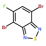


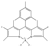
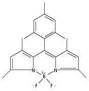


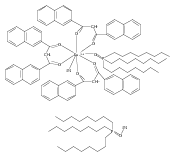


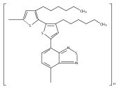


![D-Galactose, 2-[(2-azidoacetyl)amino]-2-deoxy-](/data/chemimg/115300/869186-83-4.png)
![D-Galactose, 2-[(2-azidoacetyl)amino]-2-deoxy-](/data/chemimg/115300/869186-83-4_b.png)
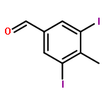
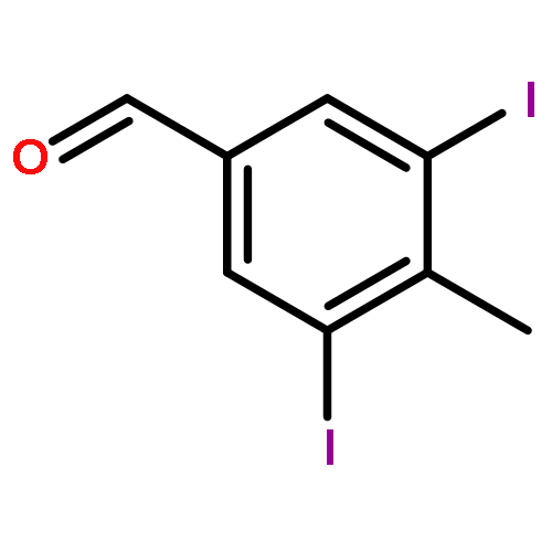


![Poly[(9,9-dihexylfluorenyl-2,7-diyl)-alt-(benzo[2,1,3]thiadiazol-4,7-diyl)]](http://img.cochemist.com/ccimg/579600/579505-53-6.png)
![Poly[(9,9-dihexylfluorenyl-2,7-diyl)-alt-(benzo[2,1,3]thiadiazol-4,7-diyl)]](http://img.cochemist.com/ccimg/579600/579505-53-6_b.png)
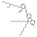
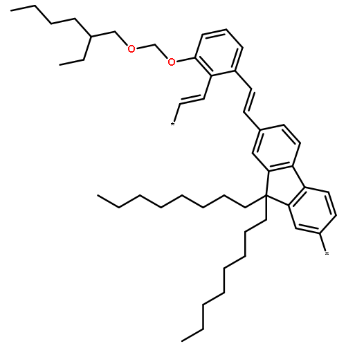

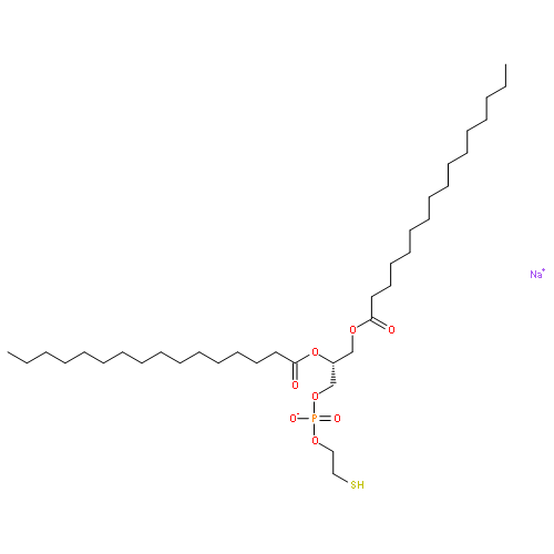


![1,3,2-Dioxaborolane,2,2'-[9,9-bis(2-ethylhexyl)-9H-fluorene-2,7-diyl]bis[4,4,5,5-tetramethyl-](http://img.cochemist.com/ccimg/357300/357219-41-1.png)
![1,3,2-Dioxaborolane,2,2'-[9,9-bis(2-ethylhexyl)-9H-fluorene-2,7-diyl]bis[4,4,5,5-tetramethyl-](http://img.cochemist.com/ccimg/357300/357219-41-1_b.png)

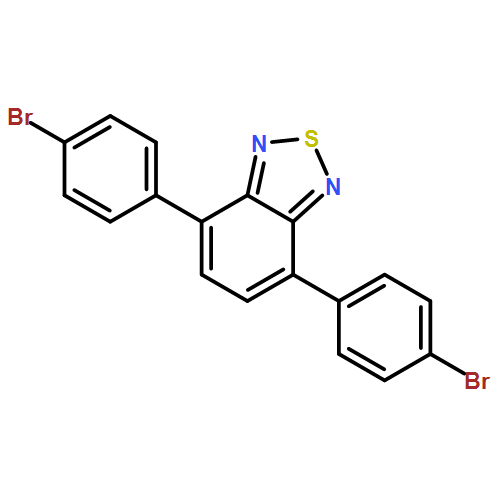


![Ethanamine, 2-[2-[2-(2-azidoethoxy)ethoxy]ethoxy]-](/data/chemimg/114300/134179-38-7.png)
![Ethanamine, 2-[2-[2-(2-azidoethoxy)ethoxy]ethoxy]-](/data/chemimg/114300/134179-38-7_b.png)
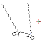


![2-[4-(aminoiminomethyl)phenyl]-1H-Indole-6-carboximidamide](http://img.cochemist.com/ccimg/47200/47165-04-8.png)
![2-[4-(aminoiminomethyl)phenyl]-1H-Indole-6-carboximidamide](http://img.cochemist.com/ccimg/47200/47165-04-8_b.png)


![3,5,9-Trioxa-4-phosphapentacosan-1-aminium,4-hydroxy-N,N,N-trimethyl-10-oxo-7-[(1-oxohexadecyl)oxy]-, inner salt, 4-oxide](http://img.cochemist.com/ccimg/2700/2644-64-6.png)
![3,5,9-Trioxa-4-phosphapentacosan-1-aminium,4-hydroxy-N,N,N-trimethyl-10-oxo-7-[(1-oxohexadecyl)oxy]-, inner salt, 4-oxide](http://img.cochemist.com/ccimg/2700/2644-64-6_b.png)


![Pyridinium,4-[6-[4-(diethylamino)phenyl]-1,3,5-hexatrien-1-yl]-1-[3-(triethylammonio)propyl]-,bromide (1:2)](http://img.cochemist.com/ccimg/162200/162112-35-8.png)
![Pyridinium,4-[6-[4-(diethylamino)phenyl]-1,3,5-hexatrien-1-yl]-1-[3-(triethylammonio)propyl]-,bromide (1:2)](http://img.cochemist.com/ccimg/162200/162112-35-8_b.png)


![3',6'-Dihydroxy-3H-spiro[isobenzofuran-1,9'-xanthen]-3-one](http://img.cochemist.com/ccimg/2400/2321-07-5.png)
![3',6'-Dihydroxy-3H-spiro[isobenzofuran-1,9'-xanthen]-3-one](http://img.cochemist.com/ccimg/2400/2321-07-5_b.png)


[2,5-bis(hexyloxy)-1,4-phenylene](2-cyano-1,2-ethenediyl)]](http://img.cochemist.com/ccimg/151900/151897-69-7.png)
[2,5-bis(hexyloxy)-1,4-phenylene](2-cyano-1,2-ethenediyl)]](http://img.cochemist.com/ccimg/151900/151897-69-7_b.png)
![Poly[(9,9-dioctylfluorenyl-2,7-diyl)-alt-(benzo[2,1,3]thiadiazol-4,7-diyl)]](/data/chemimg/107700/210347-52-7.png)
![Poly[(9,9-dioctylfluorenyl-2,7-diyl)-alt-(benzo[2,1,3]thiadiazol-4,7-diyl)]](/data/chemimg/107700/210347-52-7_b.png)

