Co-reporter:Zhonghua Mao, Chunli Gan, Jiuxin Zhu, Nan Ma, Lijun Wu, Libo Wang, Xiaobo Wang
Bioorganic & Medicinal Chemistry Letters 2017 Volume 27, Issue 12(Issue 12) pp:
Publication Date(Web):15 June 2017
DOI:10.1016/j.bmcl.2017.04.076
•An Atherosclerotic model has been established using thoracic aortas vascular ring.•7 flavonoids were got from Helichrysum arenarium L. MOENCH.•Anti-atherosclerotic activity of compounds was investigated.•The anti-inflammatory mechanism of these compounds was illustrated.We have successfully established AS model using thoracic aortas vascular ring which evaluated by the morphological changes of blood vessels, the proliferation of VSMC, and the expression of inflammation factors VEGF, CRP, JNK2 and p38. This AS model has the advantages of low cost, convenient and short period of established time. Moreover, we investigated the anti-AS activities of 7 flavonoids Narirutin (1), Naringin (2), Eriodictyol (3), Luteolin (4), Galuteolin (5), Astragalin (6), Kaempferol (7) from flowers of Helichrysum arenarium L. MOENCH by examining the vascular morphology, the inhibition on the expression of inflammation factors CRP, VEGF, JNK2, p38. In addition, we investigated the anti-AS activities of these 7 flavonoids by examining NO secretion of RAW264.7 cells in response to LPS. All above inflammation factors have been proved to be involved in the formation of AS. After comprehensive analysis of all results to discuss the structure–activity relationship, we summarized the conclusions at follow: compounds 1–7 could inhibit the expression of VEGF, CRP, JNK2, p38 and NO at different level, and we evaluated that flavonol aglycone have more significant anti-inflammation than it’s glycoside, and the anti-AS activity of flavonols were stronger than flavanones and flavones, which means that 3-group might be the effective group. Eventually, we supposed the main anti-inflammatory mechanism of these compounds was to reduce the expression of CRP, inhibit the kinases activity of JNK2 and p38, and then the MAPK pathway was suppressed, which resulted in the decrease of NO synthesis, VEGF expression and endothelial adhesion factor expression. And eventually, the scar tissue and vascular stenosis formations were prevented. This conclusion suggested flavonoids have the potential of preventing AS formation.Download high-res image (215KB)Download full-size image
Co-reporter:Da Wang, Dan Su, Xian-Zhe Li, Dan Liu, Rong-Gang Xi, Hui-Yuan Gao and Xiao-Bo Wang
RSC Advances 2016 vol. 6(Issue 33) pp:27434-27446
Publication Date(Web):11 Mar 2016
DOI:10.1039/C6RA02706G
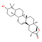
Barrigenol-like triterpenoids containing angeloyl residues in their structures are unusual natural products. To discover the chemical constituents responsible for the antitumor activity of Xanthoceras sorbifolia Bunge, the present study was carried out using the husk of this crop. Ten angeloyl barrigenol triterpenoids, including seven new (1–7) and three known compounds (8–10), were isolated from the active fraction via the bioassay-guided method. The structures of the compounds were established on the basis of spectral analysis, especially according to the data afforded by two digital-NMR and high-resolution mass spectra experiments. Compounds 1–10 exhibited varying degrees of cytotoxicity toward the human hepatoma cell line (HepG2), the human colorectal cancer cell line (HCT116) and the human glioma cell line (U87-MG). New compounds 3, 6, and 7 and known compounds 8, and 10 showed inhibitory activities similar to those of the positive control (doxorubicin hydrochloride). Cell cycle and apoptosis analysis of compound 8 revealed that it could suppress U87-MG cell proliferation by inducing apoptosis in the early period of exposure and then promote arrest at the G0/G1 phase.
Co-reporter:Chao Wang, Peipei Dong, Liyuan Zhang, Xiaokui Huo, Baojing Zhang, Changyuan Wang, Shanshan Huang, Xiaobo Wang, Jihong Yao, Kexin Liu and Xiaochi Ma
RSC Advances 2015 vol. 5(Issue 17) pp:12717-12725
Publication Date(Web):13 Jan 2015
DOI:10.1039/C4RA16459H
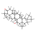
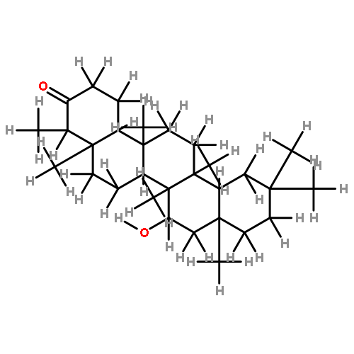
Biotransformation of 11-keto-β-boswellic acid (KBA) and acetyl-11-keto-β-boswellic acid (AKBA) catalyzed by two fungal strains (Cunninghamella elegans AS 3.1207 and Penicillium janthinellum AS 3.510) was performed in the present investigation. Eleven transformed products (1–11) were isolated, and accurately identified by various spectral methods. Among them, eight products (1–4 and 8–11) are novel. Two microorganisms used in our experiments demonstrated the favourable capability of stereo- and regio-hydroxylation at the non-active position for boswellic acid skeletons (KBA and AKBA). P. janthinellum AS 3.510 preferred to catalyze hydroxylation reaction at the C-21α position, especially for AKBA with a yield of 35.7%. Meanwhile, C. elegans AS 3.1207 preferred to catalyze the hydroxylation reaction of C-21β, especially for KBA with a yield of 55.2%. The major metabolite 1 exhibited potent anti-inflammatory activity in the in vitro bioassay.
Co-reporter:Yan Sun, Dan Liu, RongGang Xi, Xiaobo Wang, Yan Wang, Jie Hou, Baojing Zhang, Changyuan Wang, Kexin Liu, Xiaochi Ma
Bioorganic & Medicinal Chemistry Letters 2013 Volume 23(Issue 5) pp:1338-1342
Publication Date(Web):1 March 2013
DOI:10.1016/j.bmcl.2012.12.086
The capabilities of 20 strains of fungi to transform acetyl-11-keto-β-boswellic (AKBA) were screened. And biotransformation of AKBA by Cunninghamella blakesleana AS 3.970 afforded five metabolites (1–5), while two metabolites (6, 7) were isolated from biotransformation of Cunninghamella elegans AS 3.1207. The chemical structures of these metabolites were identified by spectral methods including 2D NMR and their structures were elucidated as 7β-hydroxy-3-acety-11-keto-β-boswellic acid (1), 21β-dihydroxy-3-acety-11-keto-β-boswellic acid (2), 7β,22α-dihydroxy-3-acety-11-keto-β-boswellic acid (3), 7β,16α-dihydroxy-3-acety-11-keto-β-boswellic acid (4), 7β,15α-dihydroxy-3-acety-11-keto-β-boswellic acid (5); 7β,15α,21β-trihydroxy-3-acety-11-keto-β-boswellic acid (6) and 15α,21β-dihydroxy-3-acety-11-keto-β-boswellic acid (7). All these products are previously unknown. Their primary structure–activity relationships (SAR) of inhibition activity on LPS-induced NO production in RAW 264.7 macrophage cells were evaluated.
Co-reporter:Weiru Jiang, Xiangge Tian, Yan Wang, Zheng Sun, Peipei Dong, Chao Wang, Xiaokui Huo, Baojing Zhang, Shanshan Huang, Sa Deng, Xiaobo Wang, Xiaochi Ma
Toxicology Letters (16 November 2016) Volume 262() pp:27-38
Publication Date(Web):16 November 2016
DOI:10.1016/j.toxlet.2016.09.004
•The herbs or foods containing abundant anthraquinones such as R. palmatum will cause a metabolic disorder of Mel, and should be avoided to combine application with Mel.•Five anthraquinones including: aloeemodin (1), rhein (2), emodin (3), chrysophanol (4), and physcion (5) from R. palmatum exhibited significant inhibitory effect on the melatonin metabolism.•The relationships between compounds’ inhibition activities and their structures were also investigated using computational model to further explain the inhibition mechanism.Melatonin (Mel) as an endogenous hormone, has been widely used in clinic for multiple therapeutic purposes. Further, the natural anthraquinones were widespread in various plants including herbs, foods, and some flavoring agents. The present work aims to evaluate the metabolic disorder of Mel caused by various common herbs and further identify their underlying mechanism. More importantly, the relationships between inhibitory activity and their structures were also investigated. Our results demonstrate that some herbs containing anthraquinone derivatives exhibited strong inhibition on Mel metabolism. Additionally, five anthraquinones from R. palmatum could inhibit phase I and II metabolism of Mel with a mixed inhibition kinetic model based on the mechanism of inhibiting human CYP1A1, 1A2, and SULT1A1. At last, the influence of R. palmatum and its five major components on the Mel metabolism were verified in human primary hepatocytes. In conclusion, our studies elucidated that herbs or foods containing abundant anthraquinones such as R. palmatum will cause a metabolic disorder of Mel, and should be avoided to combined application with Mel in clinic.Download high-res image (209KB)Download full-size image

![methyl 1-(beta-D-glucopyranosyloxy)-6-hydroxy-7-methyl-1,4a,5,6,7,7a-hexahydrocyclopenta[c]pyran-4-c](http://img.cochemist.com/ccimg/18600/18524-94-2.png)
![methyl 1-(beta-D-glucopyranosyloxy)-6-hydroxy-7-methyl-1,4a,5,6,7,7a-hexahydrocyclopenta[c]pyran-4-c](http://img.cochemist.com/ccimg/18600/18524-94-2_b.png)


![2H-Furo[2,3-h]chromen-2-one](http://img.cochemist.com/ccimg/600/523-50-2.png)
![2H-Furo[2,3-h]chromen-2-one](http://img.cochemist.com/ccimg/600/523-50-2_b.png)


