Co-reporter:Guobin Mao, Qin Cai, Fubing Wang, Changliang Luo, Xinghu Ji, and Zhike He
Analytical Chemistry November 7, 2017 Volume 89(Issue 21) pp:11628-11628
Publication Date(Web):October 11, 2017
DOI:10.1021/acs.analchem.7b03053
As the blood glucose concentration is an important clinical parameter of diabetes, the rapid and effective detection of blood glucose is very significant for monitoring and managing diabetes. Here, a facile method to prepare Rox-DNA functionalized CdZnTeS quantum dots (QDs) was developed. The Rox-DNA functionalized CdZnTeS QDs were prepared by a one-pot hydrothermal method through phosphorothioate DNA bound to QDs, which were employed as a ratiometric fluorescent probe for the rapid and sensitive detection of H2O2 and glucose. Compared with the traditional multistep construction of ratiometric fluorescent probes, this presented approach is simpler and more effective without chemical modification and complicated separation. The CdZnTeS QDs with green fluorescence is specifically sensitive to H2O2, while the red fluorescence of Rox is invariable. H2O2 is the product from the oxidation of glucose catalyzed by glucose oxidase (GOx). Therefore, a facile method to detect H2O2 and glucose with a detection limit of 0.075 μM for H2O2 and 0.042 μM for glucose was developed. In addition, this proposed probe has been employed for the detection of glucose in human serum with a satisfactory result. Moreover, this probe has been used for visual detection, and the health and diabetics can be distinguished by the naked eye. Meanwhile, this nanoprobe is also generalizable and can be extended to the detection of many other H2O2-mediated analytes.
Co-reporter:Jinyang Chen, Xinghu Ji, and Zhike He
Analytical Chemistry April 4, 2017 Volume 89(Issue 7) pp:3988-3988
Publication Date(Web):March 7, 2017
DOI:10.1021/acs.analchem.6b04484
On the basis of an interesting experimental phenomenon, a novel and smart composite reagent consisting of double-stranded DNA-templated copper nanoparticles (dsDNA–CuNPs) and DNA intercalator (SYBR Green I) was developed and exploited for hydrogen peroxide (H2O2) detection as well as oxidase-based biosensing. The study found that, within the composite reagent, the small molecule SYBR Green I was easily adsorbed on the surfaces of CuNPs, instead of intercalating into the dsDNA. So the composite reagent only exhibited the red fluorescence generated from dsDNA–CuNPs. However, when the solution of H2O2 was added into the composite reagent, the CuNPs were deconstructed and their fluorescence was quenched; meanwhile, the inhibition of SYBR Green I binding with dsDNA was eliminated. As a result, the mixed solution of the composite reagent with H2O2 exhibited green fluorescence generated from the intercalation of SYBR Green I into dsDNA. Since H2O2 is an important molecule and involved in various research fields, this developed composite reagent could be employed for many applications in biological analysis. As a proof-of-application demonstration, the sensitive detection of glucose was conducted. Moreover, the method was also extended to the detection of other biomolecules, such as cholesterol and horseradish peroxidase, which indicated the broad applications of the proposed sensing strategy in biomedical analysis.
Co-reporter:Jinyang Chen, Yongen Xu, Xinghu Ji, Zhike He
Sensors and Actuators B: Chemical 2017 Volume 239() pp:262-269
Publication Date(Web):February 2017
DOI:10.1016/j.snb.2016.08.017
•A simple and versatile method for nuclease assay was developed.•The proposed method is label-free, easy to operation and environment-friendly.•The method possesses high sensitivity and selectivity for nucleases assay.•The method was capable to detect nuclease in complex biological environment.In this work, inspired by the idea that the DNA fragments of nuclease cleavage exactly can serve as the primers for terminal deoxynucleotidyl transferase (TdT) mediated polymerization, a versatile and sensitive method for nuclease assay was developed based on the enzymatic polymerization and poly(thymine)-templated copper nanoparticles (CuNPs). Specifically, in the presence of target nuclease, the DNA substrate was firstly degraded as mononucleotides and oligonucleotide fragments. Employing the fragments as primers, the TdT-assisted polymerization was initiated to generate poly(thymine) templates on which fluorescent CuNPs could be synthesized. The fluorescence intensity of CuNPs was detected for the nuclease assay. So different nucleases assay could be realized just by changing the designs of DNA substrates of the corresponding nucleases. For the versatility demonstration, two nucleases, Exo III and EcoR V as the model of exonuclease and endonuclease, respectively, were detected with this method. It was proved that the proposed method was of high selectivity and sensitivity, and the limits of detections of Exo III and EcoR V were found to be 0.0138 and 0.0115 U/mL, respectively. What’s more, the satisfactory result of EcoR V detection in human serum indicates that this method is capable to detect nuclease in real sample.
Co-reporter:Yanan Zhang, Shan Guo, Shibo Cheng, Xinghu Ji, Zhike He
Biosensors and Bioelectronics 2017 Volume 94(Volume 94) pp:
Publication Date(Web):15 August 2017
DOI:10.1016/j.bios.2017.03.041
•A ratiometric fluorescent aptasensor based on label-free silicon nanodots was designed via covalent modification.•The metal-free aptasensor exhibited cell recognition, good biocompatibility, and a wide pH response range in MCF-7 cells.•Localized fluorescence imaging and precise pH measurement in living cells were performed with the ultrasmall aptasensor.The homeostasis of lysosomal pH is crucial in cell physiology. Developing small fluorescent nanosensors for lysosome imaging and ratiometric measurement of pH is highly demanded yet challenging. Herein, a pH-sensitive fluorescein tagged aptamer AS1411 has been utilized to covalently modify the label-free fluorescent silicon nanodots via a crosslinker for construction of a ratiometric pH biosensor. The established aptasensor exhibits the advantages of ultrasmall size, hypotoxicity, excellent pH reversibility and good photostability, which favors its application in an intracellular environment. Using human breast MCF-7 cancer cells and MCF-10A normal cells as the model, this aptasensor shows cell specificity for cancer cells and displays a wide pH response range of 4.5–8.0 in living cells. The results demonstrate that the pH of MCF-7 cells is 5.1, which is the expected value for acidic organelles. Lysosome imaging and accurate measurement of pH in MCF-7 cells have been successfully conducted based on this nanosensor via fluorescent microscopy and flow cytometry.
Co-reporter:Jiao Zheng, Tai Ye, Jinyang Chen, Li Xu, Xinghu Ji, Chuluo Yang, Zhike He
Biosensors and Bioelectronics 2017 Volume 90(Volume 90) pp:
Publication Date(Web):15 April 2017
DOI:10.1016/j.bios.2016.11.056
•The Z-TPE-5 could sensitively discriminate heparin from other ploysaccharides.•The Z-TPE-5 shows excellent fluorescence response to heparin with a detection limit of 1.53 ng/mL.•The high affinity between heparin and protamine is employed.•This probe can be applied for the detection of heparin in serum.•Advantages of the present strategy: label-free, simplicity, rapidity and high sensitivity.Tetraphenylethene derivatives were reported for sensitive “turn on” detection of heparin (Hep) based on the typical aggregation-induced emission (AIE). In the present strategy, three derivatives of tetraphenylethene (TPE) were designed,synthesized and compared the sensitivities for the detection of Hep. Furthermore, the (Z)-4,4′-(((1,2-diphenylethene-1,2-diyl) bis(4,1-phenylene))bis (oxy))bis(butan-1-aminium) (Z-TPE-5) was the most sensitive and chosen as the probe for Hep detection. Since Hep could be employed as a medium for inducing the aggregation of positively charged Z-TPE-5, the designed Z-TPE-5 shows excellent fluorescence response to Hep through multiple electrostatic interactions with a detection limit of 1.53 ng/mL, which is far below than most of the reported method for the detection of Hep. Due to the stronger affinity between Hep and protamine, Hep preferred to bind with it instead of Z-TPE-5 after the addition of protamine, so the fluorescence could be reduced. In comparison to prior studies, this developed strategy here not only simplifies the preparation procedure of the fluorescent probes but also can be applied in sensitive determination of Hep with good accuracy. Moreover, the detection of Hep with Z-TPE-5 was not interfered by Hep analogues, such as chondroitin sulfate and hyaluronic acid, as well as in the matrix of human serum.
Co-reporter:Ningxing Li;Jiao Zheng;Chunrong Li;Xinxin Wang;Xinghu Ji;Zhike He
Chemical Communications 2017 vol. 53(Issue 60) pp:8486-8488
Publication Date(Web):2017/07/25
DOI:10.1039/C7CC04547F
An enzyme-free stochastic DNA walker propelled by a single catalytic or double catalytic DNA assembly has been constructed. The application of the proposed DNA walking biosensor was successfully expanded to the detection of DNA and the enzymatic activity of T4 polynucleotide kinase.
Co-reporter:Ningxing Li, Jinyang Chen, Ming Luo, Chaohui Chen, Xinghu Ji, Zhike He
Biosensors and Bioelectronics 2017 Volume 87() pp:325-331
Publication Date(Web):15 January 2017
DOI:10.1016/j.bios.2016.08.067
•Two different models have been developed by changing the adding order of reagents.•The proposed strategy is of high sensitivity and selectivity, and the detection limit is as low as 9.7 fM.•The proposed biosensor was applied in complex matrix and real sample analysis.•The method was extended to DNA detection, which demonstrated the universality of the proposed sensing strategy.An ultrasensitive chemiluminescence (CL) biosensor for the detection of protein is developed in this study based on the functionalized magnetic microparticles (MMPs) and the hybridization chain reaction (HCR). First, the primer hybridized with the thrombin aptamer conjugated on the surface of MMPs. Then the HCR was triggered by part of the primer and its products were assembled on the surface of the MMPs. Through the interaction between streptavidin and biotin, the streptavidin-horseradish peroxidase (SA-HRP) was coupled with the HCR products. In the presence of thrombin, the HCR products conjugating with SA-HRP were released from the surface of MMPs after the aptamer recognized and bound to its target molecule. So the released SA-HRP in the supernatant produced a significant chemiluminescence imaging signal after the addition of H2O2-luminol. The detection limit of thrombin with this method could be as low as 9.7 fM. Besides, the sensing strategy was modified by changing the adding order of reagents that was then successfully applied in the detection of thrombin in complex sample. What's more, the DNA detection also could be carried out with this method, which demonstrated the universality of the proposed sensing strategy.
Co-reporter:Jinyang Chen, Xinghu Ji, Philip Tinnefeld, and Zhike He
ACS Applied Materials & Interfaces 2016 Volume 8(Issue 3) pp:1786
Publication Date(Web):December 31, 2015
DOI:10.1021/acsami.5b09678
In this work, a multifunctional template for selective formation of fluorescent silver nanoclusters (AgNCs) or copper nanoparticles (CuNPs) is put forward. This dumbbell-shaped (DS) DNA template is made up of two cytosine hairpin loops and an adenine–thymine-rich double-helical stem which is closed by the loops. The cytosine loops act as specific regions for the growth of AgNCs, and the double-helical stem serves as template for the CuNPs formation. By carefully investigating the sequence and length of DS DNA, we present the optimal design of the template. Benefiting from the smart design and facile synthesis, a simple, label-free, and ultrasensitive fluorescence strategy for adenosine triphosphate (ATP) detection is proposed. Through the systematic comparison, it is found that the strategy based on CuNPs formation is more sensitive for ATP assay than that based on AgNCs synthesis, and the detection limitation was found to be 81 pM. What’s more, the CuNPs formation-based method is successfully applied in the detection of ATP in human serum as well as the determination of cellular ATP. In addition to small target molecule, the sensing strategy was also extended to the detection of biomacromolecule (DNA), which illustrates the generality of this biosensor.Keywords: bioassays; copper nanoparticle; DNA; multifunctional template; silver nanocluster
Co-reporter:Chaohui Chen, Ningxing Li, Jingwen Lan, Xinghu Ji, Zhike He
Analytica Chimica Acta 2016 Volume 902() pp:154-159
Publication Date(Web):1 January 2016
DOI:10.1016/j.aca.2015.10.030
•This sensor is relied on a simple graphene/Au-NPs-based colorimetric method.•The DNA detection not only avoids any labeling but also reduces the background signal.•This present work can be expanded to detect viruses or proteins.A target-catalyzed hairpin assembly (CHA) and graphene/Au-NPs hybrids-based platform has been developed for the determination of DNA. This new sensor not only avoided any labeling but also reduced the background signal. In the absence of target, the assembly of H1 and H2 couldn't be triggered. The catalytic activity of graphene/Au-NPs hybrids was inhibited by adsorption of H1 and H2, leading to the “inactive” hybrids unable to catalyze the oxidation reaction of 3,3′,5,5′-tetramethylbenzidine (TMB). However, with the addition of target DNA, the target-catalyzed hairpin assembly was initiated and produced plenty of H1–H2 duplex, which had a weak binding affinity with the graphene/Au-NPs. Thus, the protected interface of graphene/Au-NPs hybrids became active and catalyzed the oxidation reaction of TMB accompanied with a colorless to-blue color change. This approach exhibited good sensitivity and specificity for target DNA with a detection limit of 5.74 × 10⁻¹¹ M, and realized the assay of target DNA in human serum samples. Besides, this sensor could be further expanded to detect viruses or proteins by adapting the corresponding aptamers, showing great potential in biochemical detections.
Co-reporter:Cuiling Zhang;Caiping Ding;Dongshan Xiang;Li Li;Xinghu Ji;Zhike He;Yuezhong Xian
Chinese Journal of Chemistry 2016 Volume 34( Issue 3) pp:317-325
Publication Date(Web):
DOI:10.1002/cjoc.201500906
Abstract
As a newly developed fluorescent probe, DNA functionalized quantum dots (QDs) with unique optical properties play a critical role in the bioanalytical applications. The present review provides an overview on the conjugation technologies of DNA to QDs, including electrostatic attraction, covalent attachment, specific effect and direct synthesis. In addition, we summarize recent advances on DNA functionalized QDs in the bioapplications, such as targeting nucleic acids, ions, small molecules and the protein detection, cell imaging and disease diagnosis. Finally, we envision aspects concerning current challenges on the DNA functionalized fluorescent QDs.
Co-reporter:Chen Liu, Jinyang Chen, Guobin Mao, Chen Su, Xinghu Ji and Zhike He
Analytical Methods 2016 vol. 8(Issue 30) pp:5957-5961
Publication Date(Web):04 Jul 2016
DOI:10.1039/C6AY01679K
Zeatin (ZT) is a type of cytokinin which plays important roles in the proliferation and differentiation of plants cells. Here, we describe a fluorometric method for the detection of ZT, based on target-induced structure switching of a hairpin aptamer. The signal probe, an FAM-labeled aptamer, hybridizes partially with BHQ-2-labeled DNA and the fluorescence intensity of FAM is quenched by the approaching BHQ-2. With the addition of the target, the aptamer binds to ZT with high specificity and the BHQ-2-labeled DNA is released. Thus, the fluorescence intensity of the FAM-labeled aptamer is recovered and ZT can be quantitatively determined by monitoring the fluorescence signal change. This proposed method shows good sensitivity with a detection limit as low as 135 nM for ZT detection. In comparison to the previously reported ZT detection strategies based on graphene oxide (GO), this biosensor exhibits excellent selectivity. Moreover, this method has great potential for parallel analysis of different aptamer-based target molecules.
Co-reporter:Jinyang Chen, Yucheng Liu, Xinghu Ji, Zhike He
Biosensors and Bioelectronics 2016 Volume 83() pp:221-228
Publication Date(Web):15 September 2016
DOI:10.1016/j.bios.2016.04.055
•A versatile dumbbell molecular probe was designed and employed for bioassays.•The proposed biosensor is of high sensitivity and selectivity.•The proposed strategy was applied in complex matrix and real sample analysis.•The strategy was integrated with droplet-based microfluidics for visual detection.In this work, a versatile dumbbell molecular (DM) probe was designed and employed in the sensitively homogeneous bioassay. In the presence of target molecule, the DM probe was protected from the digestion of exonucleases. Subsequently, the protected DM probe specifically bound to the intercalation dye and resulted in obvious fluorescence signal which was used to determine the target molecule in return. This design allows specific and versatile detection of diverse targets with easy operation and no sophisticated fluorescence labeling. Integrating the idea of target-protecting DM probe with adenosine triphosphate (ATP) involved ligation reaction, the DM probe with 5′-end phosphorylation was successfully constructed for ATP detection, and the limitation of detection was found to be 4.8 pM. Thanks to its excellent selectivity and sensitivity, this sensing strategy was used to detect ATP spiked in human serum as well as cellular ATP. Moreover, the proposed strategy was also applied in the visual detection of ATP in droplet-based microfluidic platform with satisfactory results. Similarly, combining the principle of target-protecting DM probe with streptavidin (SA)-biotin interaction, the DM probe with 3′-end biotinylation was developed for selective and sensitive SA determination, which demonstrated the robustness and versatility of this design.
Co-reporter:Jingwen Lan, Jinyang Chen, Ningxing Li, Xinghu Ji, Mingxia Yu, Zhike He
Talanta 2016 Volume 151() pp:126-131
Publication Date(Web):1 May 2016
DOI:10.1016/j.talanta.2016.01.024
•A microfluidic approach for magnetic-fluorescent Janus microparticle is presented.•Fluorescent labeling and magnetic separation is realized with designed particles.•Spatial separation prevents magnetic nanoparticle from quenching QDs fluorescence.•The designed Janus microparticles have been successfully applied in DNA assay.Fluorescent magnetic multifunctional microparticles were fabricated by a facile droplet microfluidic strategy. Two sodium alginate streams, one doped with Fe3O4 nanoparticles (NPs) and the other with CdSe/ZnS quantum dots, were introduced into a flow-focusing channel as a type of parallel laminar flow to form droplets containing two distinct parts. Then, at the serpentine channel, the Ca2+ in the oil phase diffused into the droplets, causing the solidification of the droplets. Thus, the Janus microparticles with excellent magnetic/fluorescent properties formed. The flow conditions were optimized and the effects of the flow rates on magnetic/fluorescent compositions were carefully investigated. Luminescent labeling and magnetic separation were simultaneously realized with the newly designed microparticles. Moreover, spatial separation between Fe3O4 NPs and QDs prevented the interference of QDs photoluminescence by the magnetic particles. The as-prepared fluorescent magnetic Janus particles were also successfully employed for DNA assay, which demonstrated the potential of the multifunctional microbeads in biological applications.
Co-reporter:Tai Ye, Chunying Li, Chen Su, Xinghu Ji, Jiao Zheng, Philip Tinnefeld and Zhike He
Chemical Communications 2015 vol. 51(Issue 41) pp:8644-8647
Publication Date(Web):14 Apr 2015
DOI:10.1039/C5CC01517K
A bottom-up strategy was developed for the enzyme mediated synthesis of Cu nanoparticles, which showed good sensing performance.
Co-reporter:Jinyang Chen, Xinghu Ji, Zhike He
Analytica Chimica Acta 2015 Volume 888() pp:110-117
Publication Date(Web):12 August 2015
DOI:10.1016/j.aca.2015.07.054
•A simple, robust and low-cost sample-introduction technique was developed.•Convenient and flexible sample changing was achieved in microfluidic system.•Novel strategy of concentration gradient generation was presented for barcoding.•High-throughput droplet screening could be realized in the integrated platform.•Multiplex DNA assay was successfully carried out in the droplet platform.In this work, a simple, flexible and low-cost sample-introduction technique was developed and integrated with droplet platform. The sample-introduction strategy was realized based on connecting the components of positive pressure input device, sample container and microfluidic chip through the tygon tubing with homemade polydimethylsiloxane (PDMS) adaptor, so the sample was delivered into the microchip from the sample container under the driving of positive pressure. This sample-introduction technique is so robust and compatible that could be integrated with T-junction, flow-focus or valve-assisted droplet microchips. By choosing the PDMS adaptor with proper dimension, the microchip could be flexibly equipped with various types of familiar sample containers, makes the sampling more straightforward without trivial sample transfer or loading. And the convenient sample changing was easily achieved by positioning the adaptor from one sample container to another. Benefiting from the proposed technique, the time-dependent concentration gradient was generated and applied for quantum dot (QD)-based fluorescence barcoding within droplet chip. High-throughput droplet screening was preliminarily demonstrated through the investigation of the quenching efficiency of ruthenium complex to the fluorescence of QD. More importantly, multiplex DNA assay was successfully carried out in the integrated system, which shows the practicability and potentials in high-throughput biosensing.
Co-reporter:Chen Su, Yufei Liu, Tai Ye, Xia Xiang, Xinghu Ji, Zhike He
Analytica Chimica Acta 2015 Volume 853() pp:495-500
Publication Date(Web):1 January 2015
DOI:10.1016/j.aca.2014.10.027
•The single-color QDs–Ru assembling dyads were applied in homogeneous DNA assay.•This biosensor exhibited high selectivity against base mismatched sequences.•This biosensor could be severed as universal platform for the detection of ssDNA.•This sensor could be used to detect the target in human serum samples.•This DNA sensor had a good selectivity under the interference of other dsDNA.In this work, a new, label-free, homogeneous, highly sensitive, and selective fluorescent biosensor for DNA detection is developed by using rolling-circle amplification (RCA) based single-color quantum dots–ruthenium complex (QDs–Ru) assembling dyads. This strategy includes three steps: (1) the target DNA initiates RCA reaction and generates linear RCA products; (2) the complementary DNA hybridizes with the RCA products to form long double-strand DNA (dsDNA); (3) [Ru(phen)2(dppx)]2+ (dppx = 7,8-dimethyldipyrido [3,2-a:2′,3′-c] phenanthroline) intercalates into the long dsDNA with strong fluorescence emission. Due to its strong binding propensity with the long dsDNA, [Ru(phen)2(dppx)]2+ is removed from the surface of the QDs, resulting in restoring the fluorescence of the QDs, which has been quenched by [Ru(phen)2(dppx)]2+ through a photoinduced electron transfer process and is overlaid with the fluorescence of dsDNA bonded Ru(II) polypyridyl complex (Ru-dsDNA). Thus, high fluorescence intensity is observed, and is related to the concentration of target. This sensor exhibits not only high sensitivity for hepatitis B virus (HBV) ssDNA with a low detection limit (0.5 pM), but also excellent selectivity in the complex matrix. Moreover, this strategy applies QDs–Ru assembling dyads to the detection of single-strand DNA (ssDNA) without any functionalization and separation techniques.A universal, label-free, homogeneous, highly sensitive, and selective fluorescent biosensor for DNA detection is developed by using rolling-circle amplification (RCA) based single-color quantum dots–ruthenium complex (QDs–Ru) assembling dyads.
Co-reporter:Chaohui Chen, Ming Luo, Tai Ye, Ningxing Li, Xinghu Ji and Zhike He
Analyst 2015 vol. 140(Issue 13) pp:4515-4520
Publication Date(Web):05 May 2015
DOI:10.1039/C5AN00485C
An ultrasensitive method for the detection of protein is critically important in fundamental research and practical applications due to the low abundance of disease markers in body fluids or tissues. To detect the trace levels of disease markers with high sensitivity and specificity, a sensitive colorimetric biosensor for protein assay was developed using gold nanoparticles (AuNPs) and rolling circle amplification (RCA). After binding the biotinylated primer/circular template to the streptavidin-conjugated sandwich ELISA immunocomplex, the biotinylated primer was isothermally extended to generate single-stranded DNA (ssDNA). Sequentially, the padlock DNA was added and hybridized with the RCA products. The aggregation of the additional AuNPs in the supernatant containing the surplus padlock DNA and a certain concentration of salt could then be observed. The established sensor allowed for the specific detection of α-fetoprotein (AFP) with a detection limit of 33.45 pg mL−1. It was also demonstrated that this method could distinguish 500 pg mL−1 AFP with the naked eye. In addition, this biosensor could be applied to complex sample analysis and could be further used as a universal method for any protein or virus determination by changing the corresponding antibodies.
Co-reporter:Tai Ye, Chunying Li, Chen Su, Xinghu Ji and Zhike He
RSC Advances 2015 vol. 5(Issue 68) pp:55336-55339
Publication Date(Web):18 Jun 2015
DOI:10.1039/C5RA07509B
An enzymatic synthesis strategy of a DNA-template alloy nanocluster is presented. In this strategy, alkaline phosphatase (ALP) catalysed pyrophosphate (PPi) hydrolysis broke the coordination between Cu2+ and PPi. The fluorescent DNA-template alloy nanoclusters were produced in situ through the reduction of the Cu2+ and Ag+ in the presence of single strand DNA (ss DNA). The fluorescence intensity of the alloy nanocluster was related to the concentration of ALP. This fluorescence turn-on strategy exhibited high sensitivity and selectivity for the ALP assay. Additionally, this strategy was also applied in an ALP-linked immuno-sorbent assay for a-fetoprotein (AFP) detection. Taking AFP as the model protein, it could be detected at a range from 10 ng mL−1 to 150 ng mL−1 and the detection limit was 4 ng mL−1 (0.042 nM). Finally, the diagnostic capability and practical application of this method was demonstrated by detecting AFP in human blood serum.
Co-reporter:Chen Liu, Guobin Mao, Chen Su, Xinghu Ji, Zilin Chen and Zhike He
Analytical Methods 2015 vol. 7(Issue 18) pp:7748-7752
Publication Date(Web):27 Jul 2015
DOI:10.1039/C5AY01728A
With the advantages of excellent optical properties and biocompatibility, DNA-functionalized quantum dots (QDs) have been widely applied in biosensing and bioimaging. Systemin is an important class of plant peptide hormone that was first identified in plants. In this paper, we have synthesized aptamer-functionalized Zn2+ doped CdTe QDs through a facile one-pot hydrothermal route, and a fluorescent aptasensor based on graphene oxide (GO) is developed for the detection of tomato systemin (TomSys) with aptamer recognition properties. In the absence of TomSys, the aptamer-functionalized QDs are adsorbed on the surface of GO and the fluorescence is efficiently quenched, while in the presence of TomSys, the specific binding of TomSys with its aptamer competitively releases aptamer-functionalized QDs from the GO surface, leading to the recovery of QDs fluorescence. The results demonstrate that the simple, rapid and cost-efficient biosensor possesses satisfactory sensitivity and selectivity for the detection of TomSys.
Co-reporter:Jinyang Chen, Yufei Liu, Tai Ye, Xia Xiang, Xinghu Ji, Zhike He
Sensors and Actuators B: Chemical 2015 215() pp: 206-214
Publication Date(Web):
DOI:10.1016/j.snb.2015.03.065
Co-reporter:Jinyang Chen, Guohua Zhou, Yufei Liu, Tai Ye, Xia Xiang, Xinghu Ji, Zhike He
Talanta 2015 Volume 134() pp:271-277
Publication Date(Web):1 March 2015
DOI:10.1016/j.talanta.2014.11.027
•A novel strategy, assembly-line manipulation of droplets (ALMD) is presented.•The microfluidic platform based on ALMD can be used to fluorescence encoding.•Based on ALMD, simultaneous multiplexed DNA on-line detection has been realized.•The method based on ALMD has potential for high-throughput DNA assay.In this article, a new mode of droplets manipulation is presented and applied for simultaneous multiplexed DNA detection. We call this droplets manipulation, “assembly-line manipulation of droplets (ALMD)”. Firstly, multiple droplets containing the same target mixtures are generated in the microchannel, and then fused with later generated different droplets containing corresponding probes, respectively. Finally, all the fused droplets were fluorescence imaged on-line and real-time. The successful implementation of droplets fluorescence encoding based on ALMD shows the reproducibility and accuracy of this manipulation mode. As a proof-of-concept application, the simultaneous multiplexed DNA detection was carried out through the model of human immunodeficiency virus (HIV) gene sequence and variola virus (small pox, VV) gene sequence based on ALMD in the microfluidic system. It is proved that this method achieves simultaneous multiplexed DNA measurements with a significantly time-saving way and without different dye-labelled probes or complex operation procedures. In addition, it reveals the possibility of high-throughput biosensing with simple chip design and detection equipment.
Co-reporter:Tai Ye, Jinyang Chen, Yufei Liu, Xinghu Ji, Guohua Zhou, and Zhike He
ACS Applied Materials & Interfaces 2014 Volume 6(Issue 18) pp:16091
Publication Date(Web):August 12, 2014
DOI:10.1021/am504035a
A simple method for preparing DNA-stabilized Ag nanoclusters (NCs) nanowires is presented. To fabricate the Ag NCs nanowires, we use just two unmodified component strands and a long enzymatically produced scaffold. These nanowires form at room temperature and have periodic sequence units that are available for fluorescence Ag NCs assembled which formed three-way junction (TWJ) structure. These Ag NCs nanowires can be clearly visualized by confocal microscopy. Furthermore, due to the high efficiency of rolling circle amplification reaction in signal amplification, the nanowires exhibit high sensitivity for the specific DNA detection with a wide linear range from 6 to 300 pM and a low detection limit of 0.84 pM, which shows good performance in the complex serum samples. Therefore, these Ag NCs nanowires might have great potential in clinical and imaging applications in the future.Keywords: rolling circle amplification; self-assembly; silver cluster
Co-reporter:Cuiling Zhang, Ji Yan, Chen Liu, Xinghu Ji, and Zhike He
ACS Applied Materials & Interfaces 2014 Volume 6(Issue 5) pp:3189
Publication Date(Web):February 10, 2014
DOI:10.1021/am405864z
DNA-functionalized quantum dots (QDs) are powerful tools for biosensing and bioimaging applications. Facile labeling methods with good fluorescence properties are desirable for the development of DNA-functionalized QDs. In this article, we describe a novel and simple approach that leads to the synthesis of DNA-functionalized CdTe:Zn2+ QDs in one step. It is the first time that DNA-functionalized QDs have been prepared using sodium tellurite as the tellurium source by a hydrothermal method. This approach will greatly reduce the synthesis time (only about 1 h) and simplify the synthesis process as well as reduce the complexity of the required experimental techniques. The as-prepared QDs exhibit high quantum yield, small size, and low toxicity. UV–vis spectra and FTIR characterization proved that the abundance of DNA on the surface of the QDs increased with the increase in the concentration of the feed DNA. Most importantly, these QDs functionalized with DNA have great potential to bind specifically to DNA, protein, and cell surface receptors.Keywords: DNA; hydrothermal route; Na2TeO3; one-step method; Zn-doped quantum dots;
Co-reporter:Yufei Liu, Ming Luo, Xia Xiang, Chaohui Chen, Xinghu Ji, Lu Chen and Zhike He
Chemical Communications 2014 vol. 50(Issue 20) pp:2679-2681
Publication Date(Web):16 Jan 2014
DOI:10.1039/C4CC00128A
We have developed a novel immuno-sensor applied to protein detection based on exonuclease III (Exo III)-induced signal amplification, multiplex binding of the biotin–streptavidin system and the fluorescence quenching ability of graphene oxide.
Co-reporter:Zhen Zhang, Yufei Liu, Xinghu Ji, Xia Xiang and Zhike He
Analyst 2014 vol. 139(Issue 19) pp:4806-4809
Publication Date(Web):01 Jul 2014
DOI:10.1039/C4AN00933A
A graphene oxide (GO) based enzyme-free signal amplification platform for homogeneous DNA sensing is developed with simplicity and high sensitivity. In the absence of the target DNA, labeled hairpin probe 1 (H1) and probe 2 (H2) were adsorbed on the surface of GO, resulting in the fluorescence quenching of the dyes and minimizing the background fluorescence. The addition of the target DNA facilitated the formation of double-stranded DNA (dsDNA) between H1 and H2, causing the probes to separate from GO and release the target DNA through a strand displacement reaction. Meanwhile, the whole reaction started anew. This is an excellent isothermal signal amplification technique without the involvement of enzymes. By monitoring the change of the fluorescence intensity, the target DNA not only can be determined in buffer solution, but also can be detected in 1% serum solution spiked with a series of concentrations of the target DNA. In addition, the consumption amount of the probes in this method is lower than that in traditional molecular beacon methods.
Co-reporter:Tai Ye, Yufei Liu, Ming Luo, Xia Xiang, Xinghu Ji, Guohua Zhou and Zhike He
Analyst 2014 vol. 139(Issue 7) pp:1721-1725
Publication Date(Web):07 Jan 2014
DOI:10.1039/C3AN02077K
We report a new sensor combined two dimensional metal–organic framework (MOF), N,N-bis(2-hydroxy-ethyl)dithiooxamidato copper(II) (H2dtoaCu), with the hairpin-structured oligonucleotides and demonstrate its feasibility in detecting multiplexed sequence-specific DNA. The key component of this sensor (MOF–MBs) is the hairpin-structured fluorescent oligonucleotide that allows the MOFs to function as both a “nanoscaffold” for the oligonucleotide and a “nanoquencher” of the fluorophore. An oligonucleotide sequence fragment of wild-type HBV (T1) and a reverse-transcription oligonucleotide sequence of RNA fragment of HIV (T2) were used as model systems. While in the presence of the targets, the fluorescence of dyes was recovered by forming a double strand structure. Multiplex DNA detection can be realized by synchronous scanning fluorescence spectrometry, and there was no cross reaction between the two probes. Under the optimum conditions, the fluorescence intensities of two dyes all exhibit good linear dependence on their target DNA concentration in the range of 1–10 nM with the detection limit of 0.87 nM and 0.22 nM for T1 and T2, respectively. As a proof of concept, the MOF–MBs have been successfully used as a potential sensing platform for simultaneous detection of multiplexed DNA.
Co-reporter:Dan Zhao, Jiao-Tian Li, Fang Gao, Cui-ling Zhang and Zhi-ke He
RSC Advances 2014 vol. 4(Issue 87) pp:47005-47011
Publication Date(Web):30 Sep 2014
DOI:10.1039/C4RA06077F
The synthesis of water-soluble blue-emitting quantum dots (QDs) in aqueous solutions has attracted much attention recently, and here we report the synthesis of high-quality core/shell ZnSe/ZnS QDs through a one-step hydrothermal route with thiol ligand N-acetyl-L-cysteine (NAC) as the stabilizer. The impacts of various synthesis factors on the fluorescence properties of the prepared QDs have been systematically investigated. Under optimal conditions, the as-prepared QDs exhibit high photoluminescence quantum yield (∼39%), narrow full-width at half-maximum, and high photostability, due to the formation of a protective ZnS shell on the ZnSe core through the decomposition of NAC under high temperature. The core/shell structure of the ZnSe/ZnS QDs was verified by UV irradiation experiment, X-ray powder diffraction, and electron diffraction spectroscopy.
Co-reporter:Cuiling Zhang, Yuanyuan Zu, Xinghu Ji and Zhike He
RSC Advances 2014 vol. 4(Issue 38) pp:20044-20047
Publication Date(Web):14 Apr 2014
DOI:10.1039/C4RA01560F
Here, a facile strategy has been developed for the synthesis of peptide-capped CdTe quantum dots (peptide–QDs) in an aqueous phase by the one-pot method. The capping ligand, a designed peptide, can form a self-assembled hydrophobic layer on the surface of QDs, which makes the peptide–QDs exhibit excellent stability in acidic and high salt solutions. The as-prepared peptide–QDs are of potential values in the applications of biosensing and bioimaging.
Co-reporter:Chaohui Chen, Xia Xiang, Yufei Liu, Guohua Zhou, Xinghu Ji, Zhike He
Biosensors and Bioelectronics 2014 Volume 58() pp:205-208
Publication Date(Web):15 August 2014
DOI:10.1016/j.bios.2014.02.060
•The QDs–ruthenium complex is first applied in TPSMLD assay.•This biosensor can avoid the interference of dsDNA to ruthenium complex.•This biosensor is promising in detecting a wide range of small molecules binding proteins.We have developed a new dual-color fluorescent biosensor for protein detection based on terminal protection of small-molecule-linked DNA and the enzymolysis of exonuclease III (Exo III). The determination of streptavidin (SA) was realized via fluorescence signals of the green color from quantum dots (QDs) and the red from [Ru(phen)2(dppx)]2+. In the absence of SA, biotin-DNA was degradated by the Exo III, thus making the [Ru(phen)2(dppx)]2+ employed as a fluorescence quencher to the QDs. With the addition of SA, dual-color response appeared because of the specific binding between SA and biotin so that the biotin-dsDNA was protected and combined with [Ru(phen)2(dppx)]2+, leading to the QDs recovery and the generating of [Ru(phen)2(dppx)]2+ fluorescence. This sensor exhibited high sensitivity with a low detection limit (2.11 ng/mL) and firstly introduced dual-color QDs–ruthenium complex dyads to protein assay.
Co-reporter:Ming Luo, Ningxing Li, Yufei Liu, Chaohui Chen, Xia Xiang, Xinghu Ji, Zhike He
Biosensors and Bioelectronics 2014 Volume 55() pp:318-323
Publication Date(Web):15 May 2014
DOI:10.1016/j.bios.2013.11.066
•A highly sensitive chemiluminescence imaging method is developed for DNA detection.•Signal amplification by circular strand-displacement polymerization is designed.•The biosensor can discriminate target DNA from one-base mismatch DNA.•The biosensor has promising in high-throughput DNA assay.A universal, highly sensitive and selective chemiluminescence (CL) imaging method has been developed for high throughput detection of DNA. After molecular beacon (MB) hybridized with the target DNA, the biotin-labeled primer was attached to a magnetic microparticle (MMP) surface by hybridization with the stem part of the MB to initiate a polymerization of DNA strand, which led to the release of the target and another polymerization cycle. Thus the polymerization produced the multiplication of biotin-labeled primer on the surface of MMPs. Sequentially, the horseradish peroxidase (HRP) was conjugated to MMPs surface through the biotin–streptavidin reaction. Then, the conjugated HRP was determined by the CL imaging method. This proposed method could detect the sequence-specific DNA as low as 0.4 pM and discriminate perfectly matched target DNA from the mismatch DNAs. All in all, this proposed method exhibited an efficient amplification performance, and would open new opportunities for sensitive and high throughput detection of DNA.
Co-reporter:Dan Zhao, Jiaotian Li, Tianming Yang, Zhike He
Biosensors and Bioelectronics 2014 Volume 52() pp:29-35
Publication Date(Web):15 February 2014
DOI:10.1016/j.bios.2013.08.031
•Reversible “turn off–on” mode was established to detect platinum drugs and DNA.•The interactions between different platinum drugs to DNA have been compared.•The influences of DNA types to DNA–cisplatin interactions were also examined.•The mode can be applied in anticancer drug screening and pharmacological study.A “turn off–on” mode has been established by using the interaction between platinum anticancer drugs and DNA as input signal and the fluorescence reversible change of quantum dots (QDs) as output signal. The QDs fluorescence can be quenched by platinum anticancer drugs via photo-induced electron transfer process, rendering the system into “turn off” status, and the system can then be “turned on” when fluorescence is restored due to covalent conjugation between DNA and platinum anticancer drugs. This dual-directional fluorescence change realized the detection of cisplatin and DNA, overcoming the selectivity problem commonly existed in the traditional mono-directional fluorescence detection mode. The reversible fluorescent “turn off-on” mode has been further employed to study the interactions between DNA and different platinum anticancer drugs (cisplatin, oxaliplatin and carboplatin). Furthermore, the impacts of different types of DNAs (different in base sequence, chain length and ssDNA/dsDNA) on the mode are also explored. This simple, fast and convenient spectroscopic method owns promising applications in the study on interaction between medical molecules and DNA, and in biochemical detections.
Co-reporter:Guohua Zhou, Xian Zhang, Xinghu Ji and Zhike He
Chemical Communications 2013 vol. 49(Issue 78) pp:8854-8856
Publication Date(Web):23 Jul 2013
DOI:10.1039/C3CC43621G
Terminal protection of small molecule linked DNA assays amplified by Exo III-aided DNA recycling is developed for the detection of streptavidin–biotin interaction, with a detection limit of 0.8 fM streptavidin in the “turn-on” state, indicating that the proposed method is an ultrasensitive platform for the detection of small molecule–protein interactions.
Co-reporter:Yufei Liu, Ming Luo, Ji Yan, Xia Xiang, Xinghu Ji, Guohua Zhou and Zhike He
Chemical Communications 2013 vol. 49(Issue 67) pp:7424-7426
Publication Date(Web):09 May 2013
DOI:10.1039/C3CC42639D
A highly sensitive and selective DNA biosensor based on hybridization chain reaction is described, which combines CdTe quantum dots (QDs) and a ruthenium complex. Based on the variation of fluorescence signals of the CdTe QDs, the target DNA is determined.
Co-reporter:Cuiling Zhang, Xinghu Ji, Yuan Zhang, Guohua Zhou, Xianliang Ke, Hanzhong Wang, Philip Tinnefeld, and Zhike He
Analytical Chemistry 2013 Volume 85(Issue 12) pp:5843
Publication Date(Web):May 20, 2013
DOI:10.1021/ac400606e
High quality and facile DNA functionalized quantum dots (QDs) as efficient fluorescence nanomaterials are of great significance for bioimaging both in vitro and in vivo applications. Herein, we offer a strategy to synthesize DNA-functionalized Zn2+ doped CdTe QDs (DNA-QDs) through a facile one-pot hydrothermal route. DNA is directly attached to the surface of QDs. The as-prepared QDs exhibit small size (3.85 ± 0.53 nm), high quantum yield (up to 80.5%), and excellent photostability. In addition, the toxicity of QDs has dropped considerably because of the Zn-doping and the existence of DNA. Furthermore, DNA has been designed as an aptamer specific for mucin 1 overexpressed in many cancer cells including lung adenocarcinoma. The aptamer-functionalized Zn2+ doped CdTe QDs (aptamer-QDs) have been successfully applied in active tumor-targeted imaging in vitro and in vivo. A universal design of DNA for synthesis of Zn2+ doped CdTe QDs could be extended to other target sequences. Owing to the abilities of specific recognition and the simple synthesis route, the applications of QDs will potentially be extended to biosensing and bioimaging.
Co-reporter:Qing Zhu, Dongshan Xiang, Cuiling Zhang, Xinghu Ji and Zhike He
Analyst 2013 vol. 138(Issue 18) pp:5194-5196
Publication Date(Web):08 Jul 2013
DOI:10.1039/C3AN00960B
The bifunctionality of graphene oxide (GO) which can highly adsorb single-stranded DNA (ssDNA) and effectively quench the emission of organic dyes is reasonably utilized in a multiplexed DNA detection system, achieving sensitive and selective detection of HIV, VV and EV, respectively.
Co-reporter:DongShan Xiang;AiHua Zheng;Ming Luo;XingHu Ji;ZhiKe He
Science China Chemistry 2013 Volume 56( Issue 3) pp:380-386
Publication Date(Web):2013 March
DOI:10.1007/s11426-012-4767-7
We have developed a multiplexed DNA detection method based on graphene oxide (GO) and molecular beacons (MBs) by synchronous fluorescence analysis, demonstrated it by an oligonucleotide sequence of wild-type HBV (T1) and a reverse-transcription oligonucleotide sequence of the RNA fragment of HIV (T2) as a model system. In the absence of targets DNA, FAM-tagged free MB probes (PHBV) and ROX-tagged free MB probes (PHIV) are adsorbed on GO via π-π interactions between DNA nucleobases and nucleosides, and the π-rich GO brings the fluorophores of MB and GO into close proximity. And then, the fluorescence of fluorophores is quenched by GO. But in the presence of targets DNA, PHBV and PHIV hybridize with their targets DNA resulting in the formation of double-stranded DNA (dsDNA), causing the separation of PHBV and PHIV from the surface of GO and the recovery of the fluorescence of fluorophores (FAM and ROX) simultaneously. The simultaneous detection of T1 and T2 can be realized by measuring fluorescence signals of FAM and ROX, respectively. Under the optimum conditions, the fluorescence intensities of two dyes all exhibit good linear dependence on their target DNA concentration in the range of 5×10⁻¹¹−5×10⁻⁹ M. The detection limit of T1 is 3×10⁻¹¹ M (3 σ), and that of T2 is 2×10⁻¹¹ M. Compared with other methods for DNA detection based on GO, the proposed method has some advantages including higher selectivity and shorter analytical time.
Co-reporter:Guohua Zhou, Yizhen Liu, Ming Luo, Xiuhong Li, Qinfeng Xu, Xinghu Ji, and Zhike He
Langmuir 2013 Volume 29(Issue 15) pp:4697-4702
Publication Date(Web):March 22, 2013
DOI:10.1021/la400404g
An assembly of gold nanoparticle through the recognition of unmodified antibody was developed. The use of peptide (Cys-Ala-Leu-Asn-Asn) as ligands to stabilize and functionalize gold nanoparticles provides technical and operational convenience. These peptide-capped particles in different sizes are recognized by antibody and assembly to form dimers and expanded hybrid material by controlling the conditions. The interparticle spacing of these assemblies was well studied with small-angle X-ray scattering measurements, and it was found that the interparticle spacing is inversely dependent on the particle size. This relationship of interparticle spacing and particle size is closely related to the structure of antibody linker. Therefore, analyzing the interparticle spacing of assemblies can reveal the equilibrium configuration of IgG. Based on the investigation, the Fab–Fab angle of IgG is obtained to be ≈102° and the Fab arms are ≈7.8 nm. These results provide new experimental data on the structure of flexible IgG.
Co-reporter:Xia Xiang, Liyang Shi, Ming Luo, Jinyang Chen, Xinghu Ji, Zhike He
Biosensors and Bioelectronics 2013 Volume 49() pp:403-409
Publication Date(Web):15 November 2013
DOI:10.1016/j.bios.2013.05.026
•A stepwise reagent introduction (SRI) is first applied in droplet platform.•The multiplexed DNA detection is realized without expensive instruments.•The DNA detection is performed based on FRET process.A stepwise reagent introduction (SRI)-based droplet platform is reported to apply in multiplexed DNA sensing via droplet imaging and manipulation techniques. The feature of SRI-based droplet platform is that the multiplexed DNA measurements are performed simultaneously without the need for expensive optical instruments. This is achieved by sequentially driving five different DNA probes labeled with carboxyfluorescein into the same chip channel to individually form droplet and fuse with target DNAs and graphene oxide droplets. Based on the variation of droplet color and fluorescence intensity, rapidly qualitative and quantitative DNA analysis are realized. The cooperation between SRI mode and droplet platform thus opens up the possibility of greatly simplified DNA sensing in high throughput format without complicated fluorescent labeling and chip design.
Co-reporter:Aihua Zheng, Ming Luo, Dongshan Xiang, Xia Xiang, Xinghu Ji, Zhike He
Talanta 2013 Volume 114() pp:49-53
Publication Date(Web):30 September 2013
DOI:10.1016/j.talanta.2013.03.080
•Signal amplification for DNA detection using Exo III is developed.•This new strategy is based on Exo III and SYBR Green I.•The assay is label-free.•The probe is easy to design, synthesize, purify and thus is much cheaper and more applicable.We have developed a new fluorescence method for specific single-stranded DNA sequences with exonuclease III (Exo III) and nucleic acid dye SYBR Green I. It is demonstrated by a reverse transcription oligonucleotide sequence (target DNA, 27 bases) of RNA fragment of human immunodeficiency virus (HIV) as a model system. In the absence of the target DNA, the hairpin-probe is in the stem-closed structure, the fluorescence of SYBR Green I is very strong. In the presence of the target DNA, the hairpin-probe hybridizes with the target DNA to form double-stranded structure with a blunt 3′-terminus. Thus, in the presence of Exo III, only the 3′-terminus of probe is subjected to digestion. Exo III catalyzes the stepwise removal of mononucleotides from this terminus, releasing the target DNA. The released target DNA then hybridizes with another probe, whence the cycle starts anew. The signal of SYBR Green I decreases greatly. This system provides a detection limit of 160 pM, which is comparable to the existing signal amplification methods that utilized Exo III as a signal amplification nuclease. Due to the unique property of Exo III, this method shows excellent detection selectivity for single-base discrimination. More importantly, superiors to other methods based on Exo III, these probes have the advantages of easier to design, synthesize, purify and thus are much cheaper and more applicable. This new approach could be widely applied to sensitive and selective nucleic acids detection.
Co-reporter:Guohua Zhou, Yizhen Liu, Ming Luo, Qinfeng Xu, Xinghu Ji, and Zhike He
ACS Applied Materials & Interfaces 2012 Volume 4(Issue 9) pp:5010
Publication Date(Web):August 21, 2012
DOI:10.1021/am301380q
The pentapeptide Cys-Ala-Leu-Asn-Asn (CALNN) has been proved to be a powerful tool to stabilize the AuNPs. These CALNN-capped AuNPs have been used to develop various bioanalysis platforms. In this paper, the CALNN-capped AuNPs are proved to be a robust tool for aggregation-based colorimetric immunoassays as well. A colorimetric immunoassay strategy based upon the antibody-induced assembly of functionalized AuNPs for Abscisic Acid glucose ester (ABA-GE) determination has been developed. The ABA-functionalized AuNPs aggregate in the presence of specific antibody, accompanied by a color change of the solution. The color change is competitively inhibited by ABA-GE. The interparticle distance in aggregates is small due to the thin peptide layer on the AuNPs surface, and it is determined by the “Y” shape antibody linker as well. As a result of that, an obvious color change in the immunoassays is observed. Under the optimized conditions, a linear response range from 5 nM to 10 μM for ABA-GE determination is obtained, and the limit of detection (LOD) is evaluated to be 2.2 nM. This method is simple, homogeneous, and has potential for visual detection of ABA-GE.Keywords: CALNN; colorimetric immunoassay; conjugated abscisic acid; gold nanoparticle; peptide; plant hormone;
Co-reporter:Ming Luo, Xi Chen, Guohua Zhou, Xia Xiang, Lu Chen, Xinghu Ji and Zhike He
Chemical Communications 2012 vol. 48(Issue 8) pp:1126-1128
Publication Date(Web):24 Nov 2011
DOI:10.1039/C2CC16868E
In this communication, we demonstrate that graphene oxide (GO) greatly inhibits the peroxidatic activity of a horseradish peroxidase-mimicking DNAzyme. Combining this observation with the unique DNA/GO interactions, an ultrasensitive GO-based chemiluminescence DNA biosensing platform is developed.
Co-reporter:Ming Luo, Xia Xiang, Dongshan Xiang, Si Yang, Xinghu Ji and Zhike He
Chemical Communications 2012 vol. 48(Issue 59) pp:7416-7418
Publication Date(Web):08 Jun 2012
DOI:10.1039/C2CC33249C
An amplified multiplexed DNA detection biosensor has been developed, which combines the unique cleavage function of exonuclease III (Exo III) with the separating ability of magnetic microparticles (MMPs). By using different fluorophores, the multiplexed detection of DNA is demonstrated.
Co-reporter:Li-Li Huang, Peng Zhou, Han-Zhong Wang, Rui Zhang, Jian Hao, Hai-Yan Xie and Zhi-Ke He
Chemical Communications 2012 vol. 48(Issue 18) pp:2424-2426
Publication Date(Web):10 Jan 2012
DOI:10.1039/C2CC17069H
Efficiently labeling nucleic acids of fully replicative viruses is a challenge. In this work, a ‘molecular light switch’ complex [Ru(phen)2(dppz)]2+, where phen = 1,10-phenanthroline and dppz = dipyrido[3,2-a:2′,3′-c]phenazine, has been exploringly used to label vaccinia virus nucleic acid. The labeled virions exhibited strong and stable fluorescence and could be imaged at the single-virion level. Moreover, they were fully infectious and can be used to study the behaviors of invasion into their host cells. The method is general and suitable for labeling various DNA viruses.
Co-reporter:Lu Chen, Xiaowei Zhang, Guohua Zhou, Xia Xiang, Xinghu Ji, Zhenhua Zheng, Zhike He, and Hanzhong Wang
Analytical Chemistry 2012 Volume 84(Issue 7) pp:3200
Publication Date(Web):March 5, 2012
DOI:10.1021/ac203172x
Human Enterovirus 71 (EV71) and Coxsackievirus B3 (CVB3) have high risks for morbidity and mortality. A virus quantitation immunoassay has been proposed by employing two colored quantum dots (QDs), antibodies of the virus, and graphene oxide (GO). The QDs are streptavidin-conjugated quantum dots (SA-QDs), and the antibodies are biotinylated antibodies. Biotinylated EV71 antibody (Ab1) was associated with 525 nm green colored SA-QDs via biotin-streptavidin interaction forming QDs-Ab1, whereas biotinylated CVB3 antibody (Ab2) was associated with 605 nm red colored SA-QDs via biotin–streptavidin interaction forming QDs-Ab2. GO was an excellent quencher to the fluorescence of both QDs-Ab1 and QDs-Ab2. The targets of EV71 and CVB3 can break up the complex of QDs-Ab and GO, recovering the fluorescence of QDs-Ab1 and QDs-Ab2, respectively. Using these two different colored QDs-Ab fluorescence recovery intensities upon the addition of targets EV71 and CVB3, the two enteroviruses can be simultaneously quantitatively determined with a single excitation light. The detection limits of EV71 and CVB3 are 0.42 and 0.39 ng mL–1 based on 3 times signal-to-noise ratio, respectively. More importantly, this strategy can be further used as a universal method for any protein or virus determination by changing the conjugated antibodies in disease early diagnosis, which can provide a fast and promising clinical approach for virus differentiation and determination. In a word, a simple, fast, sensitive, and highly selective assay for EV71 and CVB3 has been developed. It could be applied in clinical sample analysis with a satisfactory result. It was notable that the sensor could not only achieve rapid and precise quantitative determination of protein/virus by fluorescent intensity but also could be applied in semiquantitative protein/virus determination by digital visualization.
Co-reporter:Peng Zhou, Qingying Luo, Yi Lin, Lu Chen, Sha Li, Guohua Zhou, Xinghu Ji, and Zhike He
Analytical Chemistry 2012 Volume 84(Issue 17) pp:7343
Publication Date(Web):August 13, 2012
DOI:10.1021/ac300805u
Protein misfolding cycle amplification (PMCA), a novel technology on amplifying cyclically misfolded proteins in vitro, is conceptually analogous to DNA amplification by polymerase chain reaction (PCR) and has tremendous implications for the researches and diagnosis. Here we first introduce the protein amplification technology into the classic immunoassay and develop a PMCA-based immunoassay (immuno-PMCA) for highly sensitive detection of antigen. This method takes advantage of sandwich binding of two affinity aptamers for increased specificity, magnetic nanoparticles for fast magnetic separation, PMCA for signal amplification, and conjugated polyelectrolytes for visual detection, allowing the detection limit of antigen by colorimetry down to femtomolar level with a wide linear range from 10 to 104 fM. More importantly, no specialized facilities or enzymes are needed either in the amplification reaction or the evaluation of results, which indicates its great potential application in immunological research and clinical diagnostics.
Co-reporter:Xia Xiang, Ming Luo, Liyang Shi, Xinghu Ji, Zhike He
Analytica Chimica Acta 2012 Volume 751() pp:155-160
Publication Date(Web):2 November 2012
DOI:10.1016/j.aca.2012.09.008
The development of simple and inexpensive DNA detection strategy is very significant for droplet-based microfluidic system. Here, a droplet-based biosensor for multiplexed DNA analysis is developed with a common imaging device by using fluorescence-based colorimetric method and a graphene nanoprobe. With the aid of droplet manipulation technique, droplet size adjustment, droplet fusion and droplet trap are realized accurately and precisely. Due to the high quenching efficiency of graphene oxide (GO), in the absence of target DNAs, the droplet containing two single-stranded DNA probes and GO shows dark color, in which the DNA probes are labeled carboxy fluorescein (FAM) and 6-carboxy-X-rhodamine (ROX), respectively. The droplet changes from dark to bright color when the DNA probes form double helix with the specific target DNAs leading to the dyes far away from GO. This colorimetric droplet biosensor exhibits a quantitative capability for simultaneous detection of two different target DNAs with the detection limits of 9.46 and 9.67 × 10⁻⁸ M, respectively. It is also demonstrated that this biosensor platform can become a promising detection tool in high throughput applications with low consumption of reagents. Moreover, the incorporation of graphene nanoprobe and droplet technique can drive the biosensor field one more step to some extent.Graphical abstractWith a microvalve manipulate technique combined with droplet platform, a microscale fluorescence-based colorimetric sensor for multiplexed DNA analysis is developed via a graphene nanoprobe.Highlights► A quantitative detection for multiplexed DNA is first realized on droplet platform. ► The DNA detection is relied on a simple fluorescence-based colorimetric method. ► GO is served as a quencher for two different DNA fluorescent probes. ► This present work provides a rapid, sensitive, visual and convenient detection tool for droplet biosensor.
Co-reporter:Dongshan Xiang, Cuiling Zhang, Lu Chen, Xinghu Ji and Zhike He
Analyst 2012 vol. 137(Issue 24) pp:5898-5905
Publication Date(Web):19 Oct 2012
DOI:10.1039/C2AN35910C
We have developed a tricolor fluorescence quantitative method for sequence-specific DNA detection using a new molecular beacon (MB) and a nucleic acid dye TOTO-3. This new MB is designed with two fluorophores of FAM and TAMRA instead of one fluorophore and one quencher of traditional MB, and a nucleotide with guanine base is attached directly to FAM as a quencher. In the absence of target DNA, MBs are in the stem–loop state. The fluorescence of FAM is absorbed by TAMRA, and the fluorescence of TAMRA is quenched by guanine base. Meanwhile, the interaction between TOTO-3 and MBs is very weak. In the presence of target DNA, MBs hybridize with target DNA to form a double-stranded structure. TAMRA is separated from FAM and guanine base, and the fluorescence of FAM and TAMRA recovers simultaneously. At the same time TOTO-3 binds to double-stranded DNA, the fluorescence of TOTO-3 significantly enhances. In this strategy, the false-positive signals of MBs caused by non-specific interactions can be distinguished by the change of the ratio of the total fluorescence intensities of FAM and TAMRA to that of TOTO-3 at different concentrations of target DNA. In the simple sample, the detection of target DNA can be achieved with the total fluorescence intensity of three dyes, which results in a significant improvement of the detection sensitivity. In the complex sample, the detection of target DNA can be achieved with the fluorescence intensity of TOTO-3 which can overcome the false-positive signals of MBs and improve the detection accuracy.
Co-reporter:Xia Xiang, Lu Chen, Cuiling Zhang, Ming Luo, Xinghu Ji and Zhike He
Analyst 2012 vol. 137(Issue 23) pp:5586-5591
Publication Date(Web):18 Sep 2012
DOI:10.1039/C2AN36111F
Droplet methods have been successfully applied in DNA hybridization analysis and protein-protein interaction. Existing assay methods implemented in droplet platforms are severely limited by expensive and high-maintenance equipment. As a convenient detection method, colorimetry provides a new path for microscale assay since it can enhance assay efficiency and simplify the detection procedure. Here, a microscale immunoassay for α-fetoprotein (AFP) was developed for the first time by the incorporation of colorimetry and droplet platform. Ru(bpy)2(mcbpy-O-Su-ester)(PF6)2 complex (Ru) was coupled with the monoclonal antibody (Ab) of AFP to form a stable red Ru–Ab complex both as a quencher for green CdTe quantum dots (QDs) and as a capture probe for AFP. In the absence of AFP, the mixed droplet showed a red color. With the increase of AFP concentration, the color change of the droplet was from red to green as a result of the competition of AFP with QDs for Ru–Ab. The biosensor exhibited not only good sensitivity and specificity for AFP with a detection limit of 0.06 ng ml−1, but also satisfactory performance in diluted human sera with a detection limit of 0.4 ng ml−1. Notably, a visual droplet platform for screening cancer biomarkers by the naked eye based on this principle is anticipated.
Co-reporter:Dong-Shan Xiang, Guo-Hua Zhou, Ming Luo, Xing-Hu Ji and Zhi-Ke He
Analyst 2012 vol. 137(Issue 16) pp:3787-3793
Publication Date(Web):08 Jun 2012
DOI:10.1039/C2AN35262A
We have developed a dual color fluorescence quantitative detection method for specific single-stranded DNA with molecular beacons (MBs) and nucleic acid dye SYBR Green I by synchronous scanning fluorescence spectrometry. It is demonstrated by a reverse-transcription oligonucleotide sequence (target DNA, 33 bases) of RNA fragment of human immunodeficiency virus (HIV) as a model system. In the absence of target DNA, the MBs are in the stem-closed state, the fluorescence of 5-carboxy-X-rhodamine (ROX) is quenched by black hole quencher-2 (BHQ-2), and the interaction between SYBR Green I and the MBs is very weak. At this time the fluorescence signals of ROX and SYBR Green I are all very weak. In the presence of target DNA, MBs hybridize with target DNA and form a double-strand structure, the fluorophore ROX is separated from the quencher BHQ-2, and the fluorescence of ROX recovers. At the same time, SYBR Green I binds to hybridized dsDNA, whose fluorescence intensity is significantly enhanced. Thus, dual color fluorescence quantitative detection for the target DNA can be realized by synchronous scanning fluorescence spectrometry. In this strategy, the fluorescence signal of SYBR Green I is far larger than that of ROX, so the quantitative analysis of target DNA with the fluorescence intensity of SYBR Green I can significantly improve the detection sensitivity. In addition, the false-positive signals of MBs do not affect the fluorescence signals of nucleic acid dye SYBR Green I. Thereby, in the analysis of complex samples, quantitative analysis of target DNA with SYBR Green I can avoid the false-positive signals of MBs and improve the detection accuracy.
Co-reporter:Cuiling Zhang;Jing Xu;Shiming Zhang;Xinghu Ji ; Zhike He
Chemistry - A European Journal 2012 Volume 18( Issue 27) pp:8296-8300
Publication Date(Web):
DOI:10.1002/chem.201200107
Co-reporter:Xia Xiang, Lu Chen, Qinggeng Zhuang, Xinghu Ji, Zhike He
Biosensors and Bioelectronics 2012 Volume 32(Issue 1) pp:43-49
Publication Date(Web):15 February 2012
DOI:10.1016/j.bios.2011.11.013
We have developed a new luminescence-based colorimetric droplet platform for the determination of double-stranded DNAs (dsDNA). This colorimetric sensor was realized via choosing a fluorescent ensemble probe comprising water-soluble N-acetylcysteine-capped CdTe quantum dots (QDs) and Ru(bpy)2(dppx)2+ (Ru). To provide a convenient and low cost droplet platform for colorimetry, the microvalve technique was adapted to adjust droplet size precisely, achieve the desired fusion of multiple droplets and trap droplets on demand, as well as implement concentration gradients of DNA on a single chip. In the colorimetric sensor, Ru served as both an effective quencher for QDs and a reporter for dsDNA. With increasing concentration of dsDNA, a gradually enhanced color response was observed because of the competition of dsDNA with QDs for Ru. Under the optimum conditions, this biosensing system exhibited not only good sensitivity and specificity for calf thymus DNA with the detection limit of 1.0 pg, but also coincident performances in diluted human serum with the detection limit of 0.9 pg. The droplet biosensor provides a highly efficient, rapid and visual method for dsDNA analysis. The colorimetric droplet platform could be useful as a simple research tool for the study of limited and precious regents such as protein and virus samples, etc.Highlights► We develop a rapid colorimetric droplet assays platform for double-stranded DNA. ► The incorporation of QDs and Ru(bpy)2(dppx)2+ is applied in droplet for the first time. ► This platform shows great advantages in reagent consumption, detection devices and assay time.
Co-reporter:Xi Chen, Jin Chen, Fubin Wang, Xia Xiang, Ming Luo, Xinghu Ji, Zhike He
Biosensors and Bioelectronics 2012 Volume 35(Issue 1) pp:363-368
Publication Date(Web):15 May 2012
DOI:10.1016/j.bios.2012.03.018
In this work, we first employ a drying method combining with the bienzyme colorimetric detection of glucose and uric acid on microfluidic paper-based analysis devices (μPADs). The channels of 3D μPADs are also designed by us to get better results. The color results are recorded by both Gel Documentation systems and a common camera. By using Gel Documentation systems, the limits of detection (LOD) of glucose and uric acid are 3.81 × 10⁻⁵ M and 4.31 × 10⁻⁵ M, respectively one order of magnitude lower than that of the reported methods on μPADs. By using a common camera, the limits of detection (LOD) of glucose and uric acid are 2.13 × 10⁻⁴ M and 2.87 × 10⁻⁴ M, respectively. Furthermore, the effects of detection conditions have been investigated and discussed comprehensively. Human serum samples are detected with satisfactory results, which are comparable with the clinical testing results. A low-cost, simple and rapid colorimetric method for the simultaneous detection of glucose and uric acid on the μPADs has been developed with enhanced sensitivity.Highlights► A low-cost and simple colorimetric method for the simultaneous detection of glucose and uric acid has been developed. ► The detection is realized by an amplified signal based on bienzyme system. ► This paper-based biosensor can significantly reduce the high background signal by employing a drying method.
Co-reporter:Peng Zhou;Zhenhua Zheng;Wen Lu;Fuxian Zhang;Zhenfeng Zhang; Daiwen Pang; Bin Hu; Zhike He; Hanzhong Wang
Angewandte Chemie International Edition 2012 Volume 51( Issue 3) pp:670-674
Publication Date(Web):
DOI:10.1002/anie.201105701
Co-reporter:Peng Zhou;Zhenhua Zheng;Wen Lu;Fuxian Zhang;Zhenfeng Zhang; Daiwen Pang; Bin Hu; Zhike He; Hanzhong Wang
Angewandte Chemie 2012 Volume 124( Issue 3) pp:694-698
Publication Date(Web):
DOI:10.1002/ange.201105701
Co-reporter:Cuiling Zhang;Ding Gao;Guohua Zhou;Lu Chen; Xian-an Zhang; Zongqiang Cui; Zhike He
Chemistry – An Asian Journal 2012 Volume 7( Issue 8) pp:1764-1767
Publication Date(Web):
DOI:10.1002/asia.201200221
Co-reporter:Dan Zhao, Yang Fang, Haoyuan Wang and Zhike He
Journal of Materials Chemistry A 2011 vol. 21(Issue 35) pp:13365-13370
Publication Date(Web):08 Aug 2011
DOI:10.1039/C1JM11861G
Novel, water-soluble Zn2+ doped CdTe quantum dots (QDs) were prepared through a one-step hydrothermal route. Due to high temperature and high pressure in the hydrothermal route, the addition of Zn ions and employment of a new stabilizer N-acetyl-L-cysteine (NAC), the as-prepared CdTe: Zn2+ QDs (emission wavelength at 530–623 nm) exhibit a high quantum yield (up to 75.31%), high doping ratio (the actual constituent ratio reaches 2.41:1.00), high stability and excellent biocompitability. The characterization of as-prepared QDs was carried out through fluorescence spectroscopy, UV absorption spectroscopy, Fourier transform infrared spectroscopy and transmission electron microscopy. In particular, for the first time, we realized qualitative, semi-quantitative and quantitative studies on the doping of Zn to CdTe QDs through X-ray photoelectron spectroscopy, electron diffraction spectroscopy and atomic absorption spectrometry. In addition, the relation between the feed ratio of Zn to Cd and the actual constituent ratio of the prepared QDs was also examined.
Co-reporter:Xing-Hu Ji, Nan-Gang Zhang, Wei Cheng, Feng Guo, Wei Liu, Shi-Shang Guo, Zhi-Ke He and Xing-Zhong Zhao
Journal of Materials Chemistry A 2011 vol. 21(Issue 35) pp:13380-13387
Publication Date(Web):03 Aug 2011
DOI:10.1039/C1JM12253C
In this paper we describe a versatile microfluidic strategy for simultaneous preparation of quantum dot-encoded microbeads with controllable barcodes. This method involves the generation of multiple dispersed solutions carrying stepwise concentration gradients of quantum dots by using a pyramidal microfluidic network, the formation of corresponding droplets in parallel flow-focusing droplet generators, and on-chip solidification of these droplets into microbeads. Using a designed microfluidic device, we simultaneously generated five kinds of fluorescence-encoded alginate hydrogel microbeads with a five-level stepwise concentration gradient of monochromatic quantum dot or five controllable ratios of two different-sized quantum dots. This method has the following features: (1) the barcode adapter compartment substitutes the respective preparation of precursor solutions containing the correct concentrations and ratios of different quantum dots for corresponding barcodes. (2) Identical microbeads with distinct quantum dot barcodes can be simultaneously generated in homogeneous conditions. (3) The microfluidic device can be upgraded by increasing or decreasing the inlets and outlets of the pyramidal network and the number of the parallel flow-focusing droplet generators to meet the requirements for the particular barcodes. We expect that this microfluidic route will facilitate the construction and mass-production of robust and reproducible barcode materials.
Co-reporter:Lu Chen, Xiaowei Zhang, Cuiling Zhang, Guohua Zhou, Wanpo Zhang, Dongshan Xiang, Zhike He, and Hanzhong Wang
Analytical Chemistry 2011 Volume 83(Issue 19) pp:7316
Publication Date(Web):August 15, 2011
DOI:10.1021/ac201129d
We have developed a new fluorescent immune ensemble probe comprised of a conjugated lower toxic water-soluble CdTe:Zn2+ quantum dots (QDs) and Ru(bpy)2(mcbpy-O-Su-ester)(PF6)2- antibody complex (Ru-Ab) for the dual-color determination of human enterovirus 71 (EV71) in homogeneous solution. EV71 monoantibody was easily covalently conjugated with Ru(bpy)2(mcbpy-O-Su-ester)(PF6)2 to form a stable complex Ru-Ab, which acted both as an effective quencher of QDs fluorescence and the capture probe of virus antigen EV71. Herein, the target EV71 can break up the low fluorescent ionic ensemble by antigen–antibody combination to set free the fluorescent QDs and restore the fluorescence of QDs whereas the fluorescence intensity of Ru-Ab remains the same. Thus, the determination of EV71 by the complex Ru-Ab and QDs can be realized via the restoration of QDs fluorescence upon addition of EV71 and even can be directly evaluated by the ratio of green-colored QDs fluorescence intensity to Ru-Ab red-colored fluorescence intensity. The green-colored fluorescence of QDs was very sensitive to the change of EV71 concentration, and its fluorescence intensity increased with the increase of EV71 concentration between 1.8 ng/mL and 12 μg/mL. With this method, EV71 was detected at subnanogram per milliliter concentration in the presence of 160 μg/mL bovine serum albumin. More importantly, this strategy can be used as a universal method for any protein or virus by changing conjugated antibodies in disease early diagnosis providing a fast and promising clinical approach for virus determination. In a word, a simple, fast, sensitive, and highly selective assay for EV71 has been described. It could be applied in real sample analysis with a satisfactory result. It was notable that the sensor could not only achieve rapid and precise quantitative determination of protein/virus by fluorescent intensity but also could be applied in semiquantitative protein/virus determination by digital visualization.
Co-reporter:Dongshan Xiang, Guoping Zeng, Kun Zhai, Li Li and Zhike He
Analyst 2011 vol. 136(Issue 13) pp:2837-2844
Publication Date(Web):18 May 2011
DOI:10.1039/C1AN00013F
We have developed a new analytical method to detect melamine (MA) in milk powder based on the fluorescence enhancement of Au nanoparticles (AuNPs). AuNPs with the average diameter of ∼16 nm can emit stable fluorescence at 370 nm when the excitation wavelength was selected at 252 nm. The AuNPs could assemble with melamine to form larger aggregates (AuNPs–MA) through electrostatic interaction and coordinating interaction in acidic conditions, which led to the significant enhancement of the fluorescence intensity. Under the optimized conditions, the enhancement of the fluorescence intensity exhibited a good linear dependence on melamine concentration in the range from 8.0 × 10⁻¹⁰ to 8.0 × 10⁻⁸ M, and the detection limit is 6.1 × 10⁻¹⁰ M (3σ). This proposed method showed high precision and accuracy when applied to the real sample analyses. In conclusion, a simple, rapid, accurate and sensitive method to detect melamine has been suggested.
Co-reporter:Hui Huang, Li Li, Guohua Zhou, Zhihong Liu, Qiao Ma, Yuqi Feng, Guoping Zeng, Philip Tinnefeld, Zhike He
Talanta 2011 Volume 85(Issue 2) pp:1013-1019
Publication Date(Web):15 August 2011
DOI:10.1016/j.talanta.2011.05.006
Melamine that can cause serious damage to the organs of animal or human beings was found to bind to polythymine via hydrogen bonding. With this novel discovery, colorimetric detection of melamine based on label-free and labeled gold nanoparticles was developed, respectively. Both of the methods revealed good selectivity for melamine over other components that may exist in milk and good anti-influence ability. The raw milk samples were pretreated according to the National standard method combined with a solid phase extraction monolithic column. The accurate quantification of melamine as low as 41.7 nM and 46.5 nM was obtained, respectively. It also guarantees fast and reliable readout with naked eyes, making visual detection possible. Further comparison between label-free and labeled based methods was discussed in this paper.
Co-reporter:Dong-shan Xiang, Guo-ping Zeng, Zhi-ke He
Biosensors and Bioelectronics 2011 Volume 26(Issue 11) pp:4405-4410
Publication Date(Web):15 July 2011
DOI:10.1016/j.bios.2011.04.051
We have developed a new analytical method to detect multiple DNA simultaneously based on the biobarcoded CdSe/ZnS quantum dot (QD) and magnetic microparticle (MMP). It was demonstrated by using oligonucleotide sequences of 64 bases associated with human papillomavirus 16 and 18 L1 genes (HPV-16 and HPV-18) as model systems. This analytical system involves three types of probes, a MMP probe and two streptavidin-modified QD probes. The MMPs are functionalized with HPV-16 and HPV-18 captures DNA to form MMP probes. The QDs are conjugated with HPV-16 or HPV-18 probe DNA along with FAM- or Rox-labeled random DNA to form HPV-16 and HPV-18 QD probes, respectively. A one-step hybridization reaction was performed by mixing the MMP probes, HPV-16 and HPV-18 target DNA (T-16 and T-18), HPV-16 and HPV-18 QD probes. Afterwards, the hybrid-conjugated microparticles were separated by a magnet and heated to remove the MMPs. Finally, the detections of T-16 and T-18 were done by measuring fluorescence signals of FAM and Rox, respectively. Under the optimum conditions, the fluorescence intensity exhibited a good linear dependence on target DNA concentration in the range from 8 × 10⁻¹¹ to 8 × 10⁻⁹ M. The detection limit of T-16 is up to 7 × 10⁻¹¹ M (3σ), and that of T-18 is 6 × 10⁻¹¹ M. Compared with other biobarcode assay methods, the proposed method that QDs were used as the solid support has some advantages including shorter preparation time of QD probes, faster binding kinetics and shorter analytical time. Besides, it is simple and accurate.
Co-reporter:GuoHua Zhou;Ping Wang;Ju Yuan;Ting Qiu;ZhiKe He
Science China Chemistry 2011 Volume 54( Issue 8) pp:
Publication Date(Web):2011 August
DOI:10.1007/s11426-011-4326-7
Abscisic acid (ABA) is an important plant hormone. It plays a key role in regulating plant responses to abiotic stress and in controlling seed germination, growth, and stomatal aperture. A rapid, sensitive analytical method for the ABA detection is urgently required for further investigation of ABA signaling. In this work, an immunomagnetic assay combined with CdSe/ZnS amplification of chemiluminescence has been developed for the detection of ABA. The result could be read out in 30 min at least, with the simplified procedure of immunomagnetic assay. Under the optimized condition, a linear range from 1 pM to 10 nM was obtained. An unexpected result induced by the dose hook effect was discussed. This method provided the high selectivity for ABA over other components that might be contained in real samples.
Co-reporter:Qinfeng Xu, Jing Liu, Zhike He and Si Yang
Chemical Communications 2010 vol. 46(Issue 46) pp:8800-8802
Publication Date(Web):19 Oct 2010
DOI:10.1039/C0CC03349A
It was found that the chemiluminescence of acridinium ester (AE) was quenched effectively by gold nanoparticles (AuNPs) with Stern–Volmer constants approaching 1010 M−1, which was exploited to realize sequence-specific DNA detection based on the preferential absorption of AE-tagged single-strand probe on unmodified AuNPs over the hybrids of probe and target DNA.
Co-reporter:Qinfeng Xu, Xinghu Ji, Haigang Li, Jing Liu, Zhike He
Journal of Chromatography A 2010 Volume 1217(Issue 35) pp:5628-5634
Publication Date(Web):27 August 2010
DOI:10.1016/j.chroma.2010.06.071
Chemiluminescence (CL) offers a sensitive detection method for capillary electrophoresis (CE), but the implementation of CE–CL is usually under compromised operating conditions for CE, such as the prerequisite of extreme pH buffer for optimal CL reaction at the capillary outlet. This has sometimes significantly deteriorated the separation of CE. In this study, the development of a new interface makes it possible to optimize the operating conditions for CE separation and CL detection independently. The interface consists of an on-column fracture being installed in a reservoir near the capillary end to create an electrical connection and also serve as reagent addition entrance. The capillary terminal is inserted into an end-column reservoir for CL reaction and detection. In this arrangement, the applied electric field has been decoupled from the CL detection, which is proved to effectively improve CE's performance by allowing the use of optimal CE buffers. At the same time, it enables the optimization of CL detection independently. The applicability of this interface was evaluated by using acridinium ester (AE) and luminol systems. For AE system, the interfering products of CL reagent (−OH, HO2−) have been prevented, and the pH range of CE buffer can be independent to the optimal pH value of AE CL reaction, which is usually below 3. The AE was detected using running buffer at pH 8.7, giving a detection limit of 0.1 nM (S/N = 3), and the theoretical plate numbers is as high as 56 000. The on-column fracture based configuration is simple, sensitive and easy to implement.
Co-reporter:Hongliang Guan;Min Cai;Lu Chen;Yun Wang ;Zhike He
Luminescence 2010 Volume 25( Issue 4) pp:311-316
Publication Date(Web):
DOI:10.1002/bio.1151
Abstract
Water-soluble fluorescent conjugated polymers can be used as an optical platform in highly sensitive DNA sensors. Here we report a simple label-free DNA sensor using poly(3-alkoxy-4-methylthiophene) to recognize and detect different oligonucleotide targets related to the YMDD gene mutation of hepatitis B virus. The concentration of surfactant Triton X-100, NaCl, the oligonucleotide capture probe and the oligonucleotide hybridization conditions have a great impact on fluorescence intensity. Under the optimum conditions, two types of oligonucleotide targets involving YMDD gene mutation of hepatitis B virus were successfully recognized. Moreover, there was a linear relationship between fluorescence intensity and the concentration of oligonucleotide target. The detection limit of the wild-type hepatitis B virus target is 88 pmol L−1. Copyright © 2009 John Wiley & Sons, Ltd.
Co-reporter:Qi Xiao;Shan Huang;Yushu Ge;Zhike He;Yi Liu;Jiangong Liang
Journal of Fluorescence 2010 Volume 20( Issue 2) pp:541-549
Publication Date(Web):2010 March
DOI:10.1007/s10895-009-0578-4
As one of nucleic acid molecular “light switch”, Ru(bipy)2(dppx)2+ is a good probe for the determination of double-helical DNA, which displays intense fluorescence when double-helical DNA is present. However, the fluorescence of Ru(bipy)2(dppx)2+ is quenched when Ag+ is added to the Ru(bipy)2(dppx)2+-DNA system. Based on the quenching of the fluorescence of Ru(bipy)2(dppx)2+-DNA system by Ag+, a simple, rapid and specific method for Ag+ determination was proposed. In the optimum conditions, Ag+ concentration versus Ru(bipy)2(dppx)2+ fluorescence intensity gave a linear response in the range from 0.2 to 6.0 μM with a detection limit (3σ) of 3.2 × 10⁻⁸ M. The proposed method has been applied to determine the Ag+ in water samples and sulfadiazine silver cream successfully. Because of the intense fluorescence of Ru(bipy)2dppx2+ when DNA is present, the interaction between Ag+ and DNA was confirmed by fluorescence microscopy and the phenomenon of the fluorescence images agreed well with the results. The possible mechanism of the reaction was also discussed by circular dichroism spectra and isothermal titration calorimetry.
Co-reporter:Dan Zhao, W. H. Chan, Zhike He and Ting Qiu
Analytical Chemistry 2009 Volume 81(Issue 9) pp:3537
Publication Date(Web):April 7, 2009
DOI:10.1021/ac9000892
We have developed a new fluorescent ensemble probe comprising an ionic conjugate between water-soluble thioglycolic acid (TGA) capped CdTe quantum dots (QDs) and Ru(bpy)2(dppx)2+ for the dual-color detection of complementary double-stranded DNAs (dsDNA). To provide the platform for DNA detection, the Ru-complex was first employed as an effective fluorescence quencher to TGA capped QDs via photoinduced electron transfer process. Because of its strong binding affinity with Ru(bpy)2(dppx)2+, complementary dsDNA can break up the low fluoresced ionic ensemble, set free the luminescent QDs, and concomitantly generate the Ru(bpy)2(dppx)2+ intercalated DNA complex. Thus, the recognition of dsDNA by Ru(bpy)2(dppx)2+ can be realized via both the restoration of QDs fluorescence and the emergence of a new fluorescence emission signal of the quencher−substrate at 609 nm, while single-stranded DNA, ribonucleic acid, bovine albumin serum, and biological relevant metal ions cannot produce the similar results. Therefore, a simple, fast, sensitive, and highly selective assay for dsDNA has been realized.
Co-reporter:Shan Huang, Qi Xiao, Ran Li, Hong-Liang Guan, Jing Liu, Xiao-Rong Liu, Zhi-Ke He, Yi Liu
Analytica Chimica Acta 2009 Volume 645(1–2) pp:73-78
Publication Date(Web):10 July 2009
DOI:10.1016/j.aca.2009.04.034
In this contribution, a simple and sensitive method for l-cysteine detection was established based on the increment of the fluorescence intensity of mercaptoacetic acid-capped CdSe/ZnS quantum dots (QDs) in aqueous solution. Meanwhile, the fluorescence characteristics and the optimal conditions were investigated in detail. Under the optimized conditions, the linear range of QDs fluorescence intensity versus the concentration of l-cysteine was 10–800 nmol L−1, with a correlation coefficient (R) of 0.9969 and a limit of detection (3σ black) of 3.8 nmol L−1. The relative standard deviation (R.S.D.) for 0.5 μmol L−1l-cysteine was 1.1% (n = 5). There was no interference to coexisting foreign substances including common ions, carbohydrates, nucleotide acids and other 19 amino acids. The proposed method possessed the advantages of simplicity, rapidity and sensitivity. Synthetic amino acid samples, medicine sample together with human urine samples were analyzed by the methodology and the results were satisfying.
Co-reporter:Haigang Li and Zhike He
Analyst 2009 vol. 134(Issue 4) pp:800-804
Publication Date(Web):13 Feb 2009
DOI:10.1039/B819990F
Simple and sensitive chemiluminescence (CL) and CL imaging methods have been developed for the magnetic bead-based DNA hybridization assay. The assay relies on the high sensitivity and long stable light signal of the CL system in which horseradish peroxidase (HRP) catalyzes the luminol–H2O2 reaction with para-iodophenol (PIP) as the enhancer. In this protocol, a sandwich DNA hybridization is performed by mixing the target DNA with the magnetic bead-captured DNA and the biotinylated reporter DNA, followed through the biotin–streptavidin reaction with conjugated HRP, and then the conjugated HRP is determined by the CL system. The proposed CL protocol is suitable for for the detection of sequence-specific DNA related to the avian influenza A H1N1 virus at levels as low as 10 amol, and the CL imaging detection has a similar sensitivity. The sensitivities of the proposed methods with the HRP label are better than most of the metal nanoparticle-based methods, and are comparable with that of utilizing amplified techniques for DNA hybridization detection. In addition, the perfectly complementary DNA sequences and the single-base mismatched DNA sequences can be better distinguished by a thermally-stringent hybridization and washing steps. So, the proposed CL method can offer great promise for single-nucleotide polymorphism (SNP) analysis. Moreover, the proposed method may have significant potential for the simultaneous detection of various DNA sequences when different capture DNA sequences and reporter DNA sequences are used in a microarray.
Co-reporter:Hongliang Guan, Peng Zhou, Shang Zeng, Xianglei Zhou, Yun Wang, Zhike He
Talanta 2009 Volume 79(Issue 2) pp:153-158
Publication Date(Web):15 July 2009
DOI:10.1016/j.talanta.2009.03.034
In this contribution, we designed a fluorescent thiophene copolymer to detect insertion/deletion mutation in DNA by doping aldehyde group in the main chain. The fluorescence of the copolymer could be dramatically quenched on the addition of single-stranded DNA (ssDNA) via strong electrostatic interactions and electronic/energy transfer. Although the complementary ssDNA made the fluorescence recover, the hydrogen bonds and chemical coupling also played a significant role between the unpaired bases and aldehyde group, which could differentiate the subtle differences in such mutant DNA. The influence of buffer pH, concentration of NaCl, heating time and the temperature was systemically investigated and the proposed method was then successfully applied to detect real sample. With the respect to the linearity, limit of detection precision, specificity, this procedure could provide sensitive methodologies for the rapid detection and identification of nucleic acids.
Co-reporter:Shan Huang, Qi Xiao, Zhi Ke He, Yi Liu, Philip Tinnefeld, Xiong Rui Su and Xiao Niu Peng
Chemical Communications 2008 (Issue 45) pp:5990-5992
Publication Date(Web):14 Oct 2008
DOI:10.1039/B815061C
We used CdSe/ZnS quantum dots–ssDNA–fluorescent dye conjugates as bioprobes to detect micrococcal nuclease with high specificity and sensitivity, and further utilized the bioprobe to monitor the micrococcal nuclease activity in the culture medium of Staphylococcus aureus by fluorescence microscopy.
Co-reporter:Hongliang Guan, Peng Zhou, Xianglei Zhou, Zhike He
Talanta 2008 Volume 77(Issue 1) pp:319-324
Publication Date(Web):19 October 2008
DOI:10.1016/j.talanta.2008.06.040
In recent years, gold nanoparticles and water-soluble fluorescent conjugated polymers are promising materials in terms of their potential applications in a variety of fields, ranging from monitoring DNA hybridization to demonstrate the interaction between proteins, or detecting diseased cell, metal ions and small biomolecular. In order to exploit some new properties of the both, many attempts have been devoted to achieve nanoparticle–polymer composite via incorporating metal nanoparticle into polymer or vice versa, however, only few of them are put into practical application. In the present paper, we utilize the “superquenching” property of AuNPs to polythiophene derivatives for detecting aspartic acid (Asp) and glutamic acid (Glu) in pure water, and discuss the factors accounting for fluorescence quenching and recovery via modulating pH. Thus an exceptionally simple, rapid and sensitive method for detecting Asp and Glu is established with a limit of detection (LOD) is 32 nM for Asp and 57 nM for Glu, the linear range of determination for Asp is 7.5 × 10⁻⁸ M to 6 × 10⁻⁶ M and 9.0 × 10⁻⁸ M to 5 × 10⁻⁶ M for Glu. The system is applied to real sample detection and the results are satisfying. Otherwise the composite is very sensitive to pH change of solution, we expect it will be possible to use as pH sensor with wide range in the future.
Co-reporter:Dan Zhao, Juan Du, Yanguo Chen, Xinghu Ji, Zhike He and WingHong Chan
Macromolecules 2008 Volume 41(Issue 14) pp:5373-5378
Publication Date(Web):June 18, 2008
DOI:10.1021/ma8000753
Water-soluble conjugated polymers (CPs) are a versatile class of advanced organic materials with excellent photochemistry properties. Using a biotin-modified fluorescence quencher (quencher-tether-ligand, QTL) to specifically sense the presence of avidin based on CP is of great importance. However, the nonspecific interactions between various proteins and CPs greatly baffle the studies of the mechanism and applications of CP-based biosensors. We selected luminescent ruthenium(II) polypyridine biotin complex (Ru-biotin) as a proper probe, which can not only work as a fluorescent quencher but also produces fluorescence resonance energy transfer (FRET) with anionic water-soluble CP (poly[5-methoxy-2-(3-sulfopropoxy)-1,4-phenylenecinylene] (MPS-PPV)). As the emission peak of Ru-biotin produced by FRET cannot be infected by nonspecific proteins, we discussed the MPS-PPV/Ru-biotin/avidin biosensing mechanism based on the fluorescence spectra changes of polymer and Ru-biotin and consequently realized the rapid and specific detection of avidin.
Co-reporter:Chuanhui Xie;Jiwei Hu;Ruijun Tian;Zhike He;Hanfa Zou
Journal of Separation Science 2007 Volume 30(Issue 6) pp:891-899
Publication Date(Web):27 MAR 2007
DOI:10.1002/jssc.200600173
A novel monolithic silica column with zwitterionic stationary phase was prepared by in-situ covalent attachment of phenylalanine to a 3-glycidoxypropyltriethoxysilane-modified silica monolith. Due to the zwitterionic nature of the resulting stationary phase, the density and sign of the net surface charge, and accordingly the direction and magnitude of electroosmotic flow in this column during capillary electrochromatography could be manipulated by adjusting the pH values of the mobile phase. CEC separations of various acidic and basic compounds were performed on the prepared column in anodic and weakly cathodic EOF modes, respectively. The peak tailing of basic compounds in CEC on a silica column could be alleviated at optimized buffer compositions. Besides the electrophoretic mechanism and weak hydrophobic interaction, weak cation- and anion-exchange interactions are also involved in the separations of acids and bases, respectively, on the zwitterionic column.
Co-reporter:Junxia Yu, Jingyu Wang, Jian Zhang, Zhike He, Zhihong Liu, Xinping Ai
Materials Letters 2007 Volume 61(Issue 28) pp:4984-4988
Publication Date(Web):November 2007
DOI:10.1016/j.matlet.2007.03.087
Using triethylamine as a surface protective agent, a transparent and pale yellowish TiO2 sol had been prepared at 90 °C. This method was very different from the traditional methods, which produced titanium dioxide nanoparticles with anatase crystalline structure either at high acid condition or high temperature. X-ray diffraction (XRD) and transmission electron spectroscopy (TEM) demonstrated that the as-prepared TiO2 sol nanoparticles with anatase crystalline structure were uniformly distributed, and the average size was 3 nm. X-ray photoelectron spectroscopy (XPS) and UV–vis absorption spectra showed that triethylamine was adsorbed on TiO2 sol particles surface. FTIR spectroscopy noted that TiO2 sol particles had the similar spectra with Degussa P25. Photoactivity of the as-prepared TiO2 sol was studied by investigating the photodegradation of methyl violet in hydrosol reaction system under visible light irradiation.
Co-reporter:Yan-Guo Chen, Dan Zhao, Zhi-Ke He, Xin-Ping Ai
Spectrochimica Acta Part A: Molecular and Biomolecular Spectroscopy 2007 Volume 66(Issue 2) pp:448-452
Publication Date(Web):February 2007
DOI:10.1016/j.saa.2006.03.021
The effects of different metal cations on the fluorescence of water-soluble conjugated polymer (CP) and their quenching mechanism have been explored. Most transition metal cations, especially noble metal cations, such as Pd2+, Ru3+, and Pt2+ possessed higher quenching efficiency to CP fluorescence than that of the main group metal cations and other transition metal cations, which have filled or half-full outmost electron layer configurations. Base on this, rapid, sensitive detection of noble metal cations can be realized and a novel quencher-tether-ligand (QTL) probe was developed to detect avidin and streptavidin.
Co-reporter:Jiangong Liang, Zhike He, Shusheng Zhang, Shan Huang, Xinping Ai, Hanxi Yang, Heyou Han
Talanta 2007 Volume 71(Issue 4) pp:1675-1678
Publication Date(Web):15 March 2007
DOI:10.1016/j.talanta.2006.07.048
The calf thymus DNA (ctDNA) damage induced by water-soluble CdSe quantum dots (QDs) was investigated using nucleic acid molecular “light switches” as probe. It was found that little ctDNA was damaged by CdSe QDs without UV irradiation. However, under UV irradiation, ctDNA was nicked by CdSe QDs very clearly. The mechanism of ctDNA damage was also discussed. The results strongly suggested that the ctDNA damage caused by CdSe QDs was not due to photo-induced liberation of Cd2+, but due to the production of free radicals and reactive oxygen species.
Co-reporter:Yanxia Xu;Jiangong Liang;Chengguo Hu
JBIC Journal of Biological Inorganic Chemistry 2007 Volume 12( Issue 3) pp:421-427
Publication Date(Web):2007 March
DOI:10.1007/s00775-006-0198-2
Direct electron transfer of hemoglobin modified with quantum dots (QDs) (CdS) has been performed at a normal graphite electrode. The response current is linearly dependent on the scan rate, indicating the direct electrochemistry of hemoglobin in that case is a surface-controlled electrode process. UV–vis spectra suggest that the conformation of hemoglobin modified with CdS is little different from that of hemoglobin alone, and the conformation changes reversibly in the pH range 3.0–10.0. The hemoglobin in a QD film can retain its bioactivity and the modified electrode can work as a hydrogen peroxide biosensor because of its peroxidase-like activity. This biosensor shows an excellent response to the reduction of H2O2 without the aid of an electron mediator. The catalytic current shows a linear dependence on the concentration of H2O2 in the range 5 × 10⁻⁷–3 × 10⁻⁴ M with a detection limit of 6 × 10⁻⁸ M. The response shows Michaelis–Menten behavior at higher H2O2 concentrations and the apparent Michaelis–Menten constant is estimated to be 112 μM.
Co-reporter:Guanghui Yu, Jiangong Liang, Zhike He, Mengxiang Sun
Chemistry & Biology 2006 Volume 13(Issue 7) pp:723-731
Publication Date(Web):July 2006
DOI:10.1016/j.chembiol.2006.05.007
γ-Aminobutyric acid (GABA) is an inhibitory transmitter in the central nervous system of mammals. Recent investigations showed that it also plays an important role in regulating pollen tube growth and orientation in plants. To determine whether GABA receptors are also present on the membrane of pollen protoplasts, a fluorescence probe of quantum dots (QDs) was constructed and applied. The water-soluble CdSe-ZnS (core-shell) QDs were first synthesized and verified to possess good optical properties. GABA was then bioconjugated to the QDs in the presence of 1-ethyl-3-(3)-dimethylaminopropyl carbodiimide (EDC) and N-hydroxysuccinimide (NHS) to make the fluorescence probe. Using the probe, GABA binding sites were detected on the protoplast membrane of both pollen and somatic cells. Both the fluorescent signals on the surface of the protoplasts and the Ca2+ oscillation assayed via the Ca2+ probe Fluo-3/AM inside the protoplasts provided evidence that the potential GABAB receptors are present on the plant protoplast membrane.
Co-reporter:Xinghu Ji, Zhike He, Xinping Ai, Hanxi Yang, Chuanlai Xu
Talanta 2006 Volume 70(Issue 2) pp:353-357
Publication Date(Web):15 September 2006
DOI:10.1016/j.talanta.2006.02.053
A competitive immunoassay for clenbuterol (CLB) based on capillary electrophoresis with chemiluminescence (CL) detection was established. The method was based on the competitive reaction of horseradish peroxidase (HRP)-labeled CLB (CLB-HRP) and free CLB with anti-CLB antiserum. The factors affecting the electrophoresis and CL detection were systematically investigated with HRP as a model sample. Under the optimal conditions, the tracer CLB-HRP and the immunoassay complex were separated, and the linear range and the detection limit (S/N = 3) for CLB were 5.0–40 nmol l−1 and 1.2 nmol l−1, respectively. The proposed method has been applied satisfactorily in the analysis of urine sample.
Co-reporter:Jiangong Liang, Shan Huang, Danyun Zeng, Zhike He, Xinghu Ji, Xinping Ai, Hanxi Yang
Talanta 2006 Volume 69(Issue 1) pp:126-130
Publication Date(Web):15 March 2006
DOI:10.1016/j.talanta.2005.09.004
Based on the quenching of the fluorescence of CdSe quantum dots (QDs) by spironolactone, a simple, rapid and specific method for spironolactone determination was proposed. In the optimum conditions, spironolactone concentration versus quantum dot fluorescence gave a linear response with an excellent 0.997 correlation coefficient, between 2.5 and 700 mg/mL (6.0–1680 μmol/L) and the limit of detection (S/N = 3) was 0.2 μg/mL (0.48 μmol/L). The contents of spironolactone in pharmaceutical tablets were determined by the proposed method and the results agreed with the claimed values. The possible mechanism for the reaction was also discussed.
Co-reporter:Juan Xi, Xinghu Ji, Shaohong Zhang, Xinping Ai, Zhike He
Analytica Chimica Acta 2005 Volume 541(1–2) pp:191-196
Publication Date(Web):13 June 2005
DOI:10.1016/j.aca.2005.02.040
The chemiluminescence mechanism of tris-(4,7-diphenyl-1,10-phenanthrolinedisulfonic acid)ruthenium(II) (RuBPS)–Ce(IV) system and the effects of two diuretics, hydrochlorothiazide and furosemide, on its chemiluminescence intensity were investigated in detail. It was found that each of the two diuretics could enhance the chemiluminescence emission intensity of RuBPS–Ce(IV) system, based on which, they were sensitively detected by chemiluminescence analysis, respectively. Under the optimum experimental conditions, the linear range and detection limit of hydrochlorothiazide were 2.5 × 10⁻³ to 6.0 × 10⁻¹ μg ml−1 and 1.0 × 10⁻³ μg ml−1, respectively; those of furosemide were 1.0 × 10⁻² to 4.0 μg ml−1 and 8.8 × 10⁻³ μg ml−1, respectively. The proposed method has been applied to analyze the pharmaceuticals with satisfied results.
Co-reporter:Jian-gong Liang, Xin-ping Ai, Zhi-ke He, Hai-yan Xie, Dai-wen Pang
Materials Letters 2005 Volume 59(Issue 22) pp:2778-2781
Publication Date(Web):September 2005
DOI:10.1016/j.matlet.2005.04.024
A novel and simple method has been successfully developed to synthesize CdS/BSA nanocomposites. The effects of the initial molar ratios of BSA / CdS and Cd / S on size, stability and fluorescence properties of CdS/BSA nanocomposites in the solution are discussed. The as-prepared products have been characterized by absorption, fluorescence spectra, transmission electron microscopy (TEM), X-ray powder diffraction (XRD).
Co-reporter:Jian-Gong Liang, Xin-Ping Ai, Zhi-Ke He and Dai-Wen Pang
Analyst 2004 vol. 129(Issue 7) pp:619-622
Publication Date(Web):20 Apr 2004
DOI:10.1039/B317044F
CdSe quantum dots (QDs) have been prepared and modified with mercaptoacetic acid. They are water-soluble and biocompatible. To improve their fluorescence intensity and stability in water solution, bovine serum albumin (BSA) was absorbed onto their surface. Based on the quench of fluorescence signals of the functionalized CdSe QDs in the 543 nm wavelength and enhancement of them in the 570–700 nm wavelength range by Ag(I) ions at pH 5.0, a simple, rapid and specific method for Ag(I) determination was proposed. In comparison with single organic fluorophores, these nanoparticles are brighter, more stable against photobleaching, and do not suffer from blinking. Under the optimum conditions, the response is linearly proportional to the concentration of Ag(I) between 4.0 × 10⁻⁷ and 1.5 × 10⁻⁵ mol L−1, and the limit of detection is 7.0 × 10⁻⁸ mol L−1. The mechanism of reaction is also discussed.
Co-reporter:Juan Xi, Bo’an Shi, Xinping Ai, Zhike He
Journal of Pharmaceutical and Biomedical Analysis 2004 Volume 36(Issue 1) pp:237-241
Publication Date(Web):21 September 2004
DOI:10.1016/j.jpba.2004.05.021
In our experiment, it was observed that isoniazid could enhance the chemiluminescence (CL) emission of tris-(1,10-phenanthroline)ruthenium(II) (Ru(phen)32+)–cerium(IV) (Ce(IV)) system and this enhancement effect was dependent on the concentration of isoniazid, based on which, a novel CL system was established for the detection of isoniazid. Under the optimum experimental conditions, the dynamic range and detection limit are 7.0 × 10⁻² to 6.5 μg ml−1 and 2.5 × 10⁻² μg ml−1, respectively. The R.S.D. is 3.4% (n = 11). The proposed method has been applied to detect the content of isoniazid in the injection solution with satisfactory results. The possible mechanism of the CL reaction was studied.
Co-reporter:Fang Chen, Jianping Huang, Xinping Ai and Zhike He
Analyst 2003 vol. 128(Issue 12) pp:1462-1466
Publication Date(Web):30 Oct 2003
DOI:10.1039/B309625D
Base on the enhancement of Rayleigh light scattering signals of molecular “light switches” by DNA under acidic condition, a sensitive and convenient method for DNA determination was proposed. The experiments indicated that, under optimum conditions, good linear relationships were obtained between the Rayleigh light scattering intensity and the concentration of nucleic acids. The detect limits of calf thymus DNA (ctDNA) were 13.0 ng ml−1, 4.2 ng ml−1, 51.5 ng ml−1 and 3.0 ng ml−1 with four “light switches”, respectively. Plasmid DNA extracted from Bacillus subtilis were determined by the proposed method with satisfactory results, and the recovery rates of calf thymus DNA were in the range of 94.6–110.7%.
Co-reporter:Juan Xi, Xinping Ai, Zhike He
Talanta 2003 Volume 59(Issue 5) pp:1045-1051
Publication Date(Web):10 April 2003
DOI:10.1016/S0039-9140(03)00011-0
A chemiluminescence (CL) method for the determination of barbituric acid (BA) was proposed, which is based on the enhancement of BA to the CL intensity of Tris–(1,10-phenanthroline)ruthenium(II) (Ru(phen)32+)–cerium(IV) (Ce(IV)) system. The concentration of BA is proportional to the CL intensity in the range of 5.0×10⁻³–2.0 μg ml−1. The detection limit is 6.9×10⁻⁴ μg ml−1. The relative standard deviation (R.S.D.) of determining 11 samples containing 0.20 μg ml−1 BA is 3.2%. This CL method has been successfully applied to the determination of BA in the synthetic samples. The mechanism of CL reaction was studied.
Co-reporter:Lian-Sheng Ling, Zhi-Ke He, Fang Chen, Yun-E Zeng
Talanta 2003 Volume 59(Issue 2) pp:269-275
Publication Date(Web):6 February 2003
DOI:10.1016/S0039-9140(02)00499-X
A novel nucleic acid molecular ‘light switch’ method is developed for the sensitive recognition and detection of a single-base mismatched oligonucleotides. The detection limit of oligonucleotide of perfect double stranded and that with single-base, two-base and three-base mismatched are 0.11, 0.17, 0.34 and 1.5 ng ml−1, respectively. It was found that Ru(phen)2(dppx)2+ (phen=1,10-phenanthroline, dppx=7,8-dimethyl-dipyridophenazine) can be used to detect and recognize the perfect double stranded oligonucleotides from mismatched and random targets by the intensity of fluorescence and temperature. This method can be used to recognize and quantitatively detect target DNA with specific sequence. The advantage of this method is that no requisites are needed to separate the coexisting random targets in the case of a mixed solution containing perfect, mismatched and random targets, which make the recognition analysis of oligonucleotide simple and fast. Moreover, it has potential in the study of dynamic process of DNA hybridization.
Co-reporter:Yucheng Liu, Jinyang Chen, Mingyuan Du, Xinxin Wang, Xinghu Ji, Zhike He
Biosensors and Bioelectronics (15 June 2017) Volume 92() pp:
Publication Date(Web):15 June 2017
DOI:10.1016/j.bios.2017.02.004
•Hybrid nanoflowers integrating dual functions of recognition and signal amplification.•A lable free, easy to operate and environmentally friendly synthesis method.•A versatile and robust colorimetric sensor.In this work, dual-functional streptavidin (SA)-horseradish peroxidase (HRP) hybrid nanoflowers, integrating the functions of biological recognition and signal amplification, were prepared through facile one-pot green synthesis method. The prepared SA-HRP-Cu3(PO4)2 hybrid nanoflowers loaded abundant HRP and simultaneously exhibited enhanced catalytic activity, stability, and durability compared with free enzyme, which fits greatly well with the requirement of signal tag for bioassay. Besides, due to the general SA-biotin linking interaction, the SA-HRP-Cu3(PO4)2 hybrid nanoflowers possess universal capture ability to the biotinylated antibody. Hence, combined with enzyme-linked immunosorbent assay (ELISA), the dual-functional hybrid nanoflowers were used to construct a colorimetric sensor for the ultrasensitive detection of alpha-fetoprotein (AFP). The detection limit is 78 pg/mL, which is far superior to commercial ELISA kits. This presented approach holds great promise to develop on-demand hybrid system for a variety of applications ranging from biosensor and biomedicine to biocatalytic process.
Co-reporter:Yufei Liu, Ming Luo, Xia Xiang, Chaohui Chen, Xinghu Ji, Lu Chen and Zhike He
Chemical Communications 2014 - vol. 50(Issue 20) pp:NaN2681-2681
Publication Date(Web):2014/01/16
DOI:10.1039/C4CC00128A
We have developed a novel immuno-sensor applied to protein detection based on exonuclease III (Exo III)-induced signal amplification, multiplex binding of the biotin–streptavidin system and the fluorescence quenching ability of graphene oxide.
Co-reporter:Xing-Hu Ji, Nan-Gang Zhang, Wei Cheng, Feng Guo, Wei Liu, Shi-Shang Guo, Zhi-Ke He and Xing-Zhong Zhao
Journal of Materials Chemistry A 2011 - vol. 21(Issue 35) pp:NaN13387-13387
Publication Date(Web):2011/08/03
DOI:10.1039/C1JM12253C
In this paper we describe a versatile microfluidic strategy for simultaneous preparation of quantum dot-encoded microbeads with controllable barcodes. This method involves the generation of multiple dispersed solutions carrying stepwise concentration gradients of quantum dots by using a pyramidal microfluidic network, the formation of corresponding droplets in parallel flow-focusing droplet generators, and on-chip solidification of these droplets into microbeads. Using a designed microfluidic device, we simultaneously generated five kinds of fluorescence-encoded alginate hydrogel microbeads with a five-level stepwise concentration gradient of monochromatic quantum dot or five controllable ratios of two different-sized quantum dots. This method has the following features: (1) the barcode adapter compartment substitutes the respective preparation of precursor solutions containing the correct concentrations and ratios of different quantum dots for corresponding barcodes. (2) Identical microbeads with distinct quantum dot barcodes can be simultaneously generated in homogeneous conditions. (3) The microfluidic device can be upgraded by increasing or decreasing the inlets and outlets of the pyramidal network and the number of the parallel flow-focusing droplet generators to meet the requirements for the particular barcodes. We expect that this microfluidic route will facilitate the construction and mass-production of robust and reproducible barcode materials.
Co-reporter:Shan Huang, Qi Xiao, Zhi Ke He, Yi Liu, Philip Tinnefeld, Xiong Rui Su and Xiao Niu Peng
Chemical Communications 2008(Issue 45) pp:
Publication Date(Web):
DOI:10.1039/B815061C
Co-reporter:Li-Li Huang, Peng Zhou, Han-Zhong Wang, Rui Zhang, Jian Hao, Hai-Yan Xie and Zhi-Ke He
Chemical Communications 2012 - vol. 48(Issue 18) pp:NaN2426-2426
Publication Date(Web):2012/01/10
DOI:10.1039/C2CC17069H
Efficiently labeling nucleic acids of fully replicative viruses is a challenge. In this work, a ‘molecular light switch’ complex [Ru(phen)2(dppz)]2+, where phen = 1,10-phenanthroline and dppz = dipyrido[3,2-a:2′,3′-c]phenazine, has been exploringly used to label vaccinia virus nucleic acid. The labeled virions exhibited strong and stable fluorescence and could be imaged at the single-virion level. Moreover, they were fully infectious and can be used to study the behaviors of invasion into their host cells. The method is general and suitable for labeling various DNA viruses.
Co-reporter:Ming Luo, Xi Chen, Guohua Zhou, Xia Xiang, Lu Chen, Xinghu Ji and Zhike He
Chemical Communications 2012 - vol. 48(Issue 8) pp:NaN1128-1128
Publication Date(Web):2011/11/24
DOI:10.1039/C2CC16868E
In this communication, we demonstrate that graphene oxide (GO) greatly inhibits the peroxidatic activity of a horseradish peroxidase-mimicking DNAzyme. Combining this observation with the unique DNA/GO interactions, an ultrasensitive GO-based chemiluminescence DNA biosensing platform is developed.
Co-reporter:Guohua Zhou, Xian Zhang, Xinghu Ji and Zhike He
Chemical Communications 2013 - vol. 49(Issue 78) pp:NaN8856-8856
Publication Date(Web):2013/07/23
DOI:10.1039/C3CC43621G
Terminal protection of small molecule linked DNA assays amplified by Exo III-aided DNA recycling is developed for the detection of streptavidin–biotin interaction, with a detection limit of 0.8 fM streptavidin in the “turn-on” state, indicating that the proposed method is an ultrasensitive platform for the detection of small molecule–protein interactions.
Co-reporter:Yufei Liu, Ming Luo, Ji Yan, Xia Xiang, Xinghu Ji, Guohua Zhou and Zhike He
Chemical Communications 2013 - vol. 49(Issue 67) pp:NaN7426-7426
Publication Date(Web):2013/05/09
DOI:10.1039/C3CC42639D
A highly sensitive and selective DNA biosensor based on hybridization chain reaction is described, which combines CdTe quantum dots (QDs) and a ruthenium complex. Based on the variation of fluorescence signals of the CdTe QDs, the target DNA is determined.
Co-reporter:Tai Ye, Chunying Li, Chen Su, Xinghu Ji, Jiao Zheng, Philip Tinnefeld and Zhike He
Chemical Communications 2015 - vol. 51(Issue 41) pp:NaN8647-8647
Publication Date(Web):2015/04/14
DOI:10.1039/C5CC01517K
A bottom-up strategy was developed for the enzyme mediated synthesis of Cu nanoparticles, which showed good sensing performance.
Co-reporter:
Analytical Methods (2009-Present) 2015 - vol. 7(Issue 18) pp:NaN7752-7752
Publication Date(Web):2015/07/27
DOI:10.1039/C5AY01728A
With the advantages of excellent optical properties and biocompatibility, DNA-functionalized quantum dots (QDs) have been widely applied in biosensing and bioimaging. Systemin is an important class of plant peptide hormone that was first identified in plants. In this paper, we have synthesized aptamer-functionalized Zn2+ doped CdTe QDs through a facile one-pot hydrothermal route, and a fluorescent aptasensor based on graphene oxide (GO) is developed for the detection of tomato systemin (TomSys) with aptamer recognition properties. In the absence of TomSys, the aptamer-functionalized QDs are adsorbed on the surface of GO and the fluorescence is efficiently quenched, while in the presence of TomSys, the specific binding of TomSys with its aptamer competitively releases aptamer-functionalized QDs from the GO surface, leading to the recovery of QDs fluorescence. The results demonstrate that the simple, rapid and cost-efficient biosensor possesses satisfactory sensitivity and selectivity for the detection of TomSys.
Co-reporter:Ming Luo, Xia Xiang, Dongshan Xiang, Si Yang, Xinghu Ji and Zhike He
Chemical Communications 2012 - vol. 48(Issue 59) pp:NaN7418-7418
Publication Date(Web):2012/06/08
DOI:10.1039/C2CC33249C
An amplified multiplexed DNA detection biosensor has been developed, which combines the unique cleavage function of exonuclease III (Exo III) with the separating ability of magnetic microparticles (MMPs). By using different fluorophores, the multiplexed detection of DNA is demonstrated.
Co-reporter:Qinfeng Xu, Jing Liu, Zhike He and Si Yang
Chemical Communications 2010 - vol. 46(Issue 46) pp:NaN8802-8802
Publication Date(Web):2010/10/19
DOI:10.1039/C0CC03349A
It was found that the chemiluminescence of acridinium ester (AE) was quenched effectively by gold nanoparticles (AuNPs) with Stern–Volmer constants approaching 1010 M−1, which was exploited to realize sequence-specific DNA detection based on the preferential absorption of AE-tagged single-strand probe on unmodified AuNPs over the hybrids of probe and target DNA.
Co-reporter:Dan Zhao, Yang Fang, Haoyuan Wang and Zhike He
Journal of Materials Chemistry A 2011 - vol. 21(Issue 35) pp:NaN13370-13370
Publication Date(Web):2011/08/08
DOI:10.1039/C1JM11861G
Novel, water-soluble Zn2+ doped CdTe quantum dots (QDs) were prepared through a one-step hydrothermal route. Due to high temperature and high pressure in the hydrothermal route, the addition of Zn ions and employment of a new stabilizer N-acetyl-L-cysteine (NAC), the as-prepared CdTe: Zn2+ QDs (emission wavelength at 530–623 nm) exhibit a high quantum yield (up to 75.31%), high doping ratio (the actual constituent ratio reaches 2.41:1.00), high stability and excellent biocompitability. The characterization of as-prepared QDs was carried out through fluorescence spectroscopy, UV absorption spectroscopy, Fourier transform infrared spectroscopy and transmission electron microscopy. In particular, for the first time, we realized qualitative, semi-quantitative and quantitative studies on the doping of Zn to CdTe QDs through X-ray photoelectron spectroscopy, electron diffraction spectroscopy and atomic absorption spectrometry. In addition, the relation between the feed ratio of Zn to Cd and the actual constituent ratio of the prepared QDs was also examined.
Co-reporter:Ningxing Li, Jiao Zheng, Chunrong Li, Xinxin Wang, Xinghu Ji and Zhike He
Chemical Communications 2017 - vol. 53(Issue 60) pp:NaN8488-8488
Publication Date(Web):2017/07/05
DOI:10.1039/C7CC04547F
An enzyme-free stochastic DNA walker propelled by a single catalytic or double catalytic DNA assembly has been constructed. The application of the proposed DNA walking biosensor was successfully expanded to the detection of DNA and the enzymatic activity of T4 polynucleotide kinase.


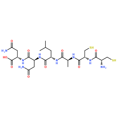
![5-[(3aR,4R,6aS)-2-oxohexahydro-1H-thieno[3,4-d]imidazol-4-yl]pentanamide](http://img.cochemist.com/ccimg/7000/6929-42-6.png)
![5-[(3aR,4R,6aS)-2-oxohexahydro-1H-thieno[3,4-d]imidazol-4-yl]pentanamide](http://img.cochemist.com/ccimg/7000/6929-42-6_b.png)


![Ethanol,2,2'-[[4-[[2-methoxy-5-methyl-4-[(4-methyl-2-nitrophenyl)azo]phenyl]azo]phenyl]imino]bis-](http://img.cochemist.com/ccimg/374600/374591-92-1.png)
![Ethanol,2,2'-[[4-[[2-methoxy-5-methyl-4-[(4-methyl-2-nitrophenyl)azo]phenyl]azo]phenyl]imino]bis-](http://img.cochemist.com/ccimg/374600/374591-92-1_b.png)


![1H,5H,11H,15H-Xantheno[2,3,4-ij:5,6,7-i'j']diquinolizin-18-ium,9-(2,5-dicarboxyphenyl)-2,3,6,7,12,13,16,17-octahydro-, inner salt](/data/chemimg/104900/194785-18-7.png)
![1H,5H,11H,15H-Xantheno[2,3,4-ij:5,6,7-i'j']diquinolizin-18-ium,9-(2,5-dicarboxyphenyl)-2,3,6,7,12,13,16,17-octahydro-, inner salt](/data/chemimg/104900/194785-18-7_b.png)
![Quinolinium,1,1'-[1,3-propanediylbis[(dimethyliminio)-3,1-propanediyl]]bis[4-[3-(3-methyl-2(3H)-benzothiazolylidene)-1-propen-1-yl]-,iodide (1:4)](/data/chemimg/104900/166196-17-4.png)
![Quinolinium,1,1'-[1,3-propanediylbis[(dimethyliminio)-3,1-propanediyl]]bis[4-[3-(3-methyl-2(3H)-benzothiazolylidene)-1-propen-1-yl]-,iodide (1:4)](/data/chemimg/104900/166196-17-4_b.png)


![Ruthenium(2+),bis(2,2'-bipyridine-kN1,kN1')(dipyrido[3,2-a:2',3'-c]phenazine-kN4,kN5)-, (OC-6-21)-](http://img.cochemist.com/ccimg/87600/87564-74-7.png)
![Ruthenium(2+),bis(2,2'-bipyridine-kN1,kN1')(dipyrido[3,2-a:2',3'-c]phenazine-kN4,kN5)-, (OC-6-21)-](http://img.cochemist.com/ccimg/87600/87564-74-7_b.png)