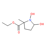
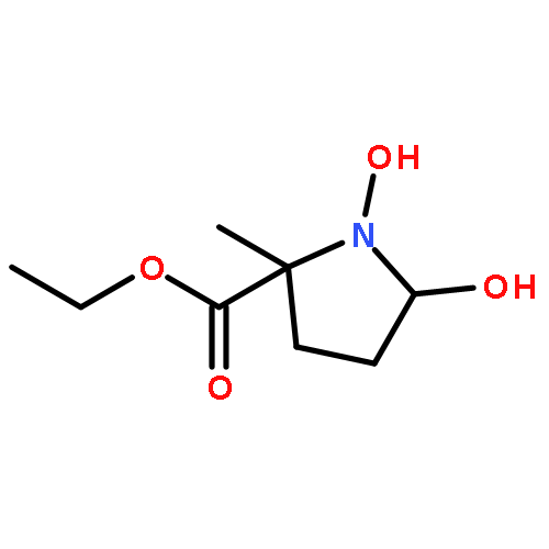
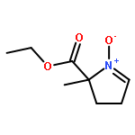
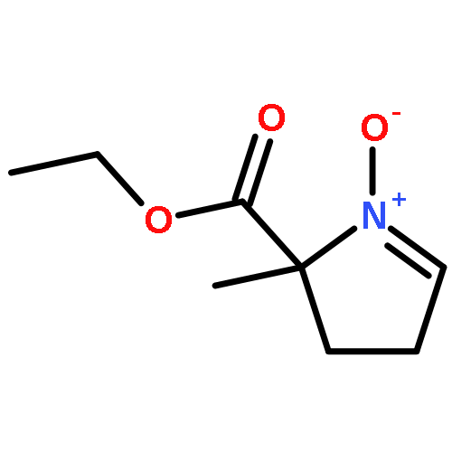
Co-reporter:James G. Roberts; Maxim A. Voinov; Andreas C. Schmidt; Tatyana I. Smirnova
Journal of the American Chemical Society 2016 Volume 138(Issue 8) pp:2516-2519
Publication Date(Web):February 3, 2016
DOI:10.1021/jacs.5b13376
Cyclic voltammetry is a widely used and powerful tool for sensitively and selectively measuring hydrogen peroxide (H2O2). Herein, voltammetry was combined with electron paramagnetic resonance spectroscopy to identify and define the role of an oxygen-centered radical liberated during the oxidation of H2O2. The spin-trap reagents, 5,5-dimethyl-1-pyrroline N-oxide (DMPO) and 2-ethoxycarbonyl-2-methyl-3,4-dihydro-2H-pyrrole-1-oxide (EMPO), were employed. Spectra exhibit distinct hyperfine patterns that clearly identify the DMPO•–OH and EMPO•–OH adducts. Multiple linear regression analysis of voltammograms demonstrated that the hydroxyl radical is a principal contributor to the voltammetry of H2O2, as signal is attenuated when this species is trapped. These data incorporate a missing, fundamental element to our knowledge of the mechanisms that underlie H2O2 electrochemistry.
Co-reporter:Lingjiao Qi, Elina Thomas, Stephanie H. White, Samantha K. Smith, Christie A. Lee, Leslie R. Wilson, and Leslie A. Sombers
Analytical Chemistry 2016 Volume 88(Issue 16) pp:8129
Publication Date(Web):July 21, 2016
DOI:10.1021/acs.analchem.6b01871
L-DOPA has been the gold standard for symptomatic treatment of Parkinson’s disease. However, its efficacy wanes over time as motor complications develop. Very little is known about how L-DOPA therapy affects the dynamics of fluctuating dopamine concentrations in the striatum on a rapid time scale (seconds). Electrochemical studies investigating the effects of L-DOPA treatment on electrically evoked dopamine release have reported conflicting results with significant variability. We hypothesize that the uncertainty in the electrochemical data is largely due to electrode fouling caused by polymerization of L-DOPA and endogenous catecholamines on the electrode surface. Thus, we have systematically optimized the procedure for fabricating cylindrical, Nafion-coated, carbon-fiber microelectrodes. This has enabled rapid and reliable detection of L-DOPA’s effects on striatal dopamine signaling in intact rat brain using fast-scan cyclic voltammetry. An acute dose of 5 mg/kg L-DOPA had no significant effect on dopamine dynamics, demonstrating the highly efficient regulatory mechanisms at work in the intact brain. In contrast, administration of 200 mg/kg L-DOPA significantly increased the amplitude of evoked dopamine release by ∼200%. Overall, this work describes a reliable tool that allows a better measure of L-DOPA augmented dopamine release in vivo, measured using fast-scan cyclic voltammetry. It provides a methodology that improves the stability and performance of the carbon-fiber microelectrode when studying the molecular mechanisms underlying L-DOPA therapy and also promises to benefit a wide variety of studies because Nafion is so commonly used in electroanalytical chemistry.
Co-reporter:Andreas C. Schmidt, Lars E. Dunaway, James G. Roberts, Gregory S. McCarty, and Leslie A. Sombers
Analytical Chemistry 2014 Volume 86(Issue 15) pp:7806
Publication Date(Web):June 26, 2014
DOI:10.1021/ac501725u
Methionine-enkephalin (M-ENK) and leucine-enkephalin (L-ENK) are small endogenous opioid peptides that have been implicated in a wide variety of complex physiological functions, including nociception, reward processing, and motivation. However, our understanding of the role that these molecules play in modulating specific brain circuits remains limited, largely due to challenges in determining where, when, and how specific neuropeptides are released in tissue. Background-subtracted fast-scan cyclic voltammetry coupled with carbon-fiber microelectrodes has proven to be sensitive and selective for detecting rapidly fluctuating neurochemicals in vivo; however, many challenges exist for applying this approach to the detection of neuropeptides. We have developed and characterized a novel voltammetric waveform for the selective quantification of small tyrosine-containing peptides, such as the ENKs, with rapid temporal (subsecond) and precise spatial (10s of micrometers) resolution. We have established that the main contributor to the electrochemical signal inherent to M-ENK is tyrosine and that conventional waveforms provide poor peak resolution and lead to fouling of the electrode surface. By employing two distinct scan rates in each anodic sweep of this analyte-specific waveform, we have selectively distinguished M-ENK from common endogenous interfering agents, such as ascorbic acid, pH shifts, and even L-ENK. Finally, we have used this approach to simultaneously quantify catecholamine and M-ENK fluctuations in live tissue. This work provides a foundation for real-time measurements of endogenous ENK fluctuations in biological locations, and the underlying concept of using multiple scan rates is adaptable to the voltammetric detection of other tyrosine-containing neuropeptides.
Co-reporter:Leyda Z. Lugo-Morales, Philip L. Loziuk, Amanda K. Corder, J. Vincent Toups, James G. Roberts, Katherine A. McCaffrey, and Leslie A. Sombers
Analytical Chemistry 2013 Volume 85(Issue 18) pp:8780
Publication Date(Web):August 6, 2013
DOI:10.1021/ac4017852
Neurotransmission occurs on a millisecond time scale, but conventional methods for monitoring nonelectroactive neurochemicals are limited by slow sampling rates. Despite a significant global market, a sensor capable of measuring the dynamics of rapidly fluctuating, nonelectroactive molecules at a single recording site with high sensitivity, electrochemical selectivity, and a subsecond response time is still lacking. To address this need, we have enabled the real-time detection of dynamic glucose fluctuations in live brain tissue using background-subtracted, fast-scan cyclic voltammetry. The novel microbiosensor consists of a simple carbon fiber surface modified with an electrodeposited chitosan hydrogel encapsulating glucose oxidase. The selectivity afforded by voltammetry enables quantitative and qualitative measurements of enzymatically generated H2O2 without the need for additional strategies to eliminate interfering agents. The microbiosensors possess a sensitivity and limit of detection for glucose of 19.4 ± 0.2 nA mM–1 and 13.1 ± 0.7 μM, respectively. They are stable, even under deviations from physiological normoxic conditions, and show minimal interference from endogenous electroactive substances. Using this approach, we have quantitatively and selectively monitored pharmacologically evoked glucose fluctuations with unprecedented chemical and spatial resolution. Furthermore, this novel biosensing strategy is widely applicable to the immobilization of any H2O2 producing enzyme, enabling rapid monitoring of many nonelectroactive enzyme substrates.
Co-reporter:Marina Spanos, Julie Gras-Najjar, Jeremy M. Letchworth, Audrey L. Sanford, J. Vincent Toups, and Leslie A. Sombers
ACS Chemical Neuroscience 2013 Volume 4(Issue 5) pp:782
Publication Date(Web):April 4, 2013
DOI:10.1021/cn4000499
The dopaminergic neurons of the nigrostriatal dopamine (DA) projection from the substantia nigra to the dorsal striatum become dysfunctional and slowly degenerate in Parkinson’s disease, a neurodegenerative disorder that afflicts more than one million Americans. There is no specific known cause for idiopathic Parkinson’s disease; however, multiple lines of evidence implicate oxidative stress as an underlying factor in both the initiation and progression of the disease. This involves the enhanced generation of reactive oxygen species, including hydrogen peroxide (H2O2), whose role in complex biological processes is not well understood. Using fast-scan cyclic voltammetry at bare carbon-fiber microelectrodes, we have simultaneously monitored and quantified H2O2 and DA fluctuations in intact striatal tissue under basal conditions and in response to the initiation of oxidative stress. Furthermore, we have assessed the effect of acute increases in local H2O2 concentration on both electrically evoked DA release and basal DA levels. Increases in endogenous H2O2 in the dorsal striatum attenuated electrically evoked DA release, and also decreased basal DA levels in this brain region. These novel results will help to disambiguate the chemical mechanisms underlying the progression of neurodegenerative disease states, such as Parkinson’s disease, that involve oxidative stress.Keywords: basal dopamine concentrations; FSCV; H2O2; oxidative stress; Parkinson’s disease; reactive oxygen species
Co-reporter:Audrey L. Sanford, Stephen W. Morton, Kelsey L. Whitehouse, Hannah M. Oara, Leyda Z. Lugo-Morales, James G. Roberts and Leslie A. Sombers
Analytical Chemistry 2010 Volume 82(Issue 12) pp:5205
Publication Date(Web):May 26, 2010
DOI:10.1021/ac100536s
Hydrogen peroxide is a reactive oxygen species that is implicated in a number of neurological disease states and that serves a critical role in normal cell function. It is commonly exploited as a reporter molecule enabling the electrochemical detection of nonelectroactive molecules at electrodes modified with substrate-specific oxidative enzymes. We present the first voltammetric characterization of rapid hydrogen peroxide fluctuations at an uncoated carbon fiber microelectrode, demonstrating unprecedented chemical and spatial resolution. The carbon surface was electrochemically conditioned on the anodic scan and the irreversible oxidation of peroxide was detected on the cathodic scan. The oxidation potential was dependent on scan rate, occurring at +1.2 V versus Ag/AgCl at a scan rate of 400 V·s−1. The relationship between peak oxidation current and concentration was linear across the physiological range tested, with deviation from linearity above 2 mM and a detection limit of 2 μM. Peroxide was distinguished from multiple interferents, both in vitro and in brain slices. The enzymatic degradation of peroxide was monitored, as was peroxide evolution in response to glucose at a glucose oxidase modified carbon fiber electrode. This novel approach provides the requisite sensitivity, selectivity, spatial and temporal resolution to study dynamic peroxide fluctuations in discrete biological locations.
Co-reporter:James G. Roberts, Benjamin P. Moody, Gregory S. McCarty and Leslie A. Sombers
Langmuir 2010 Volume 26(Issue 11) pp:9116-9122
Publication Date(Web):February 18, 2010
DOI:10.1021/la9048924
The in vivo use of carbon-fiber microelectrodes for neurochemical investigation has proven to be selective and sensitive when coupled with background-subtracted fast-scan cyclic voltammetry (FSCV). Various electrochemical pretreatments have been established to enhance the sensitivity of these sensors; however, the fundamental chemical mechanisms underlying these enhancement strategies remain poorly understood. We have investigated an electrochemical pretreatment in which an extended triangular waveform from −0.5 to 1.8 V is applied to the electrode prior to the voltammetric detection of dopamine using a more standard waveform ranging from −0.4 to 1.3 V. This pretreatment enhances the electron-transfer kinetics and significantly improves sensitivity. To gain insight into the chemical mechanism, the electrodes were studied using common analytical techniques. Contact atomic force microscopy (AFM) was used to demonstrate that the surface roughness was not altered on the nanoscale by electrochemical pretreatment. Raman spectroscopy was utilized to investigate oxide functionalities on the carbon surface and confirmed that carbonyl and hydroxyl functional groups were increased by electrochemical conditioning. Spectra collected after the selective chemical modification of these groups implicate the hydroxyl functionality, rather than the carbonyl, as the major contributor to the enhanced electrochemical signal. Finally, we have demonstrated that this electrochemical pretreatment can be used to create carbon microdisc electrodes with sensitivities comparable to those associated with larger, conventionally treated cylindrical carbon fiber microelectrodes.
.jpg)


![Methyl (3s,4r)-3-benzoyloxy-8-methyl-8-azabicyclo[3.2.1]octane-4-carboxylate](http://img.cochemist.com/ccimg/100/50-36-2.png)
![Methyl (3s,4r)-3-benzoyloxy-8-methyl-8-azabicyclo[3.2.1]octane-4-carboxylate](http://img.cochemist.com/ccimg/100/50-36-2_b.png)

