Co-reporter:Joris W. De Schutter;James P. Morrison;Michael J. Morrison;Alessio Ciulli
Journal of Medicinal Chemistry March 9, 2017 Volume 60(Issue 5) pp:2099-2118
Publication Date(Web):February 9, 2017
DOI:10.1021/acs.jmedchem.6b01869
The glycoproteins of selected microbial pathogens often include highly modified carbohydrates such as 2,4-diacetamidobacillosamine (diNAcBac). These glycoconjugates are involved in host–cell interactions and may be associated with the virulence of medically significant Gram-negative bacteria. In light of genetic studies demonstrating the attenuated virulence of bacterial strains in which modified carbohydrate biosynthesis enzymes have been knocked out, we are developing small molecule inhibitors of selected enzymes as tools to evaluate whether such compounds modulate virulence. We performed fragment-based and high-throughput screens against an amino-sugar acetyltransferase enzyme, PglD, involved in biosynthesis of UDP-diNAcBac in Campylobacter jejuni. Herein we report optimization of the hits into potent small molecule inhibitors (IC50 < 300 nM). Biophysical characterization shows that the best inhibitors are competitive with acetyl coenzyme A and an X-ray cocrystal structure reveals that binding is biased toward occupation of the adenine subpocket of the AcCoA binding site by an aromatic ring.
Co-reporter:Debasis Das;Petr Kuzmic
PNAS 2017 Volume 114 (Issue 27 ) pp:7019-7024
Publication Date(Web):2017-07-03
DOI:10.1073/pnas.1703397114
Phosphoglycosyl transferases (PGTs) are integral membrane proteins with diverse architectures that catalyze the formation
of polyprenol diphosphate-linked glycans via phosphosugar transfer from a nucleotide diphosphate-sugar to a polyprenol phosphate.
There are two PGT superfamilies that differ significantly in overall structure and topology. The polytopic PGT superfamily,
represented by MraY and WecA, has been the subject of many studies because of its roles in peptidoglycan and O-antigen biosynthesis.
In contrast, less is known about a second, extensive superfamily of PGTs that reveals a core structure with dual domain architecture
featuring a C-terminal soluble globular domain and a predicted N-terminal membrane-associated domain. Representative members
of this superfamily are the Campylobacter PglCs, which initiate N-linked glycoprotein biosynthesis and are implicated in virulence and pathogenicity. Despite the prevalence
of dual domain PGTs, their mechanism of action is unknown. Here, we present the mechanistic analysis of PglC, a prototypic
dual domain PGT from Campylobacter concisus. Using a luminescence-based assay, together with substrate labeling and kinetics-based approaches, complementary experiments
were carried out that support a ping-pong mechanism involving a covalent phosphosugar intermediate for PglC. Significantly,
mass spectrometry-based approaches identified Asp93, which is part of a highly conserved AspGlu dyad found in all dual domain
PGTs, as the active-site nucleophile of the enzyme involved in the formation of the covalent adduct. The existence of a covalent
phosphosugar intermediate provides strong support for a ping-pong mechanism of PglC, differing fundamentally from the ternary
complex mechanisms of representative polytopic PGTs.
Co-reporter:Monika Musial-Siwek; Marcie B. Jaffee
Journal of the American Chemical Society 2016 Volume 138(Issue 11) pp:3806-3812
Publication Date(Web):February 26, 2016
DOI:10.1021/jacs.5b13426
Integral membrane proteins play essential roles in all living systems; however, major technical hurdles challenge analyses of this class of proteins. Biophysical approaches that provide structural information to complement and leverage experimentally determined and computationally predicted structures are urgently needed. Herein we present the application of luminescence resonance energy transfer (LRET) for investigating the interactions of the polytopic membrane-bound oligosaccharyl transferases (OTases) with partner substrates. Monomeric OTases, such as the PglBs from Campylobacter jejuni and Campylobacter lari, catalyze transfer of glycans from membrane-associated undecaprenol diphosphate-linked substrates to proteins in the bacterial periplasm. LRET-based distance measurements are enabled by the inclusion of an encoded N-terminal lanthanide-binding tag (LBT), and LRET between the luminescent (LBT)-Tb3+ donor complex and fluorescently labeled peptide and glycan substrates provides discrete distance measurements across the span of the membrane. LRET-based measurements of detergent-solubilized PglB from C. lari allowed direct comparison with the distances based on the previously reported the C. lari PglB crystal structure, thereby validating the approach in a defined system. Distance measurements between peptide and glycan substrates and the C. jejuni PglB offer new experimental information on substrate binding to the related, but structurally uncharacterized, eukaryotic OTase.
Co-reporter:Dr. Marthe T. C. Walvoort;Dr. Vinita Lukose ; Barbara Imperiali
Chemistry - A European Journal 2016 Volume 22( Issue 11) pp:3856-3864
Publication Date(Web):
DOI:10.1002/chem.201503986
Abstract
Phosphoglycosyltransferases (PGTs) represent “gatekeeper” enzymes in complex glycan assembly pathways by catalyzing transfer of a phosphosugar from an activated nucleotide diphosphosugar to a membrane-resident polyprenol phosphate. The unique structures of selected nucleoside antibiotics, such as tunicamycin and mureidomycin A, which are known to inhibit comparable biochemical transformations, are exploited as the foundation for the development of modular synthetic inhibitors of PGTs. Herein we present the design, synthesis, and biochemical evaluation of two readily manipulatable modular scaffolds as inhibitors of monotopic bacterial PGTs. Selected compounds show IC50 values down to the 40 μm range, thereby serving as lead compounds for future development of selective and effective inhibitors of diverse PGTs of biological and medicinal interest.
Co-reporter:Vinita Lukose; Garrett Whitworth; Ziqiang Guan
Journal of the American Chemical Society 2015 Volume 137(Issue 39) pp:12446-12449
Publication Date(Web):September 9, 2015
DOI:10.1021/jacs.5b07146
The cell surfaces of bacteria are replete with diverse glycoconjugates that play pivotal roles in determining how bacteria interact with the environment and the hosts that they colonize. Studies to advance our understanding of these interactions rely on the availability of chemically defined glycoconjugates that can be selectively modified under orthogonal reaction conditions to serve as discrete ligands to probe biological interactions, in displayed arrays and as imaging agents. Herein, enzymes in the N-linked protein glycosylation (Pgl) pathway of Campylobacter jejuni are evaluated for their tolerance for azide-modified UDP-sugar substrates, including derivatives of 2,4-diacetamidobacillosamine and N-acetylgalactosamine. In vitro analyses reveal that chemoenzymatic approaches are useful for the preparation of undecaprenol diphosphate-linked glycans and glycopeptides with site-specific introduction of azide functionality for orthogonal labeling at three specific sites in the heptasaccharide glycan. The uniquely modified glycoconjugates represent valuable tools for investigating the roles of C. jejuni cell surface glycoconjugates in host pathogen interactions.
Co-reporter:Vinita Lukose, Lingqi Luo, Dima Kozakov, Sandor Vajda, Karen N. Allen, and Barbara Imperiali
Biochemistry 2015 Volume 54(Issue 50) pp:7326-7334
Publication Date(Web):November 24, 2015
DOI:10.1021/acs.biochem.5b01086
Phosphoglycosyltransferases (PGTs) catalyze the transfer of a C1′-phosphosugar from a soluble sugar nucleotide diphosphate to a polyprenol phosphate. These enzymes act at the membrane interface, forming the first membrane-associated intermediates in the biosynthesis of cell-surface glycans and glycoconjugates, including glycoproteins, glycolipids, and the peptidoglycan in bacteria. PGTs vary greatly in both their membrane topologies and their substrate preferences. PGTs, such as MraY and WecA, are polytopic, while other families of uniquely prokaryotic enzymes have only a single predicted transmembrane helix. PglC, a PGT involved in the biosynthesis of N-linked glycoproteins in the enteropathogen Campylobacter jejuni, is representative of one of the structurally most simple members of the diverse family of small bacterial PGT enzymes. Herein, we apply bioinformatics and covariance-weighted distance constraints in geometry- and homology-based model building, together with mutational analysis, to investigate monotopic PGTs. The pool of 15000 sequences that are analyzed include the PglC-like enzymes, as well as sequences from two other related PGTs that contain a “PglC-like” domain embedded in their larger structures (namely, the bifunctional PglB family, typified by PglB from Neisseria gonorrheae, and WbaP-like enzymes, typified by WbaP from Salmonella enterica). Including these two subfamilies of PGTs in the analysis highlights key residues conserved across all three families of small bacterial PGTs. Mutagenesis analysis of these conserved residues provides further information about the essentiality of many of these residues in catalysis. Construction of a structural model of the cytosolic globular domain utilizing three-dimensional distance constraints, provided by conservation covariance analysis, provides additional insight into the catalytic core of these families of small bacterial PGT enzymes.
Co-reporter:Michael J. Morrison and Barbara Imperiali
Biochemistry 2014 Volume 53(Issue 4) pp:
Publication Date(Web):January 2, 2014
DOI:10.1021/bi401546r
Prokaryote-specific sugars, including N,N′-diacetylbacillosamine (diNAcBac) and pseudaminic acid, have experienced a renaissance in the past decade because of their discovery in glycans related to microbial pathogenicity. DiNAcBac is found at the reducing end of oligosaccharides of N- and O-linked bacterial protein glycosylation pathways of Gram-negative pathogens, including Campylobacter jejuni and Neisseria gonorrhoeae. Further derivatization of diNAcBac results in the nonulosonic acid known as legionaminic acid, which was first characterized in the O-antigen of the lipopolysaccharide (LPS) in Legionella pneumophila. Pseudaminic acid, an isomer of legionaminic acid, is also important in pathogenic bacteria such as Helicobacter pylori because of its occurrence in O-linked glycosylation of flagellin proteins, which plays an important role in flagellar assembly and motility. Here, we present recent advances in the characterization of the biosynthetic pathways leading to these highly modified sugars and investigation of the roles that each plays in bacterial fitness and pathogenicity.
Co-reporter:Laura B. Peterson, Michael B. Yaffe, and Barbara Imperiali
Biochemistry 2014 Volume 53(Issue 36) pp:
Publication Date(Web):August 25, 2014
DOI:10.1021/bi500862c
Accurate and quantitative methods for measuring the dynamic fluctuations of protein kinase activities are critically needed as diagnostic tools and for the evaluation of kinase-targeted inhibitors, which represent a major therapeutic development area in the treatment of cancer and other diseases. In particular, rapid and economical methods that utilize simple instrumentation and provide quantitative data in a high throughput format will have the most impact on basic research in systems biology and medicine. There are over 500 protein kinases in the human kinome. Among these, the mitogen activated protein (MAP) kinases are recognized to be central players in key cellular signaling events and are associated with essential processes including growth, proliferation, differentiation, migration, and apoptosis. The major challenge with MAP kinase sensor development is achieving high selectivity since these kinases rely acutely on secondary interactions distal to the phosphorylation site to impart substrate specificity. Herein we describe the development and application of selective sensors for three MAP kinase subfamilies, ERK1/2, p38α/β, and JNK1/2/3. The new sensors are based on a modular design, which includes a sensing element that exploits a sulfonamido-oxine (Sox) fluorophore for reporting phosphorylation, a recognition and specificity element based on reported docking domain motifs and a variable linker, which can be engineered to optimize the intermodule distance and relative orientation. Following rigorous validation, the capabilities of the new sensors are exemplified through the quantitative analysis of the target MAP kinases in breast cancer progression in a cell culture model, which reveals a strong correlation between p38α/β activity and increased tumorgenicity.
Co-reporter:Dr. Andrew T. Krueger;Dr. Carsten Kroll;Edgar Sanchez; Linda G. Griffith; Barbara Imperiali
Angewandte Chemie International Edition 2014 Volume 53( Issue 10) pp:2662-2666
Publication Date(Web):
DOI:10.1002/anie.201307869
Abstract
Described is the development and application of a versatile semisynthetic strategy, based on a combination of sortase-mediated coupling and tetrazine ligation chemistry, which can be exploited for the efficient incorporation of tunable functionality into chimeric recombinant proteins. To demonstrate the scope of the method, the assembly of a set of bivalent ligands, which integrate members of the epidermal growth factor (EGF) ligand family, is described. By using a series of bivalent EGFs with variable intraligand spacing, the differences in structure were correlated with the ability to bias signaling in the ErbB receptor family in a cell motility assay. Biasing away from EGFR-HER2 dimerization with a bivalent EGF was observed to reduce cell motility in an intraligand distance-dependent fashion, thus demonstrating the utility of the approach for acutely perturbing receptor-mediated cell signaling pathways.
Co-reporter:Dr. Andrew T. Krueger;Dr. Carsten Kroll;Edgar Sanchez; Linda G. Griffith; Barbara Imperiali
Angewandte Chemie 2014 Volume 126( Issue 10) pp:2700-2704
Publication Date(Web):
DOI:10.1002/ange.201307869
Abstract
Described is the development and application of a versatile semisynthetic strategy, based on a combination of sortase-mediated coupling and tetrazine ligation chemistry, which can be exploited for the efficient incorporation of tunable functionality into chimeric recombinant proteins. To demonstrate the scope of the method, the assembly of a set of bivalent ligands, which integrate members of the epidermal growth factor (EGF) ligand family, is described. By using a series of bivalent EGFs with variable intraligand spacing, the differences in structure were correlated with the ability to bias signaling in the ErbB receptor family in a cell motility assay. Biasing away from EGFR-HER2 dimerization with a bivalent EGF was observed to reduce cell motility in an intraligand distance-dependent fashion, thus demonstrating the utility of the approach for acutely perturbing receptor-mediated cell signaling pathways.
Co-reporter:Matthieu Sainlos ; Wendy S. Iskenderian-Epps ; Nelson B. Olivier ; Daniel Choquet
Journal of the American Chemical Society 2013 Volume 135(Issue 12) pp:4580-4583
Publication Date(Web):March 8, 2013
DOI:10.1021/ja309870q
We report a general method for light-assisted control of interactions of PDZ domain binding motifs with their cognate domains by the incorporation of a photolabile caging group onto the essential C-terminal carboxylate binding determinant of the motif. The strategy was implemented and validated for both simple monovalent and biomimetic divalent ligands, which have recently been established as powerful tools for acute perturbation of native PDZ domain-dependent interactions in live cells.
Co-reporter:Alan M. McLean, Elke Socher, Oleg Varnavski, Travis B. Clark, Barbara Imperiali, and Theodore Goodson III
The Journal of Physical Chemistry B 2013 Volume 117(Issue 50) pp:15935-15942
Publication Date(Web):November 18, 2013
DOI:10.1021/jp407321g
We report detailed photophysical studies on the two-photon fluorescence processes of the solvatochromic fluorophore 4-DMN as a conjugate of the calmodulin (CaM) and the associated CaM-binding peptide M13. Strong two-photon fluorescence enhancement has been observed which is associated with calcium binding. It is found that the two-photon absorption cross-section is strongly dependent on the local environment surrounding the 4-DMN fluorophore in the CaM conjugates, providing sensitivity between sites of fluorophore attachment. Utilizing time-resolved measurements, the emission dynamics of 4-DMN under various environmental (solvent) conditions are analyzed. In addition, anisotropy measurements reveal that the 4-DMN–S38C–CaM system has restricted rotation in the calcium-bound calmodulin. To establish the utility for cellular imaging, two-photon fluorescence microscopy studies were also carried out with the 4-DMN-modified M13 peptide in cells. Together, these studies provide strong evidence that 4-DMN is a useful probe in two-photon imaging, with advantageous properties for cellular experiments.
Co-reporter:Dr. Elke Socher ; Barbara Imperiali
ChemBioChem 2013 Volume 14( Issue 1) pp:53-57
Publication Date(Web):
DOI:10.1002/cbic.201200700
Co-reporter:Dr. Andrew T. Krueger; Dr. Barbara Imperiali
ChemBioChem 2013 Volume 14( Issue 7) pp:788-799
Publication Date(Web):
DOI:10.1002/cbic.201300079
Abstract
Fluorescence spectroscopy is a powerful tool for probing complex biological processes. The ubiquity of peptide–protein and protein–protein interactions in these processes has made them important targets for fluorescence labeling, and to allow sensitive readout of information concerning location, interactions with other biomolecules, and macromolecular dynamics. This review describes recent advances in design, properties and applications in the area of fluorescent amino acids (FlAAs). The ability to site-selectively incorporate fluorescent amino acid building blocks into a protein or peptide of interest provides the advantage of closely retaining native function and appearance. The development of an array of fluorescent amino acids with a variety of properties, such as environment sensitivity, chelation-enhanced fluorescence, and profluorescence, has allowed researchers to gain insights into biological processes, including protein conformational changes, binding events, enzyme activities, and protein trafficking and localization.
Co-reporter:Meredith D. Hartley;Philipp E. Schneggenburger
PNAS 2013 Volume 110 (Issue 52 ) pp:20850-20851
Publication Date(Web):2013-12-24
DOI:10.1073/pnas.1320852110
Membrane-bound polyprenol-dependent pathways are important for the assembly of essential glycoconjugates in all domains of
life. However, despite their prevalence, the functional significance of the extended linear polyprenyl groups in the interactions
of the glycan substrates, the biosynthetic enzymes that act upon them, and the membrane bilayer in which they are embedded
remains a mystery. These interactions are investigated simultaneously and uniquely through application of the nanodisc membrane
technology. The Campylobacter jejuni N-linked glycosylation pathway has been chosen as a model pathway in which all of the enzymes and substrates are biochemically
accessible. We present the functional reconstitution of two enzymes responsible for the early membrane-committed steps in
glycan assembly. Protein stoichiometry analysis, fluorescence-based approaches, and biochemical activity assays are used to
demonstrate the colocalization of the two enzymes in nanodiscs. Isotopic labeling of the substrates reveals that undecaprenyl-phosphate
is coincorporated into discs with the two enzymes, and furthermore, that both enzymes are functionally reconstituted and can
sequentially convert the coembedded undecaprenyl-phosphate into undecaprenyl-diphosphate-linked disaccharide. These studies
provide a proof-of-concept demonstrating that the nanodisc model membrane system represents a promising experimental platform
for analyzing the multifaceted interactions among the enzymes involved in polyprenol-dependent glycan assembly pathways, the
membrane-associated substrates, and the lipid bilayer. The stage is now set for exploration of the roles of the conserved
polyprenols in promoting protein–protein interactions among pathway enzymes and processing of substrates through sequential
steps in membrane-associated glycan assembly.
Co-reporter:Cliff I. Stains, Nathan C. Tedford, Traci C. Walkup, Elvedin Luković, Brenda N. Goguen, Linda G. Griffith, Douglas A. Lauffenburger, Barbara Imperiali
Chemistry & Biology 2012 Volume 19(Issue 2) pp:210-217
Publication Date(Web):24 February 2012
DOI:10.1016/j.chembiol.2011.11.012
Protein kinases catalyze protein phosphorylation and thereby control the flow of information through signaling cascades. Currently available methods for concomitant assessment of the enzymatic activities of multiple kinases in complex biological samples rely on indirect proxies for enzymatic activity, such as posttranslational modifications to protein kinases. Our laboratories have recently described a method for directly quantifying the enzymatic activity of kinases in unfractionated cell lysates using substrates containing a phosphorylation-sensitive unnatural amino acid termed CSox, which can be monitored using fluorescence. Here, we demonstrate the utility of this method using a probe set encompassing p38α, MK2, ERK1/2, Akt, and PKA. This panel of chemosensors provides activity measurements of individual kinases in a model of skeletal muscle differentiation and can be readily used to generate individualized kinase activity profiles for tissue samples from clinical cancer patients.Graphical AbstractFigure optionsDownload full-size imageDownload high-quality image (195 K)Download as PowerPoint slideHighlights► A method for profiling kinase activities in biological samples is presented ► Kinase activity profiles identify positive regulators of myogenesis ► Reproducible activity profiles for tissues samples from individual cancer patients
Co-reporter:Dr. Kelly D. Daughtry;Dr. Langdon J. Martin;Ashish Sarraju; Barbara Imperiali; Karen N. Allen
ChemBioChem 2012 Volume 13( Issue 17) pp:2567-2574
Publication Date(Web):
DOI:10.1002/cbic.201200448
Abstract
Lanthanide-binding tags (LBTs), peptide-based coexpression tags with high affinity for lanthanide ions, have previously been applied as luminescent probes to provide phasing for structure determination in X-ray crystallography and to provide restraints for structural refinement and distance information in NMR. The native affinity of LBTs for Gd3+ indicates their potential as the basis for engineering of peptide-based MRI agents. However, the lanthanide coordination state that enhances luminescence and affords tightest binding would not be ideal for applications of LBTs as contrast agents, due to the exclusion of water from the inner coordination sphere. Herein, we use structurally defined LBTs as the starting point for re-engineering the first coordination shell of the lanthanide ion to provide for high contrast through direct coordination of water to Gd3+ (resulting in the single LBT peptide, m-sLBT). The effectiveness of LBTs as MRI contrast agents was examined in vitro through measurement of binding affinity and proton relaxivity. For imaging applications that require targeted observation, fusion to specific protein partners is desirable. However, a fusion protein comprising a concatenated double LBT (dLBT) as an N-terminal tag for the model protein ubiquitin had reduced relaxivity compared with the free dLBT peptide. This limitation was overcome by the use of a construct based on the m-sLBT sequence (q-dLBT–ubiquitin). The structural basis for the enhanced contrast was examined by comparison of the X-ray crystal structure of xq-dLBT–ubiquitin (wherein two tryptophan residues are replaced with serine), to that of dLBT-ubiquitin. The structure shows that the backbone conformational dynamics of the MRI variant may allow enhanced water exchange. This engineered LBT represents a first step in expanding the current base of specificity-targeted agents available.
Co-reporter:Brenda N. Goguen ; Andreas Aemissegger
Journal of the American Chemical Society 2011 Volume 133(Issue 29) pp:11038-11041
Publication Date(Web):June 21, 2011
DOI:10.1021/ja2028074
Photolabile caging groups, including the 1-(2-nitrophenyl)ethyl (NPE) group, have been applied to probe many biological processes, including protein phosphorylation. Although studies with NPE-caged phosphoamino acids have provided valuable information, these investigations have been limited to the use of only one caged species in a single experiment. To expand the scope of these tools, we have developed an approach for sequentially uncaging two different phosphopeptides in one system, enabling interrogation of multiple phosphorylation events. We present the synthesis of [7-(diethylamino)coumarin-4-yl]methyl (DEACM)-caged phosphorylated serine, threonine, and tyrosine building blocks for Fmoc-based solid-phase peptide synthesis to allow convenient incorporation of these residues into peptides and proteins. Exposure of DEACM- and NPE-caged phosphopeptides to 420 nm light selectively releases the DEACM group without affecting the NPE-caged peptide. This then enables a subsequent irradiation event at 365 nm to remove the NPE group and liberate a second phosphopeptide. We demonstrate the versatility of this general sequential uncaging approach by applying it to control Wip1 phosphatase with two wavelengths of light.
Co-reporter:Cliff I. Stains, Elvedin Luković, and Barbara Imperiali
ACS Chemical Biology 2011 Volume 6(Issue 1) pp:101
Publication Date(Web):September 16, 2010
DOI:10.1021/cb100230y
Recent efforts have identified the p38α Ser/Thr kinase as a potential target for the treatment of inflammatory diseases as well as non-small cell lung carcinoma. Despite the significance of p38α, no direct activity probe compatible with cell lysate analysis exists. Instead, proxies for kinase activation, such as phosphospecific antibodies, which do not distinguish between p38 isoforms, are often used. Our laboratory has recently developed a sulfonamido-oxine (Sox) fluorophore that undergoes a significant increase in fluorescence in response to phosphorylation at a proximal residue, allowing for real-time activity measurements. Herein we report the rational design of a p38α-selective chemosensor using this approach. We have validated the selectivity of this sensor using specific inhibitors and immunodepletions and show that p38α activity can be monitored in crude lysates from a variety of cell lines, allowing for the potential use of this sensor in both clinical and basic science research applications.
Co-reporter:Brenda N. Goguen and Barbara Imperiali
ACS Chemical Biology 2011 Volume 6(Issue 11) pp:1164
Publication Date(Web):October 3, 2011
DOI:10.1021/cb200299k
Cell migration is required for many physiological processes, including wound repair and embryogenesis, and relies on precisely orchestrated events that are regulated in a spatially and temporally controlled manner. Most traditional approaches for studying migration, such as genetic methods or the use of chemical inhibitors, do not offer insight into these important components of protein function. However, chemical tools, which respond on a more rapid time scale and in localized regions of the cell, are capable of providing more detailed, real-time information. This Review describes these recent approaches to investigate cell migration and focuses on proteins that are activated by light or small molecules, as well as fluorescent sensors of protein activity.
Co-reporter:Angelyn Larkin and Barbara Imperiali
Biochemistry 2011 Volume 50(Issue 21) pp:
Publication Date(Web):April 20, 2011
DOI:10.1021/bi200346n
Asparagine-linked glycosylation involves the sequential assembly of an oligosaccharide onto a polyisoprenyl donor, followed by the en bloc transfer of the glycan to particular asparagine residues within acceptor proteins. These N-linked glycans play a critical role in a wide variety of biological processes, such as protein folding, cellular targeting and motility, and the immune response. In the past decade, research in the field of N-linked glycosylation has achieved major advances, including the discovery of new carbohydrate modifications, the biochemical characterization of the enzymes involved in glycan assembly, and the determination of the biological impact of these glycans on target proteins. It is now firmly established that this enzyme-catalyzed modification occurs in all three domains of life. However, despite similarities in the overall logic of N-linked glycoprotein biosynthesis among the three kingdoms, the structures of the appended glycans are markedly different and thus influence the functions of elaborated proteins in various ways. Though nearly all eukaryotes produce the same nascent tetradecasaccharide (Glc3Man9GlcNAc2), heterogeneity is introduced into this glycan structure after it is transferred to the protein through a complex series of glycosyl trimming and addition steps. In contrast, bacteria and archaea display diversity within their N-linked glycan structures through the use of unique monosaccharide building blocks during the assembly process. In this review, recent progress toward gaining a deeper biochemical understanding of this modification across all three kingdoms will be summarized. In addition, a brief overview of the role of N-linked glycosylation in viruses will also be presented.
Co-reporter:Marcie B. Jaffee and Barbara Imperiali
Biochemistry 2011 Volume 50(Issue 35) pp:
Publication Date(Web):August 3, 2011
DOI:10.1021/bi201018d
The central enzyme in N-linked glycosylation is the oligosaccharyl transferase (OTase), which catalyzes glycan transfer from a polyprenyldiphosphate-linked carrier to select asparagines within acceptor proteins. PglB from Campylobacter jejuni is a single-subunit OTase with homology to the Stt3 subunit of the complex multimeric yeast OTase. Sequence identity between PglB and Stt3 is low (17.9%); however, both have a similar predicted architecture and contain the conserved WWDxG motif. To investigate the relationship between PglB and other Stt3 proteins, sequence analysis was performed using 28 homologues from evolutionarily distant organisms. Since detection of small conserved motifs within large membrane-associated proteins is complicated by divergent sequences surrounding the motifs, we developed a program to parse sequences according to predicted topology and then analyze topologically related regions. This approach identified three conserved motifs that served as the basis for subsequent mutagenesis and functional studies. This work reveals that several inter-transmembrane loop regions of PglB/Stt3 contain strictly conserved motifs that are essential for PglB function. The recent publication of a 3.4 Å resolution structure of full-length C. lari OTase provides clear structural evidence that these loops play a fundamental role in catalysis [Lizak, C.; (2011) Nature474, 350−355]. The current study provides biochemical support for the role of the inter-transmembrane domain loops in OTase catalysis and demonstrates the utility of combining topology prediction and sequence analysis for exposing buried pockets of homology in large membrane proteins. The described approach allowed detection of the catalytic motifs prior to availability of structural data and reveals additional catalytically relevant residues that are not predicted by structural data alone.
Co-reporter:Meredith D. Hartley, Michael J. Morrison, Finn Erik Aas, Bente Børud, Michael Koomey, and Barbara Imperiali
Biochemistry 2011 Volume 50(Issue 22) pp:
Publication Date(Web):May 4, 2011
DOI:10.1021/bi2003372
The O-linked protein glycosylation pathway in Neisseria gonorrhoeae is responsible for the synthesis of a complex oligosaccharide on undecaprenyl diphosphate and subsequent en bloc transfer of the glycan to serine residues of select periplasmic proteins. Protein glycosylation (pgl) genes have been annotated on the basis of bioinformatics and top-down mass spectrometry analysis of protein modifications in pgl-null strains [Aas, F. E., et al. (2007) Mol. Microbiol. 65, 607–624; Vik, A., et al. (2009) Proc. Natl. Acad. Sci. U.S.A. 106, 4447–4452], but relatively little biochemical analysis has been performed to date. In this report, we present the expression, purification, and functional characterization of seven Pgl enzymes. Specifically, the enzymes studied are responsible for synthesis of an uncommon uridine diphosphate (UDP)-sugar (PglD, PglC, and PglB-acetyltransferase domain), glycan assembly (PglB-phospho-glycosyltransferase domain, PglA, PglE, and PglH), and final oligosaccharide transfer (PglO). UDP-2,4-diacetamido-2,4,6-trideoxy-α-d-hexose (DATDH), which is the first sugar in glycan biosynthesis, was produced enzymatically, and the stereochemistry was assigned as uridine diphosphate N′-diacetylbacillosamine (UDP-diNAcBac) by nuclear magnetic resonance characterization. In addition, the substrate specificities of the phospho-glycosyltransferase, glycosyltransferases, and oligosaccharyltransferase (OTase) were analyzed in vitro, and in most cases, these enzymes exhibited strong preferences for the native substrates relative to closely related glycans. In particular, PglO, the O-linked OTase, and PglB(Cj), the N-linked OTase from Campylobacter jejuni, preferred the native N. gonorrhoeae and C. jejuni substrates, respectively. This study represents the first comprehensive biochemical characterization of this important O-linked glycosylation pathway and provides the basis for further investigations of these enzymes as antibacterial targets.
Co-reporter:Brenda N. Goguen, Galen S. Loving, Barbara Imperiali
Bioorganic & Medicinal Chemistry Letters 2011 Volume 21(Issue 17) pp:5058-5061
Publication Date(Web):1 September 2011
DOI:10.1016/j.bmcl.2011.04.051
Cdc42, a member of the Rho GTPase family, is a fundamental regulator of the actin cytoskeleton during cell migration. To generate a sensor for Cdc42 activation, we employed a multi-pronged approach, utilizing cysteine labeling and expressed protein ligation, to incorporate the environment sensitive fluorophore 4-N,N-dimethylamino-1,8-naphthalimide (4-DMN) into the GTPase binding domain of the WASP protein. These constructs bind only the active, GTP-bound conformation of Cdc42 to produce a fluorescence signal. Studies with a panel of five sensor analogs revealed a derivative that exhibits a 32-fold increase in fluorescence intensity in the presence of activated Cdc42 compared to incubation with the inactive GDP-bound form of the protein. We demonstrate that this sensor can be exploited to monitor Cdc42 nucleotide exchange and GTPase activity in a continuous, fluorescence assay.
Co-reporter:Brenda N. Goguen;Dr. Brenton D. Hoffman;Dr. James R. Sellers; Martin A. Schwartz; Barbara Imperiali
Angewandte Chemie 2011 Volume 123( Issue 25) pp:5785-5788
Publication Date(Web):
DOI:10.1002/ange.201100674
Co-reporter:Brenda N. Goguen;Dr. Brenton D. Hoffman;Dr. James R. Sellers; Martin A. Schwartz; Barbara Imperiali
Angewandte Chemie International Edition 2011 Volume 50( Issue 25) pp:5667-5670
Publication Date(Web):
DOI:10.1002/anie.201100674
Co-reporter:Katja Barthelmes ; Anne M. Reynolds ; Ezra Peisach ; Hendrik R. A. Jonker ; Nicholas J. DeNunzio ; Karen N. Allen ; Barbara Imperiali ;Harald Schwalbe
Journal of the American Chemical Society 2010 Volume 133(Issue 4) pp:808-819
Publication Date(Web):December 23, 2010
DOI:10.1021/ja104983t
Lanthanide-binding tags (LBTs) are valuable tools for investigation of protein structure, function, and dynamics by NMR spectroscopy, X-ray crystallography, and luminescence studies. We have inserted LBTs into three different loop positions (denoted L, R, and S) of the model protein interleukin-1β (IL1β) and varied the length of the spacer between the LBT and the protein (denoted 1−3). Luminescence studies demonstrate that all nine constructs bind Tb3+ tightly in the low nanomolar range. No significant change in the fusion protein occurs from insertion of the LBT, as shown by two X-ray crystallographic structures of the IL1β-S1 and IL1β-L3 constructs and for the remaining constructs by comparing the 1H−15N heteronuclear single-quantum coherence NMR spectra with that of the wild-type IL1β. Additionally, binding of LBT-loop IL1β proteins to their native binding partner in vitro remains unaltered. X-ray crystallographic phasing was successful using only the signal from the bound lanthanide. Large residual dipolar couplings (RDCs) could be determined by NMR spectroscopy for all LBT-loop constructs and revealed that the LBT-2 series were rigidly incorporated into the interleukin-1β structure. The paramagnetic NMR spectra of loop-LBT mutant IL1β-R2 were assigned and the Δχ tensor components were calculated on the basis of RDCs and pseudocontact shifts. A structural model of the IL1β-R2 construct was calculated using the paramagnetic restraints. The current data provide support that encodable LBTs serve as versatile biophysical tags when inserted into loop regions of proteins of known structure or predicted via homology modeling.
Co-reporter:Karen N Allen, Barbara Imperiali
Current Opinion in Chemical Biology 2010 Volume 14(Issue 2) pp:247-254
Publication Date(Web):April 2010
DOI:10.1016/j.cbpa.2010.01.004
Lanthanide-tagged proteins are valuable for exploiting the unique properties of Ln ions for investigating protein structure, function, and dynamics. Introduction of the Ln into the target is accomplished via chemical modification with synthetic lanthanide-chelating prosthetic groups or by coexpression with peptide-based binding tags. Complexed Ln-tags offer a heavy-atom site for solving the phase problem in X-ray crystallography. In NMR, paramagnetic lanthanide ions induce residual dipolar couplings and pseudo-contact shifts that yield valuable distance constraints for structural analysis. Lanthanide luminescence-based techniques and Ln-tagged proteins are valuable for investigating the functions and dynamics of large proteins and protein complexes and have been applied in vivo. Overall, the reach of Ln-tagged proteins will increase our ability to understand cellular functions on the molecular level.
Co-reporter:Angelyn Larkin, Nelson B. Olivier and Barbara Imperiali
Biochemistry 2010 Volume 49(Issue 33) pp:
Publication Date(Web):July 6, 2010
DOI:10.1021/bi100805b
In recent years, the opportunistic pathogen Pseudomonas aeruginosa has emerged as a major source of hospital-acquired infections. Effective treatment has proven increasingly difficult due to the spread of multidrug resistant strains and thus requires a deeper understanding of the biochemical mechanisms of pathogenicity. The central carbohydrate of the P. aeruginosa PAO1 (O5) B-band O-antigen, ManNAc(3NAc)A, has been shown to be critical for virulence and is produced in a stepwise manner by five enzymes in the Wbp pathway (WbpA, WbpB, WbpE, WbpD, and WbpI). Herein, we present the crystal structure of the aminotransferase WbpE from P. aeruginosa PAO1 in complex with the cofactor pyridoxal 5′-phosphate (PLP) and product UDP-GlcNAc(3NH2)A as the external aldimine at 1.9 Å resolution. We also report the structures of WbpE in complex with PMP alone as well as the PLP internal aldimine and show that the dimeric structure of WbpE observed in the crystal structure is confirmed by analytical ultracentrifugation. Analysis of these structures reveals that the active site of the enzyme is composed of residues from both subunits. In particular, we show that a key residue (Arg229), which has previously been implicated in direct interactions with the α-carboxylate moiety of α-ketoglutarate, is also uniquely positioned to bestow specificity for the 6′′-carboxyl group of GlcNAc(3NH2)A through a salt bridge. This finding is intriguing because while an analogous basic residue is present in WbpE homologues that do not process 6′′-carboxyl-modified saccharides, recent structural studies reveal that this side chain is retracted to accommodate a neutral C6′′ atom. This work represents the first structural analysis of a nucleotide sugar aminotransferase with a bound product modified at the C2′′, C3′′, and C6′′ positions and provides insight into a novel target for treatment of P. aeruginosa infection.
Co-reporter:James P. Morrison, Jerry M. Troutman, Barbara Imperiali
Bioorganic & Medicinal Chemistry 2010 Volume 18(Issue 23) pp:8167-8171
Publication Date(Web):1 December 2010
DOI:10.1016/j.bmc.2010.10.020
The human pathogen Campylobacter jejuni possesses a general N-linked glycosylation system that is known to play a role in pathogenicity; however, a detailed understanding of this role remains elusive. A considerable hindrance to studying bacterial N-glycosylation in vivo is the absence of small molecule inhibitors to reversibly control the process. This report describes a pathway-screening assay that targets the early enzymes of C. jejuni N-glycan biosynthesis that would enable identification of inhibitors to the first four steps in the pathway. The assay includes PglF, PglE, PglD, PglC, and PglA; the enzymes involved in the biosynthesis of an undecaprenyl diphosphate-linked disaccharide and monitors the transfer of [3H]GalNAc from the hydrophilic UDP-linked carrier to the lipophilic UndPP-diNAcBac (2,4-diacetamido-2,4,6-trideoxyglucose). The optimized assay has a Z′-factor calculated to be 0.77, indicating a robust assay suitable for screening. The diacylglycerol kinase from Streptococcus mutans, which provides a convenient method for phosphorylating undecaprenol, has been included in a modified version of the assay thereby allowing the screen to be conducted with entirely commercially available substrates.
Co-reporter:Wendy S. Iskenderian-Epps; Barbara Imperiali
ChemBioChem 2010 Volume 11( Issue 14) pp:1979-1984
Publication Date(Web):
DOI:10.1002/cbic.201000246
Co-reporter:Mark M. Chen;Alice I. Bartlett;Paul S. Nerenberg;Claire T. Friel;Christian P. R. Hackenberger;Collin M. Stultz;Sheena E. Radford
PNAS 2010 107 (52 ) pp:22528-22533
Publication Date(Web):2010-12-28
DOI:10.1073/pnas.1015356107
N-linked glycosylation modulates protein folding and stability through a variety of mechanisms. As such there is considerable
interest in the development of general rules to predict the structural consequences of site-specific glycosylation and to
understand how these effects can be exploited in the design and development of modified proteins with advantageous properties.
In this study, expressed protein ligation is used to create site-specifically glycosylated variants of the bacterial immunity
protein Im7 modified with the chitobiose disaccharide (GlcNAc-GlcNAc). Glycans were introduced at seven solvent exposed sites
within the Im7 sequence and the kinetic and thermodynamic consequences of N-linked glycosylation analyzed. The values for glycan incorporation were found to range from +5.2 to -3.8 kJ·mol-1. In several cases, glycosylation influences folding by modulating the local conformational preferences of the glycosylated
sequence. These locally mediated effects are most prominent in the center of α-helices where glycosylation negatively effects
folding and in compact turn motifs between segments of ordered secondary structure where glycosylation promotes folding and
enhances the overall stability of the native protein. The studies also provide insight into why glycosylation is commonly
identified at the transition between different types of secondary structure and when glycosylation may be used to elaborate
protein structure to protect disordered sequences from proteolysis or immune system recognition.
Co-reporter:Matthieu Sainlos ; Wendy S. Iskenderian
Journal of the American Chemical Society 2009 Volume 131(Issue 19) pp:6680-6682
Publication Date(Web):April 23, 2009
DOI:10.1021/ja900371q
A systematic and general approach for identifying efficient probes for class I PDZ domains based on environment-sensitive chromophores is presented. A series of peptides derived from the C-terminal sequence of Stargazin was first used with PDZ domains of PSD-95 and Shank3 to identify the optimal position and linker length for the 4-DMAP chromophore. The results were applied to well-characterized ligand sequences for each set of domains to generate high affinity probes that retain their native sequence specificity and yield remarkable fluorescence increases upon binding. These probes constitute efficient tools to study the dynamics and regulatory mechanisms of PDZ domain-mediated interactions.
Co-reporter:Galen Loving and Barbara Imperiali
Bioconjugate Chemistry 2009 Volume 20(Issue 11) pp:2133
Publication Date(Web):October 12, 2009
DOI:10.1021/bc900319z
The solvatochromic fluorophore 4-N,N-dimethylamino-1,8-naphthalimide (4-DMN) possesses extremely sensitive emission properties due largely to the low intrinsic fluorescence it exhibits in polar protic solvents such as water. This makes it well suited as a probe for the detection of a wide range of biomolecular interactions. Herein we report the development and evaluation of a new series of thiol-reactive agents derived from this fluorophore. The members of this series vary according to linker type and the electrophilic group required for the labeling of proteins and other biologically relevant molecules. Using the calcium-binding protein calmodulin as a model system, we compare the performance of the 4-DMN derivatives to that of several commercially available solvatochromic fluorophores identifying many key factors important to the successful application of such tools. This study also demonstrates the power of this new series of labeling agents by yielding a fluorescent calmodulin construct capable of producing a greater than 100-fold increase in emission intensity upon binding to calcium.
Co-reporter:Angelyn Larkin and Barbara Imperiali
Biochemistry 2009 Volume 48(Issue 23) pp:
Publication Date(Web):April 6, 2009
DOI:10.1021/bi900186u
The B-band O-antigen of the lipopolysaccharide found in the opportunistic pathogen Pseudomonas aeruginosa PAO1 (serotype O5) comprises a repeating trisaccharide unit that is critical for virulence and protection from host defense systems. One of the carbohydrates in this repeating unit, the rare diacetylated aminuronic acid derivative 2,3-diacetamido-2,3-dideoxy-β-d-mannuronic acid (ManNAc(3NAc)A), is thought to be produced by five enzymes (WbpA, WbpB, WbpE, WbpD, and WbpI) in a stepwise manner starting from UDP-GlcNAc. Although the genes responsible for the biosynthesis of this sugar are known, only two of the five encoded proteins (WbpA and WbpI) have been thoroughly investigated. In this report, we describe the cloning, overexpression, purification, and biochemical characterization of the three central enzymes in this pathway, WbpB, WbpE, and WbpD. Using a combination of capillary electrophoresis, RP-HPLC, and NMR spectroscopy, we show that WbpB and WbpE are a dehydrogenase/aminotransferase pair that converts UDP-GlcNAcA to UDP-GlcNAc(3NH2)A in a coupled reaction via a unique NAD+ recycling pathway. In addition, we confirm that WbpD catalyzes the acetylation of UDP-GlcNAc(3NH2)A to give UDP-GlcNAc(3NAc)A. Notably, WbpA, WbpB, WbpE, WbpD, and WbpI can be combined in vitro to generate UDP-ManNAc(3NAc)A in a single reaction vessel, thereby providing supplies of this complex glycosyl donor for future studies of lipopolysaccharide assembly. This work completes the biochemical characterization of the enzymes in this pathway and provides novel targets for potential therapeutics to combat infections with drug resistant P. aeruginosa strains.
Co-reporter:Jerry M. Troutman and Barbara Imperiali
Biochemistry 2009 Volume 48(Issue 12) pp:
Publication Date(Web):January 21, 2009
DOI:10.1021/bi802284d
Asparagine-linked protein glycosylation is essential for the virulence of the human gut mucosal pathogen Campylobacter jejuni. The heptasaccharide that is transferred to proteins is biosynthesized via the glycosyltransferase-catalyzed addition of sugar units to an undecaprenyl diphosphate-linked carrier. Genetic studies on the heptasaccharide assembly enzymes have shown that PglH, which transfers three terminal N-acetyl-galactosamine (GalNAc) residues to the carrier polyisoprene, is essential for chick colonization by C. jejuni. While it is now clear that PglH catalyzes multiple transfer reactions, the mechanism whereby the reactions cease after the addition of just three GalNAc residues has yet to be understood. To address this issue, a series of mechanistic biochemical studies was conducted with purified native PglH. This enzyme was found to follow a processive mechanism under initial rate conditions; however, product inhibition and product accumulation led to PglH release of intermediate products prior to complete conversion to the native ultimate product. Point mutations of an essential EX7E sequence motif were used to demonstrate that a single active site was responsible for all three transferase reactions, and a homology model with the mannosyltransferase PimA, from Mycobacteria smegmatis, establishes the requirement of the EX7E motif in catalysis. Finally, increased binding affinity with increasing glycan size is proposed to provide PglH with a counting mechanism that does not allow the transfer of more than three GalNAc residues. These results provide important mechanistic insights into the function of the glycosyl transfer polymerase that is related to the virulence of C. jejuni.
Co-reporter:Juan A. González-Vera, Elvedin Luković, Barbara Imperiali
Bioorganic & Medicinal Chemistry Letters 2009 Volume 19(Issue 4) pp:1258-1260
Publication Date(Web):15 February 2009
DOI:10.1016/j.bmcl.2008.12.090
A novel screening method to identify selective Sox-based fluorescent probes for Ser/Thr kinases has been developed. Peptide libraries were exposed to a kinase of interest and the products of the timed reaction were analyzed by MALDI-TOF. To demonstrate the potential of this methodology, a selective substrate for Aurora A kinase was identified that showed a 7-fold improvement in catalytic efficiency over the best substrate described to date in the literature.
Co-reporter:Elvedin Lukovi&x107;;Elizabeth VogelTaylor Dr.
Angewandte Chemie 2009 Volume 121( Issue 37) pp:6960-6963
Publication Date(Web):
DOI:10.1002/ange.200902374
Co-reporter:Elvedin Lukovi&x107;;Elizabeth VogelTaylor Dr.
Angewandte Chemie International Edition 2009 Volume 48( Issue 37) pp:6828-6831
Publication Date(Web):
DOI:10.1002/anie.200902374
Co-reporter:Anne M. Reynolds, Bianca R. Sculimbrene and Barbara Imperiali
Bioconjugate Chemistry 2008 Volume 19(Issue 3) pp:588
Publication Date(Web):February 15, 2008
DOI:10.1021/bc700426c
Lanthanide-binding tags (LBTs) are small, genetically encoded, versatile protein fusion partners that selectively bind lanthanide ions with high affinity. The LBT motif features a strategically positioned tryptophan residue that sensitizes Tb3+ luminescence upon excitation at 280 nm. Herein, we describe the preparation of new LBT peptides that incorporate unnatural amino acids in place of tryptophan, and which sensitize both Tb3+ and Eu3+ luminescence at lower energies. We also report the semisynthesis of proteins tagged with these new LBTs using native chemical ligation. This expands the scope of LBTs and will enable their wider use in luminescence applications.
Co-reporter:
Nature Protocols 2007 2(12) pp:
Publication Date(Web):2007-12-13
DOI:10.1038/nprot.2007.442
Compounds such as 2-propionyl-6-dimethylaminonaphthalene (PRODAN) and its derivatives13, 14, 15, 16, 5-dimethylamino-1-naphthalenesulfonyl (dansyl)17, 18, 19, 20 and 7-nitrobenz-2-oxa-1,3-diazole (NBD)21, 22, 23, 24, 25, are among the most commonly used environment-sensitive fluorophores. Despite their widespread use, a number of limitations are associated with the probes derived from these fluorophores. For example, PRODAN derivatives and the dansyl group exhibit relatively intense fluorescence in water and require short excitation wavelengths, which can stimulate autofluorescence in cells. NBD is less sensitive and displays a minimal emission wavelength shift upon environment modification. Furthermore, the mode of insertion of many other solvatochromic species into peptides or proteins generally relies on the use of reactive electrophilic species such as α-halocarbonyl or maleimides derivatives for labeling uniquely reactive amino acid side chains such as the sulfhydryl group of Cys. These approaches generally confer considerable conformational flexibility to the probe by extending the distance between the fluorophore and the peptide backbone, which may result in a lower degree of environment sensitivity.Efforts in our laboratory target the development of new environment-sensitive fluorophores and methods for their selective introduction into peptides. We have focused on the dimethylaminophthalimide-based, environment-sensitive fluorophores, 4-dimethylaminophthalimide (4-DMAP) and 6-dimethylaminonaphthalimide (6-DMN). The 4-DMAP fluorophore demonstrates remarkable sensitivity to the surrounding environment, which is conferred by changes in the dipole moment between the ground and the excited states26, 27. The 6-DMN fluorophore further exploits this sensitivity by integrating the advantageous properties of PRODAN via an extension of the ring system of 4-DMAP28. The two fluorophores can be prepared in the corresponding anhydride forms (see ref. 29 for relevant synthetic procedures), which can then be easily coupled to primary amino groups, facilitating the synthesis of the corresponding amino acids, 4-DAPA (4-N,N,-dimethylaminophthalimidoalanine) and 6-DMNA (6-N,N-dimethylamino-2,3-naphthalimidoalanine) (1 and 2 respectively, Fig. 1). Furthermore, use of diaminopropionic acid (Dap) for the synthesis of fluorescent peptide building block leads to solvatochromic amino acids comparable in size to the encoded amino acid Trp (see Fig. 2).These fluorescent amino acids have been successfully used to develop peptide-based probes for investigating various biological events, including phosphorylation-dependent peptide–protein interactions, for example, with 14-3-3 (a phosphothreonine- and phosphoserine-binding domain)30 and SH2 domains (phosphotyrosine-binding domains)28, as well as with fluorescent ligands binding to the C-terminal peptide-binding PDZ domains (M.S., W. Iskenderian and B.I., unpublished data), class II MHC proteins on the cell surface31 and δ-opioid receptors32. The extreme environment sensitivity and advantageous spectral properties of 4-DAPA and 6-DMNA have provided solvatochromic peptides that uniquely report specific interactions with the domains characterized by shallow binding grooves and minimal recognition sequences exemplified by the PDZ and SH2 domains. In these cases, binding occurs at the surface of the protein and leaves the peptide ligand largely solvent-exposed33, 34, 35, 36, which translates into only subtle changes of polarity. In this context, not only does the presence of the probes that we have studied lead to significant fluorescence increases upon binding, enabling the determination of quantitative information including binding constants, but also the derivatization of the ligands with fluorescent moieties does not appear to affect significantly their selectivity and affinity for the substrates28.The potential utility of 4-DAPA and 6-DMNA has been further illustrated by incorporation into antigenic peptides binding class II MHC proteins31. Binding of the environment-sensitive peptides to HLA-DR1 resulted in a dramatic modification of the spectral properties of the probe with a >1,000-fold increase of the fluorescence intensity and a 115-nm shift of the emission wavelength with the 6-DMNA-containing peptide. With such large changes in spectroscopic properties, the signal from the unbound peptide is negligible, which alleviates the need for extensive washes of excess probe. Hence, in a cellular context, the resulting fluorescence signal can be associated with a binding event rather than just the presence of the fluorophore. These properties have been conveniently used for cell sorting using fluorescence-activated flow cytometry in the same study31, and for cellular imaging of δ-opioid receptors32.The small size of the fluorophores is a clear advantage over larger systems, as illustrated by the crystallographic analysis of a 4-DAPA-peptide bound to HLA-DR1 (PDB ID:2IPK). The structure reveals that the fluorophore is easily accommodated in the P1 binding pocket, which would otherwise be occupied by the Tyr residue that it replaces in the native ligand sequence. Therefore, as also confirmed by binding analysis, there is only a minimal perturbation of the peptide–protein interaction resulting from the use of 4-DAPA and 6-DMNA in this case31. In ongoing work, we are developing approaches to incorporate these fluorophores into proteins in order to further exploit them in the context of studying protein–protein interactions.Spectroscopic properties of 4-DMAP and 6-DMN incorporated into peptides are presented in ANTICIPATED RESULTS. More detailed studies of the photophysical characteristics of these solvatochromic compounds can be found in the studies mentioned earlier. Here, the reported data focus essentially on the fluorescence changes from polar aqueous buffer to a less polar solvent, dioxane, as an illustration of the dynamic range of the resulting environment-sensitive peptidic probes. For comparative purposes, the data obtained from a peptide with the same sequence and derivatized with the dansyl group (introduced via the commercially available dansyl-chloride) are also presented. The excitation spectra of 4-DMAP and 6-DMN are relatively insensitive to the solvent polarity; 6-DMN is excited at 375 nm and 4-DMAP at 416 nm. Fluorescence emission wavelength blue-shifts of 80 and 60 nm can be observed, with 6-DMN and 4-DMAP, respectively, going from PBS buffer to dioxane, which is comparable or better than the dansyl group. More interestingly, when submitted to the same change of polarity, fluorescence intensity undergoes a >100-fold increase, which is an order of magnitude larger than dansyl (or NBD, data not shown). Importantly, the fluorescence intensities of all chromophores are of similar magnitude in a more apolar environment (dioxane).Building-block approach:Interassembly approach:In both approaches the anhydride precursors of the fluorophores are utilized. The syntheses of the anhydrides are covered in a companion Nature Protocols paper (ref. 29). We currently use both fluorophores and routinely screen for the optimal species when designing new probes. The properties and characteristics of 4-DMAP and 6-DMN are relatively comparable overall, and hence the ease of synthesis and small size of 4-DMAP make it particularly attractive for rapid initial screening. Nonetheless, despite the longer synthetic pathway that is required to obtain the 6-DMN anhydride precursor, the higher degree of environment sensitivity of the fluorophore and its slightly different structure can compensate for, and justify, additional synthetic efforts.The building-block approach consists of incorporating the fluorophore during the SPPS process (Fig. 3). It therefore requires as an initial step the synthesis of Fmoc-4-DAPA-OH or Fmoc-6-DMNA-OH, the Fmoc-protected amino acids derivatized with 4-DMAP and 6-DMN, respectively. Insertion of the environment-sensitive amino acids is then conducted using standard SPPS conditions. It is important to note that using this strategy, partial degradation of the fluorophore may be observed because of nucleophilic attack of the imide ring by piperidine (or 4-methyl-piperidine38) used for each Fmoc-deprotection step. In order to fully avoid this side reaction of the peptide, we alternatively employ a non-nucleophilic base such as 1,8-diazabicyclo[5.4.0]undec-7-ene (DBU)39, 40.In parallel with the incorporation of the environment-sensitive fluorophores via the corresponding Fmoc-protected building-block strategy, we also developed an interassembly approach in which the fluorophores are directly incorporated into the fully synthesized peptide sequences (Fig. 3). For this purpose, a commercially available, orthogonally protected diamino acid such as Fmoc-Dap(Alloc)-OH, is incorporated at the desired position in the peptide sequence. After completion of the peptide synthesis, the Alloc protecting group is conveniently and selectively removed by treatment with Pd(PPh3)4 and phenylsilane to reveal a primary amino group that can undergo condensation with either of the anhydride derivatives. Ring closure is achieved by repeated treatment with 2-(1-H-benzotriazol-1-yl)-1,1,3,3-tetramethyluronium hexafluorophosphate (HBTU)/1-hydroxy-benzotriazole hydrate (HOBt). This approach has been successfully implemented to vary the fluorophore and to introduce various linker lengths between the peptide backbone and the fluorophore using Dap, diaminobutyric acid, ornithine or Lys as diamino acids, which differ in the number of methylene units in the side chain. Please note that the protocol reported herein is identical for the incorporation of 4-DMAP and 6-DMN.Overall, the building-block and interassembly approaches are general and complement each other. Both can be applied to introduce either of the environment-sensitive fluorophores, 4-DMAP or 6-DMN, and can furthermore be implemented for use with other fluorophores. In our hands, the two approaches have been applied successfully to various solid supports such as the PAL-PEG and TGT resins, the latter being useful for generating fluorescent peptide C-terminal thioesters for application in semi-synthetic protein modifications via native chemical ligation41, 42. It is noteworthy that both fluorophores are fully compatible with the conditions used for thioester generation and subsequent ligation to expressed proteins (M.S. and B.I., unpublished data). The building-block approach benefits from being straightforward and fully amenable to automated synthesis. It requires initial synthesis of the Fmoc-protected fluorescent amino acids and special attention concerning the partial degradation of the fluorophore when piperidine or its derivatives are used as bases for Fmoc-deprotection. The interassembly approach, while requiring more steps, completely circumvents the potential fluorophore degradation that may occur during repeated Fmoc-cleavage steps, should it be necessary to avoid use of the non-nucleophilic base DBU. The strategy covered in this protocol offers the additional advantage of being adaptable for parallel screening to identify optimal probes for a given interaction by allowing fine-tuning via the use of various linker lengths and the introduction of either fluorophore in the late steps of the synthesis. When deciding which procedure to embark on, one should consider determining elements such as the targeted peptide sequence (e.g., its propensity to aggregate during synthesis, which might complicate later on-resin modifications, or the presence of DBU-sensitive residues such as Asx) and the position of the fluorescent amino acid in the sequence as well as the final application of the probes (screening, on-bead libraries, large-scale production). The detailed procedures described herein and in the 'sibling' protocol37 should therefore benefit researchers who wish to synthesize environment-sensitive, peptide-based probes by allowing them to select the approach that best suits their system of interest.Direct anhydride condensation to Alloc-protected peptide: 2–3 d (including peptide cleavage/deprotection/purification steps, but not considering the Alloc-protected peptide synthesis)Steps 1 and 2: depends on the sequence and the method used for peptide synthesisSteps 3–13: 20 hSteps 14–17: 6 hSteps 18 and 19: 1–2 dTroubleshooting advice can be found in Table 1.Ac-ETPpYSHP(4-DAPA)G-NH2. HPLC tR = 35.8 min. MS(MALDI-TOF, DHB) (m/z): [M+H+] calcd for C54H72N14O20P+, 1267.48, found 1267.44.Ac-ETPpYSHP(6-DMNA)G-NH2. HPLC tR = 39.8 min. MS(MALDI-TOF, DHB) (m/z): [M+H+] calcd for C58H74N14O20P+, 1317.49, found 1317.46.For the fluorescence emission spectra, the peptides were dissolved into two solutions of different polarity, PBS (pH 7.4) and 1,4-dioxane, at 20 μM. Excitation (λex = 421 nm for 4-DAPA, 375 nm for 6-DMNA, 337 nm for dansyl), and the recording parameters are identical for each set of solutions.
Co-reporter:
Nature Protocols 2007 2(12) pp:
Publication Date(Web):2007-12-13
DOI:10.1038/nprot.2007.443
Solvatochromic fluorophores, because of their ability to report changes in the polarity of the local microenvironment, constitute potent tools for the investigation of ligand–protein1, 2, 3 or protein–protein interactions4, 5, 6, conformational changes of proteins7, 8, 9 or post-translational modifications10, 11, 12. The present series of protocols by the same authors describe the synthesis and insertion of environment-sensitive fluorophores—derived from the dimethylaminophthalimide family: 4-dimethylaminophthalimide (4-DMAP)3, 13 and 6-dimethylaminonaphthalimide (6-DMN)3, 14—into peptides. Two complementary approaches have been developed for the site-specific incorporation of the fluorophores into peptide sequences. They consist of on-resin derivatization of the peptide (termed the 'inter-assembly' approach) and a solid phase peptide synthesis (SPPS)-based 'building block' approach (see Fig. 1). Both methods involve the initial synthesis of the 4-DMAP or 6-DMN precursor anhydrides, which are later condensed with primary amino groups to generate the chromophores. The synthesis of each anhydride is described in a companion Nature Protocols article by the same authors15. The inter-assembly approach relies on the use of N-Alloc-protected N-α-Fmoc-diamino acids during peptide synthesis and subsequent on-resin modification of the peptide with one of the fluorescent anhydrides after selective Alloc-deprotection. Alternatively, 4-DMAP and 6-DMN can be directly inserted as building blocks through their corresponding Fmoc-protected amino acids using standard Fmoc-based SPPS procedures. This protocol describes the 'building block' approach, that is, the synthesis of fluorescent amino acids and their insertion into peptides using standard SPPS techniques. The interassembly procedure is presented in a parent Nature Protocols article16. In addition, a more thorough presentation of the 4-DMAP and 6-DMN fluorophores and of the two insertion methods can be found in the aforementioned paper16.It is important to note that partial degradation of the fluorophore has been observed in cases where the unnatural amino acids are incorporated at the beginning of long peptide sequences. The side reaction results from the nucleophilic attack of the base used for Fmoc-deprotection (piperidine or 4-methyl-piperidine17) on the imide ring of the fluorophore leading to the corresponding diamide product. Phthalimides have been reported to be sensitive to attack by secondary amines and undergo similar reactions18, 19. Because exposure to Fmoc-cleavage conditions increases with the number of amino acids to be coupled downstream of 4-N,N,-dimethylaminophthalimidoalanine (4-DAPA) or 6-N,N-dimethylamino-2,3-naphthalimidoalanine (6-DMNA), diamide formation can become a concern. To avoid peptide degradation, it is advantageous to modify Fmoc-deprotection conditions and use a non-nucleophilic base such as 1,8-diazabicyclo[5.4.0]undec-7-ene (DBU)20, 21. These conditions proved efficient for incorporating Fmoc-6-DMNA-OH into peptides. It should be noted, however, that DBU can promote the formation of aspartimide in peptides sequences containing Asp residues22 and backbone amide-bond protection should be considered23.This protocol is common for the synthesis of the environment-sensitive Fmoc-protected amino acids, 4-DAPA (1) and 6-DMNA (2), and for their respective insertion into peptides. The specific procedure presented herein is for Fmoc-6-DMNA-OH (1) and differences in the protocol for preparation and incorporation of Fmoc-4-DAPA-OH (2) are given in brackets.Steps 1–6: 7–8 hSteps 11–26: 2 d (overnight step)Steps 27–31: 6 hSteps 32 and 33: depends on the sequence and the method used for peptide synthesisSteps 34–38: 2–4 hSteps 39–41: depends on the sequence and the method used for peptide synthesisSteps 42–45: 6 hSteps 46–47: 1–2 dTroubleshooting advice can be found in Table 1.Example of 4-DAPA- and 6-DMNA-containing peptides and their fluorescence characteristics can be found in the companion Nature Protocols article covering the interassembly approach16.Compound 7—allyl N-α-Fmoc-N-β-(4-N,N-dimethylaminophthalimidoyl)-L-diaminopropionate. Yield ~63%. Yellow solid. 1H NMR (400 MHz, CDCl3) δ: 2.93 (s, 6H), 4.1-4.2 (m, 3H), 4.35 (dd, 1H, J1 = 7.1 Hz, J2 = 3.2 Hz), 4.6-4.8 (m, 3H), 5.25 (d, 1H J = 10.3 Hz), 5.35 (d, 1H, J = 16.3 Hz), 5.9-6.1 (m, 2H), 6.9 (d, 1H, J = 2.0 Hz), 7.29 (m, 3H), 7.37 (t, 2H, J = 7.4 Hz), 7.6 (t, 3H, J = 7.7 Hz), 7.7 (d, 2H, J = 7.4 Hz). 13C NMR (400 MHz, CDCl3) δ: 38.87, 40.45, 47.11, 53.74, 66.72, 67.47, 105.96, 114.84, 117.07, 119.22, 119.93, 125.49, 127.14, 127.69, 131.53, 134.42, 141.19, 143.79, 144.17, 154.37, 155.93, 168.63, 169.17, 169.82.Compound 1—N-α-Fmoc-N-β-(4-N,N-dimethylaminophthalimidoyl)-L-diaminopropionic acid (Fmoc-4-DAPA-OH). Yield quantitative. Yellow solid. 1H NMR (400 MHz, CDCl3) δ: 2.97 (s, 6H), 3.79 (t, 1H, J = 7.1 Hz), 4.00 (d, 2H, J = 1.7 Hz), 4.27 (t, 1H, J = 4.0 Hz), 4.62 (t, 1H, J = 6.3 Hz), 6.71 (dd, 1H, J1 = 2.4 Hz, J2 = 8.6 Hz), 6.8 (d, 1H, J = 2.3 Hz), 7.21 (td, 1H, J1 = 0.9 Hz, J2 = 7.5 Hz), 7.26 (td, 1H J1 = 1.0 Hz, J2 = 8.6 Hz), 7.32 (d, 2H, J = 7.4 Hz), 7.46 (d, 2H, J = 8.4 Hz), 7.58 (d, 1H, J = 7.2 Hz), 7.70 (dd, 2H, J = 7.4 Hz). 13C NMR (400 MHz, CDCl3) δ: 38.90, 40.25, 47.03, 53.59, 105.97, 114.64, 116.79, 119.81, 125.31, 125.51, 127.12, 127.60, 134.31, 141.16, 143.69, 144.17, 154.26, 156.23, 169.28, 172.45, 172.68.Compound 8—allyl N-α-Fmoc-N-β-[6-(dimethylamino)-2,3-naphthalimido]-L-diaminopropionate. Yield ~95%. Bright yellow oil. 1H NMR (400 MHz, CDCl3) δ: 3.12 (s, 6H), 4.1-4.4 (m, 5H), 4.65-4.83 (m, 3H), 5.26 (d, 1H, J = 10.3 Hz), 5.38 (d, 1H, J = 17.1 Hz), 5.9-6.1 (m, 1H), 7.20 (dd, 1H, J1 = 2.0 Hz, J2 = 9.1 Hz), 7.29 (q, 2H, J = 7.4 Hz), 7.29 (td, 2H, J1 = 1.7 Hz, J2 = 5.2 Hz), 7.57 (d, 1H, J = 7.6 Hz), 7.60 (d, 1H, J = 7.5 Hz), 7.73 (d, 2H, J = 7.5 Hz), 7.80 (d, 1H, J = 9.1 Hz), 8.03 (s, 1H), 8.10 (s, 1H). 13C NMR (100 MHz, CDCl3) δ: 169.9, 168.6, 168.5, 156.0, 150.5, 144.1, 143.8, 141.3, 137.7, 131.5, 128.2, 127.7, 127.4, 127.2, 125.2, 125.5, 123.1, 122.5, 120.0, 119.3, 117.9, 107.9, 67.5, 66.8, 53.8, 47.1, 40.4, 39.2.Compound 2—N-α-Fmoc-N-β-[6-(dimethylamino)-2,3-naphthalimido]-L-diaminopropionic acid (Fmoc-6-DMNA-OH). Yield ~95%. Yellow solid. 1H NMR (400 MHz, CDCl3) δ: 3.06 (s, 6H), 4.10 (t, 1H, J = 7.2 Hz), 4.25-4.13 (m, 2H), 4.35-4.26 (m, 1H), 4.87-4.76 (m, 1H), 6.14 (d, 1H, J = 7.8 Hz), 6.86 (s, 1H), 7.12 (d, 1H, J = 9.0 Hz), 7.23 (q, 3H, J = 7.8 Hz), 7.33 (t, 2H, J = 7.1 Hz), 7.52 (dt, 2H, J = 13.8, 5.1 Hz), 7.70 (dd, 3H, J = 14.5, 8.3 Hz), 7.96 (s, 1H), 8.03 (s, 1H). MS(ESI) (m/z): [M+H+] calcd for C32H28N3O6+, 550.2, found 550.1 (55%); [M+Na+] calcd for C32H27N3NaO6+, 572.2, found 572.3 (45%).
Co-reporter:Melissa D. Shults, Dora Carrico-Moniz, Barbara Imperiali
Analytical Biochemistry 2006 Volume 352(Issue 2) pp:198-207
Publication Date(Web):15 May 2006
DOI:10.1016/j.ab.2006.03.003
Fluorescent chemosensors of protein kinase activity provide a continuous, high-throughput sensing format for the study of the roles of these enzymes, which are crucial for regulating cellular function. Specifically, chemosensors using the nonnatural amino acid, Sox, and physiological Mg2+ levels report phosphorylation with dramatic fluorescence changes that are amenable to real-time and high-throughput analysis. In this article, we report 15 probes for a total of six distinct serine/threonine kinases with large fluorescence increases and good reactivity toward the target kinase. The sensing mechanism is detailed, and the optimal sensing motif is determined. These versatile and powerful sensors provide tools for researchers studying the roles of the targeted kinases in signal transduction, and the design principles provide guidelines for the generation of future fluorescent chemosensors for any serine/threonine kinase.
Co-reporter:Christina N. Carrigan, Barbara Imperiali
Analytical Biochemistry 2005 Volume 341(Issue 2) pp:290-298
Publication Date(Web):15 June 2005
DOI:10.1016/j.ab.2005.03.026
Reversible lipid attachment was investigated as a means to deliver small peptides into cells. Two labile straight chain alkyl motifs were developed: a cysteine dodecane disulfide (Cdd) building block and a tyrosine- or serine-myristate ester. Both moieties are cleaved on cell internalization and are compatible with Fmoc solid phase peptide synthesis. A series of fluorophore-labeled peptides that varied in lipophilic content, net charge, and charge distribution were synthesized. The peptides were screened for cellular uptake efficiency as monitored by fluorescence microscopy. Effective peptide transport is based on a distributed net positive charge introduced as lysine residues at the C and/or N terminus of the peptide and the presence of a hydrophobic domain exhibiting an estimated log P ⩾ 4.0. The incorporation of labile lipid motifs into peptides enhances lipophilic character of the peptides and contributes to cellular uptake with minimal alteration to the native sequence.
Co-reporter:Kerney Jebrell Glover;Eranthie Weerapana;
Proceedings of the National Academy of Sciences 2005 102(40) pp:14255-14259
Publication Date(Web):September 26, 2005
DOI:10.1073/pnas.0507311102
Campylobacter jejuni has a general N-linked glycosylation pathway (encoded by the pgl gene cluster), which culminates in the transfer of a heptasaccharide: GalNAc-α1,4-GalNAc-α1,4-(Glcβ1,3)-GalNAc-α1,4-GalNAc-α1,4-GalNAc-α1,3-Bac
[where Bac is bacillosamine (2,4-diacetamido-2,4,6-trideoxyglucose)] from a membrane-anchored undecaprenylpyrophosphate (Und-PP)-linked
donor to the asparagine side chain of proteins at the Asn-X-Ser/Thr motif. Herein we report, the cloning, overexpression,
and purification of four of the glycosyltransferases (PglA, PglH, PglI, and PglJ) responsible for the biosynthesis of the
Und-PP-linked heptasaccharide. Starting with chemically synthesized Und-PP-linked Bac and various combinations of enzymes,
we have deduced the precise functions of these glycosyltransferases. PglA and PglJ add the first two GalNAc residues on to
the isoprenoid-linked Bac carrier, respectively. Elongation of the trisaccharide with PglH results in a hexasaccharide revealing
the polymerase activity of PglH. The final branching glucose is then added by PglI, which prefers native lipids for optimal
activity. The sequential activities of the glycosyl transferases in the pathway can be reconstituted in vitro. This pathway represents an ideal venue for investigating the integrated functions of a series of enzymatic processes that
occur at a membrane interface.
Co-reporter:M. Eugenio Vázquez, Deborah M. Rothman and Barbara Imperiali
Organic & Biomolecular Chemistry 2004 vol. 2(Issue 14) pp:1965-1966
Publication Date(Web):22 Jun 2004
DOI:10.1039/B408001G
A new 4-(N,N-dimethylamino) phthalimide-based environment-sensitive fluorescent building block for solid phase peptide synthesis 3, has been synthesized and incorporated into peptides. Peptides incorporating this residue show great potential for biological applications in sensing protein/protein interactions.
Co-reporter:Mayssam H. Ali;Ezra Peisach;Karen N. Allen
PNAS 2004 101 (33 ) pp:12183-12188
Publication Date(Web):2004-08-17
DOI:10.1073/pnas.0401245101
The x-ray crystal structure of an oligomeric miniprotein has been determined to a 1.2-Å resolution by means of multiwavelength
anomalous diffraction phasing with selenomethionine analogs that retain the biophysical characteristics of the native peptide.
Peptide 1, comprising α and β secondary structure elements with only 21 aa per monomer, associates as a discrete tetramer.
The peptide adopts a previously uncharacterized quaternary structure in which α and β components interact to form a tightly
packed and well defined hydrophobic core. The structure provides insight into the origins of the unusual thermal stability
of the oligomer. The miniprotein shares many characteristics of larger proteins, including cooperative folding, lack of 1-anilino-8-naphthalene
sulfonate binding, and limited deuterium exchange, and possesses a buried surface area typical of native proteins.
Co-reporter:Mark Nitz Dr.;Manashi Sherawat;Katherine J. Franz Dr.;Ezra Peisach Dr.;Karen N. Allen Dr. Dr.
Angewandte Chemie 2004 Volume 116(Issue 28) pp:
Publication Date(Web):7 JUL 2004
DOI:10.1002/ange.200460028
Fangen spielen mit Terbium: Neue hydrophobe Kontakte und bindende Aminosäuren wurden bei der Röntgenstrukturanalyse (2.0 Å Auflösung) eines chemisch entwickelten, 17 Aminosäurereste umfassenden Lanthanoid-bindenden Peptids im Komplex mit Tb3+ nachgewiesen (siehe Bild). Diese Befunde sind in Einklang mit denen von Lumineszenzhalbwertszeit-Messungen in Lösung, nach denen der Komplex keine Wassermoleküle in der ersten Koordinationssphäre enthält.
Co-reporter:Mark Nitz Dr.;Manashi Sherawat;Katherine J. Franz Dr.;Ezra Peisach Dr.;Karen N. Allen Dr. Dr.
Angewandte Chemie International Edition 2004 Volume 43(Issue 28) pp:
Publication Date(Web):7 JUL 2004
DOI:10.1002/anie.200460028
Playing tag with terbium: New hydrophobic contacts and ligating amino acids are revealed in the 2.0-Å resolution X-ray crystal structure of a chemically evolved 17-residue lanthanide-binding peptide complexed with a Tb3+ ion (see ribbon diagram). The crystal structure agrees well with luminescence-lifetime measurements in solution, which indicate that no first-shell-coordinating water molecules are present in the complex.
Co-reporter:Eranthie Weerapana and Barbara Imperiali
Organic & Biomolecular Chemistry 2003 vol. 1(Issue 1) pp:93-99
Publication Date(Web):26 Nov 2002
DOI:10.1039/B209342A
Oligosaccharyl transferase (OT) is the enzyme responsible for asparagine-linked glycosylation in the lumen of the endoplasmic reticulum, which is a subcellular compartment within eukaryotic cells. Inhibition of this enzyme within a cellular environment would provide a valuable investigative tool for glycobiology. Due to the limitations of peptides, none of the existing peptide-based inhibitors of OT demonstrate activity in cell-based enzyme assays. We report herein the design, synthesis and preliminary biological characterization of a family of peptidomimetics that inhibit OT with Ki values in the nanomolar range. The hexapeptide Bz-Dab-Ala-Thr-Val-Thr-Nph-NH2
(Ki
= 69 nM) was used as the prototype for the design of bioavailable inhibitors. Several aminobenzoic acid spacer groups were evaluated as potential isosteres of the Val-Thr dipeptide unit and the peptidomimetic incorporating 3-aminobenzoic acid proved to inhibit OT with similar potency to the parent compound (Ki
= 84 nM). Further modifications explored the effects of size, hydrophobicity and conformational rigidity on enzyme affinity. This study yielded a family of potent non-peptidic inhibitors that are viable candidates for the in vivo inhibition of OT.
Co-reporter:Carlos J. Bosques
PNAS 2003 Volume 100 (Issue 13 ) pp:7593-7598
Publication Date(Web):2003-06-24
DOI:10.1073/pnas.1232504100
It is now accepted that the structural transition from cellular prion protein (PrPC) to proteinase K-resistant prion protein scrapie (PrPSc) is the major event leading to transmissible spongiform encephalopathies. Although the mechanism of this transition remains
elusive, glycosylation has been proposed to impede the PrPC to PrPSc conversion. To address the role of glycosylation, we have prepared glycosylated and unglycosylated peptides derived from
the 175–195 fragment of the human prion protein. Comparison of the structure, aggregation kinetics, fibril formation capabilities,
and redox susceptibility of Cys-179 has shown that the N-linked glycan (at Asn-181) significantly reduces the rate of fibrillization
by promoting intermolecular disulfide formation via Cys-179. Further-more, the aggressive fibrillization of a C179S mutant
of this fragment highlights the significant role of disulfide stability in retarding the rate of fibril formation. The implications
of these studies are discussed in the context of fibril formation in the intact prion protein.
Co-reporter:Katherine J. Franz Dr.;Mark Nitz Dr.
ChemBioChem 2003 Volume 4(Issue 4) pp:
Publication Date(Web):26 MAR 2003
DOI:10.1002/cbic.200390046
Comprehensive proteomic analyses require new methodologies to accelerate the correlation of gene sequence with protein function. Key tools for such efforts include biophysical probes that integrate into the covalent architecture of proteins. Lanthanide-binding tags (LBTs) are expressible, multitasking fusion partners that are optimized to bind lanthanide ions and have several desirable attributes, which include long-lived luminescence, excellent X-ray scattering power for phase determination, and magnetic properties to facilitate NMR spectroscopic structure elucidation. Herein, we present peptide sequences with a 40-fold higher affinity for Tb3+ions and significantly brighter luminescence intensity compared with existing peptides. Incorporation of an LBT onto ubiquitin as a prototype fusion protein allows the use of powerful protein-visualization techniques, which include rapid luminescence detection of LBT-tagged proteins in SDS-PAGE gels, as well as determination of protein concentrations in complex mixtures. The LBT strategy is a new alternative for expressing fluorescent fusion proteins by routine molecular biological techniques.
Co-reporter:Mark Nitz Dr.;Katherine J. Franz Dr.;Rebecca L. Maglathlin
ChemBioChem 2003 Volume 4(Issue 4) pp:
Publication Date(Web):26 MAR 2003
DOI:10.1002/cbic.200390047
Lanthanide-binding tags (LBTs) are protein fusion partners consisting of encoded amino acids that bind lanthanide ions with high affinity. Herein, we present a new screening methodology for the identification of new LBT sequences with high affinity for Tb3+ions and intense luminescence properties. This methodology utilizes solid-phase split-and-pool combinatorial peptide synthesis. Orthogonally cleavable linkers allow an efficient two-step screening procedure. The initial screen avoids the interference caused by on-bead screening by photochemically releasing a portion of the peptides into an agarose matrix for evaluation. The secondary screen further characterizes each winning sequence in a defined aqueous solution. Employment of this methodology on a series of focused combinatorial libraries yielded a linear peptide sequence of 17 encoded amino acids that demonstrated a 140-fold increase in affinity (57 nM dissociation constant, KD) over previously reported lanthanide-binding peptides. This linear sequence was macrocyclized by introducing a disulfide bond between flanking cysteine residues to produce a peptide with a 2-nM apparent dissociation constant for Tb3+ions.
Co-reporter:Mark Nitz Dr.;Katherine J. Franz Dr.;Rebecca L. Maglathlin
ChemBioChem 2003 Volume 4(Issue 4) pp:
Publication Date(Web):26 MAR 2003
DOI:10.1002/cbic.200390042
The cover picture shows the chemistry and biology used to develop a new protein labeling strategy based on lanthanide-binding peptides. Focused peptide libraries are screened to identify oligopeptide motifs of dual function: the peptides bind TbIII with remarkably high affinity and promote intense TbIII luminescence. The peptide sequences are then genetically encoded to create recombinant fusion proteins that possess a site-specific and minimally invasive fluorophore. Further information can be found in the two articles by Imperiali and co-workers on pp. 265–271 and pp. 272–276.
Co-reporter:Mark Nitz, Adam R. Mezo, Mayssam H. Ali and Barbara Imperiali
Chemical Communications 2002 (Issue 17) pp:1912-1913
Publication Date(Web):29 Jul 2002
DOI:10.1039/B205224E
6-(2-Dimethylaminonaphthoyl) alanine (DANA) was prepared via an enantioselective synthesis and incorporated into the S-peptide of RNase S establishing the large changes in fluorescence that can occur upon peptide–protein interaction.
Co-reporter:
Nature Structural and Molecular Biology 2001 8(6) pp:535-539
Publication Date(Web):
DOI:10.1038/88604
Here we report the creation of a predominantly -structured mini-protein motif. The design target is based on the naturally occurring toxin hand (TH) motifs that are composed of four disulfide bonds and three loops that form a 'hand'. Analysis and subsequent modification of several generations of mini-proteins produced the final 29-residue mini-protein. The structured motif of this new mini-protein provides insight into the compensatory changes that result in the formation of a tightly packed hydrophobic core in a small, globular -structure motif. Additionally, this mini-motif represents a new, distinct surface topology for protein design and a valuable, yet compact, model system for the study of -sheet structure in water.
Co-reporter:Anne Dell, Michael Famulok, Barbara Imperiali
Current Opinion in Chemical Biology 2000 Volume 4(Issue 6) pp:599-601
Publication Date(Web):1 December 2000
DOI:10.1016/S1367-5931(00)00155-1
Co-reporter:Marcie B. Jaffee, Barbara Imperiali
Protein Expression and Purification (June 2013) Volume 89(Issue 2) pp:241-250
Publication Date(Web):1 June 2013
DOI:10.1016/j.pep.2013.04.001
•A protocol for recombinant production of PglB, a bacterial oligosaccharyl transferase.•PglB was overexpressed in Escherichia coli using high-density autoinduction.•An average procedure gives 550-fold purification with 64% yield.•Pure enzyme retains full catalytic activity for over two months .Asparagine-linked glycosylation (NLG) plays a significant role in a diverse range of cellular processes, including protein signaling and trafficking, the immunologic response, and immune system evasion by pathogens. A major impediment to NLG-related research is an incomplete understanding of the central enzyme in the biosynthetic pathway, the oligosaccharyl transferase (OTase). Characterization of the OTase is critical for developing ways to inhibit, engineer, and otherwise manipulate the enzyme for research and therapeutic purposes. The minimal understanding of this enzyme can be attributed to its complex, transmembrane structure, and the resulting instability and resistance to overexpression and purification. The following article describes an optimized procedure for recombinant expression and purification of PglB, a bacterial OTase, in a stably active form. The conditions screened at each step, the order of screening, and the method of comparing conditions are described. Ultimately, the following approach increased expression levels from tens of micrograms to several milligrams of active protein per liter of Escherichia coli culture, and increased stability from several hours to greater than six months post-purification. This represents the first detailed procedure for attaining a pure, active, and stable OTase in milligram quantities. In addition to presenting an optimized protocol for expression and purification of PglB, these results present a general guide for the systematic optimization of the expression, purification, and stability of a large, transmembrane protein.
Co-reporter:Meredith D. Hartley, Barbara Imperiali
Archives of Biochemistry and Biophysics (15 January 2012) Volume 517(Issue 2) pp:83-97
Publication Date(Web):15 January 2012
DOI:10.1016/j.abb.2011.10.018
Co-reporter:Michael J. Morrison, Barbara Imperiali
Archives of Biochemistry and Biophysics (1 August 2013) Volume 536(Issue 1) pp:
Publication Date(Web):1 August 2013
DOI:10.1016/j.abb.2013.05.011
Highlights•The A. baumannii enzymes WeeK, J, and I produce UDP-diNAcBac.•A. baumannii UDP-diNAcBac pathway enzymes are kinetically characterized.•UDP-diNAcBac enzymes exhibit surprising diversity in N- and O-linked glycosylation.•The terminal acetyltransferase may promote flux through the UDP-diNAcBac pathway.The Gram-negative, opportunistic pathogen Acinetobacter baumannii has recently captured headlines due to its ability to circumvent current antibiotic therapies. Herein we show that the multi-drug resistant (MDR) AYE strain of A. baumannii contains a gene locus that encodes three enzymes responsible for the biosynthesis of the highly-modified bacterial nucleotide sugar, UDP-N,N′-diacetylbacillosamine (UDP-diNAcBac). Previously, this UDP-sugar has been implicated in the pgl pathway of Campylobacter jejuni. Here we report the overexpression, purification, and biochemical characterization of the A. baumannii enzymes WeeK, WeeJ, and WeeI that are responsible for the production of UDP-diNAcBac. We also demonstrate the function of the phosphoglycosyltransferase (WeeH), which transfers the diNAcBac moiety to undecaprenyl-phosphate. UDP-diNAcBac biosynthesis in A. baumannii is also directly compared to the homologous pathways in the pathogens C. jejuni and Neisseria gonorrhoeae. This work demonstrates for the first time the ability of A. baumannii to generate the highly-modified, UDP-diNAcBac nucleotide sugar found previously in other bacteria adding to the growing list of pathogens that assemble glycoconjugates including bacillosamine. Additionally, characterization of these pathway enzymes highlights the opportunity for investigating the significance of highly-modified sugars in bacterial pathogenesis.Download high-res image (235KB)Download full-size image

![2-Naphthalenesulfonic acid,6-[[4-(2,5-dihydro-2,5-dioxo-1H-pyrrol-1-yl)phenyl]amino]-](http://img.cochemist.com/ccimg/73400/73376-23-5.png)
![2-Naphthalenesulfonic acid,6-[[4-(2,5-dihydro-2,5-dioxo-1H-pyrrol-1-yl)phenyl]amino]-](http://img.cochemist.com/ccimg/73400/73376-23-5_b.png)


![Uridine, 5'-[(2-aminoethyl)amino]-5'-deoxy-](http://img.cochemist.com/ccimg/917800/917756-79-7.png)
![Uridine, 5'-[(2-aminoethyl)amino]-5'-deoxy-](http://img.cochemist.com/ccimg/917800/917756-79-7_b.png)
![L-Alanine, 3-azido-N-[(9H-fluoren-9-ylmethoxy)carbonyl]-](/data/chemimg/127000/684270-46-0.png)
![L-Alanine, 3-azido-N-[(9H-fluoren-9-ylmethoxy)carbonyl]-](/data/chemimg/127000/684270-46-0_b.png)
![Ethanone,2-bromo-1-[6-(dimethylamino)-2-naphthalenyl]-](http://img.cochemist.com/ccimg/210900/210832-86-3.png)
![Ethanone,2-bromo-1-[6-(dimethylamino)-2-naphthalenyl]-](http://img.cochemist.com/ccimg/210900/210832-86-3_b.png)
![2-[methyl(7-nitro-2,1,3-benzoxadiazol-4-yl)amino]ethyl iodoacetate](/data/chemimg/1486800/67013-48-3.png)
![2-[methyl(7-nitro-2,1,3-benzoxadiazol-4-yl)amino]ethyl iodoacetate](/data/chemimg/1486800/67013-48-3_b.png)
![1-Naphthalenesulfonicacid, 5-[[2-[(2-iodoacetyl)amino]ethyl]amino]-](http://img.cochemist.com/ccimg/37000/36930-63-9.png)
![1-Naphthalenesulfonicacid, 5-[[2-[(2-iodoacetyl)amino]ethyl]amino]-](http://img.cochemist.com/ccimg/37000/36930-63-9_b.png)


![1H-Thieno[3,4-d]imidazole-4-pentanamide, hexahydro-2-oxo-N-3,6,9,12-tetraoxapentadec-14-yn-1-yl-, (3aS,4S,6aR)-](/data/chemimg/186200/1262681-31-1.png)
![1H-Thieno[3,4-d]imidazole-4-pentanamide, hexahydro-2-oxo-N-3,6,9,12-tetraoxapentadec-14-yn-1-yl-, (3aS,4S,6aR)-](/data/chemimg/186200/1262681-31-1_b.png)


![L-Norleucine, 6-azido-N-[(9H-fluoren-9-ylmethoxy)carbonyl]-](/data/chemimg/116900/159610-89-6.png)
![L-Norleucine, 6-azido-N-[(9H-fluoren-9-ylmethoxy)carbonyl]-](/data/chemimg/116900/159610-89-6_b.png)


![L-Tyrosine, N-[(9H-fluoren-9-ylmethoxy)carbonyl]-, 2-propenyl ester](/data/chemimg/1728900/143674-78-6.png)
![L-Tyrosine, N-[(9H-fluoren-9-ylmethoxy)carbonyl]-, 2-propenyl ester](/data/chemimg/1728900/143674-78-6_b.png)


![L-Serine, N-[(9H-fluoren-9-ylmethoxy)carbonyl]-, 2-propen-1-yl ester](/data/chemimg/1685700/136497-85-3.png)
![L-Serine, N-[(9H-fluoren-9-ylmethoxy)carbonyl]-, 2-propen-1-yl ester](/data/chemimg/1685700/136497-85-3_b.png)

![(1R,2S,4R)-Bicyclo[2.2.1]hept-5-ene-2-carboxylic acid](http://img.cochemist.com/ccimg/68000/67999-53-5.png)
![(1R,2S,4R)-Bicyclo[2.2.1]hept-5-ene-2-carboxylic acid](http://img.cochemist.com/ccimg/68000/67999-53-5_b.png)


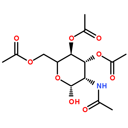
![(2WEI ,5WEI ,7WEI ,13WEI )-4-ACETOXY-13-{[(2R,3S)-3-(BENZOYLAMINO)-2-HYDROXY-<WBR />3-PHENYLPROPANOYL]OXY}-1,7-DIHYDROXY-9,10-DIOXO-5,20-EPOXYTAX-11-<WBR />EN-2-YL BENZOATE](http://img.cochemist.com/ccimg/26300/26296-50-4.png)
![(2WEI ,5WEI ,7WEI ,13WEI )-4-ACETOXY-13-{[(2R,3S)-3-(BENZOYLAMINO)-2-HYDROXY-<WBR />3-PHENYLPROPANOYL]OXY}-1,7-DIHYDROXY-9,10-DIOXO-5,20-EPOXYTAX-11-<WBR />EN-2-YL BENZOATE](http://img.cochemist.com/ccimg/26300/26296-50-4_b.png)

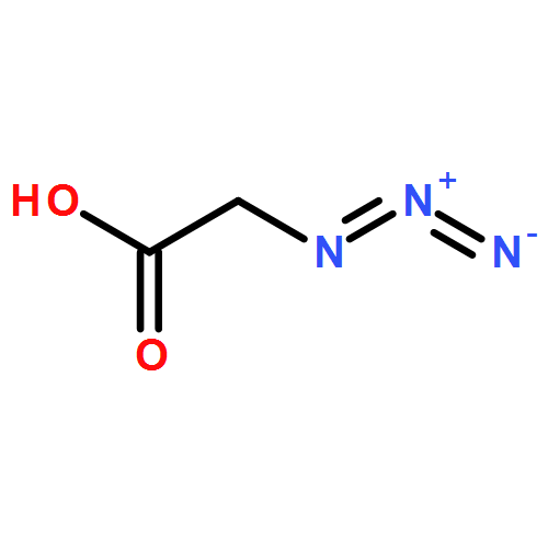

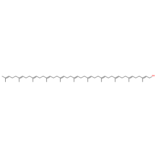


![1H-Benz[de]isoquinoline-1,3(2H)-dione, 6-(dimethylamino)-](/data/chemimg/3433200/82616-18-0.png)
![1H-Benz[de]isoquinoline-1,3(2H)-dione, 6-(dimethylamino)-](/data/chemimg/3433200/82616-18-0_b.png)
![PHOSPHONO [(2E,6E,10E,14E,18E,22E,26E,30E,34E,38E)-3,7,11,15,19,23,27,31,35,39,43-UNDECAMETHYLTETRATETRACONTA-2,6,10,14,18,22,26,30,34,38,42-UNDECAENYL] HYDROGEN PHOSPHATE](http://img.cochemist.com/ccimg/31900/31867-59-1.png)
![PHOSPHONO [(2E,6E,10E,14E,18E,22E,26E,30E,34E,38E)-3,7,11,15,19,23,27,31,35,39,43-UNDECAMETHYLTETRATETRACONTA-2,6,10,14,18,22,26,30,34,38,42-UNDECAENYL] HYDROGEN PHOSPHATE](http://img.cochemist.com/ccimg/31900/31867-59-1_b.png)


![[4-(aminomethyl)-5-hydroxy-6-methylpyridin-3-yl]methyl Dihydrogen Phosphate](http://img.cochemist.com/ccimg/600/529-96-4.png)
![[4-(aminomethyl)-5-hydroxy-6-methylpyridin-3-yl]methyl Dihydrogen Phosphate](http://img.cochemist.com/ccimg/600/529-96-4_b.png)
![Uridine 5'-(trihydrogendiphosphate), P'-[2-(acetylamino)-2-deoxy-a-D-glucopyranosyl] ester](http://img.cochemist.com/ccimg/600/528-04-1.png)
![Uridine 5'-(trihydrogendiphosphate), P'-[2-(acetylamino)-2-deoxy-a-D-glucopyranosyl] ester](http://img.cochemist.com/ccimg/600/528-04-1_b.png)
![(3AR,4R,5R,6AS)-4-FORMYL-2-OXOHEXAHYDRO-2H-CYCLOPENTA[B]FURAN-5-Y<WBR />L 4-BIPHENYLCARBOXYLATE](http://img.cochemist.com/ccimg/100/72-89-9.png)
![(3AR,4R,5R,6AS)-4-FORMYL-2-OXOHEXAHYDRO-2H-CYCLOPENTA[B]FURAN-5-Y<WBR />L 4-BIPHENYLCARBOXYLATE](http://img.cochemist.com/ccimg/100/72-89-9_b.png)