Co-reporter:Xing Zhang, Dixy E. Green, Victor L. Schultz, Lei Lin, Xiaorui Han, Ruitong Wang, Alexander Yaksic, So Young Kim, Paul L. DeAngelis, and Robert J. Linhardt
The Journal of Organic Chemistry September 15, 2017 Volume 82(Issue 18) pp:9910-9910
Publication Date(Web):August 16, 2017
DOI:10.1021/acs.joc.7b01787
Unnatural chemically modified nucleotide sugars UDP-4-N3-GlcNAc and UDP-4-N3-GalNAc were chemically synthesized for the first time. These unnatural UDP sugar products were then tested for incorporation into hyaluronan, heparosan, or chondroitin using polysaccharide synthases. UDP-4-N3-GlcNAc served as a chain termination substrate for hyaluronan or heparosan synthases; the resulting 4-N3-GlcNAc-terminated hyaluronan and heparosan were then successfully conjugated with Alexa Fluor 488 DIBO alkyne, demonstrating that this approach is generally applicable for labeling and detection of suitable glycosaminoglycans.
Co-reporter:Lijuan Hou, Xing Zhang, Paiyz E. Mikael, Lei Lin, Wenjun Dong, Yingying Zheng, Trevor John Simmons, Fuming Zhang, and Robert J. Linhardt
ACS Omega October 2017? Volume 2(Issue 10) pp:6321-6321
Publication Date(Web):October 2, 2017
DOI:10.1021/acsomega.7b00460
Poly(glycerol sebacate) (PGS) has increasingly become a desirable biomaterial due to its elastic mechanical properties, biodegradability, and biocompatibility. Here, we report microfibrous core–shell mats of polycaprolactone (PCL)–PGS prepared using wet–wet coaxial electrospinning. The anticoagulant heparin was immobilized onto the surface of these electrospun fiber mats, and they were evaluated for their chemical, mechanical, and biological properties. The core–shell structure of PCL–PGS provided tunable degradation and mechanical properties. The slowly degrading PCL provided structural integrity, and the fast degrading PGS component increased fiber elasticity. Young’s modulus of PCL–PGS ranged from 5.6 to 15.7 MPa. The ultimate tensile stress ranged from 2.0 to 2.9 MPa, and these fibers showed elongation from 290 to 900%. The addition of PGS and grafting of heparin improved the attachment and proliferation of human umbilical vein endothelial cells. Core–shell PCL–PGS fibers demonstrate improved performance as three-dimensional fibrous mats for potential tissue-engineering applications.Topics: Biodegradable materials; Biodegradable materials; Cell and Molecular biology; Fibers; Materials processing; Mechanical properties; Tissue engineering;
Co-reporter:Yanlei Yu, Jiana Duan, Franklin E. Leach III, Toshihiko Toida, Kyohei Higashi, Hong Zhang, Fuming Zhang, I. Jonathan Amster, and Robert J. Linhardt
Journal of the American Chemical Society November 22, 2017 Volume 139(Issue 46) pp:16986-16986
Publication Date(Web):November 7, 2017
DOI:10.1021/jacs.7b10164
Glycomics represents one of the last frontiers and most challenging in omic analysis. Glycosylation occurs in the endoplasmic reticulum and the Golgi organelle and its control is neither well-understood nor predictable based on proteomic or genomic analysis. One of the most structurally complex classes of glycoconjugates is the proteoglycans (PGs) and their glycosaminoglycan (GAG) side chains. Previously, our laboratory solved the structure of the chondroitin sulfate chain of the bikunin PG. The current study examines the much more complex structure of the dermatan sulfate GAG chain of decorin PG. By utilizing sophisticated separation methods followed by compositional analysis, domain mapping, and tandem mass spectrometry coupled with analysis by a modified genetic algorithm approach, the structural motif for the decorin dermatan sulfate chain was determined. This represents the second example of a GAG with a prominent structural motif, suggesting that the structural variability of this class of glycoconjugates is somewhat simpler than had been expected.
Co-reporter:Victor L. Schultz, Xing Zhang, Kathryn Linkens, Jenna Rimel, Dixy E. Green, Paul L. DeAngelis, and Robert J. Linhardt
The Journal of Organic Chemistry 2017 Volume 82(Issue 4) pp:
Publication Date(Web):January 27, 2017
DOI:10.1021/acs.joc.6b02929
Unnatural uridine diphosphate (UDP)-sugar donors, UDP-4-deoxy-4-fluoro-N-acetylglucosamine (4FGlcNAc) and UDP-4-deoxy-4-fluoro-N-acetylgalactosamine (4FGalNAc), were prepared using both chemical and chemoenzymatic syntheses relying on N-acetylglucosamine-1-phosphate uridylyltransferase (GlmU). The resulting unnatural UDP-sugar donors were then tested as substrates in glycosaminoglycan synthesis catalyzed by various synthases. UDP-4FGlcNAc was transferred onto an acceptor by Pastuerella multocida heparosan synthase 1 and subsequently served as a chain terminator.
Co-reporter:Guoyun Li;Lingyun Li;Eun Ji Joo;Ji Woong Son;Young Jin Kim
Glycoconjugate Journal 2017 Volume 34( Issue 5) pp:661-669
Publication Date(Web):18 August 2017
DOI:10.1007/s10719-017-9790-7
In this report, we used liquid chromatography-mass spectrometry and Western blotting to analyze the content and structure of glycosaminoglycans, glycolipids and selected proteins to compare differences between patient-matched normal and cancerous lung tissues obtained from lung cancer patients. The cancer tissue samples contained over twice as much chondroitin sulfate (CS)/dermatan sulfate (DS) as did the normal tissue samples, while the amount of heparan sulfate (HS) and hyaluronan (HA) in normal and cancer tissues were not significantly different. In HS, several minor disaccharide components, including NS6S, NS2S and 2S were significantly lower in cancer tissues, while the levels of major disaccharides, TriS, NS and 0S disaccharides were not significantly different in normal and cancer tissues. In regards to CS/DS, the level of 4S disaccharide (the major component of CS-type A and DS) decreased and the level of 6S disaccharide (the major component of CS- type C) increased in cancer tissues. We also compared the content and structure of GAGs in lung tissues from smoking and non-smoking patients. Analysis of the glycolipids showed all lipids present in these lung tissues, with the exception of sphingomyelin were higher in cancer tissues than in normal tissues. Western analysis showed that syndecan 1 and 2 proteoglycans displayed much higher expression in cancer tissue/biopsy samples. This investigation begins to provide an understanding of patho-physiological roles on glycosaminoglycans and glycolipids and might be useful in identifying potential biomarkers in lung cancer.
Co-reporter:Haisheng Wang;Wenqin He;Peixia Jiang;Yanlei Yu;Lei Lin
Glycoconjugate Journal 2017 Volume 34( Issue 5) pp:643-649
Publication Date(Web):27 July 2017
DOI:10.1007/s10719-017-9786-3
There is a need for degradative enzymes in the study of glycosaminoglycans. Many of these enzymes are currently available either in their natural or recombinant forms. Unfortunately, progress in structure-activity studies of keratan sulfate (KS) have been impeded by the lack of a commercially available endo-β-N-acetylglucosaminidase, keratantase II. The current study uses a recently published sequence of a highly thermostable keratanase II identified in Bacillus circulans to clone and express a series of truncation mutants in Escherichia coli BL21. The resulting truncated forms of keratanase II exhibit activity and excellent storage and thermal stability making these useful tools for glycobiology research.
Co-reporter:Yanlei Yu;Makoto Hirakane;Daisuke Mori;Lei Lin;Fuming Zhang
Glycoconjugate Journal 2017 Volume 34( Issue 4) pp:545-552
Publication Date(Web):29 May 2017
DOI:10.1007/s10719-017-9774-7
Heparin is a structurally complex polysaccharide used as a clinical anticoagulant. It is comprised of a heterogeneous mixture of polysaccharide chains having a variety of sequences and lengths. The production methods and regulatory controls of pharmaceutical heparins have changed over the years. This study assesses the structural and activity uniformity of the polysaccharide chains comprising two contemporary heparin products. The heparin fractions with different sizes and charges were separated with size exclusion and ion exchange chromatography. The fractions were analyzed for their molecular weight properties, di- and tetrasaccharide compositions, and anti-factor IIa and anti-factor-Xa activities. The distribution of these properties through chains of different lengths and ones with different charge density were compared. The results demonstrate that with the increase in heparin purity, activity and molecular weight required by the current pharmacopeia, the uniformity of pharmaceutical heparin products have increased.
Co-reporter:Xinyue Liu, Kalib St. Ange, Lei Lin, Fuming Zhang, Lianli Chi, Robert J. Linhardt
Journal of Chromatography A 2017 Volume 1480(Volume 1480) pp:
Publication Date(Web):13 January 2017
DOI:10.1016/j.chroma.2016.12.021
•Four types of commercial enoxaparins were analyzed by integrated LC–MS and NMR analysis.•Heparinase treatment was used before bottom-up analysis and disaccharide analysis.•Intact chain, oligosaccharide, and disaccharide analyses relied on LC–MS.•Monosaccharide compositional analysis relied on top-down NMR analysis.•Generic enoxaparins and innovator product are similar, differences are observed due to parent heparin and process conditions.A strategy for the comprehensive analysis of low molecular weight (LMW) heparins is described that relies on using an integrated top-down and bottom-up approach. Liquid chromatography-mass spectrometry, an essential component of this approach, is rapid, robust, and amenable to automated processing and interpretation. Nuclear magnetic resonance spectroscopy provides complementary top-down information on the chirality of the uronic acid residues comprising a low molecular weight heparin. Using our integrated approach four different low molecular weight heparins prepared from porcine heparin through chemical β-eliminative cleavage were comprehensively analyzed. Lovenox™ and Clexane™, the innovator versions of enoxaparin marketed in the US and Europe, respectively, and two generic enoxaparins, from Sandoz and Teva, were analyzed. The results which were supported by analysis of variation (ANOVA), while showing remarkable similarities between different versions of the product and good lot-to-lot consistency of each product, also detects subtle differences that may result from differences in their manufacturing processes or differences in the source (or parent) porcine heparin from which each product is prepared.
Co-reporter:Junhui Li, Shan Li, Lufeng Yan, Tian Ding, Robert J. Linhardt, Yanlei Yu, Xinyue Liu, Donghong Liu, Xingqian Ye, Shiguo Chen
European Journal of Medicinal Chemistry 2017 Volume 139(Volume 139) pp:
Publication Date(Web):20 October 2017
DOI:10.1016/j.ejmech.2017.07.065
•fCS oligosaccharides with different sulfation patterns were obtained from sea cucumbers.•Free radical depolymerization of fCSs was highly selective.•fCSs exert anticoagulant activity by selective inhibition of intrinsic tenase.•Depolymerization of fCSs should decrease adverse effects of activation FXII.•Sulfation pattern and molecular size are important to anticoagulant activity.Fucosylated chondroitin sulfates (fCSs) are structurally unusual glycosaminoglycans isolated from sea cucumbers that exhibit potent anticoagulant activity. These fCSs were isolated from sea cucumber, Isostichopus badionotus and Pearsonothuria graeffei. Fenton reaction followed by gel filtration chromatography afforded fCS oligosaccharides, with different sulfation patterns identified by mass and NMR spectroscopy, and these were used to clarify the relationship between the structures and the anticoagulant activities of fCSs. In vitro activities were measured by activated partial thromboplastin time (APTT), thrombin time (TT), thrombin and factor Xa inhibition, and activation of FXII. The results showed that free radicals preferentially acted on GlcA residues affording oligosaccharides that were purified from both fCSs. The inhibition of thrombin and factor X activities, mediated through antithrombin III and heparin cofactor II of fCSs oligosaccharides were affected by their molecular weight and fucose branches. Oligosaccharides with different sulfation patterns of the fucose branching had a similar ability to inhibit the FXa by the intrinsic factor Xase (factor IXa-VIIIa complex). Oligosaccharides with 2,4-O-sulfo fucose branches from fCS-Ib showed higher activities than ones with 3,4-O-disulfo branches obtained from fCS-Pg. Furthermore, a heptasaccharide is the minimum size oligosaccharide required for anticoagulation and FXII activation. This activity was absent for fCS oligosaccharides smaller than nonasaccharides. Molecular size and fucose branch sulfation are important for anticoagulant activity and reduction of size can reverse the activation of FXII caused by native fCSs.Download high-res image (189KB)Download full-size image
Co-reporter:Jing Zhao, Xinyue Liu, Anju Malhotra, Quanhong Li, Fuming Zhang, Robert J. Linhardt
Analytical Biochemistry 2017 Volume 526(Volume 526) pp:
Publication Date(Web):1 June 2017
DOI:10.1016/j.ab.2017.03.013
A novel method has been developed for the easy measurement of heparin's anticoagulant activity using surface plasmon resonance. The anticoagulant activity of target heparin was evaluated by measuring the competitive antithrombin III binding of analyte heparin in the solution phase and USP heparin immobilized on chip surface. Heparins, obtained from different animal sources, and low molecular weight heparins were analyzed. The results were reproducible and correlated well with the results of chromogenic assays (correlation coefficient r = 0.98 for anti-Xa and r = 0.94 for anti-IIa). This protocol provides many advantages, significantly minimizing time, cost and the complications of chromogenic assay methods.
Co-reporter:Xing Zhang;Yongmei Xu;Po-Hung Hsieh;Jian Liu;Lei Lin;Eric P. Schmidt
Organic & Biomolecular Chemistry 2017 vol. 15(Issue 5) pp:1222-1227
Publication Date(Web):2017/02/01
DOI:10.1039/C6OB02603F
A heparin oligosaccharide having a completely natural structure was successfully synthesized through a chemoenzymatic approach using an unnatural glycosyl acceptor, p-nitrophenyl glucuronide (GlcA-pNP). The use of an inexpensive and commercially available GlcA-pNP acceptor facilitates oligosaccharide recovery and purification on C-18 resin during chemoenzymatic synthesis. Oligosaccharide chain extension and modification afforded a heptasaccharide with gluconic acid residues at its reducing and non-reducing ends. Treatment with periodate oxidation followed by Smith degradation or alkaline elimination resulted in the selective cleavage of vicinal diol-containing glucuronic acid residues affording highly sulfated heparin pentasaccharides having a completely natural structure. This methodology should facilitate the chemoenzymatic synthesis of a family of highly sulfated heparin oligosaccharides with unmodified structures for biological evaluation.
Co-reporter:Xing Zhang;Vijayakanth Pagadala;Hannah M. Jester;Andrew M. Lim;Truong Quang Pham;Anna Marie P. Goulas;Jian Liu
Chemical Science (2010-Present) 2017 vol. 8(Issue 12) pp:7932-7940
Publication Date(Web):2017/11/20
DOI:10.1039/C7SC03541A
Heparan sulfate (HS) is a member of the glycosaminoglycans (GAG) family that plays essential roles in biological processes from animal sources. Heparin, a highly sulfated form of HS, is widely used as anticoagulant drug worldwide. The high diversity and complexity of HS and heparin represent a roadblock for structural characterization and biological activity studies. Access to structurally defined oligosaccharides is critical for the successful development of HS and heparin structure–activity relationships. In this study, a library of 66 HS and heparin oligosaccharides covering different sulfation patterns and sizes was prepared through an efficient method of chemoenzymatic synthesis. A systematic nuclear magnetic resonance spectroscopy study was firstly undertaken for every oligosaccharide in the library. In addition to the availability of different oligosaccharides, this work also provides spectroscopic data helpful for characterizing more complicated polysaccharide structures providing a safeguard to ensure the quality of the drug heparin. This HS/heparin library will be useful for activity screening and facilitate future structure–activity relationship studies.
Co-reporter:Yin Chen;Megan Reddy;Yanlei Yu;Fuming Zhang
Glycoconjugate Journal 2017 Volume 34( Issue 1) pp:119-126
Publication Date(Web):2017 February
DOI:10.1007/s10719-016-9737-4
Glycosaminoglycans (GAGs) were prepared from the muscular stomach or gizzard of the chicken. The content of GAGs on a dry weight basis contains 0.4 wt.% a typical value observed for a muscle tissue. The major GAG components were chondroitin-6-sulfate and chondroitin-4-sulfate (~64 %) of molecular weight 21–22 kDa. Hyaluronan (~24 %) had a molecular weight 120 kDa. Smaller amounts (12 %) of heparan sulfate was also present which was made of more highly sulfated chains of molecular weight of 21-22 kDa and a less sulfated low molecular weight (< 10 kDa) heterogeneous partially degraded heparan sulfate. Chicken gizzard represents an inexpensive and readily available source of muscle tissue-derived GAGs.
Co-reporter:So Young Kim, Jing Zhao, Xinyue Liu, Keith Fraser, Lei Lin, Xing Zhang, Fuming ZhangJonathan S. Dordick, Robert J. Linhardt
Biochemistry 2017 Volume 56(Issue 8) pp:
Publication Date(Web):February 2, 2017
DOI:10.1021/acs.biochem.6b01056
In February 2016, the World Health Organization declared a Public Health Emergency of International Concern on Zika Virus (ZIKV), because of its association with severe fetal anomalies of congenitally infected humans. This has led to urgent efforts by academic, federal, and industry research groups to improve our understanding of the pathogenesis of ZIKV and to develop detection methods, therapeutic strategies, and vaccines. Although we still do not have the entire picture of the pathogenesis of ZIKV, extensive research has been conducted on related pathogenic flaviviruses (i.e., dengue virus, West Nile virus, and yellow fever virus). Binding to glycosaminoglycans (GAGs) through its envelope protein is the first step in successful host cell invasion of dengue virus. In this study, we examined ZIKV envelope protein (ZIKV E) binding to GAGs in a real time interaction study using surface plasmon resonance (SPR) to explore the role of GAGs in host cell entry of ZIKV into placenta and brain. ZIKV E strongly binds (KD = 443 nM) pharmaceutical heparin (HP), a highly sulfated GAG, and binds with lower avidity to less sulfated GAGs, suggesting that the ZIKV E–GAG interaction may be electrostatically driven. Using SPR competition assays with various chain length HP oligosaccharides (from 4 to 18 saccharide units in length), we observed that ZIKV E preferentially binds to longer HP oligosaccharides (with 8–18 saccharides). Next, we examined GAGs prepared from human placentas to determine if they bound ZIKV E, possibly mediating placental cell invasion of ZIKV. Compositional analysis of these GAGs as well as SPR binding studies showed that both chondroitin sulfate and heparan sulfate GAGs, present on the placenta, showed low-micromolar interactions with ZIKV E. Both porcine brain CS and HS also showed micromolar binding with ZIKV E. Moreover, heparan sulfate with a higher TriS content, the dominant repeating unit of HP, shows a high affinity for ZIKV E. These results suggest that GAGs may be utilized as attachment factors for host cell entry of Zika virus as they do in other pathogenic flaviviruses. They may also assist us in advancing our understanding of the pathogenesis of ZIKV and guide us in designing therapeutics to combat ZIKV with more insight.
Co-reporter:Jacob A. Englaender;Yuanyuan Zhu
Applied Microbiology and Biotechnology 2017 Volume 101( Issue 7) pp:2843-2851
Publication Date(Web):2017 April
DOI:10.1007/s00253-016-8047-x
Heparin, an anticoagulant drug, is biosynthesized in selected animal cells. The heparin biosynthetic enzymes mainly consist of sulfotransferases and all are integral transmembrane glycoproteins. These enzymes are generally produced in engineered Escherichia coli as without their transmembrane domains as non-glycosylated fusion proteins. In this study, we used the yeast, Komagataella pastoris, to prepare four sulfotransferases involved in heparin biosynthesis as glycoproteins. While the yields of these yeast-expressed enzymes were considerably lower than E. coli-expressed enzymes, these enzymes were secreted into the fermentation media simplifying their purification and were endotoxin free. The activities of these sulfotransferases, expressed as glycoproteins in yeast, were compared to the bacterially expressed proteins. The yeast-expressed sulfotransferase glycoproteins showed improved kinetic properties than the bacterially expressed proteins.
Co-reporter:Xiaojun Sun, Lei Lin, Xinyue Liu, Fuming Zhang, Lianli Chi, Qiangwei Xia, and Robert J. Linhardt
Analytical Chemistry 2016 Volume 88(Issue 3) pp:1937
Publication Date(Web):December 29, 2015
DOI:10.1021/acs.analchem.5b04405
Heparins, highly sulfated, linear polysaccharides also known as glycosaminoglycans, are among the most challenging biopolymers to analyze. Hyphenated techniques in conjunction with mass spectrometry (MS) offer rapid analysis of complex glycosaminoglycan mixtures, providing detailed structural and quantitative data. Previous analytical approaches have often relied on liquid chromatography (LC)–MS, and some have limitations including long separation times, low resolution of oligosaccharide mixtures, incompatibility of eluents, and often require oligosaccharide derivatization. This study examines the analysis of glycosaminoglycan oligosaccharides using a novel electrokinetic pump-based capillary electrophoresis (CE)–MS interface. CE separation and electrospray were optimized using a volatile ammonium bicarbonate electrolyte and a methanol–formic acid sheath fluid. The online analyses of highly sulfated heparin oligosaccharides, ranging from disaccharides to low molecular weight heparins, were performed within a 10 min time frame, offering an opportunity for higher-throughput analysis. Disaccharide compositional analysis as well as top-down analysis of low molecular weight heparin was demonstrated. Using normal polarity CE separation and positive-ion electrospray ionization MS, excellent run-to-run reproducibility (relative standard deviation of 3.6–5.1% for peak area and 0.2–0.4% for peak migration time) and sensitivity (limit of quantification of 2.0–5.9 ng/mL and limit of detection of 0.6–1.8 ng/mL) could be achieved.
Co-reporter:Jing Zhao, Xinyue Liu, Chelsea Kao, Emily Zhang, Quanhong Li, Fuming Zhang, and Robert J. Linhardt
Biochemistry 2016 Volume 55(Issue 32) pp:4552-4559
Publication Date(Web):July 22, 2016
DOI:10.1021/acs.biochem.6b00555
Langerin, a C-type lectin, is expressed in Langerhans cells. It was reported that langerin binds sulfated glycans, which is an important initial step for its role in blocking human immunodeficiency virus (HIV) transmission by capturing HIV pathogens and mediating their internalization into Birbeck granules for their elimination. It is fundamentally important to understand these interactions at the molecular level for the design of new highly specific therapeutic agents for HIV. Surface plasmon resonance (SPR), which allows for the real-time, direct, quantitative analysis of the label-free molecular interactions, has been used successfully for biophysical characterization of glycosaminoglycan (GAG)–protein interactions. In this study, we report kinetics, structural analysis, and the effects of physiological conditions (e.g., pH, salt concentration, and Ca2+ and Zn2+concentrations) on the interactions between GAGs and langerin using SPR. SPR results revealed that langerin binds to heparin with high affinity (KD ∼ 2.4 nM) and the oligosaccharide length required for the interactions is larger than a tetrasaccharide. This heparin/heparan sulfate-binding protein also interacts with other GAGs, including dermatan sulfate, chondroitin sulfates C–E and KS. In addition, liquid chromatography–mass spectrometry analysis was used to characterize the structure of sulfated glycans that bound to langerin.
Co-reporter:Yi Jia Wang, Lei Lin, Xing Zhang, Victor Schultz, Fuming Zhang, Jing Zhi Sun, Robert J. Linhardt
Analytical Biochemistry 2016 Volume 514() pp:48-54
Publication Date(Web):1 December 2016
DOI:10.1016/j.ab.2016.09.007
Abstract
A positively charged tetraphenylethene (TPE) derivative, TPE-4MN, was synthesized as a probe for heparin based on aggregation induced emission. On the addition of 5.0 μg/mL of heparin, TPE-4MN showed an enhanced emission of about 10-fold. The change in fluorescence at 475 nm was linear over a range of heparin concentrations of 0–1.0 μg/mL with an R = 0.99988 and the limit of detection (LOD) was calculated to be 0.75 μg/mL. The mechanism of the detection was proven to be through an ion pairing interaction. TPE-4MN showed good selectivity for heparin over other types of polysaccharides and could easily distinguish heparin from heparan sulfate, a glycosaminoglycan having a similar structure to that of heparin.
Co-reporter:Ebru Ucakturk;Orkun Akman;Xiaojun Sun;Dilek Ertoy Baydar
Glycoconjugate Journal 2016 Volume 33( Issue 1) pp:103-112
Publication Date(Web):2016 February
DOI:10.1007/s10719-015-9643-1
Glycosaminoglycans (GAGs) are heterogeneous, linear, highly charged, anionic polysaccharides consisting of repeating disaccharides units. GAGs have some biological significance in cancer progression (invasion and metastasis) and cell signaling. In different cancer types, GAGs undergo specific structural changes. In the present study, in depth investigation of changes in sulfation pattern and composition of GAGs, heparan sulfate (HS)/heparin (HP), chondroitin sulfate (CS)/dermatan sulfate and hyaluronan (HA) in normal renal tissue (NRT) and renal cell carcinoma tissue (RCCT) were evaluated. The statistical evaluation showed that alteration of the HS (HSNRT = 415.1 ± 115.3; HSRCCT = 277.5 ± 134.3), and CS (CSNRT = 35.3 ± 12.3; CSRCCT = 166.7 ± 108.8) amounts (in ng/mg dry tissue) were statistically significant (p < 0.05). Sulfation pattern in NRT and RCCT was evaluated to reveal disaccharide profiles. Statistical analyses showed that RCCT samples contain significantly increased amounts (in units of ng/mg dry tissue) of 4SCS (NRT = 25.7 ± 9.4; RCCT = 117.1 ± 73.9), SECS (NRT = 0.7 ± 0.3; RCCT = 4.7 ± 4.5), 6SCS (NRT = 6.1 ± 2.7; RCCT = 39.4 ± 34.7) and significantly decreased amounts (in units of ng/mg dry tissue) of NS6SHS (RCCT = 28.6 ± 6.5, RCCT = 10.2 ± 8.0), NS2SHS (RCCT = 44.2 ± 13.8; RCCT = 27.2 ± 15.0), NSHS (NRT = 68.4 ± 15.8; RCCT = 50.4 ± 21.2), 2S6SHS (NRT = 1.0 ± 0.4; RCCT = 0.4 ± 0.3), and 6SHS (NRT = 60.6 ± 17.5; RCCT = 24.9 ± 12.3). If these changes in GAGs are proven to be specific and sensitive, they may serve as potential biomarkers in RCC. Our findings are likely to help us to show the direction for further investigations to be able to bring different diagnostic and prognostic approaches in renal tumors.
Co-reporter:Xing Zhang, Omar Khalidi, So Young Kim, Ruitong Wang, Victor Schultz, Brady F. Cress, Richard A. Gross, Mattheos A.G. Koffas, Robert J. Linhardt
Bioorganic & Medicinal Chemistry Letters 2016 26(13) pp: 3089-3092
Publication Date(Web):1 July 2016
DOI:10.1016/j.bmcl.2016.05.003
A series of 5,7-dihydroxyflavanone derivatives were efficiently synthesized. Their antimicrobial efficacy on Gram-negative, Gram-positive bacteria and yeast were evaluated. Among these compounds, most of the halogenated derivatives exhibited the best antimicrobial activity against Gram-positive bacteria, the yeast Saccharomyces cerevisiae, and the Gram-negative bacterium Vibrio cholerae. The cytotoxicities of these compounds were low as evaluated on HepG2 cells using a cell viability assay. This study suggests that halogenated flavanones might represent promising pharmacological candidates for further drug development.Download high-res image (86KB)Download full-size image
Co-reporter:Angeles Farrán, Chao Cai, Manuel Sandoval, Yongmei Xu, Jian Liu, María J. Hernáiz, and Robert J. Linhardt
Chemical Reviews 2015 Volume 115(Issue 14) pp:6811
Publication Date(Web):June 29, 2015
DOI:10.1021/cr500719h
Co-reporter:Xiaojun Sun, Lingyun Li, Katherine H. Overdier, Lee Anne Ammons, Ivor S. Douglas, Clay Cothren Burlew, Fuming Zhang, Eric P. Schmidt, Lianli Chi, and Robert J. Linhardt
Analytical Chemistry 2015 Volume 87(Issue 12) pp:6220
Publication Date(Web):May 25, 2015
DOI:10.1021/acs.analchem.5b00913
The determination of complex analytes, present at low concentrations, in biological fluids poses a difficult challenge. This study relies on an optimized method of recovery, enzymatic treatment, and disaccharide analysis by liquid chromatography–tandem mass spectrometry to rapidly determine low concentrations of glycosaminoglycans in human urine. The approach utilizes multiple reaction monitoring (MRM) of glycosaminoglycan disaccharides obtained from treating urine samples with recombinant heparin lyases and chondroitin lyase. This rapid and sensitive method allows the analysis of glycosaminoglycan content and disaccharide composition in urine samples having concentrations 10- to 100-fold lower than those typically analyzed from patients with metabolic diseases, such as mucopolysaccharidosis. The current method facilitates the analysis low (ng/mL) levels of urinary glycosaminoglycans present in healthy individuals and in patients with pathological conditions, such as inflammation and cancers, that can subtly alter glycosaminoglycan content and composition.
Co-reporter:Sashka Dimitrievska, Chao Cai, Amanda Weyers, Jenna L. Balestrini, Tylee Lin, Sumati Sundaram, Go Hatachi, David A. Spiegel, Themis R. Kyriakides, Jianjun Miao, Guoyun Li, Laura E. Niklason, Robert J. Linhardt
Acta Biomaterialia 2015 Volume 13() pp:177-187
Publication Date(Web):February 2015
DOI:10.1016/j.actbio.2014.11.015
Abstract
A novel method enabling the engineering of a dense and appropriately oriented heparin-containing layer on decellularized aortas has been developed. Amino groups of decellularized aortas were first modified to azido groups using 3-azidobenzoic acid. Azide-clickable dendrons were attached onto the azido groups through “alkyne–azide” click chemistry, affording a tenfold amplification of adhesions sites. Dendron end groups were finally decorated with end-on modified heparin chains. Heparin chains were oriented like heparan sulfate groups on native endothelial cells surface. X-ray photoelectron spectroscopy, nuclear magnetic resonance imaging, mass spectrometry and Fourier transform infrared FTIR spectroscopy were used to characterize the synthesis steps, building the final heparin layered coatings. The continuity of the heparin coating was verified using fluorescent microscopy and histological analysis. The efficacy of heparin linkage was demonstrated with factor Xa anti-thrombogenic assay and platelet adhesion studies. The results suggest that oriented heparin immobilization to decellularized aortas may improve the in vivo blood compatibility of decellularized aortas and vessels.
Co-reporter:Guoyun Li, Lingyun Li, Fang Tian, Linxia Zhang, Changhu Xue, and Robert J. Linhardt
ACS Chemical Biology 2015 Volume 10(Issue 5) pp:1303
Publication Date(Web):February 13, 2015
DOI:10.1021/acschembio.5b00011
Glycosaminoglycans (GAGs), a family of polysaccharides widely distributed in eukaryotic cells, are responsible for a wide array of biological functions. Quantitative disaccharide compositional analysis is one of the primary ways to characterize the GAG structure. This structural analysis is typically time-consuming (1–2 weeks) and labor intensive, requiring GAG recovery and multistep purification, prior to the enzymatic/chemical digestion of GAGs, and finally their analysis. Moreover, 105–107 cells are usually required for compositional analysis. We report a sensitive, rapid, and quantitative analysis of GAGs present in a small number of cells. Commonly studied cell lines were selected based on phenotypic properties related to the biological functions of GAGs. These cells were lysed using a commercial surfactant reagent, sonicated, and digested with polysaccharide lyases. The resulting disaccharides were recovered by centrifugal filtration, labeled with 2-aminoacridone, and analyzed by liquid chromatography (LC)-mass spectrometry (MS). Using a highly sensitive MS method, multiple reaction monitoring (MRM), the limit of detection for each disaccharide was reduced to 0.5–1.0 pg, as compared with 1.0–5.0 ng obtained using standard LC-MS analysis. Sample preparation time was reduced to 1–2 days, and the cell number required was reduced to 5000 cells for complete GAG characterization to as few as 500 cells for the characterization of the major GAG disaccharide components. Our survey of the glycosaminoglycanomes of the 20 selected cell lines reveals major differences in their GAG amounts and compositions. Structure–function relationships are explored using these data, suggesting the utility of this method in cellular glycobiology.
Co-reporter:Ujjwal Bhaskar, Guoyun Li, Li Fu, Akihiro Onishi, Mathew Suflita, Jonathan S. Dordick, Robert J. Linhardt
Carbohydrate Polymers 2015 Volume 122() pp:399-407
Publication Date(Web):20 May 2015
DOI:10.1016/j.carbpol.2014.10.054
•A simple one pot method for heparin synthesis.•Bioengineered heparin with activity similar to USP heparin.•2-O-sulfation must occur before 6-O-sulfation.Contamination in heparin batches during early 2008 has resulted in a significant effort to develop a safer bioengineered heparin using bacterial capsular polysaccharide heparosan and recombinant enzymes derived from the heparin/heparan sulfate biosynthetic pathway. This requires controlled chemical N-deacetylation/N-sulfonation of heparosan followed by epimerization of most of its glucuronic acid residues to iduronic acid and O-sulfation of the C2 position of iduronic acid and the C3 and C6 positions of the glucosamine residues. A combinatorial study of multi-enzyme, one-pot, in vitro biocatalytic synthesis, carried out in tandem with sensitive analytical techniques, reveals controlled structural changes leading to heparin products similar to animal-derived heparin active pharmaceutical ingredients. Liquid chromatography–mass spectrometry and nuclear magnetic resonance spectroscopy analysis confirms an abundance of heparin's characteristic trisulfated disaccharide, as well as 3-O-sulfo containing residues critical for heparin binding to antithrombin III and its anticoagulant activity. The bioengineered heparins prepared using this simplified one-pot chemoenzymatic synthesis also show in vitro anticoagulant activity.
Co-reporter:Guoyun Li, Lingyun Li, Changhu Xue, Dustin Middleton, Robert J. Linhardt, Fikri Y. Avci
Journal of Chromatography A 2015 Volume 1397() pp:43-51
Publication Date(Web):5 June 2015
DOI:10.1016/j.chroma.2015.04.009
•Pneumococcal type 3 polysaccharide (PnP3) is depolymerized.•Pn3P derived oligosacchides are separated by liquid chromatography (LC).•Hydrophilic interaction LC and reverse phase LC are used in their separation.•LC–mass spectrometry (MS) analysis determined oligosaccharide composition.•LC–tandem MS analysis determined oligosaccharide sequence.Pneumococcal type-3 polysaccharide (Pn3P) is considered a major target for the development of a human vaccine to protect against Streptococcus pneumoniae infection. Thus, it is critical to develop methods for the preparation and analysis of Pn3P-derived oligosaccharides to better understand its immunological properties. In this paper, we profile oligosaccharides, generated by the free radical depolymerization of Pn3P, using liquid chromatography (LC)–tandem mass spectrometry (MS/MS). Hydrophilic liquid interaction chromatography (HILIC)–mass spectrometry (MS) revealed a series of oligosaccharides with an even- and odd-number of saccharide residues, ranging from monosaccharide, degree of polymerization (dp1) to large oligosaccharides up to dp 20, generated by free radical depolymerization. Isomers of oligosaccharides with an even number of sugar residues were easily separated on a HILIC column, and their sequences could be distinguished by comparing MS/MS of these oligosaccharides and their reduced alditols. Fluorescent labeling with 2-aminoacridone (AMAC) followed by reversed phase (RP)-LC–MS/MS was applied to analyze and sequence poorly separated product mixtures, as RP-LC affords higher resolution of AMAC-labeled oligosaccharides than does HILIC-based separation. The present methodology can be potentially applied to profiling other capsular polysaccharides.
Co-reporter:John P. Jasper;Fuming Zhang;Russell B. Poe
Journal of Pharmaceutical Sciences 2015 Volume 104( Issue 2) pp:457-463
Publication Date(Web):
DOI:10.1002/jps.24134
The assessment of provenance of heparin is becoming a major concern for the pharmaceutical industry and its regulatory bodies. Batch-specific [carbon (δ13C), nitrogen (δ15N), oxygen (δ18O), sulfur (δ34S), and hydrogen (δD)] stable isotopic compositions of five different animal-derived heparins were performed. Measurements readily allowed their differentiation into groups and/or subgroups based on their isotopic provenance. Principle component analysis showed that a bivariate plot of δ13C and δ18O is the best single, bivariate plot that results in the maximum discrimination ability when only two stable isotopes are used to describe the variation in the data set. Stable isotopic analyses revealed that (1) stable isotope measurements on these highly sulfated polysaccharide (molecular weight ∼15 kDa) natural products (“biologics”) were feasible; (2) in bivariate plots, the δ13C versus δ18O plot reveals a well-defined relationship for source differentiation of hogs raised in the United States from hogs raised in Europe and China; (3) the δD versus δ18O plot revealed the most well-defined relationship for source differentiation based on the hydrologic environmental isotopes of water (D/H and 18O/16O); and (4) the δ15N versus δ18O and δ34S versus δ18O relationships are both very similar, possibly reflecting the food sources used by the different heparin producers. © 2014 Wiley Periodicals, Inc. and the American Pharmacists Association J Pharm Sci 104:457–463, 2015
Co-reporter:Yingying Zheng, Jonathan Monty, Robert J. Linhardt
Carbohydrate Research 2015 Volume 405() pp:23-32
Publication Date(Web):20 March 2015
DOI:10.1016/j.carres.2014.07.016
•Polysaccharide nanocomposites can be prepared using a number of methods.•Polysaccharide nanocomposites often rely on green chemistry for their preparation.•Commonly used preparation methods include electrospinning and film casting.•Polysaccharide nanocomposites are commonly used as biomaterials.•Other applications of nanocomposites are electrical devices and packaging materials.Polysaccharide nanocomposites have become increasingly important materials over the past decade. Polysaccharides offer a green alternative to synthetic polymers in the preparation of soft nanomaterials. They have also been used in composites with hard nanomaterials, such as metal nanoparticles and carbon-based nanomaterials. This mini review describes methods for polysaccharide nanocomposite preparation and reviews the various types and diverse applications for these novel materials.
Co-reporter:Matthew Suflita;Li Fu;Wenqin He
Applied Microbiology and Biotechnology 2015 Volume 99( Issue 18) pp:7465-7479
Publication Date(Web):2015 September
DOI:10.1007/s00253-015-6821-9
Glycosaminoglycans are linear anionic polysaccharides that exhibit a number of important biological and pharmacological activities. The two most prominent members of this class of polysaccharides are heparin/heparan sulfate and the chondroitin sulfates (including dermatan sulfate). These polysaccharides, having complex structures and polydispersity, are biosynthesized in the Golgi of most animal cells. The chemical synthesis of these glycosaminoglycans is precluded by their structural complexity. Today, we depend on food animal tissues for their isolation and commercial production. Ton quantities of these glycosaminoglycans are used annually as pharmaceuticals and nutraceuticals. The variability of animal-sourced glycosaminoglycans, their inherent impurities, the limited availability of source tissues, the poor control of these source materials, and their manufacturing processes suggest a need for new approaches for their production. Over the past decade, there have been major efforts in the biotechnological production of these glycosaminoglycans. This mini-review focuses on the use of recombinant enzymes and metabolic engineering for the production of heparin and chondroitin sulfates.
Co-reporter:Li Fu, Scott A. McCallum, Jianjun Miao, Courtney Hart, Gregory J. Tudryn, Fuming Zhang, Robert J. Linhardt
Fuel 2015 Volume 141() pp:39-45
Publication Date(Web):1 February 2015
DOI:10.1016/j.fuel.2014.10.039
•Solid-state 13C NMR for biomass analysis.•Extractive preparation of pristine lignin.•A direct measurement for lignin quantification.Biofuels and biomaterials, produced from lignocellulosic feedstock, require facile access to cellulose and hemicellulose to be competitive with petroleum processing and sugar-based fermentation. Physical-chemical barriers resulting from lignin complicates the hydrolysis biomass into fermentable sugars. Thus, the amount of lignin within a substrate is critical in determining biomass processing. The application of 13C cross-polarization, magic-angle spinning, and solid-state nuclear magnetic resonance for the direct quantification of lignin content in biomass is examined. Using a standard curve constructed from pristine lignin and cellulose, the lignin content of a biomass sample is accurately determined through direct measurement without chemical or enzymatic pre-treatment.
Co-reporter:Fuming Zhang;Jianhua Zhang
Glycoconjugate Journal 2015 Volume 32( Issue 9) pp:695-702
Publication Date(Web):2015 December
DOI:10.1007/s10719-015-9620-8
Nattokinase (NK) is a serine protease extracted from a traditional Japanese food called natto. Due to its strong fibrinolytic and thrombolytic activity, NK is regarded as a valuable dietary supplement or nutraceutical for the oral thrombolytic therapy. In addition, NK has been investigated for some other medical applications including treatment of hypertension, Alzheimer’s disease, and vitreoretinal disorders. The most widely used clinical anticoagulants are heparin and low molecular weight heparins. The interactions between heparin and proteins modulate diverse patho-physiological processes and heparin modifies the activity of serine proteases. Indeed, heparin plays important roles in almost all of NK’s potential therapeutically applications. The current report relies on surface plasmon resonance spectroscopy to examine NK interacting with heparin as well as other glycosaminoglycans (GAGs). These studies showed that NK is a heparin binding protein with an affinity of ~250 nM. Examination with differently sized heparin oligosaccharides indicated that the interaction between NK and heparin is chain-length dependent and the minimum size for heparin binding is a hexasaccharide. Studies using chemically modified heparin showed the 6-O-sulfo as well as the N-sulfo groups but not the 2-O-sulfo groups within heparin, are essential for heparin’s interaction with NK. Other GAGs (including HS, DS, and CSE) displayed modest binding affinity to NK. NK also interfered with other heparin-protein interactions, including heparin’s interaction with antithrombin and fibroblast growth factors.
Co-reporter:Chao Cai, Demetria M. Dickinson, Lingyun Li, Sayaka Masuko, Matt Suflita, Victor Schultz, Shawn D. Nelson, Ujjwal Bhaskar, Jian Liu, and Robert J. Linhardt
Organic Letters 2014 Volume 16(Issue 8) pp:2240-2243
Publication Date(Web):April 4, 2014
DOI:10.1021/ol500738g
The chemoenzymatic synthesis of heparan sulfate tetrasaccharide (1) and hexasaccharide (2) with a fluorous tag attached at the reducing end is reported. The fluorous tert-butyl dicarbonate (FBoc) tag did not interfere with enzymatic recognition for both elongation and specific sulfation, and flash purification was performed by standard fluorous solid-phase extraction (FSPE). Based on an FBoc attached disaccharide as acceptor, a series of partial N-sulfated, 6-O-sulfated heparan sulfate oligosaccharides were successfully synthesized employing fluorous techniques.
Co-reporter:Guoyun Li, Chao Cai, Lingyun Li, Li Fu, Yuqing Chang, Fuming Zhang, Toshihiko Toida, Changhu Xue, and Robert J. Linhardt
Analytical Chemistry 2014 Volume 86(Issue 1) pp:326
Publication Date(Web):December 17, 2013
DOI:10.1021/ac403625a
Heparin is a critically important anticoagulant drug that was contaminated with a persulfonated polysaccharide in 2008, resulting in a number of severe adverse reactions, some leading to death. Controversy remains as to the precise composition of the 2008 contaminant, and new information suggests that heparin may now be subject to adulteration with a new, difficult to detect, contaminant, N-sulfo oversulfated chondroitin sulfate. This study synthesizes this new potential contaminant and describes the use of radical depolymerization followed by liquid chromatography–mass spectrometry to detect N-sulfo oversulfated chondroitin sulfate and to confirm the structure of the 2008 contaminant as oversulfated chondroitin sulfate and not oversulfated heparan sulfate.
Co-reporter:Seok Joon Kwon, Eun Ji Jeong, Yung Choon Yoo, Chao Cai, Gi-Hyeok Yang, Jae Chul Lee, Jonathan S. Dordick, Robert J. Linhardt, and Kyung Bok Lee
Analytical Chemistry 2014 Volume 86(Issue 5) pp:2279
Publication Date(Web):February 8, 2014
DOI:10.1021/ac500262d
The sensitive detection of highly toxic botulinum neurotoxin (BoNT) from Clostridium botulinum is of critical importance because it causes human illnesses if foodborne or introduced in wounds and as an iatrogenic substance. Moreover, it has been recently considered a possible biological warfare agent. Over the past decade, significant progress has been made in BoNT detection technologies, including mouse lethality assays, enzyme-linked immunosorbent assays, and endopeptidase assays and by mass spectrometry. Critical assay requirements, including rapid assay, active toxin detection, sensitive and accurate detection, still remain challenging. Here, we present a novel method to detect active BoNTs using a Glyco-quantitative polymerase chain-reaction (qPCR) approach. Sialyllactose, which interacts with the binding-domain of BoNTs, is incorporated into a sialyllactose-DNA conjugate as a binding-probe for active BoNT and recovered through BoNT-immunoprecipitation. Glyco-qPCR analysis of the bound sialyllactose-DNA is then used to detect low attomolar concentrations of BoNT and attomolar to femtomolar concentrations of BoNT in honey, the most common foodborne source of infant botulism.
Co-reporter:Li Fu;Fuming Zhang;Guoyun Li;Akihiro Onishi;Ujjwal Bhaskar;Peilong Sun
Journal of Pharmaceutical Sciences 2014 Volume 103( Issue 5) pp:1375-1383
Publication Date(Web):
DOI:10.1002/jps.23939
The standard process for preparing the low-molecular-weight heparin (LMWH) tinzaparin, through the partial enzymatic depolymerization of heparin, results in a reduced yield because of the formation of a high content of undesired disaccharides and tetrasaccharides. An enzymatic ultrafiltration reactor for LMWH preparation was developed to overcome this problem. The behavior, of the heparin oligosaccharides and polysaccharides using various membranes and conditions, was investigated to optimize this reactor. A novel product, LMWH-II, was produced from the controlled depolymerization of heparin using heparin lyase II in this optimized ultrafiltration reactor. Enzymatic ultrafiltration provides easy control and high yields (>80%) of LMWH-II. The molecular weight properties of LMWH-II were similar to other commercial LMWHs. The structure of LMWH-II closely matched heparin's core structural features. Most of the common process artifacts, present in many commercial LWMHs, were eliminated as demonstrated by 1D and 2D nuclear magnetic resonance spectroscopy. The antithrombin III and platelet factor-4 binding affinity of LMWH-II were comparable to commercial LMWHs, as was its in vitro anticoagulant activity. © 2014 Wiley Periodicals, Inc. and the American Pharmacists Association J Pharm Sci 103:1375–1383, 2014
Co-reporter:Li Fu, Lingyun Li, Chao Cai, Guoyun Li, Fuming Zhang, Robert J. Linhardt
Analytical Biochemistry 2014 Volume 461() pp:46-48
Publication Date(Web):15 September 2014
DOI:10.1016/j.ab.2014.05.028
Abstract
The thermal instability of the anticoagulant heparin is associated, in part, with the solvolytic loss of N-sulfo groups. This study describes a new method to assess the increased content of unsubstituted amino groups present in thermally stressed and autoclave-sterilized heparin formulations. N-Acetylation of heparin samples with acetic anhydride-d6 is followed by exhaustive heparinase treatment and disaccharide analysis by hydrophilic interaction chromatography mass spectrometry (HILIC–MS). The introduction of a stable isotopic label provides a sensitive probe for the detection and localization of the lost N-sulfo groups, potentially providing valuable insights into the degradation mechanism and the reasons for anticoagulant potency loss.
Co-reporter:Guoyun Li, Bo Yang, Lingyun Li, Fuming Zhang, Changhu Xue, Robert J. Linhardt
Analytical Biochemistry 2014 Volume 455() pp:3-9
Publication Date(Web):15 June 2014
DOI:10.1016/j.ab.2014.02.033
Abstract
Complete heparin digestion with heparin lyase 2 affords a mixture of disaccharides and resistant tetrasaccharides with 3-O-sulfo group-containing glucosamine residues at their reducing ends. Quantitative online liquid chromatography–mass spectrometric analysis of these resistant tetrasaccharides is described in this article. The disaccharide and tetrasaccharide compositions of seven porcine intestinal heparins and five low-molecular-weight heparins were analyzed by this method. These resistant tetrasaccharides account for from 5.3 to 7.3 wt% of heparin and from 6.2 to 8.3 wt% of low-molecular-weight heparin. Because these tetrasaccharides are derived from heparin’s antithrombin III-binding sites, we examined whether this method could be applied to estimate the anticoagulant activity of heparin. The content of 3-O-sulfo group-containing tetrasaccharides in a heparin correlated positively (r = 0.8294) to heparin’s anticoagulant activity.
Co-reporter:Fuming Zhang;Heather A. Moniz;Benjamin Walcott
Glycoconjugate Journal 2014 Volume 31( Issue 4) pp:299-307
Publication Date(Web):2014 May
DOI:10.1007/s10719-014-9522-1
Green fluorescent proteins (GFPs) and their derivatives are widely used as markers to visualize cells, protein localizations in in vitro and in vivo studies. The use of GFP fusion protein for visualization is generally thought to have negligible effects on cellular function. However, a number of reports suggest that the use of GFP may impact the biological activity of these proteins. Heparin is a glycosaminoglycan (GAG) that interacts with a number of proteins mediating diverse patho-physiological processes. In the heparin-based interactome studies, heparin-binding proteins are often prepared as GFP fusion proteins. In this report, we use surface plasmon resonance (SPR) spectroscopy to study the impact of the GFP tagging on the binding interaction between heparin and a heparin-binding protein, the Roundabout homolog 1 (Robo1). SPR reveals that heparin binds with higher affinity to Robo1 than GFP-tagged Robo1 and through a different kinetic mechanism. A conformational change is observed in the heparin-Robo1 interaction, but not in the heparin-Robo1-GFP interaction. Furthermore the GFP-tagged Robo1 requires a shorter (hexasaccharide) than the tag-free Robo1 (octadecasaccharide). These data demonstrate that GFP tagging can reduce the binding affinity of Robo1 to heparin and hinder heparin binding-induced Robo1 conformation change.
Co-reporter:Julie M. Beaudet;Leandra Mansur;Eun Ji Joo;Eyal Kamhi;Bo Yang
Glycoconjugate Journal 2014 Volume 31( Issue 2) pp:109-116
Publication Date(Web):2014 February
DOI:10.1007/s10719-013-9506-6
Placental malaria is a serious problem in sub-Saharan Africa. Young women are particular susceptible to contracting this form of malaria during their first or second pregnancy despite previously acquired immunity from past infections. Placental malaria is caused by Plasmodium falciparum parasites expressing VAR2CSA on the erythrocyte surface. This protein adheres to a low-sulfated chondroitin sulfate-A found in placental tissue causing great harm to both mother and developing fetus. In rare cases, the localization of infected erythrocytes to the placenta can even result in the vertical transmission of malaria. In an effort to better understand this infection, chondroitin sulfate was isolated from the cotyledon part of the placenta, which should be accessible for parasite adhesion, as well as two non-accessible parts of the placenta to serve as controls. The placental chondroitin sulfate structures and their VAR2CSA binding were characterized. All portions of human placenta contained sufficient amounts of the appropriate low-sulfated chondroitin sulfate-A to display high-affinity binding to a recombinant truncated VAR2CSA construct, as determined using surface plasmon resonance. The cotyledon is the only placental tissue accessible to parasites in the bloodstream, suggesting it is the primary receptor for parasite infected red blood cells.
Co-reporter:Zhangguo Liu;Fuming Zhang;Lingyun Li;Guoyun Li;Wenqing He
Glycoconjugate Journal 2014 Volume 31( Issue 8) pp:593-602
Publication Date(Web):2014 November
DOI:10.1007/s10719-014-9557-3
Glycosaminoglycans (GAGs) have numerous applications in the fields of pharmaceuticals, cosmetics, nutraceuticals, and foods. GAGs are also critically important in the developmental biology of all multicellular animals. GAGs were isolated from chicken egg components including yolk, thick egg white, thin egg white, membrane, calcified shell matrix supernatant, and shell matrix deposit. Disaccharide compositional analysis was performed using ultra high-performance liquid chromatography-mass spectrometry. The results of these analyses showed that all four families of GAGs were detected in all egg components. Keratan sulfate was found in egg whites (thick and thin) and shell matrix (calcified shell matrix supernatant and deposit) with high level. Chondroitin sulfates were much more plentiful in both shell matrix components and membrane. Hyaluronan was plentiful in both shell matrix components and membrane, but was only present in a trace of quantities in the yolk. Heparan sulfate was plentiful in the shell matrix deposit but was present in a trace of quantities in the egg content components (yolk, thick and thin egg whites). Most of the chondroitin and heparan sulfate disaccharides were present in the GAGs found in chicken eggs with the exception of chondroitin and heparan sulfate 2,6-disulfated disaccharides. Both CS and HS in the shell matrix deposit contained the most diverse chondroitin and heparan sulfate disaccharide compositions. Eggs might provide a potential new source of GAGs.
Co-reporter:Xue Zhao, Bo Yang, Lingyun Li, Fuming Zhang, Robert J. Linhardt
Carbohydrate Polymers 2013 Volume 96(Issue 2) pp:503-509
Publication Date(Web):25 July 2013
DOI:10.1016/j.carbpol.2013.04.009
•Hyaluronan is an unsulfated glycosaminoglycan.•Hyaluronan can be broken down enzymatically into even numbered oligosaccharides number.•Depolymerization of hyaluronan using reactive oxygen species is possible.•Controlled depolymerization can afford hyaluronan oligosaccharides for mass spectral analysis.•Reactive oxygen species breakdown of hyaluronan offers an insight into its breakdown in disease.Hydroxyl radicals are widely implicated in the oxidation of carbohydrates in biological and industrial processes and are often responsible for their structural modification resulting in functional damage. In this study, the radical depolymerization of the polysaccharide hyaluronan was studied in a reaction with hydroxyl radicals generated by Fenton Chemistry. A simple method for isolation and identification of the resulting non-sulfated oligosaccharide products of oxidative depolymerization was established. Hyaluronan oligosaccharides were analyzed using ion-pairing reversed phase high performance liquid chromatography coupled with tandem electrospray mass spectrometry. The sequence of saturated hyaluronan oligosaccharides having even- and odd-numbers of saccharide units, afforded through oxidative depolymerization, were identified. This study represents a simple, effective ‘fingerprinting’ protocol for detecting the damage done to hyaluronan by oxidative radicals. This study should help reveal the potential biological outcome of reactive-oxygen radical-mediated depolymerization of hyaluronan.
Co-reporter:Li Fu;Guoyun Li;Bo Yang;Akihiro Onishi;Lingyun Li;Peilong Sun;Fuming Zhang
Journal of Pharmaceutical Sciences 2013 Volume 102( Issue 5) pp:1447-1457
Publication Date(Web):
DOI:10.1002/jps.23501
Abstract
Although most pharmaceutical heparin used today is obtained from porcine intestine, heparin has historically been prepared from bovine lung and ovine intestine. There is some regulatory concern about establishing the species origin of heparin. This concern began with the outbreak of mad cow disease in the 1990s and was exacerbated during the heparin shortage in the 2000s and the heparin contamination crisis of 2007–2008. Three heparins from porcine, ovine, and bovine were characterized through state-of-the-art carbohydrate analysis methods with a view profiling their physicochemical properties. Differences in molecular weight, monosaccharide and disaccharide composition, oligosaccharide sequence, and antithrombin III-binding affinity were observed. These data provide some insight into the variability of heparins obtained from these three species and suggest some analytical approaches that may be useful in confirming the species origin of a heparin active pharmaceutical ingredient. © 2013 Wiley Periodicals, Inc. and the American Pharmacists Association J Pharm Sci 102:1447–1457, 2013
Co-reporter:Fuming Zhang, Javier Aguilera, Julie M. Beaudet, Qing Xie, Thomas F. Lerch, Omar Davulcu, Wilfredo Colón, Michael S. Chapman, and Robert J. Linhardt
Biochemistry 2013 Volume 52(Issue 36) pp:
Publication Date(Web):August 19, 2013
DOI:10.1021/bi4008676
Adeno-associated virus (AAV) is a key candidate in the development of gene therapy. In this work, we used surface plasmon resonance spectroscopy to study the interaction between AAV and heparin and other glycosaminoglycans (GAGs). Surface plasmon resonance results revealed that heparin binds to AAV with an extremely high affinity. Solution competition studies showed that binding of AAV to heparin is chain length-dependent. AAV prefers to bind full chain heparin. All sulfo groups (especially N-sulfo and 6-O-sulfo groups) on heparin are important for the AAV–heparin interaction. Higher levels of sulfo group substitution in GAGs enhance their binding affinities. Atomic force microscopy was also performed to image AAV-2 in a complex with heparin.
Co-reporter:Eric Sterner, Luciana Meli, Seok-Joon Kwon, Jonathan S. Dordick, and Robert J. Linhardt
Biochemistry 2013 Volume 52(Issue 50) pp:
Publication Date(Web):November 29, 2013
DOI:10.1021/bi401284r
Fibroblast growth factor (FGF) signals cell growth through its interaction with a fibroblast growth factor receptor (FGFR) and a glycosaminoglycn (GAG) coreceptor. Here, we examine the signaling of five different FGFs (1, 2, 6, 8, and 8b) through FGFR3c. A small library of GAG and GAG-derivative coreceptors are screened to understand better the structure–activity relationship of these coreceptors on signaling. Initially, data were collected in a microtiter plate well-based cell proliferation assay. In an effort to reduce reagent requirements and improve assay throughput, a cell-based microarray platform was developed. In this cell-based microarray, FGFR3c-expressing cells were printed in alginate hydrogel droplets of ∼30 nL and incubated with FGF and GAG. Heparin was the most effective GAG coreceptor for all FGFs studied. Other GAGs, such as 2-O-desulfated heparin and chondroitin sulfate B, were also effective coreceptors. Signaling by FGF 8 and FGF 8b showed the widest tolerance for coreceptor structure. Finally, this on-chip cell-based microarray provides comparable data to a microtiter plate well-based assay, demonstrating that the coreceptor assay can be converted into a high-throughput assay.
Co-reporter:Martial Webster;Jianjun Miao;Bron Lynch;Da'Sean Green;Reginald Jones-Sawyer;Juana Mendenhall
Macromolecular Materials and Engineering 2013 Volume 298( Issue 4) pp:447-453
Publication Date(Web):
DOI:10.1002/mame.201200081
Co-reporter:Fuming Zhang, Benjamin Walcott, Dongwen Zhou, Alla Gustchina, Yi Lasanajak, David F. Smith, Rodrigo S. Ferreira, Maria Tereza S. Correia, Patrícia M. G. Paiva, Nicolai V. Bovin, Alexander Wlodawer, Maria L. V. Oliva, and Robert J. Linhardt
Biochemistry 2013 Volume 52(Issue 12) pp:
Publication Date(Web):February 28, 2013
DOI:10.1021/bi400077b
CrataBL, a protein isolated from Crataeva tapia bark, which is both a serine protease inhibitor and a lectin, has been previously shown to exhibit a number of interesting biological properties, including anti-inflammatory, analgesic, antitumor, and insecticidal activities. Using a glycan array, we have now shown that only sulfated carbohydrates are effectively bound by CrataBL. Because this protein was recently shown to delay clot formation by impairing the intrinsic pathway of the coagulation cascade, we considered that its natural ligand might be heparin. Heparin is a glycosaminoglycan (GAG) that interacts with a number of proteins, including thrombin and antithrombin III, which have a critical, essential pharmacological role in regulating blood coagulation. We have thus employed surface plasmon resonance to improve our understanding of the binding interaction between the heparin polysaccharide and CrataBL. Kinetic analysis shows that CrataBL displays strong heparin binding affinity (KD = 49 nM). Competition studies using different size heparin-derived oligosaccharides showed that the binding of CrataBL to heparin is chain length-dependent. Full chain heparin with 40 saccharides or large oligosaccharides, having 16–18 saccharide residues, show strong binding affinity for CrataBL. Heparin-derived disaccharides through tetradecasaccharides show considerably lower binding affinity. Other highly sulfated GAGs, including chondroitin sulfate E and dermatan 4,6-disulfate, showed CrataBL binding affinity comparable to that of heparin. Less highly sulfated GAGs, heparan sulfate, chondroitin sulfate A and C, and dermatan sulfate displayed modest binding affinity as did chondroitin sulfate D. Studies using chemically modified heparin show that N-sulfo and 6-O-sulfo groups on heparin are essential for CrataBL–heparin interaction.
Co-reporter:Chao Cai, Kristi Edgar, Jian Liu, Robert J. Linhardt
Carbohydrate Research 2013 Volume 372() pp:30-34
Publication Date(Web):3 May 2013
DOI:10.1016/j.carres.2013.02.010
A ‘clickable’ disaccharide was prepared by treating the aldehyde precursor with hydroxylamine, followed by the catalytic hydrogenation and diazotransfer reaction. This disaccharide was successfully applied to the elongation of the backbone construction of ultralow molecular weight (ULMW) heparins using two bacterial glycosyl transferases, N-acetyl glucosaminyl transferase from Escherichia coli K5 (KfiA) and heparosan synthase-2 (pmHS2) from Pasteurella multocida.Graphical abstractTetrasaccharide as an acceptor with an ‘clickable’ azido group.Highlights► A disaccharide was prepared by chemical treatment of heparosan. ► An azido group was introduced into this heparosan disaccharide. ► The azido heparosan disaccharide was used as a glycosylation acceptor. ► A heparin tetrasaccharide was enzymatically synthesized.
Co-reporter:Chao Cai, Lingyun Li, Cate Harvey, Jian Liu, Robert J. Linhardt
Tetrahedron Letters 2013 Volume 54(Issue 33) pp:4471-4474
Publication Date(Web):14 August 2013
DOI:10.1016/j.tetlet.2013.06.044
We have developed an efficient chemoenzymatic synthesis of heparan sulfate oligosaccharides employing the para-nitrophenyl (p-NP) β-glucuronide as an acceptor compatible with enzymatic elongation and one that significantly simplifies oligosaccharide purification on C-18 resin. Employing ceric ammonium nitrate as oxidative reagent to remove the p-NP group unexpectedly also removed the glucuronic acid residue at the reducing-end, affording a smaller oligosaccharide. The application of ceric ammonium sulfate allowed the removal of the p-NP without concomitant loss of the adjacent glucuronic acid offering a route to longer heparin sulfate oligosaccharide products.
Co-reporter:Xue Zhao, Bo Yang, Kathryn Linkens, Payel Datta, Akihiro Onishi, Fuming Zhang, Robert J. Linhardt
Analytical Biochemistry 2013 Volume 434(Issue 2) pp:215-217
Publication Date(Web):15 March 2013
DOI:10.1016/j.ab.2012.12.009
The separation and quantification of glycosaminoglycan (GAG) chains with different levels of sulfation from cells and media, and prepared through chemoenzymatic synthesis or metabolic engineering, pose a major challenge in glycomics analysis. A method for microscale separation and quantification of heparin, heparan sulfate, and heparosan from cells is reported. This separation relies on a mini strong anion exchange spin column eluted stepwise with various concentrations of sodium chloride. Disaccharide analysis by LC–MS was used to monitor the chemical structure of the various GAG chains that were recovered.
Co-reporter:Odile Francesca Restaino;Ujjwal Bhaskar
Applied Microbiology and Biotechnology 2013 Volume 97( Issue 9) pp:3893-3900
Publication Date(Web):2013 May
DOI:10.1007/s00253-012-4682-z
A bioengineered heparin, as a replacement for animal-derived heparin, is under development that relies on the fermentative production of heparosan by Escherichia coli K5 and its subsequent chemoenzymatic modification using biosynthetic enzymes. A critical enzyme in this pathway is the mammalian 6-O-sulfotransferase (6-OST-1) which specifically sulfonates the glucosamine residue in a heparin precursor. This mammalian enzyme, previously cloned and expressed in E. coli, is required in kilogram amounts if an industrial process for bioengineered heparin is to be established. In this study, high cell density cultivation techniques were exploited to obtain recombinant 6-OST-1. Physiological studies were performed in shake flasks to establish optimized growth and production conditions. Induction strategies were tested in fed-batch experiments to improve yield and productivity. High cell density cultivation in 7-l culture, together with a coupled inducer strategy using isopropyl β-d-1-thiogalactopyranoside and galactose, afforded 482 mg l−1 of enzyme with a biomass yield of 16.2 mg gcdw−1 and a productivity of 10.5 mg l−1 h−1.
Co-reporter:Wenya Wang;Jacob A. Englaender;Peng Xu
Applied Biochemistry and Biotechnology 2013 Volume 171( Issue 4) pp:954-962
Publication Date(Web):2013 October
DOI:10.1007/s12010-013-0415-8
A key enzyme for the biosynthesis and bioengineering of heparin, 3-O-sulfotransferase-1 (3-OST-1), was expressed and purified in Gram-positive Bacillus subtilis and Bacillus megaterium. Western blotting, protein sequence analysis, and enzyme activity measurement confirmed the expression. The enzymatic activity of 3-OST-1 expressed in Bacillus species were found to be similar to those found when expressed in Escherichia coli. The endotoxin level in 3-OST-1 from B. subtilis and B. megaterium were 104–105-fold lower than that of the E. coli-expressed 3-OST-1, which makes the Bacillus expression system of particular interest for the generation of pharmaceutical grade raw heparin from nonanimal sources.
Co-reporter:Guoyun Li;Sayaka Masuko;Dixy E. Green;Yongmei Xu;Lingyun Li;Fuming Zhang;Changhu Xue;Jian Liu;Paul L. DeAngelis
Biopolymers 2013 Volume 99( Issue 10) pp:675-685
Publication Date(Web):
DOI:10.1002/bip.22263
ABSTRACT
Testosteronan, an unusual glycosaminoglycan (GAG) first isolated from the microbe Comamonas testosteroni, was enzymatically synthesized in vitro by transferring uridine diphosphate sugars on β-p-nitrophenyl glucuronide acceptor. After chemically converting testosteronan to N-sulfotestosteronan it was tested as a substrate for sulfotransferases involved in the biosynthesis of the GAG, heparan sulfate. Studies using 35S-labeled 3′-phosphoadenosine-5′-phosphosulfate (PAPS) showed that only 6-O-sulfotransferases acted on N-sulfotestosteronan. An oxidative depolymerization reaction was explored to generate oligosaccharides from 34S-labeled 6-O-sulfo-N-sulfotestosteroran using 34S-labeled PAPS because testosteronan was resistant to all of the tested GAG-degrading enzymes. Liquid chromotography-mass spectrometric analysis of the oxidatively depolymerized polysaccharides confirmed the incorporation of 34S into ∼14% of the glucosamine residues. Nuclear magnetic resonance spectroscopy also showed that the sulfo groups were transferred to ∼20% of the 6-hydroxyl groups in the glucosamine residue of N-sulfotestosteronan. The bioactivity of 6-O-sulfo-N-sulfotestosteronan was examined by performing protein-binding studies with fibroblast growth factors and antithrombin (AT) III using a surface plasmon resonance competition assay. The introduction of 6-O-sulfo groups enhanced N-sulfotestosteronan binding to the fibroblast growth factors, but not to AT III. © 2013 Wiley Periodicals, Inc. Biopolymers 99: 675–685, 2013.
Co-reporter:Leyla Gasimli;Hope E. Stansfield;Alison V. Nairn;Haiying Liu
Glycoconjugate Journal 2013 Volume 30( Issue 5) pp:497-510
Publication Date(Web):2013 July
DOI:10.1007/s10719-012-9450-x
Pluripotent and multipotent cells become increasingly lineage restricted through differentiation. Alterations to the cellular proteoglycan composition and structure should accompany these changes to influence cell proliferation, delineation of tissues and acquisition of cell migration capabilities. Retinoic acid plays an important role in pre-patterning of the early embryo. Retinoic acid can be used in vitro to induce differentiation, causing pluripotent and multipotent cells to become increasingly lineage restricted. We examined retinoic acid-induced changes in the cellular proteoglycan composition of the well-characterized teratocarcinoma line NCCIT. Our analysis revealed changes in the abundance of transcripts for genes encoding core proteins, enzymes that are responsible for early and late linkage region biosynthesis, as well as enzymes for GAG chain extension and modification. Transcript levels for genes encoding core proteins used as backbones for polysaccharide synthesis revealed highly significant increases in expression of lumican and decorin, 1,500-fold and 2,800-fold, respectively. Similarly, glypican 3, glypican 5, versican and glypican 6 showed increases between 5 and 70-fold. Significant decreases in biglycan, serglycin, glypican 4, aggrecan, neurocan, CD74 and glypican 1 were observed. Disaccharide analysis of the glycans in heparin/heparan sulfate and chondroitin/dermatan sulfate revealed retinoic acid-induced changes restricted to chondroitin/dermatan sulfate glycans. Our study provides the first detailed analysis of changes in the glycosaminoglycan profile of human pluripotent cells upon treatment with the retinoic acid morphogen.
Co-reporter:Amanda Weyers;Bo Yang;Jong-Hwan Park;Yong-Seok Kim
Glycoconjugate Journal 2013 Volume 30( Issue 7) pp:701-707
Publication Date(Web):2013 October
DOI:10.1007/s10719-013-9476-8
Glycosaminoglycans (GAGS) are anionic, linear, polysaccharides involved in cell signaling. The GAG content, composition and structure of human tissue have been suggested to play a role in cancer and might provide useful diagnostic or prognostic markers. The current study examines 17 stomach tissue biopsy samples taken from normal individuals and from patients with gastric cancers. An ultrasensitive liquid chromatography (LC) – mass spectrometry assay was applied to individual biopsy samples as small 250 μg providing GAG content and disaccharide composition. The results of these analyses show a significant increase in non-sulfated chondroitin/dermatan sulfate concentration in all cancer samples when compared to normal tissues. In addition in advanced gastric cancer, a significant decrease is observed in hyaluronan.
Co-reporter:Lingyun Li, Fuming Zhang, Joseph Zaia, and Robert J. Linhardt
Analytical Chemistry 2012 Volume 84(Issue 20) pp:8822
Publication Date(Web):September 17, 2012
DOI:10.1021/ac302232c
Low molecular heparins (LMWHs) are structurally complex, heterogeneous, polydisperse, and highly negatively charged mixtures of polysaccharides. The direct characterization of LMWH is a major challenge for currently available analytical technologies. Electrospray ionization (ESI) liquid chromatography-mass spectrometry (LC-MS) is a powerful tool for the characterization complex biological samples in the fields of proteomics, metabolomics, and glycomics. LC-MS has been applied to the analysis of heparin oligosaccharides, separated by size exclusion, reversed phase ion-pairing chromatography, and chip-based amide hydrophilic interaction chromatography (HILIC). However, there have been limited applications of ESI-LC-MS for the direct characterization of intact LMWHs (top-down analysis) due to their structural complexity, low ionization efficiency, and sulfate loss. Here we present a simple and reliable HILIC-Fourier transform (FT)-ESI-MS platform to characterize and compare two currently marketed LMWH products using the top-down approach requiring no special sample preparation steps. This HILIC system relies on cross-linked diol rather than amide chemistry, affording highly resolved chromatographic separations using a relatively high percentage of acetonitrile in the mobile phase, resulting in stable and high efficiency ionization. Bioinformatics software (GlycReSoft 1.0) was used to automatically assign structures within 5-ppm mass accuracy.
Co-reporter:Bo Yang, Yuqing Chang, Amanda M. Weyers, Eric Sterner, Robert J. Linhardt
Journal of Chromatography A 2012 Volume 1225() pp:91-98
Publication Date(Web):17 February 2012
DOI:10.1016/j.chroma.2011.12.063
Glycosaminoglycans are a family of polysaccharides widely distributed in all eukaryotic cells. These polyanionic, linear chain polysaccharides are composed of repeating disaccharide units that are often differentially substituted with sulfo groups. The diversity of glycosaminoglycan structures in cells, tissues and among different organisms reflect their functional an evolutionary importance. Glycosaminoglycan composition and structure also changes in development, aging and in disease progression, making their accurate and reliable analysis a critical, albeit, challenging endeavor. Quantitative disaccharide compositional analysis is one of the primary ways to characterize glycosaminoglycan composition and structure and has a direct relationship with glycosaminoglycan biological functions. In this study, glycosaminoglycan disaccharides, prepared from heparan sulfate/heparin, chondroitin sulfate/dermatan sulfate and neutral hyaluronic acid using multiple polysaccharide lyases, were fluorescently labeled with 2-aminoacridone, fractionated into 17 well-resolved components by reverse-phase ultra-performance liquid chromatography, and analyzed by electrospray ionization mass spectrometry. This analysis was successfully applied to cell, tissue, and biological fluid samples for the picomole level detection of glycosaminoglycan composition and structure.Highlights► Glycosaminoglycans in biological samples were analyzed. ► These glycosaminoglycans were digested to disaccharides with heparinases. ► The disaccharides were fluorescently labeled by reductive amination. ► The 17 resulting disaccharides were separated by reverse phase HPLC. ► LC–MS analysis was used for picomole quantification of the disaccharide mixture.
Co-reporter:Fuming Zhang, Julie M. Beaudet, David M. Luedeke, Ryan G. Walker, Thomas B. Thompson, and Robert J. Linhardt
Biochemistry 2012 Volume 51(Issue 34) pp:
Publication Date(Web):July 19, 2012
DOI:10.1021/bi300804g
Heparin and related heparan sulfate interact with a number of cytokines and growth factors, thereby playing an essential role in many physiological and pathophysiological processes by involving both signal transduction and the regulation of the tissue distribution of cytokines/growth factors. Follistatin (FS) is an autocrine protein with a heparin-binding motif that serves to regulate the cell proliferative activity of the paracrine hormone, and member of the TGF-β family, activin A (ActA). Follistatin is currently under investigation as an antagonist of another TGF-β family member, myostatin (Mstn), for the promotion of muscle growth in diseases associated with muscle atrophy. In this study, we employ surface plasmon resonance (SPR) spectroscopy to dissect the binding interactions between the heparin polysaccharide and both free follistatin (FS288) and its complexes (FS288–ActA and FS288–Mstn). FS288 complexes show much higher heparin binding affinity than FS288 alone. SPR solution competition studies using heparin oligosaccharides showed that the binding of FS288 and its complex to heparin is dependent on chain length. Full chain heparin or large oligosaccharides, having 18–20 sugar residues, show the highest binding activity for FS288 and the FS288–ActA complex, whereas smaller heparin molecules could interact with the FS288–Mstn complex. These interactions were also analyzed in normal physiological buffers and at different salt concentrations and pH values. Unbound follistatin was much more sensitive to all salt concentrations of >150 mM. The binding of heparin to the FS288–ActA complex was disrupted at 500 mM salt, whereas it was actually strengthened for the FS288–Mstn complex. At acidic pH values, binding of heparin to FS288 and the FS288–ActA complex was enhanced. While slightly acidic pH values (pH 6.2 and 5.2) enhanced the binding of the FS288–Mstn complex to heparin, at pH 4 heparin binding was inhibited. Overall, these studies demonstrate that binding of a specific ligand to FS288 differentially regulates its affinity and behavior for heparin molecules.
Co-reporter:Lingyun Li, Mellisa Ly and Robert J. Linhardt
Molecular BioSystems 2012 vol. 8(Issue 6) pp:1613-1625
Publication Date(Web):28 Mar 2012
DOI:10.1039/C2MB25021G
Proteoglycans (PGs) are among the most structurally complex biomacromolecules in nature. They are present in all animal cells and frequently exert their critical biological functions through interactions with protein ligands and receptors. PGs are comprised of a core protein to which one or multiple, heterogeneous, and polydisperse glycosaminoglycan (GAG) chains are attached. Proteins, including the protein core of PGs, are now routinely sequenced either directly using proteomics or indirectly using molecular biology through their encoding DNA. The sequencing of the GAG component of PGs poses a considerably more difficult challenge because of the relatively underdeveloped state of glycomics and because the control of their biosynthesis in the endoplasmic reticulum and the Golgi is poorly understood and not believed to be template driven. Recently, the GAG chain of the simplest PG has been suggested to have a defined sequence based on its top-down Fourier transform mass spectral sequencing. This review examines the advances made over the past decade in the sequencing of GAG chains and the challenges the field face in sequencing complex PGs having critical biological functions in developmental biology and pathogenesis.
Co-reporter:Yuqing Chang, Bo Yang, Xue Zhao, Robert J. Linhardt
Analytical Biochemistry 2012 Volume 427(Issue 1) pp:91-98
Publication Date(Web):1 August 2012
DOI:10.1016/j.ab.2012.05.004
A quantitative and highly sensitive method for the analysis of glycosaminoglycan (GAG)-derived disaccharides that relies on capillary electrophoresis (CE) with laser-induced fluorescence detection is presented. This method enables complete separation of 17 GAG-derived disaccharides in a single run. Unsaturated disaccharides were derivatized with 2-aminoacridone to improve sensitivity. The limit of detection was at the attomole level and approximately 100-fold more sensitive than traditional CE–ultraviolet detection. A CE separation timetable was developed to achieve complete resolution and shorten analysis time. The relative standard deviations of migration time and peak areas at both low and high concentrations of unsaturated disaccharides are all less than 2.7 and 3.2%, respectively, demonstrating that this is a reproducible method. This analysis was successfully applied to cultured Chinese hamster ovary cell samples for determination of GAG disaccharides. The current method simplifies GAG extraction steps and reduces inaccuracy in calculating ratios of heparin/heparan sulfate to chondroitin sulfate/dermatan sulfate resulting from the separate analyses of a single sample.
Co-reporter:Priscilla Paul;Jiraporn Suwan;Jian Liu
Analytical and Bioanalytical Chemistry 2012 Volume 403( Issue 6) pp:1491-1500
Publication Date(Web):2012 June
DOI:10.1007/s00216-012-5944-4
Sulfotransferases are enzymes that catalyze the transfer of sulfo groups from a donor, for example 3′-phosphoadenosine 5′-phosphosulfate, to an acceptor, for example the amino or hydroxyl groups of a small molecule, xenobiotic, carbohydrate, or peptide. These enzymes are important targets in the design of novel therapeutics for treatment of a variety of diseases. This review examines assays used for this important class of enzyme, paying particular attention to sulfotransferases acting on carbohydrates and peptides and the major challenges associated with their analysis.
Co-reporter:Dr. Seok Joon Kwon; Kyung Bok Lee;Kemal Solakyildirim;Sayaka Masuko;Mellisa Ly;Dr. Fuming Zhang;Dr. Lingyun Li; Jonathan S. Dordick; Robert J. Linhardt
Angewandte Chemie International Edition 2012 Volume 51( Issue 47) pp:11800-11804
Publication Date(Web):
DOI:10.1002/anie.201205112
Co-reporter:Dr. Seok Joon Kwon; Kyung Bok Lee;Kemal Solakyildirim;Sayaka Masuko;Mellisa Ly;Dr. Fuming Zhang;Dr. Lingyun Li; Jonathan S. Dordick; Robert J. Linhardt
Angewandte Chemie 2012 Volume 124( Issue 47) pp:11970-11974
Publication Date(Web):
DOI:10.1002/ange.201205112
Co-reporter:Sayaka Masuko, Smritilekha Bera, Dixy E. Green, Michel Weïwer, Jian Liu, Paul L. DeAngelis, and Robert J. Linhardt
The Journal of Organic Chemistry 2012 Volume 77(Issue 3) pp:1449-1456
Publication Date(Web):December 28, 2011
DOI:10.1021/jo202322k
Eight N-acetylglucosamine-1-phosphate and N-acetylgalactosamine-1-phosphate analogs have been synthesized chemically and were tested for their recognition by the GlmU uridyltransferase enzyme. Among these, only substrates that have an amide linkage to the C-2 nitrogen were transferred by GlmU to afford their corresponding uridine diphosphate(UDP)-sugar nucleotides. Resin-immobilized GlmU showed comparable activity to nonimmobilized GlmU and provides a more facile final step in the synthesis of an unnatural UDP-donor. The synthesized unnatural UDP-donors were tested for their activity as substrates for glycosyltransferases in the preparation of unnatural glycosaminoglycans in vitro. A subset of these analogs was useful as donors, increasing the synthetic repertoire for these medically important polysaccharides.
Co-reporter:Jianjun Miao, Fuming Zhang, Majde Takieddin, Shaker Mousa, and Robert J. Linhardt
Langmuir 2012 Volume 28(Issue 9) pp:4396-4403
Publication Date(Web):February 7, 2012
DOI:10.1021/la3000137
Extracorporeal filter cartridges, filled with an activated carbon bead (ACB) adsorbent, have been used for removal of overdosed cancer drugs from the blood. Coatings on adsorbent matrices, poly(methyl methacrylate) (PMMA)/activated carbon bead and PMMA/chitosan/heparin/ACB composites, were tested to improve their biocompatibility and blood compatibility. PMMA coating on ACBs was accomplished in a straightforward manner using a PMMA solution in ethyl acetate. A one-step hybrid coating of ACBs with PMMA–anticoagulant heparin required the use of acetone and water co-solvents. Multilayer coatings with three components, PMMA, chitosan, and heparin, involved three steps: PMMA was first coated on ACBs; chitosan was then coated on the PMMA-coated surface; and finally, heparin was covalently attached to the chitosan coating. Surface morphologies were studied by scanning electron microscopy. X-ray photoelectron spectroscopy confirmed the −SO3– group. Adsorption, of a chemotherapy drug (doxorubicin) from both water and phosphate-buffered saline, by the coated ACBs was examined. The adsorption isotherm curves were fitted using the Freundlich model. The current adsorption system might find potential applications in the removal of high-dose regional chemotherapy drugs while maintaining high efficiency, biocompatibility, and blood compatibility.
Co-reporter:Ujjwal Bhaskar;Eric Sterner;Anne Marie Hickey
Applied Microbiology and Biotechnology 2012 Volume 93( Issue 1) pp:1-16
Publication Date(Web):2012 January
DOI:10.1007/s00253-011-3641-4
Anticoagulant heparin has been shown to possess important biological functions that vary according to its fine structure. Variability within heparin’s structure occurs owing to its biosynthesis and animal tissue-based recovery and adds another dimension to its complex polymeric structure. The structural variations in chain length and sulfation patterns mediate its interaction with many heparin-binding proteins, thereby eliciting complex biological responses. The advent of novel chemical and enzymatic approaches for polysaccharide synthesis coupled with high throughput combinatorial approaches for drug discovery have facilitated an increased effort to understand heparin’s structure–activity relationships. An improved understanding would offer potential for new therapeutic development through the engineering of polysaccharides. Such a bioengineering approach requires the amalgamation of several different disciplines, including carbohydrate synthesis, applied enzymology, metabolic engineering, and process biochemistry.
Co-reporter:Minoru Miyauchi, Trevor J. Simmons, Jianjun Miao, Jennifer E. Gagner, Zachary H. Shriver, Udayanath Aich, Jonathan S. Dordick, and Robert J. Linhardt
ACS Applied Materials & Interfaces 2011 Volume 3(Issue 6) pp:1958
Publication Date(Web):May 11, 2011
DOI:10.1021/am200187x
Electrospun polymer fibers were prepared containing mixtures of different proportions of ferromagnetic and superparamagnetic nanoparticles. The magnetic properties of these fibers were then explored using a superconducting quantum interference device. Mixed superparamagnetic/ferromagnetic fibers were examined for mesoscale magnetic exchange coupling, which was not observed as theoretically predicted. This study includes some of the highest magnetic nanoparticle loadings (up to 50 wt %) and the highest magnetization values (≈ 25 emu/g) in an electrospun fiber to date and also demonstrates a novel mixed superparamagnetic/ferromagnetic system.Keywords: electrospun fibers; ferromagnetic fibers; superconducting quantum interference device; superparamagnetic fibers
Co-reporter:Zhongping Xiao ; Britney R. Tappen ; Mellisa Ly ; Wenjing Zhao ; Lauren P. Canova ; Huashi Guan
Journal of Medicinal Chemistry 2011 Volume 54(Issue 2) pp:603-610
Publication Date(Web):December 17, 2010
DOI:10.1021/jm101381k
Seven pharmaceutical heparins were investigated by oligosaccharide mapping by digestion with heparin lyase 1, 2, or 3, followed by high performance liquid chromatography analysis. The structure of one of the prepared mapping standards, ΔUA-Gal-Gal-Xyl-O-CH2CONHCH2COOH (where ΔUA is 4-deoxy-α-l-threo-hex-4-eno-pyranosyluronic acid, Gal is β-d-galactpyranose, and Xyl is β-d-xylopyranose) released from the linkage region using either heparin lyase 2 or heparin lyase 3 digestion, is reported for the first time. A size-dependent susceptibility of site cleaved by heparin lyase 3 was also observed. Heparin lyase 3 acts on the undersulfated domains of the heparin chain and does not cleave the linkages within heparin’s antithrombin III binding site. Thus, a novel low molecular weight heparin (LMWH) is afforded on heparin lyase 3 digestion of heparin due to this unique substrate specificity, which has anticoagulant activity comparable to that of currently available LMWH.
Co-reporter:Kyohei Higashi, Mellisa Ly, Zhenyu Wang, Sayaka Masuko, Ujjwal Bhaskar, Eric Sterner, Fuming Zhang, Toshihiko Toida, Jonathan S. Dordick, Robert J. Linhardt
Carbohydrate Polymers 2011 Volume 86(Issue 3) pp:1365-1370
Publication Date(Web):30 August 2011
DOI:10.1016/j.carbpol.2011.06.042
Heparosan is a polysaccharide, which serves as the critical precursor in heparin biosynthesis and chemoenzymatic synthesis of bioengineered heparin. Because the molecular weight of microbial heparosan is considerably larger than heparin, the controlled depolymerization of microbial heparosan is necessary prior to its conversion to bioengineered heparin. We have previously reported that other acidic polysaccharides could be partially depolymerized with maintenance of their internal structure using a titanium dioxide-catalyzed photochemical reaction. This photolytic process is characterized by the generation of reactive oxygen species that oxidize individual saccharide residues within the polysaccharide chain. Using a similar approach, a microbial heparosan from Escherichia coli K5 of molecular weight >15,000 was depolymerized to a heparosan of molecular weight 8000. The 1H NMR spectra obtained showed that the photolyzed heparosan maintained the same structure as the starting heparosan. The polysaccharide chains of the photochemically depolymerized heparosan were also characterized by electrospray ionization-Fourier-transform mass spectrometry. While the chain of K5 heparosan starting material contained primarily an even number of saccharide residues, as a result of coliphage K5 lyase processing, both odd and even chain numbers were detected in the photochemically depolymerized heparosan. These results suggest that the photochemical depolymerization of heparosan was a random process that can take place at either the glucuronic acid or the N-acetylglucosamine residue within the heparosan polysaccharide.Highlights• Heparosan produced from Escherichia coli is a precursor of bioengineered heparin. • Control of heparosan molecular weight is critical to produce a generic version of heparin. • Photochemical depolymerization of heparosan using titanium dioxide is possible. • Controlled depolymerization can afford heparosan of appropriate molecular weight to prepare bioengineered heparin. • Controlled depolymerization does not alter the structure of the heparosan backbone.
Co-reporter:Preeyanat Vongchan, Yupanan Wutti-In, Warayuth Sajomsang, Pattarapond Gonil, Suchart Kothan, Robert J. Linhardt
Carbohydrate Polymers 2011 Volume 85(Issue 1) pp:215-220
Publication Date(Web):22 April 2011
DOI:10.1016/j.carbpol.2011.02.018
N,N,N-Trimethyl chitosan chloride is capable of forming nanocomplexes with protein through ionotropic gelation. A monoclonal antibody, raised against human liver heparan sulfate proteoglycan and specifically inhibiting hepatocellular carcinoma in vitro, was prepared in nanocomplexes of this modified chitosan. The smallest nanocomplexes (59 ± 17 nm, zeta-potential 16.5 ± 0.5 mV) were obtained at polysaccharide:antibody ratios of 5:0.3. Spherical particles with a smooth surface and compact structure having a mean diameter of ∼11.2 ± 0.09 nm were investigated by atomic force microscopy. Cellular uptake of fluorescently labeled nanocomplexes was studied in mouse monocyte models of cancer and normal cells. External and internal fluorescence was analyzed by flow cytometry. The results demonstrate that the nanocomplexes could enter cells and were retained for a longer period of time in cancer cells where they exhibited greater toxicity. These nanocomplexes appear safe and could potentially enhance the half-life of added antibodies.
Co-reporter:Bo Yang, Amanda Weyers, Jong Youn Baik, Eric Sterner, Susan Sharfstein, Shaker A. Mousa, Fuming Zhang, Jonathan S. Dordick, Robert J. Linhardt
Analytical Biochemistry 2011 Volume 415(Issue 1) pp:59-66
Publication Date(Web):1 August 2011
DOI:10.1016/j.ab.2011.04.003
A high-resolution method for the separation and analysis of disaccharides prepared from heparin and heparan sulfate (HS) using heparin lyases is described. Ultra-performance liquid chromatography in a reverse-phase ion-pairing mode efficiently separates eight heparin/HS disaccharides. The disaccharides can then be detected and quantified using electrospray ionization mass spectrometry. This method is particularly useful in the analysis of small amounts of biological samples, including cells, tissues, and biological fluids, because it provides high sensitivity without being subject to interference from proteins, peptides, and other sample impurities.
Co-reporter:Sayaka Masuko, Kyohei Higashi, Zhenyu Wang, Ujjwal Bhaskar, Anne Marie Hickey, Fuming Zhang, Toshihiko Toida, Jonathan S. Dordick, Robert J. Linhardt
Carbohydrate Research 2011 Volume 346(Issue 13) pp:1962-1966
Publication Date(Web):27 September 2011
DOI:10.1016/j.carres.2011.06.004
Ozone is known to add across and cleave carbon–carbon double bonds. Ozonolysis is widely used for the preparation of pharmaceuticals, for bleaching substances and for killing microorganisms in air and water sources. Some polysaccharides and oligosaccharides, such as those prepared using chemical or enzymatic β-elimination, contain a site of unsaturation. We examined ozonolysis of low-molecular-weight heparins (LMWHs), enoxaparin and logiparin, and heparosan oligo- and polysaccharides for the removal of the nonreducing terminal unsaturated uronate residue. 1D 1H NMR showed that these ozone-treated polysaccharides retained the same structure as the starting polysaccharide, except that the C4–C5 double bond in the nonreducing end unsaturated uronate had been removed. The anticoagulant activity of the resulting product from enoxaparin and logiparin was comparable to that of the starting material. These results demonstrate that ozonolysis is an important tool for the removal of unsaturated uronate residues from LMWHs and heparosan without modification of the core polysaccharide structure or diminution of anticoagulant activity. This reaction also has potential applications in the chemoenzymatic synthesis of bioengineered heparin from Escherichia coli-derived K5 heparosan.Graphical abstractHighlights► Escherichia coli heparosan has an unsaturated uronic acid at its non-reducing end. ► Some low molecular weight heparins have an unsaturated uronic acid. ► Ozone removes unsaturated uronic acid from the non-reducing end. ► Ozonolysis does not alter the structure of the heparosan or heparin backbone. ► Ozonolysis does not alter the anticoagulant activity of low molecular weight heparin.
Co-reporter:Zhenling Liu, Zhongping Xiao, Sayaka Masuko, Wenjing Zhao, Eric Sterner, Vinod Bansal, Jawed Fareed, Jonathan Dordick, Fuming Zhang, Robert J. Linhardt
Analytical Biochemistry 2011 Volume 408(Issue 1) pp:147-156
Publication Date(Web):1 January 2011
DOI:10.1016/j.ab.2010.09.015
A quantitative analysis of a recalled contaminated lot of heparin sodium injection U.S. Pharmacopeia (USP) was undertaken in response to the controversy regarding the exact nature of the contaminant involved in the heparin (HP) crisis. A mass balance analysis of the formulated drug product was performed. After freeze-drying, a 1-ml vial for injection afforded 54.8 ± 0.3 mg of dry solids. The excipients, sodium chloride and residual benzyl alcohol, accounted for 11.4 ± 0.5 and 0.9 ± 0.5 mg, respectively. Active pharmaceutical ingredient (API) represented 41.5 ± 1.0 mg, corresponding to 75.7 wt% of dry mass. Exhaustive treatment of API with specific enzymes, heparin lyases, and/or chondroitin lyases was used to close mass balance. HP represented 30.5 ± 0.5 mg, corresponding to 73.5 wt% of the API. Dermatan sulfate (DS) impurity represented 1.7 ± 0.3 mg, corresponding to 4.1 wt% of API. Contaminant, representing 9.3 ± 0.1 mg corresponding to 22.4 wt% of API, was found in the contaminated formulated drug product. The recovery of contaminant was close to quantitative (95.6–100 wt%). A single contaminant was unambiguously identified as oversulfated chondroitin sulfate (OSCS).
Co-reporter:Tatiana N. Laremore, Franklin E. Leach III, I. Jonathan Amster, Robert J. Linhardt
International Journal of Mass Spectrometry 2011 Volume 305(2–3) pp:109-115
Publication Date(Web):15 August 2011
DOI:10.1016/j.ijms.2010.09.020
A mixture of glycosaminoglycan (GAG) chains from a plasma proteoglycan bikunin was fractionated using native, continuous-elution polyacrylamide gel electrophoresis, and the resulting fractions were analyzed by electrospray ionization Fourier transform mass spectrometry (ESI FTMS). Molecular mass analysis of the intact GAG afforded information about the length and composition of GAG chains in the mixture. Ambiguity in the interpretation of the intact GAG mass spectra was eliminated by conducting an additional experiment in which the GAG chains of known molecular mass were treated with a GAG-degrading enzyme, chondroitinase ABC, and the digestion products were analyzed by ESI FTMS. The plasma bikunin GAG chains consisted predominantly of odd number of saccharides, although few chains consisting of even number of saccharides were also detected. Majority of the analyzed chains were tetrasulfated or pentasulfated and comprised by 29–41 monosaccharides.Graphical abstractThe length and number of sulfo groups of intact glycosaminoglycan chains of a plasma proteoglycan bikunin are determined by ESI FTMS.Research highlights▶ Continuous-elution polyacrylamide gel electrophoresis to purify bikunin proteoglycan. ▶ Electrospray ionization Fourier transform mass spectrometry for intact bikunin glcosaminoglycan chains. ▶ Plasma bikunin GAG chains consisted predominantly of odd number of saccharides.
Co-reporter:Smritilekha Bera and Robert J. Linhardt
The Journal of Organic Chemistry 2011 Volume 76(Issue 9) pp:3181-3193
Publication Date(Web):March 25, 2011
DOI:10.1021/jo200076z
Triazole linked heparosan and chondroitin disaccharide and tetrasaccharide building blocks were synthesized in a stereoselective manner by applying a very efficient copper catalyzed azide−alkyne cycloaddition (CuAAC) reaction of appropriately substituted azido-glucuronic acid and propargyluted N-acetyl glucosamine and N-acetyl galactosamine derivative, respectively. The resulting suitably substituted tetrasaccharide analogues can be easily converted into azide and alkyne unit for further synthesis of higher oligosaccharide analogues.
Co-reporter:Jianjun Miao, Ravindra C. Pangule, Elena E. Paskaleva, Elizabeth E. Hwang, Ravi S. Kane, Robert J. Linhardt, Jonathan S. Dordick
Biomaterials 2011 32(36) pp: 9557-9567
Publication Date(Web):
DOI:10.1016/j.biomaterials.2011.08.080
Co-reporter:Yongmei Xu;Majde Takieddin;Haoming Xu;Renpeng Liu;Shaker A. Mousa;Sayaka Masuko;Juliana Jing;Jian Liu
Science 2011 Volume 334(Issue 6055) pp:498-501
Publication Date(Web):28 Oct 2011
DOI:10.1126/science.1207478
An enzymatic synthesis may ultimately offer a more efficient means of producing an important class of anticoagulant drugs.
Co-reporter:Bo Yang;Kemal Solakyildirim;Yuqing Chang
Analytical and Bioanalytical Chemistry 2011 Volume 399( Issue 2) pp:541-557
Publication Date(Web):2011 January
DOI:10.1007/s00216-010-4117-6
The elucidation of the structure of glycosaminoglycan has proven to be challenging for analytical chemists. Molecules of glycosaminoglycan have a high negative charge and are polydisperse and microheterogeneous, thus requiring the application of multiple analytical techniques and methods. Heparin and heparan sulfate are the most structurally complex of the glycosaminoglycans and are widely distributed in nature. They play critical roles in physiological and pathophysiological processes through their interaction with heparin-binding proteins. Moreover, heparin and low-molecular weight heparin are currently used as pharmaceutical drugs to control blood coagulation. In 2008, the health crisis resulting from the contamination of pharmaceutical heparin led to considerable attention regarding their analysis and structural characterization. Modern analytical techniques, including high-performance liquid chromatography, capillary electrophoresis, mass spectrometry, and nuclear magnetic resonance spectroscopy, played critical roles in this effort. A successful combination of separation and spectral techniques will clearly provide a critical advantage in the future analysis of heparin and heparan sulfate. This review focuses on recent efforts to develop hyphenated techniques for the analysis of heparin and heparan sulfate.
Co-reporter:Trevor J. Simmons;Sang Hyun Lee;Jianjun Miao
Wood Science and Technology 2011 Volume 45( Issue 4) pp:719-733
Publication Date(Web):2011 November
DOI:10.1007/s00226-010-0395-6
Synthetic wood composite films containing cellulose, hemicelluloses, and lignin, the three major components of natural wood, were prepared in a room temperature ionic liquid solvent, 1-ethyl-3-methylimidazolium acetate, [EMIM][Ac]. Various synthetic wood composites were obtained by dissolution of individual wood components together with additives, including polyethylene glycol (PEG), chitosan, and multi-wall carbon nanotubes (MWNTs) in [EMIM][Ac]. The addition of water affords a gel that was dried in either a low humidity environment or under vacuum. Synthetic wood films showed smoother surface textures, higher water resistance, and higher tensile strengths than cellulose films formed by the same methods. Tailor-made synthetic wood composites were also prepared having a variety of desirable properties, including antimicrobial activities, controlled hydro-phobicity/philicity, high relative dielectric constant, and a high degree of cohesiveness.
Co-reporter:Zhenyu Wang;Bo Yang;Zhenqing Zhang;Mellisa Ly
Applied Microbiology and Biotechnology 2011 Volume 91( Issue 1) pp:91-99
Publication Date(Web):2011 July
DOI:10.1007/s00253-011-3231-5
The production of the anticoagulant drug heparin from non-animal sources has a number of advantages over the current commercial production of heparin. These advantages include better source material availability, improved quality control, and reduced concerns about animal virus or prion impurities. A bioengineered heparin would have to be chemically and biologically equivalent to be substituted for animal-sourced heparin as a pharmaceutical. In an effort to produce bioengineered heparin that more closely resembles pharmaceutical heparin, we have investigated a key step in the process involving the N-deacetylation of heparosan. The extent of N-deacetylation directly affects the N-acetyl/N-sulfo ratio in bioengineered heparin and also impacts its molecular weight. Previous studies have demonstrated that the presence and quantity of N-acetylglucosamine in the nascent glycosaminoglycan chain, serving as the substrate for the subsequent enzymatic modifications (C5 epimerization and O-sulfonation), can impact the action of these enzymes and, thus, the content and distribution of iduronic acid and O-sulfo groups. In this study, we control the N-deacetylation of heparosan to produce a bioengineered heparin with an N-acetyl/N-sulfo ratio and molecular weight that is similar to animal-sourced pharmaceutical heparin. The structural composition and anticoagulant activity of the resultant bioengineered heparin was extensively characterized and compared to pharmaceutical heparin obtained from porcine intestinal mucosa.
Co-reporter:Hari G. Garg;Hicham Mrabat;Lunyin Yu;Charles A. Hales
Glycoconjugate Journal 2011 Volume 28( Issue 6) pp:
Publication Date(Web):2011 August
DOI:10.1007/s10719-011-9341-6
Heparin (HP) inhibits the growth of several cell types in vitro including bovine pulmonary artery (BPA) smooth muscle cells (SMCs). In initial studies we discovered that an O-hexanoylated low-molecular-weight (LMW) HP derivative having acyl groups with 6-carbon chain length was more potent inhibitor of BPA-SMCs than the starting HP. We prepared several O-acylated LMWHP derivatives having 4-, 6-, 8-, 10-, 12-, and 18- carbon acyl chain lengths to determine the optimal acyl chain length for maximum anti-proliferative properties of BPA-SMCs. The starting LMWHP was prepared from unfractionated HP by sodium periodate treatment followed by sodium borohydride reduction. The tri-n-butylammonium salt of this LMWHP was O-acylated with butanoic, hexanoic, octanoic, decanoic, dodecanoic, and stearyl anhydrides separately to give respective O-acylated LMWHP derivatives. Gradient polyacrylamide gel electrophoresis (PAGE) was used to examine the average molecular weights of those O-acylated LMWHP derivatives. NMR analysis indicated the presence of one O-acyl group per disaccharide residue. Measurement of the inhibition of BPA-SMCS as a function of O-acyl chain length shows two optima, at a carbon chain length of 6 (O-hexanoylated LMWHP) and at a carbon chain length 12-18 (O-dodecanoyl and O-stearyl LMWHPs). A solution competition SPR study was performed to test the ability of different O-acylated LMWHP derivatives to inhibit fibroblast growth factor (FGF) 1 and FGF2 binding to surface-immobilized heparin. All the LMWHP derivatives bound to FGF1 and FGF2 but each exhibited slightly different binding affinity.
Co-reporter:Luciana Meli, Jianjun Miao, Jonathan S. Dordick and Robert J. Linhardt
Green Chemistry 2010 vol. 12(Issue 11) pp:1883-1892
Publication Date(Web):12 Oct 2010
DOI:10.1039/C0GC00283F
Polymer electrospinning has emerged as a powerful technique for the fabrication of nanofibrous materials with high specific surface areas, controllable compositions, and high porosities for a wide range of applications. The electrospinning of biopolymers for fiber formation is of particular interest not only because the resources are renewable, but also because of the desirable characteristics of these biomacromolecules, including biocompatibility, biodegradability, and exquisite specificity. Electrospinning has routinely relied on organic solvents for the dissolution of polymeric materials, which are evaporated in the course of nanofiber formation. Most biopolymers, however, are insoluble in organic solvents so they cannot be electrospun using conventional approaches. Room temperature ionic liquids (RTILs) offer a solution to overcome these difficulties due to their exceptional solvent properties, allowing the electrospinning of recalcitrant biopolymers like cellulose. Moreover, non-volatile RTILs can provide a ‘greener’ processing alternative by preventing the release of harmful volatile compounds to the environment. This review provides an overview of the advantages and challenges of polymer electrospinning from highly conductive, non-volatile RTIL solutions, emphasizing the utility of RTILs in the dissolution of biopolymers, and the fabrication of advanced functional biopolymer composite fibers.
Co-reporter:Zhenyu Wang, Zhenqing Zhang, Scott A. McCallum, Robert J. Linhardt
Analytical Biochemistry 2010 Volume 398(Issue 2) pp:275-277
Publication Date(Web):15 March 2010
DOI:10.1016/j.ab.2009.12.005
Traditional chromatographic quantification methods for heparosan produced from the Escherichia coli K5 strain rely on extensive purification requiring laborious sample preparation. These methods are time-consuming, often resulting in sample loss during purification, and thus might not accurately reflect the amount of heparosan in the original mixture. A simple, sensitive 1H nuclear magnetic resonance (NMR) quantification method that directly quantifies heparosan K5 polysaccharide present in E. coli fermentation supernatant is described.
Co-reporter:Kemal Solakyildirim, Zhenqing Zhang, Robert J. Linhardt
Analytical Biochemistry 2010 Volume 397(Issue 1) pp:24-28
Publication Date(Web):1 February 2010
DOI:10.1016/j.ab.2009.09.031
Chondroitin sulfate (CS) has an important role in cell division, in the central nervous system, and in joint-related pathologies such as osteoarthritis. Due to the complex chemical structure and biological importance of CS, simple, sensitive, high resolution, and robust analytical methods are needed for the analysis of CS disaccharides and oligosaccharides. An ion-pairing, reversed-phase, ultraperformance liquid chromatography (IPRP–UPLC) separation, coupled to electrospray ionization mass spectrometry with an ion trap mass analyzer, was applied for the analyses of CS-derived disaccharides. UPLC separation technology uses small particle diameter, short column length, and elevated column temperature to obtain high resolution and sensitivity. Hexylamine (15 mM) was selected as the optimal ion-pairing reagent.
Co-reporter:Tatiana N. Laremore, Mellisa Ly, Kemal Solakyildirim, Dmitri V. Zagorevski, Robert J. Linhardt
Analytical Biochemistry 2010 Volume 401(Issue 2) pp:236-241
Publication Date(Web):15 June 2010
DOI:10.1016/j.ab.2010.03.004
Separation of milligram amounts of heparin oligosaccharides ranging in degree of polymerization from 4 to 32 is achieved within 6 h using continuous elution polyacrylamide gel electrophoresis (CE–PAGE) on commercially available equipment. The purity and structural integrity of CE–PAGE-separated oligosaccharides are confirmed by strong anion exchange high-pressure liquid chromatography, electrospray ionization Fourier transform mass spectrometry, and two-dimensional nuclear magnetic resonance spectroscopy. The described method is straightforward and time-efficient, affording size-homogeneous oligosaccharides that can be used in sequencing, protein binding, and other structure–function relationship studies.
Co-reporter:Minoru Miyauchi, Jianjun Miao, Trevor J. Simmons, Jong-Won Lee, Thomas V. Doherty, Jonathan S. Dordick, and Robert J. Linhardt
Biomacromolecules 2010 Volume 11(Issue 9) pp:
Publication Date(Web):August 6, 2010
DOI:10.1021/bm1006129
Core−sheath multiwalled carbon nanotube (MWNT)−cellulose fibers of diameters from several hundreds of nanometers to several micrometers were prepared by coaxial electrospinning from a nonvolatile, nonflammable ionic liquid (IL) solvent, 1-methyl-3-methylimidazolium acetate ([EMIM][Ac]). MWNTs were dispersed in IL to form a gel solution. This gel core solution was electrospun surrounded by a sheath solution of cellulose dissolved in the same IL. Electrospun fibers were collected in a coagulation bath containing ethanol−water to remove the IL completely and dried to form core−sheath MWNT−cellulose fibers having a cable structure with a conductive core and insulating sheath. Enzymatic treatment of a portion of a mat of these fibers with cellulase selectively removed the cellulose sheath exposing the MWNT core for connection to an electrode. These MWNT−cellulose fiber mats demonstrated excellent conductivity because of a conductive pathway of bundled MWNTs. Fiber mat conductivity increased with increasing ratio of MWNT in the fibers with a maximum conductivity of 10.7 S/m obtained at 45 wt % MWNT loading.
Co-reporter:A-Rang Im;Youmie Park;Joon-Soo Sim;Zhenqing Zhang
Glycoconjugate Journal 2010 Volume 27( Issue 2) pp:249-257
Publication Date(Web):2010 February
DOI:10.1007/s10719-009-9273-6
The whole tissue of the earthworm (Eisenia andrei) was lyophilized and extracted to purify glycosaminoglycans. Fractions, eluting from an anion-exchange column at 1.0 M and 2.0 M NaCl, showed the presence of acidic polysaccharides on agarose gel electrophoresis. Monosaccharide compositional analysis showed that galactose and glucose were most abundant monosaccharides in both fractions. Depolymerization of the polysaccharide mixture with glycosaminoglycan-degrading enzymes confirmed the presence of chondroitin sulfate/dermatan sulfate and heparan sulfate in the 2.0 M NaCl fraction. The content of GAGs (uronic acid containing polysaccharide) in the 2.0 M NaCl fraction determined by carbazole assay was 2%. Disaccharide compositional analysis using liquid chromatography–electrospray ionization mass spectrometry (LC–ESI–MS) analysis after chondroitinase digestion (ABC and ACII), showed that the chondroitin sulfate/dermatan sulfate contained a 4-O-sulfo (76%), 2,4-di-O-sulfo (15%), 6-O-sulfo (6%), and unsulfated (4%) uronic acid linked N-acetylgalactosamine residues. LC–ESI–MS analysis of heparin lyase I/II/III digests demonstrated the presence of N-sulfo (69%), N-sulfo-6-O-sulfo (25%) and 2-O-sulfo-N-sulfo-6-O-sulfo (5%) uronic acid linked N-acetylglucosamine residues.
Co-reporter:Jeffrey G. Martin ; Megha Gupta ; Yongmei Xu ; Srinivas Akella ; Jian Liu ; Jonathan S. Dordick
Journal of the American Chemical Society 2009 Volume 131(Issue 31) pp:11041-11048
Publication Date(Web):July 10, 2009
DOI:10.1021/ja903038d
Using digital microfluidics, recombinant enzyme technology, and magnetic nanoparticles, we have created a functional prototype of an artificial Golgi organelle. Analogous to the natural Golgi, which is responsible for the enzymatic modification of glycosaminoglycans immobilized on proteins, this artificial Golgi enzymatically modifies glycosaminoglycans, specifically heparan sulfate (HS) chains immobilized onto magnetic nanoparticles. Sulfo groups were transferred from adenosine 3′-phosphate 5′-phosphosulfate to the 3-hydroxyl group of the d-glucosamine residue in an immobilized HS chain using d-glucosaminyl 3-O-sulfotransferase. After modification, the nanoparticles with immobilized HS exhibited increased affinity for fluorescently labeled antithrombin III as detected by confocal microscopy. Since the biosynthesis of HS involves an array of specialized glycosyl transferases, epimerase, and sulfotransferases, this approach should mimic the synthesis of HS in vivo. Furthermore, our method demonstrates the feasibility of investigating the effects of multienzyme systems on the structure of final glycan products for HS-based glycomic studies.
Co-reporter:Haiying Liu, Zhenqing Zhang and Robert J. Linhardt
Natural Product Reports 2009 vol. 26(Issue 3) pp:313-321
Publication Date(Web):19 Jan 2009
DOI:10.1039/B819896A
Covering: up to November 2008
Co-reporter:F. Javier Muñoz, Sabine André, Hans-Joachin Gabius, José V. Sinisterra, María J. Hernáiz and Robert J. Linhardt
Green Chemistry 2009 vol. 11(Issue 3) pp:373-379
Publication Date(Web):24 Dec 2008
DOI:10.1039/B814171A
Several simple glycosides of D-glucose (Glc) and N-acetyl-D-galactosamine (GalNAc) were prepared in a single step glycosylation reaction using unprotected and unactivated sugar donors. The resulting GalNAc glycoside, containing a bifunctional linker, was used to immobilize this glycoconjugate to a self assembled monolayer on a gold biosensor chip. Surface plasmon resonance (SPR) experiments demonstrated that this immobilized glycoconjugate bound to GalNAc specific lectin, Viscum album agglutinin.
Co-reporter:Tatiana N Laremore, Fuming Zhang, Jonathan S Dordick, Jian Liu, Robert J Linhardt
Current Opinion in Chemical Biology 2009 Volume 13(5–6) pp:633-640
Publication Date(Web):December 2009
DOI:10.1016/j.cbpa.2009.08.017
Heparin, the focus of this review, is a crucially important anticoagulant drug produced from animal sources, which was contaminated last year leading to a number of adverse side effects, some resulting in death. Heparin is a highly acidic polysaccharide and a member of a family of biopolymers called glycosaminoglycans. The structure and activities of heparin are detailed along with recent advances in heparin structural analysis and biological evaluation. Current state-of-the-art chemical and chemoenzymatic synthesis of heparin and new approaches for its metabolic engineering are described. New technologies, including microarrays and digital microfluidics, are proposed for high-throughput synthesis and screening of heparin and for the fabrication of an artificial Golgi.
Co-reporter:Zhenqing Zhang, Jin Xie, Haiying Liu, Jian Liu and Robert J. Linhardt
Analytical Chemistry 2009 Volume 81(Issue 11) pp:4349
Publication Date(Web):April 29, 2009
DOI:10.1021/ac9001707
The glycosaminoglycan (GAG) family of biomacromolecules is composed acidic and linear chains of repeating disaccharide units. Quantitative disaccharide composition analysis is essential for the study and characterization of GAGs. Heparan sulfate and heparin consist of multiple disaccharide units and can be well-separated by ion-pairing reversed-phase microflow high-performance liquid chromatography (IPRP-Mf-HPLC). Each disaccharide can be detected and its mass confirmed by electrospray ionization mass spectrometry (ESI-MS). Isotopically enriched disaccharides were prepared chemoenzymatically from a uniformly 13C,15N-labeled N-acetylheparosan (−GlcA(1→4)GlcNAc−) obtained from the fermentation of E. coli K5. These isotopically enriched disaccharides have identical HPLC retention times and mass spectra as their unlabeled counterparts and were used in liquid chromatography−mass spectrometry (LC−MS) as internal standards. The ratio of intensities between each pair of enriched and nonenriched disaccharides showed a linear relationship as a function of concentration. With the use of these calibration curves, the amount of each disaccharide (≥2 ng/disaccharide) could be quantified in four heparan sulfate samples analyzed by this method.
Co-reporter:T.J. Simmons, S.-H. Lee, T.-J. Park, D.P. Hashim, P.M. Ajayan, R.J. Linhardt
Carbon 2009 Volume 47(Issue 6) pp:1561-1564
Publication Date(Web):May 2009
DOI:10.1016/j.carbon.2009.02.005
Single wall carbon nanotubes (SWCNTs) are coated with polyvinylpyrrolidone-iodine (povidone-iodine or PVPI) in water. This solution of SWCNT and PVPI is deposited as a composite film, composed of individual and bundled SWCNTs with a PVPI coating. This material acts as a conductive nanotextured bandage with high flexibility and self contained slow-release antiseptic iodine. Antibacterial properties were tested on Escherichia coli, showing high efficacy over 48 h. Four-probe resistance tests showed a sheet resistance of approximately 10 kΩ/□. This material show promise for wound healing applications where regeneration of nervous tissue connections is desired, as it will act to prevent infection, allow oxygen to the wound site through micron sized pores, provide a nanotextured substrate material for nervous and tissue growth, and stimulate reconnection of nerve cells by electrical pulsing.
Co-reporter:Mauricio Mora-Pale, Michel Weïwer, Jingjing Yu, Robert J. Linhardt, Jonathan S. Dordick
Bioorganic & Medicinal Chemistry 2009 Volume 17(Issue 14) pp:5146-5152
Publication Date(Web):15 July 2009
DOI:10.1016/j.bmc.2009.05.061
Enzymatic oxidation of apocynin, which may mimic in vivo metabolism, affords a large number of oligomers (apocynin oxidation products, AOP) that inhibit vascular NADPH oxidase. In vitro studies of NADPH oxidase activity were performed to identify active inhibitors, resulting in a trimer hydroxylated quinone (IIIHyQ) that inhibited NADPH oxidase with an IC50 = 31 nM. Apocynin itself possessed minimal inhibitory activity. NADPH oxidase is believed to be inhibited through prevention of the interaction between two NADPH oxidase subunits, p47phox and p22phox. To that end, while apocynin was unable to block the interaction of his-tagged p47phox with a surface immobilized biotinylated p22phox peptide, the IIIHyQ product strongly interfered with this interaction (apparent IC50 = 1.6 μM). These results provide evidence that peroxidase-generated AOP, which consist of oligomeric phenols and quinones, inhibit critical interactions that are involved in the assembly and activation of human vascular NADPH oxidase.
Co-reporter:Michel Weïwer;Chi-Chang Chen;Melissa M. Kemp
European Journal of Organic Chemistry 2009 Volume 2009( Issue 16) pp:2611-2620
Publication Date(Web):
DOI:10.1002/ejoc.200900117
Abstract
α-Sialic acid azide 1 has been used as a substrate for the efficient preparation of 1,2,3-triazole derivatives of sialic acid using the copper-catalyzed azide–alkyne Huisgen cycloaddition (“click chemistry”). Our approach is to generate non-natural N-glycosides of sialic acid that are resistant to neuraminidase-catalyzed hydrolysis as opposed to the natural O-glycosides. These N-glycosides would act as neuraminidase inhibitors to prevent the release of new virions. As a preliminary study, a small library of 1,2,3-triazole-linked sialic acid derivatives has been synthesized in 71–89 % yield. A disaccharide mimic of sialic acid has also been prepared using the α-sialic acid azide 1 and a C-8 propargyl sialic acid acceptor in 68 % yield. A model sialic acid coated dendrimer was also synthesized from a perpropargylated pentaerythritol acceptor. These novel sialic acid derivatives were then evaluated as potential neuraminidase inhibitors using a 96-well plate fluorescence assay; micromolar IC50 values wereobserved, comparable to the known sialidase inhibitorNeu5Ac2en.(© Wiley-VCH Verlag GmbH & Co. KGaA, 69451 Weinheim, Germany, 2009)
Co-reporter:Michel Weïwer;Chi-Chang Chen;Melissa M. Kemp
European Journal of Organic Chemistry 2009 Volume 2009( Issue 16) pp:
Publication Date(Web):
DOI:10.1002/ejoc.200990041
Abstract
The cover picture shows the binding of an influenza virus to the surface of a host cell. The hemaglutinin (blue spikes) binds to the sialic acid (green hexagons) residues present on the non-reducing end of the surface glycoprotein to gain entry into the cell. Once the cell is infected, the viral neuraminidases (pink pinwheels) cleave the sialic acids to escape. The 1,2,3-triazole-linked sialic acid derivatives (green spheres) were designed to act as non-hydrolyzable inhibitors to block the virus. Details are discussed in the article by R. J. Linhardt et al. on p. 2611 ff.
Co-reporter:Zhenqing Zhang;Boyangzi Li;Jiraporn Suwan;Fuming Zhang;Zhenyu Wang;Haiying Liu;Barbara Mulloy
Journal of Pharmaceutical Sciences 2009 Volume 98( Issue 11) pp:4017-4026
Publication Date(Web):
DOI:10.1002/jps.21729
Abstract
In 2008, heparin (active pharmaceutical ingredient, API) lots were associated with anaphylactoid-type reactions. Oversulfated chondroitin sulfate (OSCS), a semi-synthetic glycosaminoglycan (GAG), was identified as a contaminant and dermatan sulfate (DS) as an impurity. While DS has no known toxicity, OSCS was toxic leading to patient deaths. Heparins, prepared before these adverse reactions, needed to be screened for impurities and contaminants. Heparins were analyzed using high-field 1H-NMR spectroscopy. Heparinoids were mixed with a pure heparin and analyzed by 1H-NMR to assess the utility of 1H-NMR for screening heparin adulterants. Sensitivity of heparinoids to deaminative cleavage, a method widely used to depolymerize heparin, was evaluated with polyacrylamide gel electrophoresis to detect impurities and contaminants, giving limits of detection (LOD) ranging from 0.1% to 5%. Most pharmaceutical heparins prepared between 1941 and 2008 showed no impurities or contaminants. Some contained DS, CS, and sodium acetate impurities. Heparin prepared in 2008 contained OSCS contaminant. Heparin adulterated with heparinoids showed additional peaks in their high-field 1H-NMR spectra, clearly supporting NMR for monitoring of heparin API with an LOD of 0.5–10%. Most of these heparinoids were stable to nitrous acid treatment suggesting its utility for evaluating impurities and contaminants in heparin API. © 2009 Wiley-Liss, Inc. and the American Pharmacists Association J Pharm Sci 98:4017–4026, 2009
Co-reporter:Jiraporn Suwan, Zhenqing Zhang, Boyangzi Li, Preeyanat Vongchan, Puttinan Meepowpan, Fuming Zhang, Shaker A. Mousa, Shaymaa Mousa, Bhusana Premanode, Prachya Kongtawelert, Robert J. Linhardt
Carbohydrate Research 2009 Volume 344(Issue 10) pp:1190-1196
Publication Date(Web):6 July 2009
DOI:10.1016/j.carres.2009.04.016
The novel low-molecular-weight chitosan polysulfate (MW 5120–26,200 Da) was prepared using the depolymerization of chitosan with papain (EC. 3.4.22.2). The sulfonation of depolymerized products was performed using chlorosulfonic acid in N,N-dimethylformamide under semi-heterogeneous conditions. The structures of the products were characterized by FTIR, 13C NMR, and 1H NMR (1D, 2D NMR) spectroscopy. The present study sheds light on the mechanism of anticoagulant activity of chitosan polysulfate. Anticoagulant activity was investigated by an activated partial thromboplastin assay, a thrombin time assay, a prothrombin time assay, and thrombelastography. Surface plasmon resonance also provided valuable data for understanding the relationship between the molecular binding of sulfated chitosan to two important blood clotting regulators, antithrombin III and heparin cofactor II. These results show that the principal mechanism by which this chitosan polysulfate exhibits anticoagulant activity is mediated through heparin cofactor II and is dependent on polysaccharide molecular weight.
Co-reporter:Zhenqing Zhang, Youmie Park, Melissa M. Kemp, Wenjing Zhao, A-Rang Im, David Shaya, Miroslaw Cygler, Yeong Shik Kim, Robert J. Linhardt
Analytical Biochemistry 2009 Volume 385(Issue 1) pp:57-64
Publication Date(Web):1 February 2009
DOI:10.1016/j.ab.2008.10.014
Liquid chromatography–mass spectrometry was applied to determine the action pattern of different chondroitin lyases. Two commercial enzymes, chondroitinase ABC (Proteus vulgaris) and chondroitinase ACII (Arthrobacter aurescens), having action patterns previously determined by viscosimetry and gel electrophoresis were first examined. Next, the action patterns of recombinant lyases, chondroitinase ABC from Bacteroides thetaiotaomicron (expressed in Escherichia coli) and chondroitinase AC from Flavobacterium heparinum (expressed in its original host), were examined. Chondroitin sulfate A (CS-A, also known as chondroitin-4-sulfate) was used as the substrate for these four lyases. Aliquots taken at various time points were analyzed. The products of chondroitinase ABC (P. vulgaris) and chondroitinase AC (F. heparinum) contained unsaturated oligosaccharides of sizes ranging from disaccharide to decasaccharide, demonstrating that both are endolytic enzymes. The products afforded by chondroitinase ABC (B. thetaiotaomicron) and chondroitinase ACII (A. aurescens) contained primarily unsaturated disaccharide. These two exolytic enzymes showed different minor products, suggesting some subtle specificity differences between the actions of these two exolytic lyases on chondroitin sulfate A.
Co-reporter:Melissa M. Kemp, Ashavani Kumar, Shaymaa Mousa, Tae-Joon Park, Pulickel Ajayan, Natsuki Kubotera, Shaker A. Mousa and Robert J. Linhardt
Biomacromolecules 2009 Volume 10(Issue 3) pp:
Publication Date(Web):February 18, 2009
DOI:10.1021/bm801266t
Metal nanoparticles have been studied for their anticoagulant and anti-inflammatory efficacy in various models. Specifically, gold and silver nanoparticles exhibit properties that make these ideal candidates for biological applications. The typical synthesis of gold and silver nanoparticles incorporates contaminants that could pose further problems. Here we demonstrate a clean method of synthesizing gold and silver nanoparticles that exhibit biological functions. These nanoparticles were prepared by reducing AuCl4 and AgNO3 using heparin and hyaluronan as both reducing and stabilizing agents. The particles show stability under physiological conditions and narrow size distributions for heparin particles and wider distribution for hyaluronan particles. Studies show that the heparin nanoparticles exhibit anticoagulant properties. Additionally, either gold− or silver−heparin nanoparticles exhibit local anti-inflammatory properties without any significant effect on systemic hemostasis upon administration in carrageenan-induced paw edema models. In conclusion, gold and silver nanoparticles complexed with heparin demonstrated effective anticoagulant and anti-inflammatory efficacy, having potential in various local applications.
Co-reporter:Fuming Zhang;Zhenqing Zhang;Robert Thistle;Lindsey McKeen
Glycoconjugate Journal 2009 Volume 26( Issue 2) pp:211-218
Publication Date(Web):2009 February
DOI:10.1007/s10719-008-9177-x
The zebrafish (Danio rerio) is a popular model organism for the study of developmental biology, disease mechanisms, and drug discovery. Glycosaminoglycans (GAGs), located on animal cell membranes and in the extracellular matrix, are important molecules in cellular communication during development, in normal physiology and pathophysiology. Vertebrates commonly contain a variety of GAGs including chondroitin/dermatan sulfates, heparin/heparan sulfate, hyaluronan and keratan sulfate. Zebrafish might represent an excellent experimental organism to study the biological roles of GAGs. A recent study showing the absence of heparan sulfate in adult zebrafish, suggested a more detailed evaluation of the GAGs present in this important model organism needed to be undertaken. This report aimed at examining the structural alterations of different GAGs at the molecular level at different developmental stages. GAGs were isolated and purified from zebrafish in different stages in development ranging from 0.5 days to adult. The content and disaccharide composition of chondroitin sulfate and heparan sulfate were determined using chemical assays, liquid chromotography and mass spectrometry. The presence of HS in adult fish was also confirmed using 1H-NMR.
Co-reporter:Tae-Joon Park, Sang-Hyun Lee, Trevor J. Simmons, Jeffrey G. Martin, Shaker A. Mousa, Elisaveta A. Snezhkova, Veronika V. Sarnatskaya, Vladimir G. Nikolaev and Robert J. Linhardt
Chemical Communications 2008 (Issue 40) pp:5022-5024
Publication Date(Web):02 Sep 2008
DOI:10.1039/B809791G
We report novel heparin–cellulose–charcoal composites prepared using room temperature ionic liquids (RTILs) to enhance the biocompatibility and blood compatibility of activated charcoal beads while decreasing the size of their active pores.
Co-reporter:Zhenqing Zhang ; Michel Weïwer ; Boyangzi Li ; Melissa M. Kemp ; Tyler H. Daman
Journal of Medicinal Chemistry 2008 Volume 51(Issue 18) pp:5498-5501
Publication Date(Web):August 29, 2008
DOI:10.1021/jm800785t
Heparin, a widely used anticoagulant, is being rapidly displaced by low-molecular-weight heparins. Recently, certain lots of heparin have been associated with anaphylactoid-type reactions resulting from contamination with oversulfated chondroitin sulfate. This impurity has also contaminated low-molecular-weight heparins obtained by chemical and enzymatic depolymerization of heparin. The sensitivity of oversulfated chondroitin sulfate to five different depolymerization processes similar to ones used in preparing low-molecular-weight heparins is reported.
Co-reporter:Tae-Joon Park, Moo-Yeal Lee, Jonathan S. Dordick, Robert J. Linhardt
Analytical Biochemistry 2008 Volume 383(Issue 1) pp:116-121
Publication Date(Web):1 December 2008
DOI:10.1016/j.ab.2008.07.037
A heparin glycan chip (HepGlyChip) with a 4800-fold enhanced signal-to-noise ratio as compared with the control without heparin was developed for high-throughput analysis of heparin–protein interactions for new drug development and for screening biological samples in diagnostic applications. As a proof of concept, a heparin glycan microarray was prepared on a poly(styrene-co-maleic anhydride) (PS–MA)-coated glass slide. Heparin was covalently immobilized on poly-l-lysine (PLL) layer with multiple binding sites by sulfo-ethylene glycol bis(succinimidylsuccinate) (sulfo-EGS), increasing the signal-to-noise ratio, minimizing nonspecific binding of target proteins, and resulting in a three-dimensional (3D) structure on the HepGlyChip. This on-chip signal amplification platform was successfully demonstrated by probing the heparin microarray with the highly specific heparin-binding protein antithrombin III (AT III).
Co-reporter:Mohamad Warda;Fuming Zhang;Moustafa Radwan;Zhenqing Zhang
Glycoconjugate Journal 2008 Volume 25( Issue 5) pp:441-450
Publication Date(Web):2008 July
DOI:10.1007/s10719-007-9090-8
Impaired placento-fetal communication is a coherent symptom of exaggerated pre-eclampsia. The impact of the cellular expression of different glycosaminoglycans (GAGs) in this event on the placenta in pre-eclampsia is still obscure. This is the first study aimed at discovering the relationship between structural alterations of different sulfated GAGs at the molecular level and the development of pre-eclampsia in inflicted placenta. Sulfated GAGs were isolated and purified from control and pre-eclampsia placentas. The amount and the molecular weight of GAG in each tissue sample were measured. The polydispersity of the recovered GAG samples were determined by polyacrylamide gel electrophoresis. The disaccharide composition of chondroitin sulfate, dermatan sulfate and heparan sulfate were deduced by chondroitinase and heparinase depolymerization followed by liquid chromatography–mass spectrometry. The in vivo sulfo-modulation of GAGs in pre-eclampsia and control placenta were examined using RT-PCR to determine the transcription levels of different sulfotransferases involved in GAG biosynthesis. Marked differences in GAG sulfation patterns and mRNA level of encoding selected GAG O-sulfotransferases were observed in pre-eclampsia. These data suggest a linkage between pre-eclampsia and the observed alterations in placental GAGs and could provide new insights about the modulating role of GAGs in the development and the severity of placental pre-eclampsia.
Co-reporter:Youmie Park;Zhenqing Zhang;Tatiana N. Laremore;Boyangzi Li
Glycoconjugate Journal 2008 Volume 25( Issue 9) pp:863-877
Publication Date(Web):2008 December
DOI:10.1007/s10719-008-9149-1
Acharan sulfate content from African giant snail (Achatina fulica) was compared in eggs and snails of different ages. Acharan sulfate was not found in egg. Acharan sulfate disaccharide →4)-α-d-GlcNpAc (1→4)-α-l-IdoAp2S(1→, analyzed by SAX (strong-anion exchange)–HPLC was observed soon after hatching and increases as the snails grow. Monosaccharide compositional analysis showed that mole % of glucosamine, a major monosaccharide of acharan sulfate, increased with age while mole % of galactose decreased with age. These results suggest that galactans represent a major energy source during development, while acharan sulfate appearing immediately after hatching, is essential for the snail growth. The structures of neutral N-glycans released from eggs by peptide N-glycosidase F (PNGase F), were next elucidated using ESI-MS/MS, MALDI-MS/MS, enzyme digestion, and monosaccharide composition analysis. Three types of neutral N-glycan structures were observed, truncated (Hex2–4-HexNAc2), high mannose (Hex5–9-HexNAc2), and complex (Hex3-HexNAc2–10) types. None showed core fucosylation.
Co-reporter:Alba Silipo Dr.;Zhenqing Zhang Dr.;F. Javier Cañada Dr.;Antonio Molinaro Dr. Dr.;Jesús Jiménez-Barbero Dr.
ChemBioChem 2008 Volume 9( Issue 2) pp:240-252
Publication Date(Web):
DOI:10.1002/cbic.200700400
Abstract
The solution conformation behavior of a dermatan-derived tetrasaccharide—ΔHexA-(13)-GalNAc4S-β-(14)-IdoA-α-(13)-red-GalNAc4S (S is a sulfate group)—has been explored by means of NMR spectroscopy, especially by NOE-based conformational analysis. The tetrasaccharide was present as four species, two of which are chemically different in the anomeric orientation of the reducing 2-deoxy-2-acetamido-galactose (red-GalNAc) residue, while the other two are the result of different conformations of the iduronic acid (IdoA) unit. The two α–β-interconverting anomers were present in a 0.6:1 ratio. Ring conformations have been defined by analysis of 3JH,H coupling constants and interresidual NOE contacts. Both 2-deoxy-2-acetamido-galactose (GalNAc) residues were found in the 4C1 chair conformation, the unsaturated uronic acid (Δ-Hex A) adopts a strongly predominant half-chair 1H2 conformation, while the IdoA residue exists either in the 1C4 chair or in the 2S0 skewed boat geometries, in a 4:1 ratio. There is a moderate flexibility of Φ and Ψ torsions as suggested by nuclear Overhauser effects (NOEs), molecular modeling (MM), and molecular dynamics (MD) studies. This was further investigated by residual dipolar couplings (RDCs). One-bond CH RDCs (1DC,H) and long-range H–H (3DH,H) RDCs were measured for the tetrasaccharide in a phage solution and interpreted in combination with restrained MD simulation. The RDC-derived data substantially confirmed the validity of the conformer distribution resulting from the NOE-derived simulations, but allowed an improved definition of the conformational behavior of the oligosaccharides in solution. In summary, the data show a moderate flexibility of the four tetrasaccharide species at the central glycosidic linkage. Differences in the shapes of species with the IdoA in skew and in chair conformations and in the distribution of the sulfate groups have also been highlighted.
Co-reporter:Zhenqing Zhang;Jin Xie;Jian Liu
Journal of The American Society for Mass Spectrometry 2008 Volume 19( Issue 1) pp:82-90
Publication Date(Web):2008 January
DOI:10.1016/j.jasms.2007.10.012
Isobaric oligosaccharides enzymatically prepared from hyaluronic acid (HA) and N-acetylheparosan (NAH), were distinguished using tandem mass spectrometry. The only difference between the two series of oligosaccharides was the linkage pattern (in HA 1→3 and in NAH 1→4) between glucuronic acid and N-acetylglucosamine residues. Tandem mass spectrometry afforded spectra in which glycosidic cleavage fragment ions were observed for both HA and NAH oligosaccharides. Cross-ring cleavage ions 0,2An and 0,2An-h (n is even number) were observed only in GlcNAc residues of NAH oligosaccharides. One exception was an 0,2A2 ion fragment observed for the disaccharide from HA. These cross-ring cleavage fragment ions are useful to definitively distinguish HA and NAH oligosaccharides.
Co-reporter:Robert J. Linhardt, Jin-Hwan Kim
Chemistry & Biology 2007 Volume 14(Issue 9) pp:972-973
Publication Date(Web):21 September 2007
DOI:10.1016/j.chembiol.2007.09.002
Escherichia coli K5 heparosan was enzymatically modified by Chen and colleagues [10] to construct a library of heparan sulfate polysaccharides for evaluation, leading to the discovery that a 2-O-sulfoiduronic acid residue is not essential for antithrombin-mediated anticoagulant activity in larger oligosaccharide and polysaccharide structures.
Co-reporter:Xuejun Yuan;Yi-Zhi Li;Yan Xu;Xiaozeng You
European Journal of Inorganic Chemistry 2007 Volume 2007(Issue 6) pp:
Publication Date(Web):8 JAN 2007
DOI:10.1002/ejic.200600912
A novel layered framework of tin(II) phosphate, [Sn4(PO4)3(OH)]2–·[trans-1,2-C6H10(NH3)2]2+, has been synthesized under hydrothermal conditions with tin(II) oxalate, phosphoric acid and a mixture of trans- and cis-1,2-diaminocyclohexane (1,2-DACH). The compound crystallizes in the monoclinic space group P21/n (No. 14). The channels of the trans-1,2-DACH are occluded by hydrogen bonding in the doubly protonated form, which is leached out from the framework. Reaction using only tran-DACH gives the same compound. For comparison, when cis-DACH is used as template under the same conditions instead of the mixture of trans and cis isomers, the neutral inorganic phosphate framework (Sn3O)2(Sn2O)2(PO4)4 is formed. It crystallizes in the triclinic space group P (No. 2). These results suggest that trans-1,2-DACH is selectively occluded in the framework [Sn4(PO4)3(OH)]2–·[trans-1,2-C6H10(NH3)2]2+. (© Wiley-VCH Verlag GmbH & Co. KGaA, 69451 Weinheim, Germany, 2007)
Co-reporter:Tae-Joon Park, Michel Weïwer, Xuejun Yuan, Sultan N. Baytas, Eva M. Munoz, Saravanababu Murugesan, Robert J. Linhardt
Carbohydrate Research 2007 Volume 342(3–4) pp:614-620
Publication Date(Web):26 February 2007
DOI:10.1016/j.carres.2006.11.022
Glycosylation in room temperature ionic liquid is demonstrated using unprotected and unactivated donors. Modest yields of simple benzyl glycosides and disaccharides of glucose, mannose and N-acetylgalactosamine were obtained in 1-ethyl-3-methylimidazolium benzoate with Amberlite IR-120 (H+) resin or p-toluenesulfonic acid as promoters.
Co-reporter:Zhenqing Zhang, Jin Xie, Fuming Zhang, Robert J. Linhardt
Analytical Biochemistry 2007 Volume 371(Issue 1) pp:118-120
Publication Date(Web):1 December 2007
DOI:10.1016/j.ab.2007.07.003
Co-reporter:Saravanababu Murugesan, John M. Wiencek, Rex X. Ren, Robert J. Linhardt
Carbohydrate Polymers 2006 Volume 63(Issue 2) pp:268-271
Publication Date(Web):3 February 2006
DOI:10.1016/j.carbpol.2005.09.022
Benzoate-based room temperature ionic liquids (RTILs) have been synthesized with cations of varying structures. The thermal properties of these RTILs have been characterized to understand their structure–property relationship. A subset of these benzoate-RTILs was found to be capable of dissolving highly charged polysaccharides, glycosaminoglycans (GAGs) to an extent of 10 mg/ml while PF6 and BF4 RTILs were not able to dissolve GAGs in significant quantities. The impact of the nature of the GAG counter-ion on GAG solubility is also addressed. The imidazolium salts of GAGs dissolved easier when compared to the sodium salts.
Co-reporter:Lianli Chi, Eva M. Munoz, Hyung Seok Choi, Young Wan Ha, Yeong Shik Kim, Toshihiko Toida, Robert J. Linhardt
Carbohydrate Research 2006 Volume 341(Issue 7) pp:864-869
Publication Date(Web):22 May 2006
DOI:10.1016/j.carres.2006.02.030
The structures of a series of large oligosaccharides derived from acharan sulfate were characterized. Acharan sulfate is an unusual glycosaminoglycan isolated from the giant African snail, Achatina fulica. Oligosaccharides from decasaccharide to hexadecasaccharide were enzymatically prepared using heparin lyase II and purified. Capillary electrophoresis and gel electrophoresis confirmed the purity of these oligosaccharides. Their structures, determined by ESI-MS and NMR, were consistent with the major repeating sequence in acharan sulfate, →4)-α-d-GlcNpAc-(1→4)-α-l-IdoAp2S-(1→, terminated by 4-linked α-d-GlcNpAc residue at the reducing end and by 4,5-unsaturated pyranosyluronic acid 2-sulfate at the non-reducing end.
Co-reporter:Fuming Zhang, Peilong Sun, Eva Muñoz, Lianli Chi, Shinobu Sakai, Toshihiko Toida, Haifeng Zhang, Shaker Mousa, Robert J. Linhardt
Analytical Biochemistry 2006 Volume 353(Issue 2) pp:284-286
Publication Date(Web):15 June 2006
DOI:10.1016/j.ab.2006.01.040
Co-reporter:Shaker Mousa;Aravind Vijayaraghavan;Pulickel M. Ajayan;Saravanababu Murugesan
Journal of Biomedical Materials Research Part B: Applied Biomaterials 2006 Volume 79B(Issue 2) pp:298-304
Publication Date(Web):24 APR 2006
DOI:10.1002/jbm.b.30542
A novel heparin- and cellulose-based biocomposite is fabricated by exploiting the enhanced dissolution of polysaccharides in room temperature ionic liquids (RTILs). This represents the first reported example of using a new class of solvents, RTILs, to fabricate blood-compatible biomaterials. Using this approach, it is possible to fabricate the biomaterials in any form, such as films or membranes, fibers (nanometer- or micron-sized), spheres (nanometer- or micron-sized), or any shape using templates. In this work, we have evaluated a membrane film of this composite. Surface morphological studies on this biocomposite film showed the uniformly distributed presence of heparin throughout the cellulose matrix. Activated partial thromboplastin time and thromboelastography demonstrate that this composite is superior to other existing heparinized biomaterials in preventing clot formation in human blood plasma and in human whole blood. Membranes made of these composites allow the passage of urea while retaining albumin, representing a promising blood-compatible biomaterial for renal dialysis, with a possibility of eliminating the systemic administration of heparin to the patients undergoing renal dialysis. © 2006 Wiley Periodicals, Inc. J Biomed Mater Res Part B: Appl Biomater, 2006
Co-reporter:Eva M. Muñoz, Haining Yu, Jeannette Hallock, R. Erik Edens, Robert J. Linhardt
Analytical Biochemistry 2005 Volume 343(Issue 1) pp:176-178
Publication Date(Web):1 August 2005
DOI:10.1016/j.ab.2005.04.031
Co-reporter:Nur Sibel Gunay, Robert J. Linhardt
Journal of Chromatography A 2003 Volume 1014(1–2) pp:225-233
Publication Date(Web):3 October 2003
DOI:10.1016/S0021-9673(03)01288-3
A capillary electrophoresis method for the separation of high-molecular-mass heparin oligosaccharides compatible with mass spectral detection was developed. Structurally defined heparin oligosaccharides ranging in size from tetrasaccharide to tetradecasaccharide were used to optimize the conditions. Applying normal and reversed polarity modes, these oligosaccharides were separated by CE under various conditions. Ammonium hydrogencarbonate (30 mM at pH 8.50) used as the running electrolyte system gave good separation efficiency and resolution in the normal polarity mode. Application of this method to the separation of complicated heparin oligosaccharide mixtures required the addition of electrolyte additives. Ammonium hydrogencarbonate (30 mM), containing triethylamine (10 mM), was useful for the separation of complex oligosaccharide mixtures. Run-to-run and day-to-day precision and limits of detection were established for these separations.
Co-reporter:Fikri Y. Avci, Toshihiko Toida, Robert J. Linhardt
Carbohydrate Research 2003 Volume 338(Issue 20) pp:2101-2104
Publication Date(Web):26 September 2003
DOI:10.1016/S0008-6215(03)00348-3
Chondroitin O-methyl ester was depolymerized by chondroitin AC lyase (EC 4.2.2.5) from Flavobacterium heparinum. The major product isolated from the depolymerization reaction was found to be methyl α-l-threo-hex-4-enopyranosyluronate-(1→4)-2-acetamido-2-deoxy-α,β-d-galactopyranoside.Graphic
Co-reporter:Li Fu, Fuming Zhang, Guoyun Li, Akihiro Onishi, ... Robert J. Linhardt
Journal of Pharmaceutical Sciences (May 2014) Volume 103(Issue 5) pp:1375-1383
Publication Date(Web):1 May 2014
DOI:10.1002/jps.23939
The standard process for preparing the low-molecular-weight heparin (LMWH) tinzaparin, through the partial enzymatic depolymerization of heparin, results in a reduced yield because of the formation of a high content of undesired disaccharides and tetrasaccharides. An enzymatic ultrafiltration reactor for LMWH preparation was developed to overcome this problem. The behavior, of the heparin oligosaccharides and polysaccharides using various membranes and conditions, was investigated to optimize this reactor. A novel product, LMWH-II, was produced from the controlled depolymerization of heparin using heparin lyase II in this optimized ultrafiltration reactor. Enzymatic ultrafiltration provides easy control and high yields (>80%) of LMWH-II. The molecular weight properties of LMWH-II were similar to other commercial LMWHs. The structure of LMWH-II closely matched heparin's core structural features. Most of the common process artifacts, present in many commercial LWMHs, were eliminated as demonstrated by 1D and 2D nuclear magnetic resonance spectroscopy. The antithrombin III and platelet factor-4 binding affinity of LMWH-II were comparable to commercial LMWHs, as was its in vitro anticoagulant activity. © 2014 Wiley Periodicals, Inc. and the American Pharmacists Association.
Co-reporter:Li Fu, Guoyun Li, Bo Yang, Akihiro Onishi, ... Robert J. Linhardt
Journal of Pharmaceutical Sciences (May 2013) Volume 102(Issue 5) pp:1447-1457
Publication Date(Web):1 May 2013
DOI:10.1002/jps.23501
Although most pharmaceutical heparin used today is obtained from porcine intestine, heparin has historically been prepared from bovine lung and ovine intestine. There is some regulatory concern about establishing the species origin of heparin. This concern began with the outbreak of mad cow disease in the 1990s and was exacerbated during the heparin shortage in the 2000s and the heparin contamination crisis of 2007–2008. Three heparins from porcine, ovine, and bovine were characterized through state-of-the-art carbohydrate analysis methods with a view profiling their physicochemical properties. Differences in molecular weight, monosaccharide and disaccharide composition, oligosaccharide sequence, and antithrombin III-binding affinity were observed. These data provide some insight into the variability of heparins obtained from these three species and suggest some analytical approaches that may be useful in confirming the species origin of a heparin active pharmaceutical ingredient. © 2013 Wiley Periodicals, Inc. and the American Pharmacists Association J Pharm Sci 102:1447–1457, 2013
Co-reporter:Zhenqing Zhang, Jin Xie, Jian Liu, Robert J. Linhardt
Journal of the American Society for Mass Spectrometry (January 2008) Volume 19(Issue 1) pp:82-90
Publication Date(Web):1 January 2008
DOI:10.1016/j.jasms.2007.10.012
Isobaric oligosaccharides enzymatically prepared from hyaluronic acid (HA) and N-acetylheparosan (NAH), were distinguished using tandem mass spectrometry. The only difference between the two series of oligosaccharides was the linkage pattern (in HA 1→3 and in NAH 1→4) between glucuronic acid and N-acetylglucosamine residues. Tandem mass spectrometry afforded spectra in which glycosidic cleavage fragment ions were observed for both HA and NAH oligosaccharides. Cross-ring cleavage ions 0,2An and 0,2An-h (n is even number) were observed only in GlcNAc residues of NAH oligosaccharides. One exception was an 0,2A2 ion fragment observed for the disaccharide from HA. These cross-ring cleavage fragment ions are useful to definitively distinguish HA and NAH oligosaccharides.
Co-reporter:John P. Jasper, Fuming Zhang, Russell B. Poe, Robert J. Linhardt
Journal of Pharmaceutical Sciences (February 2015) Volume 104(Issue 2) pp:457-463
Publication Date(Web):1 February 2015
DOI:10.1002/jps.24134
The assessment of provenance of heparin is becoming a major concern for the pharmaceutical industry and its regulatory bodies. Batch-specific [carbon (δ13C), nitrogen (δ15N), oxygen (S18O), sulfur (δ34S), and hydrogen (δD)] stable isotopic compositions of five different animal-derived heparins were performed. Measurements readily allowed their differentiation into groups and/or subgroups based on their isotopic provenance. Principle component analysis showed that a bivariate plot of δ13C and δ18O is the best single, bivariate plot that results in the maximum discrimination ability when only two stable isotopes are used to describe the variation in the data set. Stable isotopic analyses revealed that (1) stable isotope measurements on these highly sulfated polysaccharide (molecular weight ~ 15 kDa) natural products (“biologics”) were feasible; (2) in bivariate plots, the δ13C versus δ18O plot reveals a well-defined relationship for source differentiation of hogs raised in the United States from hogs raised in Europe and China; (3) the δD versus δ18O plot revealed the most well-defined relationship for source differentiation based on the hydrologic environmental isotopes of water (D/H and 18O/16O); and (4) the δ15N versus δ18O and δ34S versus δ18O relationships are both very similar, possibly reflecting the food sources used by the different heparin producers.
Co-reporter:Yin Chen, Lei Lin, Isaac Agyekum, Xing Zhang, ... Robert J. Linhardt
Journal of Pharmaceutical Sciences (April 2017) Volume 106(Issue 4) pp:973-981
Publication Date(Web):1 April 2017
DOI:10.1016/j.xphs.2016.11.023
Heparin is a polysaccharide that is widely used as an anticoagulant drug. The mechanism for heparin’s anticoagulant activity is primarily through its interaction with a serine protease inhibitor, antithrombin III (AT), that enhances its ability to inactivate blood coagulation serine proteases, including thrombin (factor IIa) and factor Xa. The AT-binding site in the heparin is one of the most well-studied carbohydrate-protein binding sites and its structure is the basis for the synthesis of the heparin pentasaccharide drug, fondaparinux. Despite our understanding of the structural requirements for the heparin pentasaccharide AT-binding site, there is a lack of data on the natural variability of these binding sites in heparins extracted from animal tissues. The present work provides a detailed study on the structural variants of the tetrasaccharide fragments of this binding site afforded following treatment of a heparin with heparin lyase II. The 5 most commonly observed tetrasaccharide fragments of the AT-binding site are fully characterized, and a method for their quantification in heparin and low-molecular-weight heparin products is described.
Co-reporter:Xinyue Liu, Kalib St Ange, Xiaohua Wang, Lei Lin, Fuming Zhang, Lianli Chi, Robert J. Linhardt
Analytica Chimica Acta (8 April 2017) Volume 961() pp:
Publication Date(Web):8 April 2017
DOI:10.1016/j.aca.2017.01.042
•Low molecular weight heparins prepared from different heparin parents were analyzed.•An integrated analytical approach relied on LC-MS and NMR analysis.•Monosaccharide compositional analysis relied on top-down NMR analysis.•Intact chain, oligosaccharide, and disaccharide analyses relied on LC-MS.•Differences due to parent heparin were observed using principal component analysis.Heparin is a structurally complex, polysaccharide anticoagulant derived from livestock, primarily porcine intestinal tissues. Low molecular weight (LMW) heparins are derived through the controlled partial depolymerization of heparin. Increased manufacturing and regulatory concerns have provided the motivation for the development of more sophisticated analytical methods for determining both their structure and pedigree. A strategy, for the comprehensive comparison of parent heparins and their LMW heparin daughters, is described that relies on the analysis of monosaccharide composition, disaccharide composition, and oligosaccharide composition. Liquid chromatography-mass spectrometry is rapid, robust, and amenable to automated processing and interpretation of both top-down and bottom-up analyses. Nuclear magnetic resonance spectroscopy provides complementary top-down information on the chirality of the uronic acid residues and glucosamine substitution. Principal component analysis (PCA) was applied to the normalized abundance of oligosaccharides, calculated in the bottom-up analysis, to show parent and daughter correlation in oligosaccharide composition. Using these approaches, six pairs of parent heparins and their daughter generic enoxaparins from two different manufacturers were comprehensively analyzed. Enoxaparin is the most widely used LMW heparin and is prepared through controlled chemical β-eliminative cleavage of porcine intestinal heparin. Lovenox®, the innovator version of enoxaparin marketed in the US, was analyzed as a reference for the daughter LMW heparins. The results, show similarities between LMW heparins from two different manufacturers with Lovenox®, excellent lot-to-lot consistency of products from each manufacturer, and detects a correlation between each parent heparin and daughter LMW heparin.
Co-reporter:Wenjing Zhao, Marie-Line Garron, Bo Yang, Zhongping Xiao, ... Robert J. Linhardt
FEBS Letters (4 August 2011) Volume 585(Issue 15) pp:2461-2466
Publication Date(Web):4 August 2011
DOI:10.1016/j.febslet.2011.06.023
Heparin and heparan sulfate contain a rare 3-O-sulfoglucosamine residue critical for anticoagulation and virus recognition, respectively. The glycosidic linkage proximate to this 3-O-sulfoglucosamine is resistant to cleavage by all heparin lyases (Heps). HepII has a broad specificity. The crystal structure of the wild type HepII identified its active site and showed a close spatial proximity between Asn405 and the 3-OH group of the bound glucosamine residue. In this study, we mutated Asn405 to the less sterically demanding Ala405 or Gly405, which broadened the substrate specificity of HepII and caused it to cleave the resistant linkage proximate to the 3-O-sulfoglucosamine residue.Highlights► Heparin and heparan sulfate contain a rare 3-O-sulfoglucosamine residue. ► 3-O-Sulfoglucosamine is critical for anticoagulation and virus recognition. ► The glycosidic linkage proximate to this 3-O-sulfoglucosamine is resistant to heparin lyase cleavage. ► Protein engineering of heparin lyase II broadened its specificity towards 3-O-sulfoglucosamine containing substrates.
Co-reporter:Saravanababu Murugesan, Shaker A. Mousa, Laura J. O’Connor, David W. Lincoln, Robert J. Linhardt
FEBS Letters (20 March 2007) Volume 581(Issue 6) pp:1157-1160
Publication Date(Web):20 March 2007
DOI:10.1016/j.febslet.2007.02.022
Angiogenesis is important for normal growth and wound healing processes. An imbalance of the growth factors involved in this process, however, causes the acceleration of several diseases including malignant, ocular, and inflammatory diseases. Inhibiting angiogenesis through interfering with its pathway is a promising methodology to hinder the progression of these diseases. Herein, we studied the anti-angiogenic effects of various carbon materials such as graphite, multiwalled carbon nanotubes and fullerenes in vascular endothelial growth factor (VEGF) and basic fibroblast growth factor (FGF2)-induced angiogenesis evaluated in the chick chorioallantoic membrane (CAM) model. All the carbon materials tested showed substantial anti-angiogenic activity against either FGF2- or VEGF-induced angiogenesis in the CAM model. Those carbon materials did not have any significant effects on basal angiogenesis in the absence of the added growth factors.
Co-reporter:Jian Xiong, Ujjwal Bhaskar, Guoyun Li, Li Fu, Lingyun Li, Fuming Zhang, Jonathan S. Dordick, Robert J. Linhardt
Journal of Biotechnology (10 September 2013) Volume 167(Issue 3) pp:241-247
Publication Date(Web):10 September 2013
DOI:10.1016/j.jbiotec.2013.06.018
•Immobilized three enzymes required for chemoenzymatic synthesis.•Facilitated PAPS cofactor recycling using immobilized enzyme.•Simultaneously, epimerize, sulfonate and regenerate cofactors.•Optimized enzymatic step in the synthesis of bioengineered heparin.•Produced critical intermediate for bioengineered heparin.Heparin is a critically important anticoagulant drug that is prepared from pig intestine. In 2007–2008, there was a crisis in the heparin market when the raw material was adulterated with the toxic polysaccharide, oversulfated chondroitin sulfate, which was associated with 100 deaths in the U.S. alone. As the result of this crisis, our laboratory and others have been actively pursuing alternative sources for this critical drug, including synthetic heparins and bioengineered heparin. In assessing the bioengineering processing costs it has become clear that the use of both enzyme-catalyzed cofactor recycling and enzyme immobilization will be needed for commercialization. In the current study, we examine the use of immobilization of C5-epimerase and 2-O-sulfotransferase involved in the first enzymatic step in the bioengineered heparin process, as well as arylsulfotransferase-IV involved in cofactor recycling in all three enzymatic steps. We report the successful immobilization of all three enzymes and their use in converting N-sulfo, N-acetyl heparosan into N-sulfo, N-acetyl 2-O-sulfo heparin.
Co-reporter:Yin Chen, Kalib St.Ange, Lei Lin, Xinyue Liu, Xing Zhang, Robert J. Linhardt
Carbohydrate Polymers (10 February 2017) Volume 157() pp:
Publication Date(Web):10 February 2017
DOI:10.1016/j.carbpol.2016.09.081
•Heparin contains both glycosaminoglycan and peptidoglycosaminoglycan chains.•The reducing end structures of peptidoglycosaminoglycan chains are not well known.•Depolymerization with heparins affords reducing end tetrasaccharides.•New reducing end structures are reported.•Reducing end structures reflect manufacturing processes.Heparin is a polysaccharide based anticoagulant drug composed of a complex mixture of glycosaminoglycan chains and peptidoglycosaminoglycan chains. In an effort to better characterize this important polysaccharide based drug, we examined the peptide components of the minor peptidoglycosaminoglycan chains comprising heparin. Three different the glycan-peptide linkage regions tetrasaccharide fragments were isolated from pharmaceutical heparin using heparin lyase II and characterized the structure of these tetrasaccharides using nuclear magnetic resonance spectroscopy and mass spectrometry. A sensitive and quantitative assay was developed for these linkage regions using multiple reaction-monitoring tandem mass spectrometry. These three different linkage regions were found in heparins coming from porcine intestine and bovine lung. Two of these were also present in the low molecular weight heparin, enoxaparin.
Co-reporter:Zhenyu Wang, Jennifer Li, Samantha Cheong, Ujjwal Bhaskar, Onishi Akihiro, Fuming Zhang, Jonathan S. Dordick, Robert J. Linhardt
Journal of Biotechnology (10 December 2011) Volume 156(Issue 3) pp:188-196
Publication Date(Web):10 December 2011
DOI:10.1016/j.jbiotec.2011.08.013
The chemical step in the chemoenzymatic synthesis of bioengineered heparin has been examined and optimized statistically using a response surface methodology. A four factor, two level full factorial design experiment and a three factor Box–Behnken design were carried out. The goal was to establish a method to prepare N-sulfo, N-acetyl heparosan of the desired N-acetyl content, number average molecular weight, and in maximum yield by controlling the reactant concentrations, reaction time and reaction temperature. The response surface models obtained were used to predict the reaction conditions required to optimally prepare N-sulfo, N-acetyl heparosan from Escherichia coli generated heparosan starting material of different molecular weights.Highlights► Optimized chemical step in the chemoenzymatic synthesis of bioengineered heparin. ► Optimizedusing a response surface methodology. ► A 4-factor, 2-level full factorial design experiment was used. ► A 3-factor Box–Behnken design was carried out. ► Established method to prepare N-sulfo, N-acetyl heparosan. ► Response surface models obtained were used to predict the reaction conditions.
Co-reporter:Jing Zhao, Fuming Zhang, Xinyue Liu, Kalib St. Ange, Anqiang Zhang, Quanhong Li, Robert J. Linhardt
Carbohydrate Polymers (1 May 2017) Volume 163() pp:
Publication Date(Web):1 May 2017
DOI:10.1016/j.carbpol.2017.01.067
•Rhamnogalacturonan-I containing pectic polysaccharde was isolated from pumpkin.•2D NMR was used to determine the structure of pumpkin pectic polysaccharde.•Pumpkin pectic polysaccharde binds Ricinus communis agglutinin lectin and Galectin-3.•Pumpkin pectic polysaccharde as a micromolar inhibitor of Gal-3 in functional food.A rhamnogalacturonan-I (RG-I) containing pectic polysaccharide (PPc) was isolated from pumpkin following a low-temperature alkali treatment and a combination of gradual alcohol precipitation and ion-exchange. Monosaccharide compositional analysis of PPc revealed the presence of rhamnose, galacturonic acid, galactose, and arabinose in a molar ratio of 7.4: 25: 28: 2.6. Structural and linkage analysis by 1D NMR (1H NMR and 13C NMR), and 2D NMR (COSY, TOCSY, HSQC, and elevated temperature HMBC) suggested that PPc was a RG-I-like pectic polysaccharide, branched at the C-4 of some of the (about 29% of) rhamnosyl units, with relatively long β-1,4-d-galactan side chains to which were attached, through the C-3 of β-d-Gal, terminal non reducing α-Araf units. The results of surface plasmon resonance (SPR) show that PPc binds to two types of lectin, Ricinus communis agglutinin 120 (RCA120) and Galectin-3 (Gal-3). These binding studies show quick association and slow dissociation with a moderate binding affinity between PPc and Gal-3 of 1.26 μM. The interaction between PPc and Gal-3 suggest the potential use of pumpkin pectic polysaccharide as a Gal-3 inhibitor in functional food or drug development applications.
Co-reporter:Tae-Joon Park, Sang-Hyun Lee, Trevor J. Simmons, Jeffrey G. Martin, Shaker A. Mousa, Elisaveta A. Snezhkova, Veronika V. Sarnatskaya, Vladimir G. Nikolaev and Robert J. Linhardt
Chemical Communications 2008(Issue 40) pp:NaN5024-5024
Publication Date(Web):2008/09/02
DOI:10.1039/B809791G
We report novel heparin–cellulose–charcoal composites prepared using room temperature ionic liquids (RTILs) to enhance the biocompatibility and blood compatibility of activated charcoal beads while decreasing the size of their active pores.
Co-reporter:Xing Zhang, Yongmei Xu, Po-Hung Hsieh, Jian Liu, Lei Lin, Eric P. Schmidt and Robert J. Linhardt
Organic & Biomolecular Chemistry 2017 - vol. 15(Issue 5) pp:NaN1227-1227
Publication Date(Web):2017/01/05
DOI:10.1039/C6OB02603F
A heparin oligosaccharide having a completely natural structure was successfully synthesized through a chemoenzymatic approach using an unnatural glycosyl acceptor, p-nitrophenyl glucuronide (GlcA-pNP). The use of an inexpensive and commercially available GlcA-pNP acceptor facilitates oligosaccharide recovery and purification on C-18 resin during chemoenzymatic synthesis. Oligosaccharide chain extension and modification afforded a heptasaccharide with gluconic acid residues at its reducing and non-reducing ends. Treatment with periodate oxidation followed by Smith degradation or alkaline elimination resulted in the selective cleavage of vicinal diol-containing glucuronic acid residues affording highly sulfated heparin pentasaccharides having a completely natural structure. This methodology should facilitate the chemoenzymatic synthesis of a family of highly sulfated heparin oligosaccharides with unmodified structures for biological evaluation.


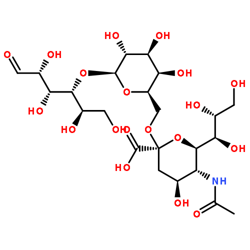


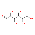
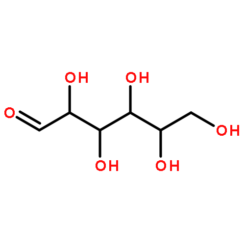

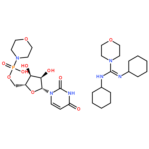


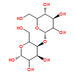
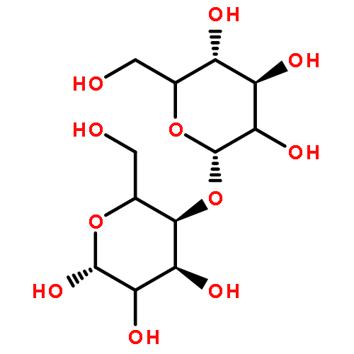


![Indeno[2,1-a]indene-1,3,6,8-tetrol,4b,5,9b,10-tetrahydro-5,10-bis(4-hydroxyphenyl)-, (4bR,5R,9bR,10R)-](http://img.cochemist.com/ccimg/105100/105037-88-5.png)
![Indeno[2,1-a]indene-1,3,6,8-tetrol,4b,5,9b,10-tetrahydro-5,10-bis(4-hydroxyphenyl)-, (4bR,5R,9bR,10R)-](http://img.cochemist.com/ccimg/105100/105037-88-5_b.png)


![Ethanone, 1-[2-hydroxy-4,6-bis(methoxymethoxy)phenyl]-](http://img.cochemist.com/ccimg/65500/65490-09-7.png)
![Ethanone, 1-[2-hydroxy-4,6-bis(methoxymethoxy)phenyl]-](http://img.cochemist.com/ccimg/65500/65490-09-7_b.png)
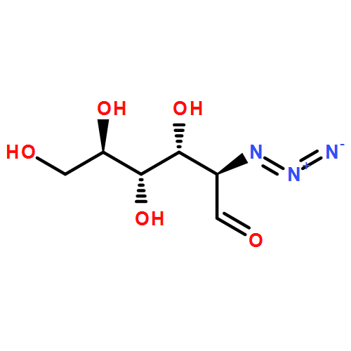
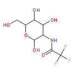
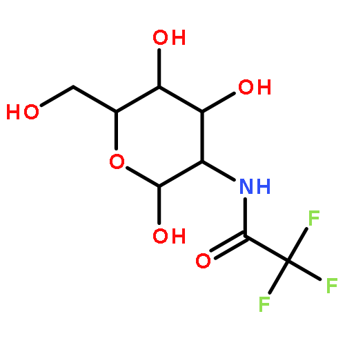
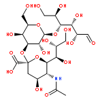
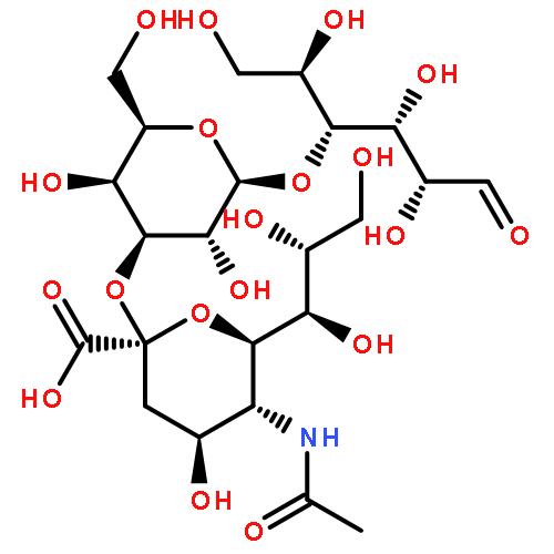


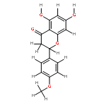
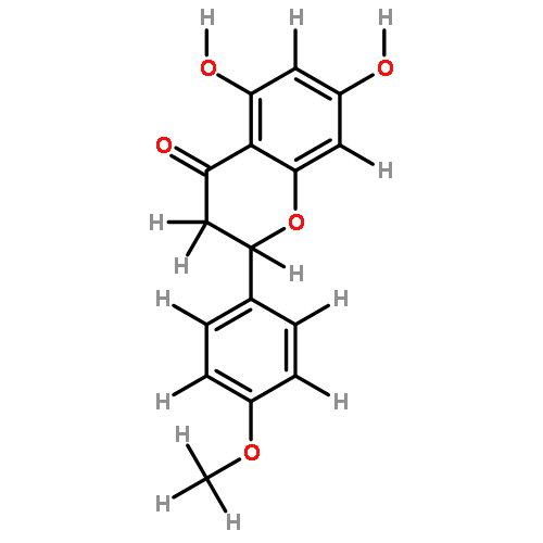
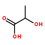


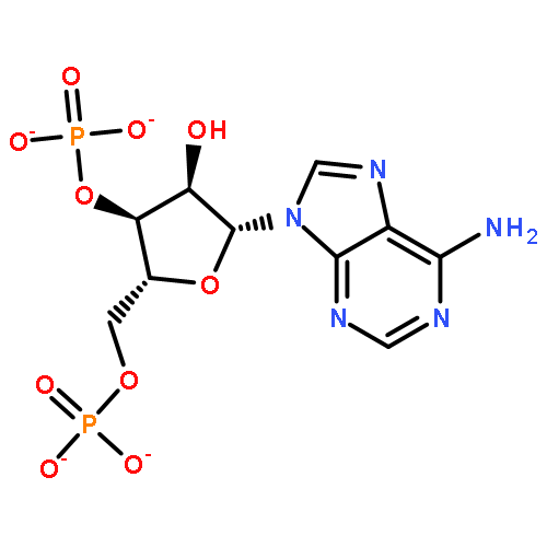


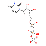
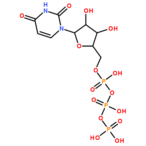


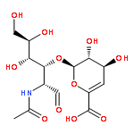


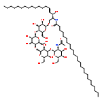
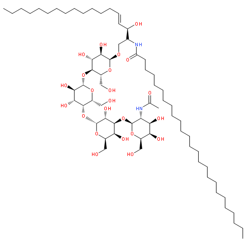


![(3Z)-3-[5-(5-HYDROXY-1H-INDOL-3-YL)-2-OXO-1H-PYRROL-3-YLIDENE]-1H-INDOL-2-ONE](http://img.cochemist.com/ccimg/600/548-54-9.png)
![(3Z)-3-[5-(5-HYDROXY-1H-INDOL-3-YL)-2-OXO-1H-PYRROL-3-YLIDENE]-1H-INDOL-2-ONE](http://img.cochemist.com/ccimg/600/548-54-9_b.png)
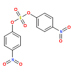
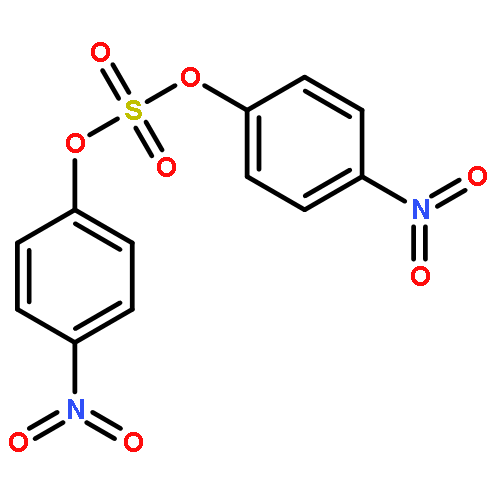


![5'-O-[hydroxy(sulfooxy)phosphoryl]adenosine 3'-(dihydrogen phosphate)](http://img.cochemist.com/ccimg/500/482-67-7.png)
![5'-O-[hydroxy(sulfooxy)phosphoryl]adenosine 3'-(dihydrogen phosphate)](http://img.cochemist.com/ccimg/500/482-67-7_b.png)