Co-reporter:Colin M. Rathbun and Jennifer A. Prescher
Biochemistry October 3, 2017 Volume 56(Issue 39) pp:5178-5178
Publication Date(Web):July 26, 2017
DOI:10.1021/acs.biochem.7b00435
Bioluminescence with luciferase–luciferin pairs is an attractive method for surveying cells in live tissues and whole organisms. Recent advances in luciferin chemistry and luciferase engineering are further expanding the scope of the technology. It is now possible to spy on cells in a variety of deep tissues and visualize multicellular interactions, feats that are enabling new questions to be asked and new ideas to be explored. This perspective piece highlights recent successes in bioluminescent probe development and their applications to imaging in live cells, tissues, and animals.
Co-reporter:R. David Row, Hui-Wen Shih, Austin T. Alexander, Ryan A. Mehl, and Jennifer A. Prescher
Journal of the American Chemical Society May 31, 2017 Volume 139(Issue 21) pp:7370-7370
Publication Date(Web):May 8, 2017
DOI:10.1021/jacs.7b03010
Cyclopropenones are attractive motifs for bioorthogonal chemistry, owing to their small size and unique modes of reactivity. Unfortunately, the fastest-reacting cyclopropenones are insufficiently stable for routine intracellular use. Here we report cyclopropenones with improved stability that maintain robust reactivity with bioorthogonal phosphines. Functionalized cyclopropenones were synthesized and their lifetimes in aqueous media and cellular environments were analyzed. The most robust cyclopropenones were further treated with a panel of phosphine probes, and reaction rates were measured. Two of the phosphine scaffolds afforded ∼100-fold rate enhancements compared to previously reported reagents. Importantly, the stabilized cyclopropenones were suitable for recombinant protein production via genetic code expansion. The products of the cyclopropenone ligation were also amenable to traceless Staudinger ligations, setting the stage for tandem labeling experiments.
Co-reporter:Colin M. Rathbun, William B. Porterfield, Krysten A. Jones, Marian J. Sagoe, Monique R. Reyes, Christine T. Hua, and Jennifer A. Prescher
ACS Central Science December 27, 2017 Volume 3(Issue 12) pp:1254-1254
Publication Date(Web):November 15, 2017
DOI:10.1021/acscentsci.7b00394
Bioluminescence imaging with luciferase enzymes and luciferin small molecules is a well-established technique for tracking cells and other biological features in rodent models. Despite its popularity, bioluminescence has long been hindered by a lack of distinguishable probes. Here we present a method to rapidly identify new substrate-selective luciferases for multicomponent imaging. Our strategy relies on parallel screening of luciferin analogues with panels of mutant enzymes. The compiled data set is then analyzed in silico to uncover mutually orthogonal sets. Using this approach, we screened 159 mutant enzymes with 12 luciferins. Thousands of orthogonal pairs were revealed with sufficient selectivity for use in biological environments. Over 100 pairs were validated in vitro, and three were applied in cell and animal models. The parallel screening method is both generalizable and scalable and will streamline the search for larger collections of orthogonal probes.
Co-reporter:Rachel C. Steinhardt;Jessica M. O'Neill;Colin M. Rathbun;Dr. David C. McCutcheon;Dr. Mira A. Paley;Dr. Jennifer A. Prescher
Chemistry - A European Journal 2016 Volume 22( Issue 11) pp:3671-3675
Publication Date(Web):
DOI:10.1002/chem.201503944
Abstract
Herein, the synthesis and characterization of an alkyne-modified luciferin is reported. This bioluminescent probe was accessed using C−H activation methodology and was found to be stable in solution and capable of light production with firefly luciferase. The luciferin analogue was also cell permeant and emitted more redshifted light than d-luciferin, the native luciferase substrate. Based on these features, the alkynyl luciferin will be useful for a variety of imaging applications.
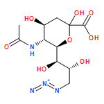
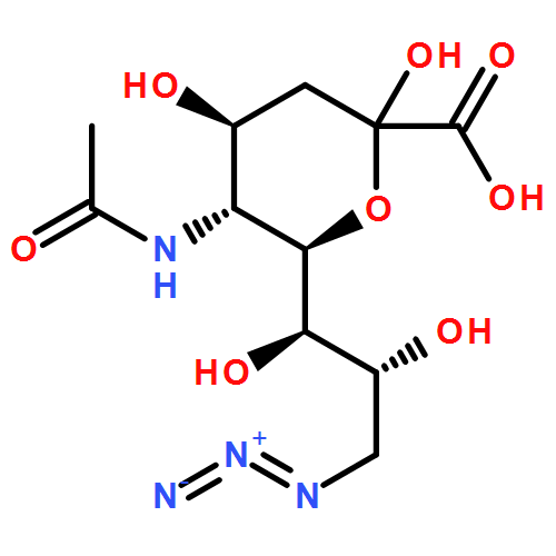
Co-reporter:David N. Kamber; Yong Liang; Robert J. Blizzard; Fang Liu; Ryan A. Mehl; K. N. Houk;Jennifer A. Prescher
Journal of the American Chemical Society 2015 Volume 137(Issue 26) pp:8388-8391
Publication Date(Web):June 18, 2015
DOI:10.1021/jacs.5b05100
A new class of bioorthogonal reagents, 1,2,4-triazines, is described. These scaffolds are stable in biological media and capable of robust reactivity with trans-cyclooctene (TCO). The enhanced stability of the triazine scaffold enabled its direct use in recombinant protein production. The triazine–TCO reaction can also be used in tandem with other bioorthogonal cycloaddition reactions. These features fill current voids in the bioorthogonal toolkit.
Co-reporter:William B. Porterfield; Krysten A. Jones; David C. McCutcheon;Jennifer A. Prescher
Journal of the American Chemical Society 2015 Volume 137(Issue 27) pp:8656-8659
Publication Date(Web):June 22, 2015
DOI:10.1021/jacs.5b02774
Cell–cell interactions underlie fundamental biological processes but remain difficult to visualize over long times and large distances in tissues and live organisms. Bioluminescence imaging with luciferase–luciferin pairs is sufficiently sensitive to image cells in vivo but lacks the spatial resolution to identify cellular locations and interactions. To repurpose this technology for visualizing cellular networks, we developed a “caged” luciferin that produces light only when cells are in close contact. This molecule comprises a nitroaromatic core that can be selectively reduced (“uncaged”) by one cell type, liberating a luciferin that can be selectively consumed by neighboring, luciferase-expressing cells. When the two cell types are in contact, robust light emission is observed. This imaging strategy will enable the noninvasive visualization of cell–cell interactions relevant to organismal biology.
Co-reporter:Hui-Wen Shih;Jennifer A. Prescher
Journal of the American Chemical Society 2015 Volume 137(Issue 32) pp:10036-10039
Publication Date(Web):August 7, 2015
DOI:10.1021/jacs.5b06969
Bioorthogonal chemistries have been widely used to probe biopolymers in living systems. To date, though, only a handful of broadly useful transformations have been identified because of the stringent requirements placed on the reactants. Here we report a novel bioorthogonal ligation between cyclopropenones and functionalized phosphines. These components are stable in physiological buffers and react rapidly with one another to form covalent adducts. The cyclopropenone ligation is also distinct from other bioorthogonal chemistries in that it makes use of readily accessible, commercially available reagents and proceeds via a nucleophilic reaction pathway. On the basis of these features, the cyclopropenone ligation is poised to join the ranks of chemistries with utility in living systems.
Co-reporter:William B Porterfield, Jennifer A Prescher
Current Opinion in Chemical Biology 2015 Volume 24() pp:121-130
Publication Date(Web):February 2015
DOI:10.1016/j.cbpa.2014.11.006
•New fluorescent proteins and small molecule fluorophores enable sensitive imaging of multicellular networks in vivo.•Split fluorescent proteins and enzymatic tagging strategies can illuminate direct cell-to-cell contacts.•Synthetic luciferins and engineered luciferases are expanding the scope of macroscopic bioluminescence imaging.•Caged probes can report on intercellular interactions in whole organisms.Cellular communication drives diverse aspects of organismal biology ranging from immune function to memory formation. The mechanisms by which cells transact information in vivo, though, are not completely understood. This is due, in part, to a lack of tools for observing collections of cells in their native habitats. New optical probes are being crafted to image networks of cell–cell interactions (i.e., ‘interactomes’) in tissues and live organisms. Examples of these probes — and their use in visualizing cell contacts and macroscopic cell networks — are highlighted.
Co-reporter:David M Patterson, Jennifer A Prescher
Current Opinion in Chemical Biology 2015 Volume 28() pp:141-149
Publication Date(Web):October 2015
DOI:10.1016/j.cbpa.2015.07.006
•Mutually orthogonal bioorthogonal reactions are described.•Current challenges in compatible reaction development are outlined.•Strategies to identify and generate orthogonal reactions are presented.•Recent applications of orthogonal bioorthogonal reactions in cellular imaging and macromolecular assembly are highlighted.Bioorthogonal reactions have long been used to examine individual biomolecules in living systems. Studies of multi-component networks demand not only reliable bioorthogonal chemistries, but also combinations of reactions that can be used in tandem. Such ‘orthogonal bioorthogonal’ transformations have been reported in recent years, and these chemistries are enabling new explorations into biology. This article highlights the development of orthogonal bioorthogonal reactions and their application in multi-target imaging and macromolecule assembly. Methods to tune and control orthogonal reactivity are also discussed, along with prospects for identifying new classes of compatible reactions.
Co-reporter:Krysten A. Jones, David J. Li, Elliot Hui, Mark A. Sellmyer, and Jennifer A. Prescher
ACS Chemical Biology 2015 Volume 10(Issue 4) pp:933
Publication Date(Web):February 2, 2015
DOI:10.1021/cb5007773
Cell–cell interactions underlie diverse physiological processes ranging from immune function to cell migration. Dysregulated cellular crosstalk also potentiates numerous pathologies, including infections and metastases. Despite their ubiquity in organismal biology, cell–cell interactions are difficult to examine in tissues and whole animals without invasive procedures. Here, we report a strategy to noninvasively image cell proximity using engineered bioluminescent probes. These tools comprise “split” fragments of Gaussia luciferase (Gluc) fused to the leucine zipper domains of Fos and Jun. When cells secreting the fragments draw near one another, Fos and Jun drive the assembly of functional, light-emitting Gluc. Photon production thus provides a readout on the distance between two cell types. We used the split fragments to visualize cell–cell interactions over time in vitro and in macroscopic models of cell migration. Further application of these tools in live organisms will refine our understanding of cell contacts relevant to basic biology and disease.
Co-reporter:David C. McCutcheon, William B. Porterfield and Jennifer A. Prescher
Organic & Biomolecular Chemistry 2015 vol. 13(Issue 7) pp:2117-2121
Publication Date(Web):17 Dec 2014
DOI:10.1039/C4OB02529F
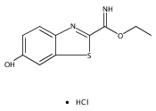
Bioluminescence imaging with luciferase–luciferin pairs is a popular method for visualizing biological processes in vivo. Unfortunately, most luciferins are difficult to access and remain prohibitively expensive for some imaging applications. Here we report cost-effective and efficient syntheses of D-luciferin and 6′-aminoluciferin, two widely used bioluminescent substrates. Our approach employs inexpensive anilines and Appel's salt to generate the luciferin cores in a single pot. Additionally, the syntheses are scalable and can provide multi-gram quantities of both substrates. The streamlined production and improved accessibility of luciferin reagents will bolster in vivo imaging efforts.
Co-reporter:Hui-Wen Shih, David N Kamber, Jennifer A Prescher
Current Opinion in Chemical Biology 2014 Volume 21() pp:103-111
Publication Date(Web):August 2014
DOI:10.1016/j.cbpa.2014.07.002
•Popular bioorthogonal reagents and reactions are highlighted.•Methods to tune bioorthogonal functional group reactivity and stability are described.•Strategies to achieve bioorthogonal reactivity ‘on demand’ are also presented.•Ongoing challenges in bioorthogonal reaction design and development are discussed.Over the past two decades, there has been intense interest in designing and implementing selective (bioorthogonal) reactions for biomolecule tracking. Here we review the most widely used bioorthogonal chemistries in live cells and animals, drawing particular attention to the unique functional groups underlying these transformations. We also describe recent efforts to tune functional group reactivities and stabilities to access even more rapid and selective chemistries. Last, we highlight ongoing challenges in identifying new bioorthogonal reagents and combinations of reactions that can be used concurrently to tag multiple biomolecules.
Co-reporter:David M. Patterson, Lidia A. Nazarova, and Jennifer A. Prescher
ACS Chemical Biology 2014 Volume 9(Issue 3) pp:592
Publication Date(Web):January 17, 2014
DOI:10.1021/cb400828a
Bioorthogonal chemistries can be used to tag diverse classes of biomolecules in cells and other complex environments. With over 20 unique transformations now available, though, selecting an appropriate reaction for a given experiment is challenging. In this article, we compare and contrast the most common classes of bioorthogonal chemistries and provide a framework for matching the reactions with downstream applications. We also discuss ongoing efforts to identify novel biocompatible reactions and methods to control their reactivity. The continued expansion of the bioorthogonal toolkit will provide new insights into biomolecule networks and functions and thus refine our understanding of living systems.
Co-reporter:David M. Patterson, Krysten A. Jones and Jennifer A. Prescher
Molecular BioSystems 2014 vol. 10(Issue 7) pp:1693-1697
Publication Date(Web):28 Feb 2014
DOI:10.1039/C4MB00092G
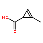
Cyclopropenes have emerged as a new class of bioorthogonal chemical reporters. These strained rings can be metabolically introduced into target biomolecules and covalently modified via mild cycloaddition chemistries. While versatile, existing cyclopropene scaffolds are inefficient reporters of protein glycosylation, owing to their branched structures and sluggish rates of reactivity. Here we describe a set of cyclopropenes for the robust detection of glycans on cell surfaces and isolated proteins. These scaffolds comprise carbamate linkages that are compatible with cellular biosynthetic pathways and exhibit rapid cycloaddition rates. Furthermore, these probes can be used in tandem with other classic bioorthogonal motifs—including azides and alkynes—to examine multiple biomolecules in tandem.
Co-reporter:Miranda A. Paley and Jennifer A. Prescher
MedChemComm 2014 vol. 5(Issue 3) pp:255-267
Publication Date(Web):13 Dec 2013
DOI:10.1039/C3MD00288H
Bioluminescence is a ubiquitous imaging modality for visualizing biological processes in vivo. This technique employs visible light and interfaces readily with most cell and tissue types, making it a versatile technology for preclinical studies. Here we review basic bioluminescence imaging principles, along with applications of the technology that are relevant to the medicinal chemistry community. These include noninvasive cell tracking experiments, analyses of protein function, and methods to visualize small molecule metabolites. In each section, we also discuss how bioluminescent tools have revealed insights into experimental therapies and aided drug discovery. Last, we highlight the development of new bioluminescent tools that will enable more sensitive and multi-component imaging experiments and, thus, expand our broader understanding of living systems.
Co-reporter:David N. Kamber ; Lidia A. Nazarova ; Yong Liang ; Steven A. Lopez ; David M. Patterson ; Hui-Wen Shih ; K. N. Houk ;Jennifer A. Prescher
Journal of the American Chemical Society 2013 Volume 135(Issue 37) pp:13680-13683
Publication Date(Web):September 3, 2013
DOI:10.1021/ja407737d
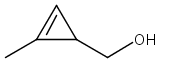
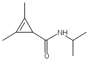
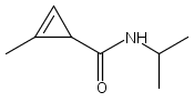
Bioorthogonal chemistries have provided tremendous insight into biomolecule structure and function. However, many popular bioorthogonal transformations are incompatible with one another, limiting their utility for studies of multiple biomolecules in tandem. We identified two reactions that can be used concurrently to tag biomolecules in complex environments: the inverse electron-demand Diels–Alder reaction of tetrazines with 1,3-disubstituted cyclopropenes, and the 1,3-dipolar cycloaddition of nitrile imines with 3,3-disubstituted cyclopropenes. Remarkably, the cyclopropenes used in these transformations differ by the placement of a single methyl group. Such orthogonally reactive scaffolds will bolster efforts to monitor multicomponent processes in cells and organisms.
Co-reporter:David C. McCutcheon ; Miranda A. Paley ; Rachel C. Steinhardt ;Jennifer A. Prescher
Journal of the American Chemical Society 2012 Volume 134(Issue 18) pp:7604-7607
Publication Date(Web):April 20, 2012
DOI:10.1021/ja301493d
Bioluminescence imaging with luciferase enzymes requires access to light-emitting, small-molecule luciferins. Here, we describe a rapid method to synthesize d-luciferin, the substrate for firefly luciferase (Fluc), along with a novel set of electronically modified analogues. Our procedure utilizes a relatively rare, but synthetically useful dithiazolium reagent to generate heteroaromatic scaffolds in a divergent fashion. Two of the luciferin analogues produced with this approach emit light with Fluc in vitro and in live cells. Collectively, our work increases the number of substrates that can be used for bioluminescence imaging and provides a general strategy for synthesizing new collections of luciferins.
Co-reporter:David M. Patterson ; Lidia A. Nazarova ; Bryan Xie ; David N. Kamber ;Jennifer A. Prescher
Journal of the American Chemical Society 2012 Volume 134(Issue 45) pp:18638-18643
Publication Date(Web):October 16, 2012
DOI:10.1021/ja3060436
Chemical reporters are unique functional groups that can be used to label biomolecules in living systems. Only a handful of broadly applicable reporters have been identified to date, owing to the rigorous demands placed on these functional groups in biological settings. We describe here a new chemical reporter—cyclopropene—that can be used to target biomolecules in vitro and in live cells. A variety of substituted cyclopropene scaffolds were synthesized and found to be stable in aqueous solution and in the presence of biological nucleophiles. Furthermore, some of the cyclopropene units were metabolically introduced into cell surface glycans and subsequently detected with covalent probes. The small size and selective reactivity of cyclopropenes will facilitate efforts to tag diverse collections of biomolecules in vivo.
Co-reporter:Lidia A. Nazarova, Roxanna J. Ochoa, Krysten A. Jones, Naomi S. Morrissette, Jennifer A. Prescher
Microbes and Infection (March 2016) Volume 18(Issue 3) pp:199-210
Publication Date(Web):1 March 2016
DOI:10.1016/j.micinf.2015.11.004
Toxoplasma gondii is an obligate intracellular parasite that infects all nucleated cell types in diverse warm-blooded organisms. Many of the surface antigens and effector molecules secreted by the parasite during invasion and intracellular growth are modified by glycans. Glycosylated proteins in the nucleus and cytoplasm have also been reported. Despite their prevalence, the complete inventory and biological significance of glycosylated proteins in Toxoplasma remain unknown. In this study, we aimed to globally profile parasite glycoproteins using a bioorthogonal chemical reporter strategy. This strategy involves the metabolic incorporation of unnatural functional groups (i.e., “chemical reporters”) into Toxoplasma glycans, followed by covalent labeling with visual probes or affinity tags. The two-step approach enables the visualization and identification of newly biosynthesized glycoconjugates in the parasite. Using a buffer that mimics intracellular conditions, extracellular Toxoplasma tachyzoites were found to metabolize and incorporate unnatural sugars (equipped with bioorthogonal functional groups) into diverse proteins. Covalent chemistries were used to visualize and retrieve these labeled structures. Subsequent mass spectrometry analysis revealed 89 unique proteins. This survey identified novel proteins as well as previously characterized proteins from lectin affinity analyses.
Co-reporter:David C. McCutcheon, William B. Porterfield and Jennifer A. Prescher
Organic & Biomolecular Chemistry 2015 - vol. 13(Issue 7) pp:NaN2121-2121
Publication Date(Web):2014/12/17
DOI:10.1039/C4OB02529F
Bioluminescence imaging with luciferase–luciferin pairs is a popular method for visualizing biological processes in vivo. Unfortunately, most luciferins are difficult to access and remain prohibitively expensive for some imaging applications. Here we report cost-effective and efficient syntheses of D-luciferin and 6′-aminoluciferin, two widely used bioluminescent substrates. Our approach employs inexpensive anilines and Appel's salt to generate the luciferin cores in a single pot. Additionally, the syntheses are scalable and can provide multi-gram quantities of both substrates. The streamlined production and improved accessibility of luciferin reagents will bolster in vivo imaging efforts.
.jpg)


![4,8-Dioxaspiro[2.5]oct-1-ene, 6,6-dimethyl-1-octyl-](http://img.cochemist.com/ccimg/453000/452922-89-3.png)
![4,8-Dioxaspiro[2.5]oct-1-ene, 6,6-dimethyl-1-octyl-](http://img.cochemist.com/ccimg/453000/452922-89-3_b.png)


![4,8-Dioxaspiro[2.5]oct-1-ene, 6,6-dimethyl-](http://img.cochemist.com/ccimg/61000/60935-22-0.png)
![4,8-Dioxaspiro[2.5]oct-1-ene, 6,6-dimethyl-](http://img.cochemist.com/ccimg/61000/60935-22-0_b.png)


![6-Nitrobenzo[d]thiazole-2-carbonitrile](http://img.cochemist.com/ccimg/188700/188672-83-5.png)
![6-Nitrobenzo[d]thiazole-2-carbonitrile](http://img.cochemist.com/ccimg/188700/188672-83-5_b.png)


![Dibenz[b,f]azocine, 11,12-didehydro-5,6-dihydro-](/data/chemimg/174600/1369862-03-2.png)
![Dibenz[b,f]azocine, 11,12-didehydro-5,6-dihydro-](/data/chemimg/174600/1369862-03-2_b.png)


![METHYL 2-(BENZYLOXY)-5-[(1R)-2-BROMO-1-HYDROXYETHYL]BENZOATE](http://img.cochemist.com/ccimg/75400/75318-49-9.png)
![METHYL 2-(BENZYLOXY)-5-[(1R)-2-BROMO-1-HYDROXYETHYL]BENZOATE](http://img.cochemist.com/ccimg/75400/75318-49-9_b.png)


![(S)-2-(6-Hydroxybenzo[d]thiazol-2-yl)-4,5-dihydrothiazole-4-carboxylic acid](/data/chemimg/473800/2591-17-5.png)
![(S)-2-(6-Hydroxybenzo[d]thiazol-2-yl)-4,5-dihydrothiazole-4-carboxylic acid](/data/chemimg/473800/2591-17-5_b.png)