Co-reporter:Fei Peng, Yinjue Wang, Lijing Sun, Yongdong Liu, Tao Hu, Guifeng Zhang, Guanghui Ma, and Zhiguo Su
Bioconjugate Chemistry 2012 Volume 23(Issue 9) pp:1812
Publication Date(Web):August 9, 2012
DOI:10.1021/bc300081f
Conventional protein PEGylation is carried out in aqueous solution. However, some hydrophobic proteins seem to be stable in organic solution. In this study, a novel approach of PEGylating IFN-β-1b in an organic solution of 2-butanol (2-BuOH) was investigated. Compared with protein PEGylation in aqueous solution, the overall modification yields increased more than 37%, while the yield of mono-PEGylated products could be increased by 36%. Furthermore, the PEGylated IFN-β-1b, which was obtained in organic solution, demonstrated 18% more antiviral potency than those derived from aqueous solution. The PEGylation step could be directly connected to the previous protein separation step for process integration. Dynamic light scattering (DLS) and atomic force microscope (AFM) analysis revealed that IFN-β-1b formed aggregates both in water and in 2-BuOH solutions. However, the aggregates were much smaller and more homogeneous in 2-BuOH than those in aqueous solution, thereby providing larger solvent accessible protein surfaces, which resulted in a more productive PEGylation process. In addition, the results of circular dichroism (CD), fluorescence spectra, and peptide mapping suggested that the increased bioactivity came from the difference in PEGylation site distribution due to solution environment that induced conformational discrepancy. The results of this study show that PEGylation of IFN-β-1b in organic solution is a facile and efficient process, which might find applications for other hydrophobic proteins.
Co-reporter:Yin-Jue Wang, Su-Juan Hao, Yong-Dong Liu, Tao Hu, Gui-Feng Zhang, Xuan Zhang, Qing-Sheng Qi, Guang-Hui Ma, Zhi-Guo Su
Journal of Controlled Release 2010 Volume 145(Issue 3) pp:306-313
Publication Date(Web):3 August 2010
DOI:10.1016/j.jconrel.2010.04.021
Recombinant human non-glycosylated erythropoietin (rh-ngEpo) expressed in E. coli was attached to polyethylene glycol (PEG) chains with different sizes and structures. The pharmacokinetic properties and in vivo potency of the PEGylated protein were investigated and comparisons were drawn between the conjugates and glycosylated recombinant Epo (rhEpo).The rh-ngEpo was modified with linear PEG-aldehyde (PEG-ALD, 20 kDa, 30 kDa, and 40 kDa) and a branched N-hydroxysuccinimide activated PEG (PEG2-NHS, 40 kDa). The monoPEGylated proteins were isolated by ion-exchange chromatography. The purified monoPEGylated conjugates suffered 6.5–86.1% loss of in vitro bioactivity compared to the unmodified rh-ngEpo. In addition, PEGylation remarkably increased the resistance of rh-ngEpo against plasma degradation. Pharmacokinetic studies showed that the plasma half-life of rh-ngEpo was increased 9.7–17.4 times by PEGylation, with the two 40k-PEG–rh-ngEpos-treated groups exhibiting better pharmacokinetic performances than rhEpo. Moreover, all the conjugates resulted in markedly enhanced Ret% (the percentage of reticulocyte count in red blood cells) compared with rh-ngEpo after subcutaneous injection. The two 40k-PEG conjugates demonstrated comparable in vivo efficacies compared with rhEpo. Overall, this research provides opportunities for the development of more cost-effective erythropoiesis-stimulating protein drugs.
Co-reporter:Fangwei Wang;Guanghui Ma
Applied Biochemistry and Biotechnology 2009 Volume 159( Issue 3) pp:
Publication Date(Web):2009 December
DOI:10.1007/s12010-008-8495-6
Efficient refolding of recombinant proteins in the forms of inclusion bodies at higher concentration remains challenging. Here, we report a strategy of a dual-gradient hydrophobic interaction chromatography (HIC) mode to refold recombinant human granulocyte colony-stimulating factor from its inclusion bodies at high protein concentration. The strategy was taken to meet the demand of dynamic refolding proceeding by gradually decrease the denaturant (guanidine-HCl) concentration and gradually increase the hydrophilicity of media (column of Poros PE 20) with glycerol as additive to provide a mild refolding surroundings. Compared with dilution method, this dual-gradient HIC process gave about 8.5-fold of increase in specific activity and 30% increase in soluble protein recovery. Furthermore, much higher protein concentration could be obtained at the same time.
Co-reporter:Fei Peng, Yongdong Liu, Xiunan Li, Lijing Sun, Dawei Zhao, Qingqing Wang, Guanghui Ma, Zhiguo Su
Journal of Biotechnology (20 January 2014) Volume 170() pp:42-49
Publication Date(Web):20 January 2014
DOI:10.1016/j.jbiotec.2013.10.037
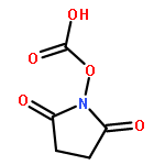
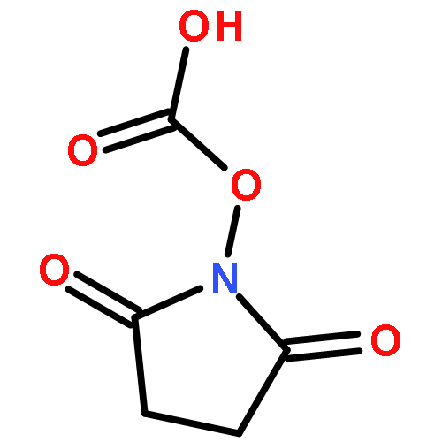
Previous studies demonstrated that hydrophobic proteins could be PEGylated in organic phase rather than water phase. It is still not known what the difference is for a hydrophilic protein's PEGylation in these two different phases. In this study, granulocyte colony stimulating factor (G-CSF) was dissolved in neat dimethyl sulfoxide (DMSO) and was PEGylated. In comparison with the PEGylation in water solution, the PEGylation degree in the organic solvent increased by 33% and 42% for PEG-maleimide (MAL-PEG) and PEG-succinimidyl carbonate (SC-PEG) respectively. Structure analysis revealed that the protein was unfolded in DMSO, which could make the PEGylated sites of G-CSF easily accessible. The hydrolysis half-life in water solution was 40 min and 9 h for SC-PEG and MAL-PEG respectively. However, in DMSO solvent, PEGs were very stable and no hydrolysis could be detected. Stopped-flow demonstrated that the conjugation speed of G-CSF by MAL-PEG and SC-PEG in DMSO were 1.6 × 10⁴ and 2 × 10² times faster than those in aqueous solution. The remarkable acceleration could mainly be attributed to an increase of protein nucleophilicity in DMSO. The results of this study could be referential to industrial application where the cost of PEG reagents and the speed of reaction on large scale are very important.