Co-reporter:Jiangtao Lei;Ruxi Qi;Luogang Xie;Wenhui Xi
RSC Advances (2011-Present) 2017 vol. 7(Issue 23) pp:13947-13956
Publication Date(Web):2017/02/28
DOI:10.1039/C6RA27608C
Increasing numbers of experiments show that carbon nanoparticles can significantly influence protein fibrillation, a process associated with many human amyloidosis. Our previous studies demonstrate that fullerenes and carbon nanotubes can markedly inhibit the formation of β-sheet-rich oligomers and amyloid fibrils of the hydrophobic KLVFFAE peptide, the central hydrophobic core fragment of Alzheimer's amyloid-β protein, mostly through hydrophobic and aromatic-stacking interactions. In this study, the effect of hydrophobic fullerene C60 on the oligomeric structures of the hydrophilic GNNQQNY peptide, the key amyloid-forming fragment of yeast prion protein Sup35, is investigated by extensive replica exchange molecular dynamics simulations. Our simulations, starting from eight random chains, show that fullerene can greatly impede the β-sheet formation of the GNNQQNY peptide and induce oligomer shifting from various β-sheet-rich structures to disordered coil conformations. Strikingly, fullerenes completely prevent fibril-like bilayer β-sheets, formed by GNNQQNY peptides alone. We find that C60 molecules interact strongly with the nonpolar aliphatic groups of polar residues N3, Q4 and Q5 and increase the water exposure of the peptide backbone, thus block the inter-peptide N3–Q4, Q4–Q4 and Q4–Q5 interactions which are crucial for oligomerization and β-sheet formation. These results, together with our previous studies on the KLVFFAE peptide, suggest that hydrophobic fullerenes could inhibit the aggregation of both hydrophobic and hydrophilic peptides. This finding may offer new clues for the design of therapeutic agents against different types of amyloidosis.
Co-reporter:Guanghong Wei, Wenhui Xi, Ruth Nussinov, and Buyong Ma
Chemical Reviews 2016 Volume 116(Issue 11) pp:6516
Publication Date(Web):January 25, 2016
DOI:10.1021/acs.chemrev.5b00562
All soluble proteins populate conformational ensembles that together constitute the native state. Their fluctuations in water are intrinsic thermodynamic phenomena, and the distributions of the states on the energy landscape are determined by statistical thermodynamics; however, they are optimized to perform their biological functions. In this review we briefly describe advances in free energy landscape studies of protein conformational ensembles. Experimental (nuclear magnetic resonance, small-angle X-ray scattering, single-molecule spectroscopy, and cryo-electron microscopy) and computational (replica-exchange molecular dynamics, metadynamics, and Markov state models) approaches have made great progress in recent years. These address the challenging characterization of the highly flexible and heterogeneous protein ensembles. We focus on structural aspects of protein conformational distributions, from collective motions of single- and multi-domain proteins, intrinsically disordered proteins, to multiprotein complexes. Importantly, we highlight recent studies that illustrate functional adjustment of protein conformational ensembles in the crowded cellular environment. We center on the role of the ensemble in recognition of small- and macro-molecules (protein and RNA/DNA) and emphasize emerging concepts of protein dynamics in enzyme catalysis. Overall, protein ensembles link fundamental physicochemical principles and protein behavior and the cellular network and its regulation.
Co-reporter:Yunxiang Sun, Zhenyu Qian and Guanghong Wei
Physical Chemistry Chemical Physics 2016 vol. 18(Issue 18) pp:12582-12591
Publication Date(Web):31 Mar 2016
DOI:10.1039/C6CP01014H
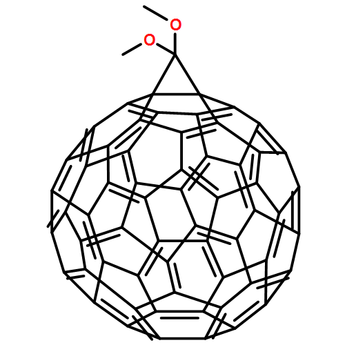
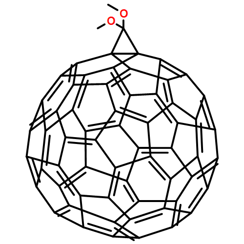
Alzheimer's disease (AD) is associated with the pathological self-assembly of amyloid-β (Aβ) peptides into β-sheet enriched fibrillar aggregates. Aβ dimers formed in the initial step of Aβ aggregation were reported to be the smallest toxic species. Inhibiting the formation of β-sheet-rich oligomers and fibrils is considered as the primary therapeutic strategy for AD. Previous studies reported that fullerene derivatives strongly inhibit Aβ fibrillation. However, the underlying inhibitory mechanism remains elusive. As a first step to understand fullerene-modulated full-length Aβ aggregation, we investigated the conformational ensemble of the Aβ1–42 dimer with and without 1,2-(dimethoxymethano)fullerene (DMF) – a more water-soluble fullerene derivative – by performing a 340 ns explicit-solvent replica exchange molecular dynamics simulation. Our simulations show that although disordered states are the most abundant conformations of the Aβ1–42 dimer, conformations containing diverse extended β-hairpins are also populated. The first most-populated β-hairpins involving residues L17–D23 and A30–V36 strongly resemble the engineered β-hairpin which is a building block of toxic Aβ oligomers. We find that the interaction of DMFs with Aβ peptides greatly impedes the formation of such β-hairpins and inter-peptide β-sheets. Binding energy analyses demonstrate that DMF preferentially binds not only to the central hydrophobic motif LVFFA of the Aβ peptide as suggested experimentally, but also to the aromatic residues including F4 and Y10 and the C-terminal hydrophobic region I31–V40. This study reveals a complete picture of the inhibitory mechanism of full-length Aβ1–42 aggregation by fullerenes, providing theoretical insights into the development of drug candidates against AD.
Co-reporter:Jiangtao Lei, Ruxi Qi, Guanghong Wei, Ruth Nussinov and Buyong Ma
Physical Chemistry Chemical Physics 2016 vol. 18(Issue 11) pp:8098-8107
Publication Date(Web):11 Feb 2016
DOI:10.1039/C5CP06538K
Recent studies suggested that p53 aggregation can lead to loss-of-function (LoF), dominant-negative (DN) and gain-of-function (GoF) effects, with adverse cancer consequences. The p53 aggregation-nucleating 251ILTIITL257 fragment is a key segment in wild-type p53 aggregation; however, an I254R mutation can prevent it. It was suggested that self-assembly of wild-type p53 and its cross-interaction with mutants differ from the classical amyloid nucleation-growth mechanism. Here, using replica exchange molecular dynamics (REMD) simulations, we studied the cross-interactions of this p53 core fragment and its aggregation rescue I254R mutant. We found that the core fragment displays strong aggregation propensity, whereas the gatekeeper I254R mutant tends to be disordered, consistent with experiments. Our cross-interaction results reveal that the wild-type p53 fragment promotes β-sheet formation of the I254R mutant by shifting the disordered mutant peptides into aggregating states. As a result, the system has similar oligomeric structures, inter-peptide interactions and free energy landscape as the wild type fragment does, revealing a prion-like process. We also found that in the cross-interaction system, the wild-type species has higher tendency to interact with the mutant than with itself. This phenomenon illustrates synergistic effects between the p53 251ILTIITL257 fragment and the mutant resembling prion cross-species propagation, cautioning against exploiting it in drug discovery.
Co-reporter:Cong Guo, Zohar A. Arnon, Ruxi Qi, Qingwen Zhang, Lihi Adler-Abramovich, Ehud Gazit, and Guanghong Wei
ACS Nano 2016 Volume 10(Issue 9) pp:8316
Publication Date(Web):August 22, 2016
DOI:10.1021/acsnano.6b02739
Molecular self-assembly is pivotal for the formation of ordered nanostructures, yet the structural diversity obtained by the use of a single type of building block is limited. Multicomponent coassembly, utilized to expand the architectural space, is principally based on empirical observations rather than rational design. Here we report large-scale molecular dynamics simulations of the coassembly of diphenylalanine (FF) and triphenylalanine (FFF) peptides at various mass ratios. Our simulations show that FF and FFF can co-organize into both canonical and noncanonical assemblies. Strikingly, toroid nanostructures, which were rarely observed for the extensively studied FF or FFF, are often seen in the FF-FFF coassembly simulations and later corroborated by scanning electron microscopy. Our simulations demonstrate a wide ratio-dependent variation of nanostructure morphologies including hollow and solid assemblies, much richer than those formed by each individual moiety. The hollow-solid structural transformation displays a discontinuous transition feature, and the toroids appear to be an obligatory intermediate for the structural transition. Interaction analysis reveals that the hollow-solid structural transition is mostly dominated by FF–FFF interactions, while the nanotoroid formation is determined by the competition between FF–water and FFF–water interactions. This study provides both structural and mechanistic insights into the coassembly of FF and FFF peptides, thus offering a molecular basis for the rational design of bionanomaterials utilizing peptide coassembly.Keywords: coassembly mechanism; controllable coassembly; diphenylalanine; geometry map; nanostructural diversity; toroid nanostructure; triphenylalanine
Co-reporter:Lin Niu, Lei Liu, Wenhui Xi, Qiusen Han, Qiang Li, Yue Yu, Qunxing Huang, Fuyang Qu, Meng Xu, Yibao Li, Huiwen Du, Rong Yang, Jacob Cramer, Kurt V. Gothelf, Mingdong Dong, Flemming Besenbacher, Qingdao Zeng, Chen Wang, Guanghong Wei, and Yanlian Yang
ACS Nano 2016 Volume 10(Issue 4) pp:4143
Publication Date(Web):March 16, 2016
DOI:10.1021/acsnano.5b07396
Inhibition of amyloid aggregation is important for developing potential therapeutic strategies of amyloid-related diseases. Herein, we report that the inhibition effect of a pristine peptide motif (KLVFF) can be significantly improved by introducing a terminal regulatory moiety (terpyridine). The molecular-level observations by using scanning tunneling microscopy reveal stoichiometry-dependent polymorphism of the coassembly structures, which originates from the terminal interactions of peptide with organic modulator moieties and can be attributed to the secondary structures of peptides and conformations of the organic molecules. Furthermore, the polymorphism of the peptide−organic coassemblies is shown to be correlated to distinctively different inhibition effects on amyloid-β 42 (Aβ42) aggregations and cytotoxicity.Keywords: amyloid cytotoxicity; amyloid β (Aβ) peptide; inhibitory effect; peptide aggregation; peptide motif; polymorphism effect; scanning tunneling microscopy
Co-reporter:Ruxi Qi; Yin Luo; Guanghong Wei; Ruth Nussinov;Buyong Ma
The Journal of Physical Chemistry Letters 2015 Volume 6(Issue 16) pp:3276-3282
Publication Date(Web):August 6, 2015
DOI:10.1021/acs.jpclett.5b01447
There are synergistic effects of Aβ and tau protein in Alzheimer’s disease. Aβ1–42 protofibril seeds induce conversion of human tau protein into β-sheet-rich toxic tau oligomers. However, the molecular mechanisms underlying such a conformational conversion are unclear. Here, we use extensive all atom replica exchange molecular dynamics simulations to investigate the effects of preformed Aβ1–42 protofibril on two monomeric tau constructs: K18 and K19. We found that Aβ oligomer stretches tau conformation and drastically reduces the metastable secondary structures/hydrogen bonding/salt-bridge networks in tau monomers and exposes their fibril nucleating motifs 275VQIINK280 and 306VQIVYK311. Aβ interacting patches around Tyr10/Ile41 contribute significantly to the interactions with K18 and K19. Aβ cross-seeded tau aggregation can adopt a “stretching-and-packing” mechanism, paving the way for the next, prion-like growth step. The results provide a mechanism on the atomic level to experimental observations that tau pathogenesis is promoted by Aβ1–42 but not by Aβ1–40.
Co-reporter:Yunxiang Sun, Wenhui Xi, and Guanghong Wei
The Journal of Physical Chemistry B 2015 Volume 119(Issue 7) pp:2786-2794
Publication Date(Web):January 22, 2015
DOI:10.1021/jp508122t
Alzheimer’s disease (AD) is associated with the aggregation of amyloid-β (Aβ) peptides into toxic prefibrillar aggregates. Recent experiments reported that small organic molecule O4 decreases the concentration of toxic oligomers by promoting fibrillation and thus reduces Aβ toxicity. However, the atomic-level details of O4–Aβ-oligomer interaction are largely unknown. In this work, we studied the structural stability of the fibrillike Aβ(17–42) trimer by performing atomistic molecular dynamics simulations of 1.5 μs in total on the trimer with and without O4. We found that the Aβ(17–42) trimer is unstable without O4, whereas its structural stability is greatly enhanced with O4. Four binding sites were found around residues F20, S26, and M35, namely the central hydrophobic core (CHC) site, the turn site, and two hydrophobic-groove sites. The two hydrophobic grooves near M35 facilitate O4 to bind through hydrophobic interaction and geometry match. The binding of O4 at the CHC site is mostly stabilized by hydrophobic and π–π stacking interactions. Hydrogen-bonding interaction between O4 and S26 plays a role in the binding of O4 to the turn site. Our work reveals the detailed stabilization mechanism of protofibrillar Aβ oligomers by O4 and may provide novel insight into the development of drug candidates against AD.
Co-reporter:Yunxiang Sun, Zhenyu Qian, Cong Guo, and Guanghong Wei
Biomacromolecules 2015 Volume 16(Issue 9) pp:
Publication Date(Web):August 24, 2015
DOI:10.1021/acs.biomac.5b00850
Amphiphilic peptides can self-assemble into ordered nanostructures with different morphologies. However, the assembly mechanism and the structures of the early assemblies prior to nanostructure formation remain elusive. In this study, we investigated the oligomeric structures of two amphiphilic heptapeptides A6K and V6K by all-atom explicit-solvent replica-exchange molecular dynamics (REMD) simulations, and then examined the assembly dynamics of large aggregates by coarse-grained (CG) MD simulations. Our 200 ns REMD simulations show that A6K peptides predominantly adopt loosely packed disordered coil aggregates, whereas V6K peptides mostly assemble into compact β-sheet-rich conformations, consistent with the signal measured experimentally in aqueous solution. Well-organized β-sheet-rich conformations, albeit with low population, are also populated for V6K octamers, including bilayer β-sheets and β-barrels. These ordered β-sheet-rich conformations are observed for the first time for amphiphilic peptides. Our 10-μs CG-MD simulations on 200 peptide chains demonstrate that A6K and V6K peptides follow two different self-assembly processes, and the former form monolayer lamellas while the latter assemble into plate-like assemblies. CG-MD simulations also show that V6K peptides display higher assembly capability than A6K, in support of our all-atom REMD simulation results. Interpeptide interaction analyses reveal that the marked differences in oligomeric structures and assembly dynamics between A6K and V6K result from the subtle interplay of competition among hydrophobic, hydrogen-bonding, and electrostatic interactions of the two peptides. Our study provides structural and mechanistic insights into the initial self-assembly process of A6K and V6K at the molecular level.
Co-reporter:Cong Guo, Sébastien Côté, Normand Mousseau, and Guanghong Wei
The Journal of Physical Chemistry B 2015 Volume 119(Issue 8) pp:3366-3376
Publication Date(Web):February 3, 2015
DOI:10.1021/jp5111357
Islet amyloid polypeptide, IAPP or amylin, is a 37-residue peptide hormone coexpressed with insulin by pancreatic β-cells. The aggregation of human IAPP (hIAPP) into amyloid deposits is associated with type II diabetes. Substantial evidence suggests that the interaction of anionic membranes with hIAPP may facilitate peptide aggregation and the N-terminal 1–19 fragment (IAPP1–19) plays an important role in peptide–membrane interaction. As a first step to understand how structural differences between human and rat IAPP peptides in membranes may influence the later oligomerization process, we have investigated the structures and orientations of hIAPP1–19 and the less toxic rIAPP1–19 (i.e., the H18R mutant of hIAPP1–19) monomers in anionic POPG bilayers by performing replica exchange molecular dynamics (REMD) simulations. On the basis of ∼20 μs REMD simulations started from a random coil conformation of the peptide placed in water, we find that unfolded h(r)IAPP1–19 can insert into the bilayers and the membrane-bound peptide stays mainly at the lipid head–tail interface. hIAPP1–19 displays higher helix propensity than rIAPP1–19, especially in the L12–L16 region. The helix is oriented parallel to the bilayer surface and buried in the membrane 0.3–0.8 nm below the phosphorus atoms, consistent with previous electron paramagnetic resonance data. The helical conformation is an amphiphilic helix with its hydrophilic and hydrophobic faces pointing, respectively, to the lipid head and tail regions. The H18R substitution enhances the electrostatic interactions of IAPP1–19 with the membrane, while it weakens the intrapeptide interactions crucial for helix formation, thus leading to lower helix propensity of rIAPP1–19. Implications of our simulation results on the membrane-mediated IAPP1–19 oligomerization are discussed.
Co-reporter:Bin Dai;Dan Li;Wenhui Xi;Fang Luo;Xiang Zhang;Man Zou;Mi Cao;Jun Hu;Wenyuan Wang;Yi Zhang;Cong Liu
PNAS 2015 112 (10 ) pp:2996-3001
Publication Date(Web):2015-03-10
DOI:10.1073/pnas.1416690112
Using and engineering amyloid as nanomaterials are blossoming trends in bionanotechnology. Here, we show our discovery of
an amyloid structure, termed “amyloid-like nanosheet,” formed by a key amyloid-forming segment of Alzheimer’s Aβ. Combining
multiple biophysical and computational approaches, we proposed a structural model for the nanosheet that is formed by stacking
the amyloid fibril spines perpendicular to the fibril axis. We further used the nanosheet for laboratorial retroviral transduction
enhancement and directly visualized the presence of virus on the nanosheet surface by electron microscopy. Furthermore, based
on our structural model, we designed nanosheet-forming peptides with different functionalities, elucidating the potential
of rational design for amyloid-based materials with novel architecture and function.
Co-reporter:Luogang Xie, Yin Luo, Dongdong Lin, Wenhui Xi, Xinju Yang and Guanghong Wei
Nanoscale 2014 vol. 6(Issue 16) pp:9752-9762
Publication Date(Web):04 Jun 2014
DOI:10.1039/C4NR01005A
Amyloid deposits are implicated in the pathogenesis of many neurodegenerative diseases such as Alzheimer's disease (AD). The inhibition of β-sheet formation has been considered as the primary therapeutic strategy for AD. Increasing data show that nanoparticles can retard or promote the fibrillation of amyloid-β (Aβ) peptides depending on the physicochemical properties of nanoparticles, however, the underlying molecular mechanism remains elusive. In this study, our replica exchange molecular dynamics (REMD) simulations show that fullerene nanoparticle – C60 (with a fullerene: peptide molar ratio greater than 1:8) can dramatically prevent β-sheet formation of Aβ(16–22) peptides. Atomic force microscopy (AFM) experiments further confirm the inhibitory effect of C60 on Aβ(16–22) fibrillation, in support of our REMD simulations. An important finding from our REMD simulations is that fullerene C180, albeit with the same number of carbon atoms as three C60 molecules (3C60) and smaller surface area than 3C60, displays an unexpected stronger inhibitory effect on the β-sheet formation of Aβ(16–22) peptides. A detailed analysis of the fullerene–peptide interaction reveals that the stronger inhibition of β-sheet formation by C180 results from the strong hydrophobic and aromatic-stacking interactions of the fullerene hexagonal rings with the Phe rings relative to the pentagonal rings. The strong interactions between the fullerene nanoparticles and Aβ(16–22) peptides significantly weaken the peptide–peptide interaction that is important for β-sheet formation, thus retarding Aβ(16–22) fibrillation. Overall, our studies reveal the significant role of fullerene hexagonal rings in the inhibition of Aβ(16–22) fibrillation and provide novel insight into the development of drug candidates against Alzheimer's disease.
Co-reporter:Cong Guo, Yin Luo, Ruhong Zhou and Guanghong Wei
Nanoscale 2014 vol. 6(Issue 5) pp:2800-2811
Publication Date(Web):02 Jan 2014
DOI:10.1039/C3NR02505E
Understanding the nature of the self-assembly of peptide nanostructures at the molecular level is critical for rational design of functional bio-nanomaterials. Recent experimental studies have shown that triphenylalanine(FFF)-based peptides can self-assemble into solid plate-like nanostructures and nanospheres, which are different from the hollow nanovesicles and nanotubes formed by diphenylalanine(FF)-based peptides. In spite of extensive studies, the assembly mechanism and the molecular basis for the structural differences between FFF and FF nanostructures remain poorly understood. In this work, we first investigate the assembly process and the structural features of FFF nanostructures using coarse-grained molecular dynamics simulations, and then compare them with FF nanostructures. We find that FFF peptides spontaneously assemble into solid nanometer-sized nanospheres and nanorods with substantial β-sheet contents, consistent with the structural properties of hundred-nanometer-sized FFF nano-plates characterized by FT-IR spectroscopy. Distinct from the formation mechanism of water-filled FF nanovesicles and nanotubes reported in our previous study, intermediate bilayers are not observed during the self-assembly process of FFF nanospheres and nanorods. The peptides in FFF nanostructures are predominantly anti-parallel-aligned, which can form larger sizes of β-sheet-like structures than the FF counterparts. In contrast, FF peptides exhibit lipid-like assembly behavior and assemble into bilayered nanostructures. Furthermore, although the self-assembly of FF and FFF peptides is mostly driven by side chain–side chain (SC–SC) aromatic stacking interactions, the main chain–main chain (MC–MC) interactions also play an important role in the formation of fine structures of the assemblies. The delicate interplay between MC–MC and SC–SC interactions results in the different nanostructures formed by the two peptides. These findings provide new insights into the structure and self-assembly pathway of di-/tri-phenylalanine peptide assemblies, which might be helpful for the design of bioinspired nanostructures.
Co-reporter:Xiaoying Zhou, Wenhui Xi, Yin Luo, Siqin Cao, and Guanghong Wei
The Journal of Physical Chemistry B 2014 Volume 118(Issue 24) pp:6733-6741
Publication Date(Web):May 23, 2014
DOI:10.1021/jp503458w
Alzheimer’s disease (AD) is associated with the pathological self-assembly of amyloid-β (Aβ) peptides into β-sheet-rich oligomers and insoluble amyloid fibrils. Experimental studies reported that 1,2-(dimethoxymethano)fullerene (DMF), a water-soluble fullerene derivative, inhibits strongly Aβ peptide aggregation at the early stage. However, the interaction and binding mechanisms are not well understood. In this study, we have investigated the detailed interaction of a DMF molecule with a fibrillar hexamer of full-length Aβ42 and the resulting structural alterations by performing multiple all-atom explicit solvent molecular dynamics (MD) simulations. Starting from different initial states with a minimum distance of 2 nm between the DMF and the Aβ protofibril, our MD simulations show that the DMF binds to the Aβ protofibril via both slow and fast binding processes. Three dominant binding sites are identified: the central hydrophobic core (CHC) site (17LVFFA21), the turn site (27NKGAI31), and the C-terminal β-sheet site consisting of the smallest side-chain residue glycine and hydrophobic residues (31IIGLMVGGVVI41). Binding energy analyses reveal the importance of π-stacking interactions, especially in the CHC site, hydrophobic interactions, and curvature matching. Strikingly, we find that the binding of DMF to the turn region can disrupt the D23–K28 salt-bridge that is important for the Aβ fibrillation. These results provide molecular insight into the binding mechanism of fullerene to Aβ protofibrils and offer new routes for the therapeutic drug design using fullerene derivatives against AD.
Co-reporter:Yin Luo, Buyong Ma, Ruth Nussinov, and Guanghong Wei
The Journal of Physical Chemistry Letters 2014 Volume 5(Issue 17) pp:3026-3031
Publication Date(Web):August 19, 2014
DOI:10.1021/jz501457f
Tau is an intrinsically disordered protein (IDP) implicated in Alzheimer’s disease. Recently, tau proteins were discovered to be able to catalyze self-acetylation, which may promote its pathological aggregation. Understanding the paradox of tau’s random-like conformations, aggregation propensity, and enzymatic activity are challenging questions. We characterized the atomic structures of two truncated tau constructs, K18 and K19, consisting of, respectively, only the four- and three-repeats of tau protein, providing structural insights into tau’s paradox. Extensive 4.8 μs replica-exchange molecular dynamics simulations of the tau proteins achieved quantitative correlation with experimental Cα chemical shifts. Our results revealed (1) dynamically ordered conformations with close lysine–cysteine distances essential for tau self-acetylation and (2) high β-sheet content and large hydrophobic surface exposure for the two critical hexapeptides (275VQIINK280 and 306VQIVYK311), crucial for tau aggregation. Together, they illuminate tau’s perplexing behavior of how its disordered state can accomplish both roles.Keywords: Alzheimer’s disease; conformational ensemble; intrinsically disordered protein; replica-exchange molecular dynamics simulations; tau protein;
Co-reporter:Zhenyu Qian and Guanghong Wei
The Journal of Physical Chemistry A 2014 Volume 118(Issue 39) pp:8922-8928
Publication Date(Web):May 15, 2014
DOI:10.1021/jp500989t
A recent study reported that confined water nanofilms may freeze continuously or discontinuously depending on their densities. In this study, we report results from molecular dynamics simulations of the structures and the phase transition of water confined between two graphene sheets with a separation of 1.0 nm under the influence of an electric (E) field applied along the direction parallel to the sheets. We find that confined water can form three kinds of ice phases at atmospheric pressure: amorphous, hexagonal, or rhombic bilayer ice, depending on the E-field strength (0–1.5 V/nm). As the E-field strength changes, these ice configurations can transform into each other through a first-order phase transition. These E-field-induced water phases are different from those induced by high pressure (under high density). In addition, we find that all of the three ice nanofilms melt through a first-order transition. The heating and cooling processes are accompanied by a hysteresis loop between the solid and liquid phases. A phase diagram of confined water between two graphene sheets is given in the temperature-E-field plane.
Co-reporter:Xiaobo Mao ; Yuanyuan Guo ; Yin Luo ; Lin Niu ; Lei Liu ; Xiaojing Ma ; Huibin Wang ; Yanlian Yang ; Guanghong Wei ;Chen Wang
Journal of the American Chemical Society 2013 Volume 135(Issue 6) pp:2181-2187
Publication Date(Web):January 18, 2013
DOI:10.1021/ja307198u
Homogeneous assemblies of the model peptides at interfaces have been achieved and observed with scanning tunneling microscopy. The dependence of the observed brightness in STM images is analyzed, and the correlation with the peptide residues is proposed. We have also investigated the conformational dynamics of the peptide assemblies adsorbed on a graphene sheet by performing all-atom molecular dynamic simulations in water at 300 K. The simulation results of the two peptide assemblies on graphite surfaces show that R4G4H8 and F4G4H8 peptide assemblies are mostly in β-sheet structure, and the interaction energy of the four different residues with graphite surfaces follows the order of Phe > His > Arg > Gly, consistent with their brightness contrasts in STM images. The insight on the distribution of residue moieties in the peptide assemblies could provide beneficial venues for studying peptide-based interfacial processes such as site-specific interactions between molecular species with peptides.
Co-reporter:Yin Luo, Paul Dinkel, Xiang Yu, Martin Margittai, Jie Zheng, Ruth Nussinov, Guanghong Wei and Buyong Ma
Chemical Communications 2013 vol. 49(Issue 34) pp:3582-3584
Publication Date(Web):15 Mar 2013
DOI:10.1039/C3CC00241A
We computationally and experimentally showed that tau protein fibrils can be formed at high temperature. When cooled, the fibrils dissociate back to monomers. Heparin promotes tau fibril formation and prevents its reversion. Our results revealed the physicochemical mechanism of reversible formation of tau fibrils.
Co-reporter:Luogang Xie, Yin Luo, and Guanghong Wei
The Journal of Physical Chemistry B 2013 Volume 117(Issue 35) pp:10149-10160
Publication Date(Web):August 8, 2013
DOI:10.1021/jp405869a
A recent experimental study reported that termini-uncapped Aβ(16–22) (with sequence KLVFFAE) peptides self-assembled into nanofibrils at pH 2.0. The oligomerization of this uncapped peptide at atomic level in acidic pH condition remains to be determined, as computational studies mainly focus on the self-assembly of capped Aβ(16–22) peptides at neutral pH condition. In this study, using replica exchange molecular dynamics (REMD) simulations with explicit solvent, we investigated the octameric structures of the uncapped Aβ(16–22) and its F19W variant at acidic pH condition. Our simulations reveal that the Aβ(16–22) octamers adopt various conformations, including closed β-barrels, bilayer β-sheets, and disordered aggregates. The closed β-barrel conformation is particularly interesting, as the cylindrical β-barrel has been reported recently as a cytotoxic species. Interpeptide contact probability analyses between all pairs of residues reveal that the hydrophobic and aromatic stacking interactions between F19 residues play an essential role in the formation of β-barrels and bilayer β-sheets. The importance of F19 and the steric effect on the structures of Aβ(16–22) octamers are further examined by REMD simulation of F19W mutant. This REMD run shows that substitution of F19 by W with a more bulky aromatic side chain significantly reduces the β-sheet content and in turn enhances the population of disordered aggregates, indicating that the steric effect significantly affect the self-assembly of low molecular weight Aβ(16–22) oligomers.
Co-reporter:Cong Guo, Yin Luo, Ruhong Zhou, and Guanghong Wei
ACS Nano 2012 Volume 6(Issue 5) pp:3907
Publication Date(Web):April 2, 2012
DOI:10.1021/nn300015g
Nanostructures, particularly those from peptide self-assemblies, have attracted great attention lately due to their potential applications in nanotemplating and nanotechnology. Recent experimental studies reported that diphenylalanine-based peptides can self-assemble into highly ordered nanostructures such as nanovesicles and nanotubes. However, the molecular mechanism of the self-organization of such well-defined nanoarchitectures remains elusive. In this study, we investigate the assembly pathway of 600 diphenylalanine (FF) peptides at different peptide concentrations by performing extensive coarse-grained molecular dynamics (MD) simulations. Based on forty 0.6–1.8 μs trajectories at 310 K starting from random configurations, we find that FF dipeptides not only spontaneously assemble into spherical vesicles and nanotubes, consistent with previous experiments, but also form new ordered nanoarchitectures, namely, planar bilayers and a rich variety of other shapes of vesicle-like structures including toroid, ellipsoid, discoid, and pot-shaped vesicles. The assembly pathways are concentration-dependent. At low peptide concentrations, the self-assembly involves the fusion of small vesicles and bilayers, whereas at high concentrations, it occurs through the formation of a bilayer first, followed by the bending and closure of the bilayer. Energetic analysis suggests that the formation of different nanostructures is a result of the delicate balance between peptide–peptide and peptide–water interactions. Our all-atom MD simulation shows that FF nanostructures are stabilized by a combination of T-shaped aromatic stacking, interpeptide head-to-tail hydrogen-bonding, and peptide–water hydrogen-bonding interactions. This study provides, for the first time to our knowledge, the self-assembly mechanism and the molecular organization of FF dipeptide nanostructures.Keywords: coarse-grained model; molecular dynamics simulations; nanostructure; phenylalanine dipeptide; self-assembly pathway; T-shaped aromatic stacking
Co-reporter:Yan Lu, Guanghong Wei, and Philippe Derreumaux
The Journal of Physical Chemistry B 2011 Volume 115(Issue 5) pp:1282-1288
Publication Date(Web):December 27, 2010
DOI:10.1021/jp110269a
The early formed oligomers of amyloid-β proteins with 40 and 42 amino acids are believed to be the culprits of Alzheimer’s disease. Aβ1−42 peptides with alanine and isoleucine mutations of glycine 33 are known to be much less toxic than the wild-type Aβ1−42 and promote the aggregation process in vitro. The fragment Aβ29−42 has also been shown to form fibrils, disrupt Aβ1−42 oligomerization, and inhibit Aβ1−42-induced neurotoxicity. As a first step toward understanding the impact of G33A and G33I mutations on the earliest steps along the Aβ1−42 aggregation pathway, we have studied the structures of the monomer and dimer of Aβ29−42 and its two G33 variants using coarse-grained replica exchange molecular dynamics simulations. These simulations, totaling 15 μs, indicate that both substitutions impact the conformational ensemble of Aβ29−42. For the monomer, the population of the β-hairpin is high for wild-type Aβ29−42, but marginal for Aβ29−42 G33I mutant. The three dimers are also stabilized by different patterns of interaction. The data are discussed in terms of the differences in the aggregation characteristics between wild-type Aβ1−42 and its two G33A and G33I variants.
Co-reporter:Luchun Ou, Yin Luo, and Guanghong Wei
The Journal of Physical Chemistry B 2011 Volume 115(Issue 32) pp:9813-9822
Publication Date(Web):June 21, 2011
DOI:10.1021/jp201474m
Recent circular dichroism spectroscopy and scanning tunneling microscopy study reported that a de novo designed α-helical peptide (with amino acid sequence DELERRIRELEARIK) would transform to β-sheet structure as well as random coil structure upon the addition of graphite particles to the peptide solution and aggregate into ordered β-sheet-rich assemblies at the graphite surface. However, the atomic-level information about the dynamics of early stage conformational transition at water–graphite interface and the driving force underlying the structural transition is largely unknown. In this study, we have investigated the conformational dynamics of two chains of the α-helical peptide in the absence and presence of a graphene sheet by performing all-atom molecular dynamic simulations in explicit solvent at 310 and 330 K. Our simulations show that consistent with the signal measured experimentally under physiological buffer conditions, two chains are mostly dimeric and keep α-helical structure in solution, whereas they unfold and assemble into an amorphous dimer at graphene surface. The β-sheet conformation is not observed in all MD runs within the 15–200 ns times scale, which indicates that the α-helix to β-sheet transition for this short peptide at graphite surface is a slow process, similar to the slow transition dynamics of globular protein reported experimentally. By analyzing all MD trajectories, we found that (1) the formation of α-helical dimer in solution is mostly driven by interpeptide hydrophobic interactions; (2) the adsorption and the α-helix unfolding of the peptide at graphene surface is initiated from the C-terminal region due to strong interactions between residues Arg13-Ile14-Lys15 and graphene surface; (3) the extent of helix unfolding strongly depends on the interaction strength between the peptide and graphene surface; and (4) the dimerization of two unfolded peptide chains at graphene surface results from the interplay between peptide–graphene and peptide–peptide interactions. This study would provide significant insight into the detailed mechanism of graphite-induced conformational transition and dimerization prior to the formation of β-sheet assemblies of this short synthetic α-helical peptide.
Co-reporter:Zhongwen Chang, Yin Luo, Yun Zhang, and Guanghong Wei
The Journal of Physical Chemistry B 2011 Volume 115(Issue 5) pp:1165-1174
Publication Date(Web):December 30, 2010
DOI:10.1021/jp107558e
Aβ25−35, a proteolytic fragment of the Alzheimer amyloid beta (Aβ) peptide, is produced in the brains of Alzheimer’s patients and retains the neurotoxicity of its full-length counterpart. The formation of pores/channels in membranes has been reported as one of the mechanisms responsible for Aβ25−35 toxicity. In addition, it has been proposed that pore/channel might be formed by the aggregation of Aβ25−35 in membranes into a β-barrel structure. However, the structure of the β-barrel and its perturbation on the ordering of lipid bilayer at atomic level remain elusive. In this study, we have investigated the interactions of three types of preformed Aβ25−35 β-barrels (labeled as barrels A, B, and C) with negatively charged palmitoyloleoylphosphatidylglycerol (POPG) lipid bilayers using all-atom molecular dynamics (MD) simulations. Each type of Aβ25−35 β-barrel consists of eight β-strands with positively charged side chains of lysine residues oriented toward the interior or exterior of the barrel. Barrels A, B, and C have respectively an out-of-register mixed parallel−antiparallel (taken from our previous study), in-register mixed parallel−antiparallel, and in-register antiparallel β-strand arrangements. Simulations have been performed by employing the initial configurations where the β-barrels are fully or partially inserted into the bilayer. On the basis of nine independent 150 ns MD runs for the full-insertion system, we found that barrels A and C slightly affect the local ordering of lipid bilayer, while barrel B perturbs the local structure of membrane and even causes membrane leakage for water by forming nanometer-sized hydrophilic pore when lysine residues on its inner side. Two 100 ns MD simulations on partial-insertion system show that partial insertion of Aβ25−35 β-barrel in the bilayer results in a tendency to stay inside for barrel B. These results suggest that barrel B with Lys residues on its inner side is the most likely Aβ25−35 pore structure leading to membrane leakage. Our MD simulations provide significant insight into the atomic resolution structure of Aβ25−35 β-sheet-rich pores and the membrane disruption mechanism induced by Aβ25−35 amyloid pores.
Co-reporter:Huiyu Li, Yin Luo, Philippe Derreumaux and Guanghong Wei
The Journal of Physical Chemistry B 2010 Volume 114(Issue 2) pp:1004-1009
Publication Date(Web):December 29, 2009
DOI:10.1021/jp908889q
There is experimental evidence that the transmembrane fragment spanning amino acids 70−86 of glycophorin A, GpA70−86, forms amyloid fibrils and the inhibitor RGTFEGKF prevents GpA70−86 fibril formation at an equimolar ratio. Both the GpA70−86 and inhibitor peptides contain a GxxxG motif as found in many amyloid proteins such as the Alzheimer’s amyloid β-peptide and prion protein. To explore the intrinsic, early interaction and inhibition mechanism, we have determined the structures of GpA70−86 in the absence and presence of the inhibitor by means of extensive molecular dynamics simulations in explicit solvent. Consistent with experiments on the fibrils, our simulations show that the two GxxxG motifs interact significantly at the monomer level. They go, however, one step beyond by indicating that the inhibitor has a significant impact on the global structure of GpA70−86, but a limited influence on the conformations of the GxxxG motif. Implications of our simulations on amyloid propagation of proteins containing GxxxG motifs are discussed.
Co-reporter:Luogang Xie, Dongdong Lin, Yin Luo, Huiyu Li, Xinju Yang, Guanghong Wei
Biophysical Journal (21 October 2014) Volume 107(Issue 8) pp:
Publication Date(Web):21 October 2014
DOI:10.1016/j.bpj.2014.08.034
The pathogenesis of Alzheimer’s disease (AD) is associated with the aggregation of amyloid-β (Aβ) peptides into toxic aggregates with β-sheet character. In a previous computational study, we showed that pristine single-walled carbon nanotubes (SWCNTs) can inhibit the formation of β-sheet-rich oligomers in the central hydrophobic core fragment of Aβ (Aβ16–22). However, the poor solubility of SWCNTs in water hinders their use in biomedical applications and nanomedicine. Here, we investigate the influence of hydroxylated SWCNT, a water-soluble SWCNT derivative, on the aggregation of Aβ16–22 peptides using all-atom explicit-water replica exchange molecular dynamics simulations. Our results show that hydroxylated SWCNTs can significantly inhibit β-sheet formation and shift the conformations of Aβ16–22 oligomers from ordered β-sheet-rich structures toward disordered coil aggregates. Detailed analyses of the SWCNT-Aβ interaction reveal that the inhibition of β-sheet formation by hydroxylated SWCNTs mainly results from strong electrostatic interactions between the hydroxyl groups of SWCNTs and the positively charged residue K16 of Aβ16–22 and hydrophobic and aromatic stacking interactions between SWCNTs and F19 and F20. In addition, our atomic force microscopy and thioflavin T fluorescence experiments confirm the inhibitory effect of both pristine and hydroxylated SWCNTs on Aβ16–22 fibrillization, in support of our previous and present replica exchange molecular dynamics simulation results. These results demonstrate that hydroxylated SWCNTs efficiently inhibit the aggregation of Aβ16–22; in addition, they offer molecular insight into the inhibition mechanism, thus providing new clues for the design of therapeutic drugs against amyloidosis.
Co-reporter:Huiyu Li, Yin Luo, Philippe Derreumaux, Guanghong Wei
Biophysical Journal (2 November 2011) Volume 101(Issue 9) pp:
Publication Date(Web):2 November 2011
DOI:10.1016/j.bpj.2011.09.046
Alzheimer's disease is associated with the abnormal self-assembly of the amyloid-β (Aβ) peptide into toxic β-rich aggregates. Experimental studies have shown that hydrophobic nanoparticles retard Aβ fibrillation by slowing down the nucleation process; however, the effects of nanoparticles on Aβ oligomeric structures remain elusive. In this study, we investigate the conformations of Aβ(16-22) octamers in the absence and presence of a single-walled carbon nanotube (SWCNT) by performing extensive all-atom replica exchange molecular-dynamics simulations in explicit solvent. Our simulations starting from eight random chains demonstrate that the addition of SWCNT into Aβ(16-22) solution prevents β-sheet formation. Simulation starting from a prefibrillar β-sheet octamer shows that SWCNT destabilizes the β-sheet structure. A detailed analysis of the Aβ(16-22)/SWCNT/water interactions reveals that both the inhibition of β-sheet formation and the destabilization of prefibrillar β-sheets by SWCNT result from the same physical forces: hydrophobic and π-stacking interactions (with the latter playing a more important role). By analyzing the stacking patterns between the Phe aromatic rings and the SWCNT carbon rings, we find that short ring–centroid distances mostly favor parallel orientation, whereas large distances allow all other orientations to be populated. Overall, our computational study provides evidence that SWCNT is likely to inhibit Aβ(16-22) and full-length Aβ fibrillation.
Co-reporter:Zhaoming Fu, Yin Luo, Philippe Derreumaux, Guanghong Wei
Biophysical Journal (16 September 2009) Volume 97(Issue 6) pp:
Publication Date(Web):16 September 2009
DOI:10.1016/j.bpj.2009.07.014
Recent experimental studies show that carbon nanotubes impact the aggregation process of proteins associated with neurodegenerative diseases. However, the details of molecular interactions between proteins and carbon nanotubes are still not well understood. In this study, we investigate the initial adsorption features and dynamics of the Alzheimer's amyloid-β peptide spanning residues 25–35 (Aβ25–35) on a single-walled carbon nanotube (SWNT) surface using fully atomic molecular dynamics simulations (MD) in explicit solvent. The initial configurations of the Aβ25–35 peptides consist of two preformed bilayer β-sheets, each with four or five β-strands in parallel or mixed antiparallel-parallel orientations. Our simulations show, for what we believe is the first time, that two disjointed Aβ25–35 β-sheets with mixed antiparallel-parallel strands can assemble into β-barrels wrapping the SWNT. In contrast, both simulations of Aβ25–35 without SWNT, and simulations of SWNT−Aβ25–35 with purely parallel β-strands, lead to disordered aggregates. We find that Aβ25–35 β-barrel formation involves at least two steps: i), curving of the Aβ25–35 β-sheets as a result of strong hydrophobic interactions with carbon nanotube concomitantly with dehydration of the SWNT-peptide interface; and ii), intersheet backbone hydrogen bond formation with fluctuating intrasheet hydrogen bonds. Detailed analysis of the conversion shows that β-barrel formation on SWNT surface results from the interplay of dehydration and peptide-SWNT/peptide-peptide interactions. Implications of our results on amyloid fibril inhibition are discussed.
Co-reporter:Chungwen Liang, Philippe Derreumaux, Normand Mousseau, Guanghong Wei
Biophysical Journal (15 July 2008) Volume 95(Issue 2) pp:
Publication Date(Web):15 July 2008
DOI:10.1529/biophysj.107.125054
Solid-state NMR study shows that the 22-residue K3 peptide (Ser20-Lys41) from β2-microglobulin (β2m) adopts a β-strand-loop-β-strand conformation in its fibril state. Residue Pro32 has a trans conformation in the fibril state of the peptide, while it adopts a cis conformation in the native state of full-length β2m. To get insights into the structural properties of the K3 peptide, and determine whether the strand-loop-strand conformation is encoded at the monomeric level, we run all-atom explicit solvent replica exchange molecular dynamics on both the cis and trans variants. Our simulations show that the conformational space of the trans- and cis-K3 peptides is very different, with 1% of the sampled conformations in common at room temperature. In addition, both variants display only 0.3–0.5% of the conformations with β-strand-loop-β-strand character. This finding, compared to results on the Alzheimer's Aβ peptide, suggests that the biases toward aggregation leading to the β-strand-loop-β-strand conformation in fibrils are peptide-dependent.
Co-reporter:Qin Qiao, Ruxi Qi, Guanghong Wei and Xuhui Huang
Physical Chemistry Chemical Physics 2016 - vol. 18(Issue 43) pp:NaN29904-29904
Publication Date(Web):2016/09/28
DOI:10.1039/C6CP05590G
Amyloid deposits of human islet amyloid polypeptide (hIAPP) are identified in 95% of type II diabetes patients. The oligomers during the early stage of hIAPP aggregation are believed to be more cytotoxic than the mature fibrils. However, the structural details during the initial stage of hIAPP aggregation are still under debate experimentally. To understand its initial nucleation mechanism, we investigate the thermodynamics and kinetics of hIAPP(11–25) dimerization, which is the first manifestation of the interplay between intra- and inter-molecular interactions, via the construction of Markov state models from extensive molecular dynamics simulations. We identified a largely populated metastable dimer state with the antiparallel cross-β structure, although tangled coil states are also observed. The dimerization process consists of two stages kinetically: the initial collision of separate monomers followed by structural rearrangements. During the collapsing stage, hydrophobic interactions are the main driving force, although electrostatic interactions also play a role. In the subsequent structural rearrangement step, there exist heterogeneous pathways from the initial collapsed complexes to the antiparallel cross-β structure, with the transition time-scales around hundreds of microseconds. Our replica-exchange molecular dynamics simulations demonstrate that this antiparallel cross-β state is negligible in the dimer ensemble of the fibril-free S20P mutant, indicating that it is an on-pathway intermediate for hIAPP(11–25) fibrillation. These results, together with those from our previous study of the monomer, prompt us to propose a generalized model with the combination of the induced-fit and conformational-selection mechanisms for this dimerization process. These findings shed light on the understanding of hIAPP(11–25) aggregation mechanisms.
Co-reporter:Jiangtao Lei, Ruxi Qi, Guanghong Wei, Ruth Nussinov and Buyong Ma
Physical Chemistry Chemical Physics 2016 - vol. 18(Issue 11) pp:NaN8107-8107
Publication Date(Web):2016/02/11
DOI:10.1039/C5CP06538K
Recent studies suggested that p53 aggregation can lead to loss-of-function (LoF), dominant-negative (DN) and gain-of-function (GoF) effects, with adverse cancer consequences. The p53 aggregation-nucleating 251ILTIITL257 fragment is a key segment in wild-type p53 aggregation; however, an I254R mutation can prevent it. It was suggested that self-assembly of wild-type p53 and its cross-interaction with mutants differ from the classical amyloid nucleation-growth mechanism. Here, using replica exchange molecular dynamics (REMD) simulations, we studied the cross-interactions of this p53 core fragment and its aggregation rescue I254R mutant. We found that the core fragment displays strong aggregation propensity, whereas the gatekeeper I254R mutant tends to be disordered, consistent with experiments. Our cross-interaction results reveal that the wild-type p53 fragment promotes β-sheet formation of the I254R mutant by shifting the disordered mutant peptides into aggregating states. As a result, the system has similar oligomeric structures, inter-peptide interactions and free energy landscape as the wild type fragment does, revealing a prion-like process. We also found that in the cross-interaction system, the wild-type species has higher tendency to interact with the mutant than with itself. This phenomenon illustrates synergistic effects between the p53 251ILTIITL257 fragment and the mutant resembling prion cross-species propagation, cautioning against exploiting it in drug discovery.
Co-reporter:Yin Luo, Paul Dinkel, Xiang Yu, Martin Margittai, Jie Zheng, Ruth Nussinov, Guanghong Wei and Buyong Ma
Chemical Communications 2013 - vol. 49(Issue 34) pp:NaN3584-3584
Publication Date(Web):2013/03/15
DOI:10.1039/C3CC00241A
We computationally and experimentally showed that tau protein fibrils can be formed at high temperature. When cooled, the fibrils dissociate back to monomers. Heparin promotes tau fibril formation and prevents its reversion. Our results revealed the physicochemical mechanism of reversible formation of tau fibrils.
Co-reporter:Yunxiang Sun, Zhenyu Qian and Guanghong Wei
Physical Chemistry Chemical Physics 2016 - vol. 18(Issue 18) pp:NaN12591-12591
Publication Date(Web):2016/03/31
DOI:10.1039/C6CP01014H
Alzheimer's disease (AD) is associated with the pathological self-assembly of amyloid-β (Aβ) peptides into β-sheet enriched fibrillar aggregates. Aβ dimers formed in the initial step of Aβ aggregation were reported to be the smallest toxic species. Inhibiting the formation of β-sheet-rich oligomers and fibrils is considered as the primary therapeutic strategy for AD. Previous studies reported that fullerene derivatives strongly inhibit Aβ fibrillation. However, the underlying inhibitory mechanism remains elusive. As a first step to understand fullerene-modulated full-length Aβ aggregation, we investigated the conformational ensemble of the Aβ1–42 dimer with and without 1,2-(dimethoxymethano)fullerene (DMF) – a more water-soluble fullerene derivative – by performing a 340 ns explicit-solvent replica exchange molecular dynamics simulation. Our simulations show that although disordered states are the most abundant conformations of the Aβ1–42 dimer, conformations containing diverse extended β-hairpins are also populated. The first most-populated β-hairpins involving residues L17–D23 and A30–V36 strongly resemble the engineered β-hairpin which is a building block of toxic Aβ oligomers. We find that the interaction of DMFs with Aβ peptides greatly impedes the formation of such β-hairpins and inter-peptide β-sheets. Binding energy analyses demonstrate that DMF preferentially binds not only to the central hydrophobic motif LVFFA of the Aβ peptide as suggested experimentally, but also to the aromatic residues including F4 and Y10 and the C-terminal hydrophobic region I31–V40. This study reveals a complete picture of the inhibitory mechanism of full-length Aβ1–42 aggregation by fullerenes, providing theoretical insights into the development of drug candidates against AD.