Co-reporter:Steven M. Asiala, Neil C. Shand, Karen Faulds, and Duncan Graham
ACS Applied Materials & Interfaces August 2, 2017 Volume 9(Issue 30) pp:25488-25488
Publication Date(Web):June 29, 2017
DOI:10.1021/acsami.7b09197
Surface-enhanced, spatially offset Raman spectroscopy (SESORS) combines the remarkable enhancements in sensitivity afforded by surface-enhanced Raman spectroscopy (SERS) with the non-invasive, subsurface sampling capabilities of spatially offset Raman spectroscopy. Taken together, these techniques show great promise for in vivo Raman measurements. Herein, we present a step forward for this technique, demonstrating SESORS through tissue analogues of six known and varied thicknesses, with a large number of distinct spatial offsets, in a backscattering optical geometry. This is accomplished by spin-coating SERS-active nanoparticles (NPs) on glass slides and monitoring the relative spectral contribution from the NPs and tissue sections, respectively, as a function of both the tissue thickness and the spatial offset of the collection probe. The results show that SESORS outperforms SERS alone for this purpose, the NP signal can be attained at tissue thicknesses of >6.75 mm, and greater tissue thicknesses require greater spatial offsets to maximize the NP signal, all with an optical geometry optimized for utility. This demonstration represents a step forward toward the implementation of SESORS for non-invasive, in vivo analysis.Keywords: nanoparticles; nanotags; Raman; SORS; tissue analysis;
Co-reporter:Kirsten Gracie;Susan Pang;Gerwyn M. Jones;Karen Faulds;Julian Braybrook
Analytical Methods (2009-Present) 2017 vol. 9(Issue 10) pp:1589-1594
Publication Date(Web):2017/03/09
DOI:10.1039/C6AY03296F
Measurement of cortisol in serum is used commonly as an indicator of stress and disease. Conventional analytical techniques have limited utility given that they remain largely laboratory based, they do not directly measure the deemed biologically active free cortisol, and there is no robust correlation between the free cortisol measurements within serum and saliva. It would therefore be desirable to measure both the free and total cortisol readily within the same matrix in a portable device in the field or at the bedside. This paper demonstrates the utility of a portable Raman approach to measure both the biologically active free cortisol as well as total cortisol in human serum, compared to a laboratory-based chemiluminescence analysis technique. This alternative portable Raman method produced results that were consistent with results obtained from previous methods, which has the potential for further miniaturisation for point of test applications.
Co-reporter:H. Kearns, S. Sengupta, I. Ramos Sasselli, L. Bromley III, K. Faulds, T. Tuttle, M. A. Bedics, M. R. Detty, L. Velarde, D. Graham and W. E. Smith
Chemical Science 2016 vol. 7(Issue 8) pp:5160-5170
Publication Date(Web):21 Apr 2016
DOI:10.1039/C6SC00068A
Infrared surface enhanced Raman scattering (SERS) is an attractive technique for the in situ detection of nanoprobes in biological samples due to the greater depth of penetration and reduced interference compared to SERS in the visible region. A key challenge is to understand the surface layer formed in suspension when a specific label is added to the SERS substrate in aqueous suspension. SERS taken at different wavelengths, theoretical calculations, and surface-selective sum frequency generation vibrational spectroscopy (SFG-VS) were used to define the surface orientation and manner of attachment of a new class of infrared SERS labels with a thiopyrylium core and four pendant 2-selenophenyl rings. Hollow gold nanospheres (HGNs) were used as the enhancing substrate and two distinct types of SERS spectra were obtained. With excitation close to resonance with both the near infrared electronic transition in the label (max 826 nm) and the plasmon resonance maximum (690 nm), surface enhanced resonance Raman scattering (SERRS) was obtained. SERRS indicates that the major axis of the core is near to perpendicular to the surface plane and SFG-VS obtained from a dried gold film gave a similar orientation with the major axis at an angle 64–85° from the surface plane. Longer excitation wavelengths give SERS with little or no molecular resonance contribution and new vibrations appeared with significant displacements between the thiopyrylium core and the pendant selenophene rings. Analysis using calculated spectra with one or two rings rotated indicates that two rings on one end are rotated towards the metal surface to give an arrangement of two selenium and one sulphur atoms directly facing the gold structure. The spectra, together with a space filled model, indicate that the molecule is strongly adsorbed to the surface through the selenium and sulphur atoms in an arrangement which will facilitate layer formation.
Co-reporter:Jugal Kishore Sahoo, Narayana M. S. Sirimuthu, Anne Canning, Mischa Zelzer, Duncan Graham and Rein V. Ulijn
Chemical Communications 2016 vol. 52(Issue 25) pp:4698-4701
Publication Date(Web):01 Mar 2016
DOI:10.1039/C5CC09189F
We report on the use of Raman spectroscopy as a tool to characterise model peptide functionalised surfaces. By taking advantage of Raman reporters built into the peptide sequence, the enzymatic hydrolysis of these peptides could be determined.
Co-reporter:Stacey Laing, Raffaella Suriano, Dimitrios A. Lamprou, Carol-Anne Smith, Matthew J. Dalby, Samuel Mabbott, Karen Faulds, and Duncan Graham
ACS Applied Materials & Interfaces 2016 Volume 8(Issue 37) pp:24844
Publication Date(Web):August 30, 2016
DOI:10.1021/acsami.6b03860
We report a novel approach for patterning thermoresponsive hydrogels based on N,N-diethylacrylamide (DEAAm) and bifunctional Jeffamine ED-600 by dip-pen nanolithography (DPN). The direct writing of micron-sized thermoresponsive polymer spots was achieved with efficient control over feature size. A Jeffamine-based ink prepared through the combination of organic polymers, such as DEAAm, in an inorganic silica network was used to print thermosensitive arrays on a thiol-silanized silicon oxide substrate. The use of a Jeffamine hydrogel, acting as a carrier matrix, allowed a reduction in the evaporation of ink molecules with high volatility, such as DEAAm, and facilitated the transfer of ink from tip to substrate. The thermoresponsive behavior of polymer arrays which swell/deswell in aqueous solution in response to a change in temperature was successfully characterized by atomic force microscopy (AFM) and Raman spectroscopy: a thermally induced change in height and hydration state was observed, respectively. Finally, we demonstrate that cells can adhere to and interact with these dynamic features and exhibit a change in behavior when cultured on the substrates above and below the transition temperature of the Jeffamine/DEAAm thermoresponsive hydrogels. This demonstrates the potential of these micropatterned hydrogels to act as a controllable surface for cell growth.Keywords: atomic force microscopy; dip-pen nanolithography; polymer arrays; smart hydrogel structures; thermoresponsive polymers
Co-reporter:Hayleigh Kearns, Matthew A. Bedics, Neil C. Shand, Karen Faulds, Michael R. Detty and Duncan Graham
Analyst 2016 vol. 141(Issue 17) pp:5062-5065
Publication Date(Web):14 Jan 2016
DOI:10.1039/C5AN02662H
Chalcogenopyrylium nanotags demonstrate an unprecedented SERS performance with a retina safe, 1550 nm laser excitation. These unique nanotags consisting of chalcogenopyrylium dyes and 100 nm gold nanoparticles produce exceptional SERS signals with picomolar detection limits obtained at this extremely red-shifted and eye-safe laser excitation.
Co-reporter:Jonathan Simpson, Derek Craig, Karen Faulds and Duncan Graham
Nanoscale Horizons 2016 vol. 1(Issue 1) pp:60-63
Publication Date(Web):20 Nov 2015
DOI:10.1039/C5NH00036J
We have produced silver glyconanoparticles for the sensitive (56 ng mL−1), low volume and rapid detection of cholera toxin B-subunit (CTB) in synthetic freshwater (simulating the ion compositions of natural waters in which CTB could be found). This is achieved by monitoring the changes in surface enhanced Raman scattering (SERS) intensity of a Raman reporter bound to the glyconanoparticle surface. The particles selectively aggregate upon interaction with CTB, causing an increase in the measured SERS signal. The particles are designed to mimic the interactions involving the cell surface GM1 ganglioside and CTB. This is achieved by using a combination of polyethylene glycol linkers terminated with either galactose or sialic acid.
Co-reporter:David G. Mackanic, Samuel Mabbott, Karen Faulds, and Duncan Graham
The Journal of Physical Chemistry C 2016 Volume 120(Issue 37) pp:20677-20683
Publication Date(Web):April 4, 2016
DOI:10.1021/acs.jpcc.6b01861
The photothermal release of single-stranded DNA (ssDNA) from the surface of gold nanoparticles of different shapes and sizes is a promising mode of delivering DNA for gene-therapy applications. Here, we demonstrate the first targeted photothermal release of ssDNA from hollow gold nanospheres (HGNs) and analyze the release of the ssDNA using quantitative surface-enhanced Raman scattering (SERS). The HGNs used demonstrate a tunable localized surface plasmon resonance (LSPR) frequency while maintaining size consistency, allowing for selective ssDNA release based on matching the excitation frequency to the plasmon resonance. It is shown that HGNs with resonances at 760 and 670 nm release significant amounts of ssDNA when excited via 785 and 640 nm lasers, respectively. When excited with a wavelength far from the LSPR of the particles, the ssDNA release is negligible. This is the first demonstration of SERS to analyze the amount of ssDNA photothermally released from the surface of HGNs. In contrast to traditional fluorescence measurements, this SERS-based approach provides quantitatively robust data for analysis of ssDNA release and lays a strong foundation for future studies exploiting plasmonically induced ssDNA release.
Co-reporter:Matthew A. Bedics, Hayleigh Kearns, Jordan M. Cox, Sam Mabbott, Fatima Ali, Neil C. Shand, Karen Faulds, Jason B. Benedict, Duncan Graham and Michael R. Detty
Chemical Science 2015 vol. 6(Issue 4) pp:2302-2306
Publication Date(Web):21 Jan 2015
DOI:10.1039/C4SC03917C
Surfaced enhanced Raman scattering (SERS) nanotags operating with 1280 nm excitation were constructed from reporter molecules selected from a library of 14 chalcogenopyrylium dyes containing phenyl, 2-thienyl, and 2-selenophenyl substituents and a surface of hollow gold nanoshells (HGNs). These 1280 SERS nanotags are unique as they have multiple chalcogen atoms available which allow them to adsorb strongly onto the gold surface of the HGN thus producing exceptional SERS signals at this long excitation wavelength. Picomolar limits of detection (LOD) were observed and individual reporters of the library were identified by principal component analysis and classified according to their unique structure and SERS spectra.
Co-reporter:Samantha Moreton, Karen Faulds, Neil C. Shand, Matthew A. Bedics, Michael R. Detty and Duncan Graham
Nanoscale 2015 vol. 7(Issue 14) pp:6075-6082
Publication Date(Web):06 Mar 2015
DOI:10.1039/C5NR00091B
Hollow Gold Nanospheres (HGNs) exhibit a unique combination of properties which provide great scope for their use in many biomedical applications. However, they are highly unstable to changes in their surrounding environment and have a tendency to aggregate, particularly when exposed to high salt concentrations or changes in pH which is not ideal for applications such as cell imaging and drug delivery where stable solutions are required for efficient cellular uptake. Therefore there is a significant need to find a suitable stabilising agent for HGNs, however potential stabilising agents for these nanostructures have not previously been compared. Within this work we present an improved method for stabilising HGNs which simultaneously shifts the SPR from around 700 nm to 800 nm or greater. Herein, we compare three different materials which are commonly used as stabilising agents; polymers, sugars and silica in order to determine the optimum stabilising agent for HGNs. Analysis was performed using extinction spectroscopy and dynamic light scattering, supported with SEM imaging. Results showed PEG to be the most suitable stabilising agent for HGNs displaying both an increased stability to changes in salt concentration and pH as well as increased long term stability in solution. Furthermore, we demonstrate that in addition to increased stability, SERS detection can be achieved at both 1064 nm and 785 nm excitation. This combination of improved stability with a SPR in the NIR region along with SERS detection demonstrates the great potential for these nanostructures to be used in applications such as biological SERS imaging and drug delivery.
Co-reporter:H. Kearns, N. C. Shand, K. Faulds and D. Graham
Chemical Communications 2015 vol. 51(Issue 38) pp:8138-8141
Publication Date(Web):10 Apr 2015
DOI:10.1039/C5CC01429H
This is the first report of SERS switching ‘on and off’ using laser induced plasmonic heating of poly(N-isopropylacrylamide) (PNIPAM) coated hollow gold nanoshells (HGNs). The degree of Raman enhancement for these thermosensitive SERS nanotags was controlled by plasmonic tuning of the properties of the HGNs.
Co-reporter:Julie Docherty, Samuel Mabbott, W. Ewen Smith, John Reglinski, Karen Faulds, Christine Davidson and Duncan Graham
Analyst 2015 vol. 140(Issue 19) pp:6538-6543
Publication Date(Web):21 Aug 2015
DOI:10.1039/C5AN01525A
Surface enhanced Raman scattering (SERS) can generate characteristic spectral “fingerprints” from metal complexes, thus providing the potential for the development of methods of analysis for the identification and quantitation of a range of metal ions in solution. The advantages include sensitivity and the use of one ligand for several metals without the need for a specific chromophore. Aqueous solutions of Fe(II), Ni(II), Zn(II), Cu(II), Cr(III) and Cd(II) in the presence of excess 2,2′-bipyridyl (bipy) were analysed using SERS. Specific marker bands enabled the identification of each metal ion and the limit of detection for each metal ion was estimated. Two of the ions, Zn(II) and Cu(II), could be detected below the World Health Organisation's (WHO) recommended limits for drinking water at levels of 0.22 and 0.6 mg L−1, respectively.
Co-reporter:H. Kearns, N. C. Shand, W. E. Smith, K. Faulds and D. Graham
Physical Chemistry Chemical Physics 2015 vol. 17(Issue 3) pp:1980-1986
Publication Date(Web):27 Nov 2014
DOI:10.1039/C4CP04281F
Surface enhanced Raman scattering (SERS) tags are in situ probes that can provide sensitive and selective probes for optical analysis in biological materials. Engineering tags for use in the near infrared (NIR) region is of particular interest since there is an uncongested spectral window for optical analysis due to the low background absorption and scattering from many molecules. An improved synthesis has resulted in the formation of hollow gold nanoshells (HGNs) with a localised surface plasmon resonance (LSPR) between 800 and 900 nm which provide effective SERS when excited at 1064 nm. Seven Raman reporters containing aromatic amine or thiol attachment groups were investigated. All were effective but 1,2-bis(4-pyridyl)ethylene (BPE) and 4,4-azopyridine (AZPY) provided the largest enhancement. At approximately monolayer coverage, these two reporters appear to pack with the main axis of the molecule perpendicular or nearly perpendicular to the surface giving strong SERS and thus providing effective 1064 nm gold SERS nanotags.
Co-reporter:Tara Donnelly, W. Ewen Smith, Karen Faulds and Duncan Graham
Chemical Communications 2014 vol. 50(Issue 85) pp:12907-12910
Publication Date(Web):08 Sep 2014
DOI:10.1039/C4CC06335J
This paper describes the first report of the combination of functionalised silver nanoparticles and silver-coated magnetic nanoparticles in a stable sandwich assay for DNA detection using SERS, providing robust multi-target recognition.
Co-reporter:Derek Craig, Sarah McAughtrie, Jonathan Simpson, Corinna McCraw, Karen Faulds, and Duncan Graham
Analytical Chemistry 2014 Volume 86(Issue 10) pp:4775
Publication Date(Web):April 20, 2014
DOI:10.1021/ac4038762
Lectin-functionalized silver nanoparticles have been successfully designed for use as molecular imaging agents to investigate carbohydrate–lectin interactions at the surface of mammalian cells, using surface-enhanced Raman scattering (SERS). Carbohydrate-lectin interactions are key to many cellular processes and are responsible for controlling an array of cellular interactions. In this study, lectin-functionalized silver nanoparticles were used to detect the expression of carbohydrate species at the cellular interface. The carbohydrate–lectin interactions were demonstrated using three different lectin species for three distinct cell types. Due to the known difference between the expressions of glycans in cancerous versus noncancerous cells of the same origin, this approach has been expanded to study both cancerous and noncancerous prostate cells. This has been achieved via confocal SERS mapping of the expression of the key glycan, sialic acid, on the surface of each of these cell types. In achieving such discrimination, a novel method has been created by which glycan expression can be reproducibly monitored. Comparative studies were performed using both fluorescence and SERS. SERS provided an increased discrimination over fluorescence when analyzing cell subsets to discriminate between cancerous and noncancerous cells. The success of this method means that it could be used to complement the current gold standard histopathological techniques.
Co-reporter:K. Gracie, V. Dhamodharan, P. I. Pradeepkumar, K. Faulds and D. Graham
Analyst 2014 vol. 139(Issue 18) pp:4458-4465
Publication Date(Web):24 Apr 2014
DOI:10.1039/C4AN00551A
Nucleic acids are of key biological importance due to their range of functions and ability to form various different structures, with an example of emerging significance being quadruplexes formed by guanine-rich sequences. These guanine rich sequences are found in different regions of the genome such as telomeres, gene promoters and introns and UTRs of mRNAs. Here a new approach has been developed that utilises surface enhanced Raman scattering (SERS) for the detection of the formation of G-quadruplexes. Three G-quadruplex stabilising ligands that each have their own unique SERS response were used in this study and their ability to act as reporters assessed. A SERS response was only obtained from the ligands in the absence of G-quadruplex formation. This resulted in an “on/off” method which was successfully used to qualitatively detect the formation of G-quadruplex using quadruplex-forming sequences such as human telomeric and C-MYC promoter DNAs. The unique SERS spectra of each stabilising ligand offer the potential for use of SERS to study higher order DNA structures. This work shows that the ligands used can act simultaneously as a potential therapeutic stabilising agent and a SERS reporter, therefore allowing the use of SERS as a method of analysis of the formation of G-quadruplex DNAs.
Co-reporter:Richard N. Cassar, Duncan Graham, Iain Larmour, Alastair W. Wark, Karen Faulds
Vibrational Spectroscopy 2014 Volume 71() pp:41-46
Publication Date(Web):March 2014
DOI:10.1016/j.vibspec.2014.01.004
Spherical and monodispersed silver nanoparticles (AgNPs) are ideal for fundamental research as the contribution from size and shape can be accounted for in the experimental design. In this paper a seeded growth method is presented, whereby varying the concentration of sodium borohydride-reduced silver nanoparticle seeds, different sizes of stable spherical nanoparticles with a low polydispersity nanoparticles are produced using hydroquinone as a selective reducing agent. The surface-enhanced Raman scattering (SERS) enhancement factor for each nanoparticle size produced (17, 26, 50, and 65 nm) was then assessed using three different analytes, rhodamine 6G (R6G), malachite green oxalate (MGO) and thiophenol (TP). The enhancement factor gives an indication of the Raman enhancement effect by the nanoparticle. Using non-aggregated conditions and two different laser excitation wavelength (633 nm and 785 nm) it is shown that an increase in particle size results in an increased enhancement for each analyte used.
Co-reporter:Sarah McAughtrie, Karen Faulds, Duncan Graham
Journal of Photochemistry and Photobiology C: Photochemistry Reviews 2014 Volume 21() pp:40-53
Publication Date(Web):December 2014
DOI:10.1016/j.jphotochemrev.2014.09.002
•A range of diseases can be detected by Raman and SERS in vitro, ex vivo and in vivo.•In vivo multiplexed detection of cancer was achieved.•SERS is also a key step in disease treatment.•Studies must be further extended to a physiologically relevant medium and in vivo.•The potential exists for the application of the techniques in a clinical setting.The implementation of Raman and surface enhanced Raman spectroscopy (SERS) for the detection of disease has increased in recent years. The reasons for their increased implementation have often been attributed to their well-known advantages, including the production of narrow spectral bands, which are characteristic of the molecular components present, their non-destructive method of analysis and the sensitivity and specificity which they can confer. This review analyses a range of diseases which can be detected by Raman or SERS, particularly those in vitro, ex vivo and in vivo. The sophistication of the investigated systems varied widely but the suitability of Raman and SERS for medical diagnostics and future implementation in a clinical environment is clearly demonstrated.
Co-reporter:Sarah McAughtrie, Katherine Lau, Karen Faulds and Duncan Graham
Chemical Science 2013 vol. 4(Issue 9) pp:3566-3572
Publication Date(Web):10 Jul 2013
DOI:10.1039/C3SC51437D
Recent studies have clearly demonstrated that Raman and surface enhanced Raman scattering (SERS) spectroscopies are information rich, non-destructive techniques for the monitoring of subtle intracellular changes. However, despite the demonstrated and sophisticated applications of these techniques in cell studies there still remains a lack of accompanying 3D images. Herein we demonstrate for the first time combined 3D Raman and SERS imaging for the simultaneous confirmation of the cellular inclusion and multiple component detection of SERS nanotags. We also report on the 3D elucidation of the cell nuclei by multivariate analysis methods. Imaging in 3D will be critical to understanding architectural changes between diseased and healthy cells, and tissues. It will also provide non-destructive definitive proof of cellular uptake whilst simultaneously confirming targeting of SERS nanotags to their intended destinations.
Co-reporter:Hai-nan Xie, Iain A. Larmour, Yi-Chieh Chen, Alastair W. Wark, Vasiliki Tileli, David W. McComb, Karen Faulds and Duncan Graham
Nanoscale 2013 vol. 5(Issue 2) pp:765-771
Publication Date(Web):05 Dec 2012
DOI:10.1039/C2NR33187J
Optical analysis in the near infrared region is of significant biological importance due to better tissue penetration and reduced autofluorescence. In this work, an improved synthesis of hollow gold nanospheres (HGNs), which provides a tunable localized surface plasmon resonance (LSPR) from 610 nm up to 1320 nm, is demonstrated. The scattering properties of these nanoparticles are shown using surface enhanced Raman scattering (SERS) at 1064 nm excitation wavelength and are compared to citrate reduced gold and silver nanoparticles of similar physical sizes and surface properties. After the addition of salts, a strong signal was observed from hollow gold with a LSPR of 650 nm and a weaker, yet observable, signal from HGNs with a LSPR of 775 nm. However, no obvious signals were observed in the case of standard citrate reduced gold, silver or HGNs with a LSPR of 1080 nm. The absorption properties of HGNs were investigated by monitoring their photothermal activity. In this case, different nanoparticle suspensions including citrate reduced gold, silver, and HGNs were illuminated by a continuous laser at 785 nm excitation wavelength and the absorption efficiency of HGNs with a LSPR of 775 nm was calculated to be 0.81% which is more than 5 times higher than the absorption efficiency of citrate reduced gold nanoparticles under similar conditions.
Co-reporter:Luca Guerrini, Lee Barrett, Jennifer A. Dougan, Karen Faulds and Duncan Graham
Nanoscale 2013 vol. 5(Issue 10) pp:4166-4170
Publication Date(Web):10 Apr 2013
DOI:10.1039/C3NR01197F
A novel fluorescent-based method for characterisation of oligonucleotide–nanoparticle conjugates (ONPCs) is reported. We exploit the ability of the double-stranded DNA-binding dye, SYBR Green I, to develop a powerful analytical tool to investigate the melting properties and hybridisation behavior of ONPCs as well as their corresponding DNA-mediated assemblies.
Co-reporter:Derek Craig, Jonathan Simpson, Karen Faulds and Duncan Graham
Chemical Communications 2013 vol. 49(Issue 1) pp:30-32
Publication Date(Web):22 Oct 2012
DOI:10.1039/C2CC36949D
An on/off SERS aggregation system has been designed to investigate carbohydrate–lectin interactions. Detection of the lectin ConA was achieved at pico molar levels. Discrimination was also demonstrated between two different carbohydrate moieties demonstrating the specific interaction between the carbohydrate and specific protein present.
Co-reporter:Danny van Lierop, Iain A. Larmour, Karen Faulds, and Duncan Graham
Analytical Chemistry 2013 Volume 85(Issue 3) pp:1408
Publication Date(Web):January 11, 2013
DOI:10.1021/ac302254h
SERS primers have been used to directly detect specific PCR products utilizing the difference in adsorption of single-stranded and double-stranded DNA onto nanoparticle surfaces. Herein, seven parameters important for improved positive SERS assays for real applications were investigated. First, we applied a model system for optimization experiments, followed by a PCR assay to detect pathogen DNA, and then the introduction of a new assay that utilizes the 5′→3′ exonuclease activity of Taq DNA polymerase to partly digest the SERS probe, generating dye-labeled single-stranded DNA increasing the SERS signals for detection of pathogen DNA. Applying the model system, it was found that uni-molecular SERS primers perform better than bi-molecular SERS primers. However, within the PCR assays, it was found that uni- and bi-molecular SERS primers performed very similarly, and the most reproducible results were obtained using the 5′→3′ exonuclease digestion assay. These SERS-based assays offer new routes over conventional fluorescence-based techniques without compromising sensitivity or selectivity.
Co-reporter:Stacey Laing, Eleanore J. Irvine, Aaron Hernandez-Santana, W. Ewen Smith, Karen Faulds, and Duncan Graham
Analytical Chemistry 2013 Volume 85(Issue 12) pp:5617
Publication Date(Web):May 22, 2013
DOI:10.1021/ac4009012
Here, we report the first use of resonance Raman scattering for the detection of miniaturized microscale arrays fabricated by dip-pen nanolithography. Antibody arrays for prostate-specific antigen (PSA) were printed, and a sandwich immunoassay was carried out. An enzyme-linked detection antibody was used to provide an insoluble and stable colored microdot in the recommended size range for microarray readers, which could be read with resonance Raman scattering. This gives quantitative detection as well as an improved detection limit and a larger dynamic range than that previously achieved by direct fluorescent detection methods. By Raman mapping across the arrayed area, the microdots were easily detected with very little background signal from surrounding areas. Levels of PSA as low as 25 pg/mL were detected using this method, which could be extended to a large number of useful biomarkers.
Co-reporter:Ross Stevenson, Sarah McAughtrie, Laura Senior, Robert J. Stokes, Helen McGachy, Laurence Tetley, Paola Nativo, James M. Brewer, James Alexander, Karen Faulds and Duncan Graham
Analyst 2013 vol. 138(Issue 21) pp:6331-6336
Publication Date(Web):29 Aug 2013
DOI:10.1039/C3AN00729D
Dysfunctional intracellular enzymatic activity is believed to be an underlying cause of a myriad of diseases. We present the first use of surface enhanced Raman scattering (SERS) as a detection technique capable of reporting intracellular activity of a specific enzyme. Careful choice of reagents allowed the preparation of high resolution cellular activity maps highlighting the specific conversion of the commonly used ELISA reagent 5-bromo-4-chloro-3-indolyl β-D-galactopyranoside (X-Gal), by wild type β-galactosidase enzymes. Further, through co-addition of X-Gal substrate and inhibitors we were able to demonstrate that intracellular substrate conversion occurred predominantly through an enzymatically specific pathway. The data presented therefore supports the application of SERS probes as sensitive, specific sensors of biochemical activity and demonstrates the use of SERS probes for the first time as beacons capable of high resolution subcellular localisation of native enzymes.
Co-reporter:Luca Guerrini and Duncan Graham
Chemical Society Reviews 2012 vol. 41(Issue 21) pp:7085-7107
Publication Date(Web):26 Jul 2012
DOI:10.1039/C2CS35118H
In recent years, Surface-Enhanced Raman Spectroscopy (SERS) has experienced a tremendous increase of attention in the scientific community, expanding to a continuously wider range of diverse applications in nanoscience, which can mostly be attributed to significant improvements in nanofabrication techniques that paved the way for the controlled design of reliable and effective SERS nanostructures. In particular, the plasmon coupling properties of interacting nanoparticles are extremely intriguing due to the concentration of enormous electromagnetic enhancements at the interparticle gaps. Recently, great efforts have been devoted to develop new nanoparticle assembly strategies in suspension with improved control over hot-spot architecture and cluster structure, laying the foundation for the full exploitation of their exceptional potential as SERS materials in a wealth of chemical and biological sensing. In this review we summarize in an exhaustive and systematic way the state-of-art of plasmonic nanoparticle assembly in suspension specifically developed for SERS applications in the last 5 years, focusing in particular on those strategies which exploited molecular linkers to engineer interparticle gaps in a controlled manner. Importantly, the novel advances in this rather new field of nanoscience are organized into a coherent overview aimed to rationally describe the different strategies and improvements in the exploitation of colloidal nanoparticle assembly for SERS application to real problems.
Co-reporter:Željka Krpetić ; Ishwar Singh ; Wu Su ; Luca Guerrini ; Karen Faulds ; Glenn A. Burley
Journal of the American Chemical Society 2012 Volume 134(Issue 20) pp:8356-8359
Publication Date(Web):May 7, 2012
DOI:10.1021/ja3014924
Traditional methods for the construction of nanoparticle arrays and lattices exploit Watson–Crick base pairing of single-stranded DNA sequences as a proxy for self-assembly. Although this approach has been utilized in a variety of applications in nanoassembly, diagnostics, and biomedicine, the diversity of this recognition lexicon could be considerably increased by developing strategies that recognize the base-pairing landscape of double-stranded DNA (dsDNA) sequences. Herein we describe the first report of programmed gold nanoparticle (GNP) aggregation directed by the recognition of dsDNA sequences using pyrrole–imidazole polyamide–GNP (PA–GNP) conjugates. We demonstrate the reversibility and selectivity of this strategy for forming GNP aggregates in the presence of fully matched dsDNA sequences relative to dsDNA sequences containing one- and two-base-pair mismatches.
Co-reporter:Luca Guerrini, Fiona McKenzie, Alastair W. Wark, Karen Faulds and Duncan Graham
Chemical Science 2012 vol. 3(Issue 7) pp:2262-2269
Publication Date(Web):19 Apr 2012
DOI:10.1039/C2SC20031G
The understanding of the relationship between plasmonic and surface-enhanced Raman scattering (SERS) properties of dynamic nanoparticle assemblies is of paramount importance for the optimal design of related plasmonic nanostructures, especially for SERS applications. In this regard, recent studies have provided new important insights for well-ordered nanoparticle assemblies but little is known about the relationship between the physical and optical properties for large ensembles of randomly aggregated metal nanoparticles in suspension, which still represents the simplest and most common route to obtain highly effective SERS substrates. Here we exploit the triplex-assembling ability of DNA-conjugated silver nanoparticles to engineer interparticle junctions with controlled interparticle distance and tune the aggregation rate to allow accurate investigation into the correlation between the averaged time-dependent plasmonic and SERS responses within a complex ensemble of nanoparticles in suspension. Solution-based single particle tracking was used to characterize the heterogeneity of the nanoparticle assembly with statistical reliability, acting as a key tool to unravel the connection between these two bulk responses. To achieve this, we report the first example of the parallel hybridization of dye-labeled locked nucleic acid (LNA) silver nanoparticle probes to double stranded DNA bridges of different lengths to form a triplex assembly, that provides SERS enhancements directly related to the interparticle distance imposed by the high structural rigidity of the double stranded linker. This is also a crucial step towards utilising SERS for the study of DNA in its natural double stranded state and, ultimately, to obtain nanoscale distance-dependent information in challenging biological environments using specially designed nanoparticles.
Co-reporter:Danny van Lierop, Željka Krpetić, Luca Guerrini, Iain A. Larmour, Jennifer A. Dougan, Karen Faulds and Duncan Graham
Chemical Communications 2012 vol. 48(Issue 66) pp:8192-8194
Publication Date(Web):30 Apr 2012
DOI:10.1039/C2CC31731A
Improved positively charged nanoparticles are described to provide a simplified SERS substrate for DNA detection. Complete flocculation of the nanoparticles is prevented due to the controlled analyte induced aggregation. This provides a stable aggregation state which significantly extends the analysis window simplifying DNA detection by SERS.
Co-reporter:Hai-nan Xie, Iain A. Larmour, W. Ewen Smith, Karen Faulds and Duncan Graham
Analyst 2012 vol. 137(Issue 10) pp:2297-2299
Publication Date(Web):21 Mar 2012
DOI:10.1039/C2AN35044K
A highly SERS-active substrate was fabricated by trapping gold “nanoworms” on commercially available filter membranes providing significant enhancement of the Raman signal as a result of the remarkable electromagnetic couplings induced by the dense packing. The resultant substrate provides a simple and cost-effective porous SERS surface for use and quantitative analytical procedures.
Co-reporter:Hai-nan Xie, Iain A. Larmour, W Ewen Smith, Karen Faulds, and Duncan Graham
The Journal of Physical Chemistry C 2012 Volume 116(Issue 14) pp:8338-8342
Publication Date(Web):March 25, 2012
DOI:10.1021/jp3014089
Hollow gold nanospheres (HGNs) provide a tunable surface plasmon resonance from 550 to 820 nm by controlling their inner diameter and wall thickness. Although they have been used for biological imaging based on their optical properties in the near-infrared region, their surface-enhanced Raman scattering (SERS) performance has not been thoroughly studied. Herein, HGNs with different surface plasmon resonances were synthesized and functionalized with different Raman reporters. HGNs coupled with Raman reporters, on and off resonant with the laser excitation wavelength, were systematically interrogated in isolated and partially aggregated situations. HGNs with thicker shells generated higher SERS responses than thinner shells no matter whether they were isolated, or partially aggregated, or whether their surface plasmon resonances were resonant with the excitation wavelength or not. This study gives insight into the basis of the SERS properties for these kinds of materials.
Co-reporter:Iain A. Larmour ; Erick A. Argueta ; Karen Faulds
The Journal of Physical Chemistry C 2012 Volume 116(Issue 4) pp:2677-2682
Publication Date(Web):January 9, 2012
DOI:10.1021/jp2092219
Surface-enhanced (resonance) Raman spectroscopy (SE(R)RS) holds great promise for the in vivo detection of multiple disease markers. Nanotags consisting of a metallic nanoparticle decorated with reporter molecules encapsulated in either an inert or biofunctionalized shell, for inactive or active targeting, have been developed. To improve the tissue depth from which the signal can be detected, it is preferable to operate with excitation in the near-infrared wavelengths; however, this reduces the inherent Raman signal intensity. The signal strength can be reestablished by matching the absorbance of the nanoparticle with the laser excitation. However, nanoparticles must get physically larger to support absorbances in the near-infrared region, which can have an adverse affect on cellular uptake. In this paper we compare the use of silver nanoparticles with plasmon absorbances at longer wavelengths with clusters (2–4 nanoparticles) formed from much smaller nanoparticles which support so-called “hot spots”. We find that the small clusters outperform the resonant single nanoparticles with respect to the observed SE(R)RS signal. It has also previously been shown in the literature that small nanoparticles are more readily taken up into cells than larger nanoparticles. This knowledge combined with the results reported here highlight an important design consideration in that new SE(R)RS active nanotags should be made from coupled small dimensional nanoparticles rather than large single nanoparticles that support absorbances in the near-infrared region.
Co-reporter:Anna F. Robson;Ted R. Hupp;Fiona Lickiss;Kathryn L. Ball;Karen Faulds
PNAS 2012 Volume 109 (Issue 21 ) pp:8073-8078
Publication Date(Web):2012-05-22
DOI:10.1073/pnas.1116637109
The tumor suppressor protein, p53, is either mutated or absent in >50% of cancers and is negatively regulated by the mouse
double minute (MDM2) protein. Understanding and inhibition of the MDM2-p53 interaction are, therefore, critical for developing
novel chemotherapeutics, which are currently limited because of a lack of appropriate study tools. We present a nanosensing
approach to investigate full-length MDM2 interactions with p53, thus providing an allosteric assay for identifying binding
ligands. Surface-enhanced Raman scattering (SERS)-active nanoparticles, functionalized with a p53 peptide mimic (peptide 12.1),
display biologically specific aggregation following addition of MDM2. Nanoparticle assembly is competitively inhibited by
the N-terminal MDM2-binding ligands peptide 12.1 and Nutlin-3. This study reports nanoparticle assembly through specific protein–peptide
interactions that can be followed by SERS. We demonstrate solution-based MDM2 allosteric interaction studies that use the
full-length protein.
Co-reporter:Hai-nan Xie;Dr. Ross Stevenson; Nicholas Stone;Dr. Aaron Hernez-Santana;Dr. Karen Faulds; Duncan Graham
Angewandte Chemie International Edition 2012 Volume 51( Issue 34) pp:8509-8511
Publication Date(Web):
DOI:10.1002/anie.201203728
Co-reporter:Joanna Wrzesien, Duncan Graham
Tetrahedron 2012 68(4) pp: 1230-1240
Publication Date(Web):
DOI:10.1016/j.tet.2011.11.053
Co-reporter:Hai-nan Xie;Dr. Ross Stevenson; Nicholas Stone;Dr. Aaron Hernez-Santana;Dr. Karen Faulds; Duncan Graham
Angewandte Chemie 2012 Volume 124( Issue 34) pp:8637-8639
Publication Date(Web):
DOI:10.1002/ange.201203728
Co-reporter:Aaron Hernandez-Santana, Eleanore Irvine, Karen Faulds and Duncan Graham
Chemical Science 2011 vol. 2(Issue 2) pp:211-215
Publication Date(Web):17 Nov 2010
DOI:10.1039/C0SC00420K
We report the first direct patterning of elastomeric PDMS structures by dip-pen nanolithography (DPN). This method involves the use of a cantilever tip to transfer a PDMS ink onto a silicon dioxide surface to create dot array patterns which are then cross-linked and bonded irreversibly to the substrate. The chemical composition of the PDMS structures deposited by DPN was characterised by Raman microspectroscopy to provide an insight into the ink transfer process. This technique offers a significant advance in the ability to rapidly and easily produce programmable surface features from a widely used polymer for use in a variety of applications.
Co-reporter:Lee Barrett, Jennifer A. Dougan, Karen Faulds and Duncan Graham
Nanoscale 2011 vol. 3(Issue 8) pp:3221-3227
Publication Date(Web):06 Jun 2011
DOI:10.1039/C1NR10310E
Metallic nanoparticles functionalized with oligonucleotides are used for a number of nucleic acid detection strategies. However, oligonucleotide-nanoparticle conjugates suffer from a lack of stability when exposed to certain conditions associated with DNA detection assays. In this study, we report the synthesis of thiol and thioctic acid-modified oligonucleotide gold nanoparticle (OGNs) conjugates functionalized with a dye label and varying spacer groups. The thioctic acid-modified conjugates exhibit increased stability when treated with dithiothreitol (DTT) compared to the more commonly used thiol modification. When the dye labelled oligonucleotide nanoparticle conjugates are exposed to the same conditions there is a pronounced increase in the stability for both thioctic acid and thiol modified sequences. These results open up the possibility of simply using a dye label to enhance the stability of oligonucleotide-nanoparticle conjugates in DNA detection assays where the enhanced stability of the conjugate system can be advantageous in more complex biological environments.
Co-reporter:Aaron Hernandez-Santana, Allan R. Mackintosh, Benoit Guilhabert, Alexander L. Kanibolotsky, Martin D. Dawson, Peter J. Skabara and Duncan Graham
Journal of Materials Chemistry A 2011 vol. 21(Issue 37) pp:14209-14212
Publication Date(Web):07 Jul 2011
DOI:10.1039/C1JM11378J
We report the controlled patterning of nano-sized oligofluorene truxenes onto silicon dioxide by dip-pen nanolithography (DPN) using a UV-curable pre-polymer as a carrier fluid. In this technique, a sharp atomic force microscope (AFM) cantilever tip is used to transfer the liquid ink onto a surface using piezo-controlled movements and excellent spatial registry. The photo-curable carrier fluid is then exposed to UV-light to produce a cross-linked, host matrix while retaining the photoluminescent properties of the truxenes and providing protection against photo-oxidation. The chemical composition of the composite structures deposited by DPN was characterised by Raman microspectroscopy and microphotoluminescence to demonstrate successful incorporation of the photoluminescent truxenes in the polymer matrix.
Co-reporter:Louise Rocks, Karen Faulds and Duncan Graham
Chemical Communications 2011 vol. 47(Issue 15) pp:4415-4417
Publication Date(Web):10 Mar 2011
DOI:10.1039/C0CC05338D
A reproducible method for the successful silica coating of silver nanoparticles is reported. A selection of tri-functional reporter molecules were designed to allow controlled synthesis of silica shelled, dye coded, SERS active silver nanoparticles.
Co-reporter:Danny van Lierop, Karen Faulds, and Duncan Graham
Analytical Chemistry 2011 Volume 83(Issue 15) pp:5817
Publication Date(Web):April 25, 2011
DOI:10.1021/ac200514e
Surface enhanced raman scattering (SERS) based molecular diagnostic assays for the detection of specific DNA sequences have been developed in recent years to compete with the more common fluorescence based approaches. Current SERS assays either require time-consuming separation steps that increase assay cost and can also increase the risk of contamination or they are negative assays, where the signal intensity decreases in the presence of target DNA. Herein, we report a new separation free SERS assay with an increase in signal intensity when target DNA is present using a specifically designed SERS primer. The presence of specific bacterial DNA from Staphylococcus epidermidis was detected using polymerase chain reaction (PCR) and SERS and indicates a new opportunity for exploration of SERS assays requiring minimal handling steps.
Co-reporter:Stacey Laing, Aaron Hernandez-Santana, Jörg Sassmannshausen, Darren L. Asquith, Iain B. McInnes, Karen Faulds, and Duncan Graham
Analytical Chemistry 2011 Volume 83(Issue 1) pp:297
Publication Date(Web):December 1, 2010
DOI:10.1021/ac1024039
Tumor necrosis factor α is an inflammatory cytokine which has been linked with many infectious and inflammatory diseases. Detection and quantification of this key biomarker is commonly achieved by use of an enzyme-linked immunosorbent assay (ELISA). This fundamental technique uses the spectroscopic detection of a chromogen such as 3,3′,5,5′-tetramethylbenzidine (TMB). Horseradish peroxidase (HRP), bound to the detection antibody, catalyzes the oxidation of TMB by hydrogen peroxide to generate colored products which may be measured spectrophotometrically. In this study we have used a conventional ELISA kit and shown that, by replacing the traditional colorimetric detection with resonance Raman spectroscopy, we can achieve 50 times lower detection limits and the potential for multiplexed analysis is increased. In this approach, the laser wavelength was tuned to be in resonance with an electronic transition of the oxidized TMB. The relative intensity of the enhanced Raman bands is proportional to the amount of TMB, thus providing a means of improved quantification. Furthermore, TMB is one of the most widely used chromogenic substrates for HRP-based detection and commercial ELISA test kits, indicating that this detection technique is applicable to a large number of target analytes.
Co-reporter:Eleanore Jane Irvine, Aaron Hernandez-Santana, Karen Faulds and Duncan Graham
Analyst 2011 vol. 136(Issue 14) pp:2925-2930
Publication Date(Web):07 Jun 2011
DOI:10.1039/C1AN15178A
Advancements in lithography methods for printing biomolecules on surfaces are proving to be potentially beneficial for disease screening and biological research. Dip-pen nanolithography (DPN) is a versatile micro and nanofabrication technique that has the ability to produce functional biomolecule arrays. The greatest advantage, with respect to the printing mechanism, is that DPN adheres to the sensitive mild conditions required for biomolecules such as proteins. We have developed an optimised, high-throughput printing technique for fabricating protein arrays using DPN. This study highlights the fabrication of a prostate specific antigen (PSA) immunoassay detectable by fluorescence. Spot sizes are typically no larger than 8 μm in diameter and limits of detection for PSA are comparable with a commercially available ELISA kit. Furthermore, atomic force microscopy (AFM) analysis of the array surface gives great insight into how the nitrocellulose substrate functions to retain protein integrity. This is the first report of protein arrays being printed on nitrocellulose using the DPN technique and the smallest feature size yet to be achieved on this type of surface. This method offers a significant advance in the ability to produce dense protein arrays on nitrocellulose which are suitable for disease screening using standard fluorescence detection.
Co-reporter:Iain A. Larmour and Duncan Graham
Analyst 2011 vol. 136(Issue 19) pp:3831-3853
Publication Date(Web):21 Jul 2011
DOI:10.1039/C1AN15452D
Surface enhancement can provide improved detection sensitivity in a range of optical spectroscopies. When applied to bioanalysis these enhanced techniques allow for the detection of disease biomarkers at lower levels, which has a clear patient benefit. However, to achieve widespread clinical use of surface enhanced techniques there remain several “grand challenges”. In this review we consider the substrates employed to achieve enhancement before reviewing each enhanced optical technique in detail; surface plasmon resonance, localised surface plasmon resonance, surface enhanced fluorescence, surface enhanced infrared absorption spectroscopy and surface enhanced (resonance) Raman spectroscopy. Finally we set out the “grand challenges” currently facing the field.
Co-reporter:Hai-nan Xie ; Iain A. Larmour ; Vasiliki Tileli ; Ai Leen Koh ; David W. McComb ; Karen Faulds
The Journal of Physical Chemistry C 2011 Volume 115(Issue 42) pp:20515-20522
Publication Date(Web):September 15, 2011
DOI:10.1021/jp207645b
Surface enhanced Raman scattering (SERS) “hot spots” are the regions where the electromagnetic field is significantly enhanced, resulting in much greater SERS activity than other areas. Therefore, the engineering and characterization of “hot spots” have attracted much attention. Herein, we have synthesized nanoworms formed by connecting hollow gold nanospheres and investigated their composition as well as their SERS properties, including their potential for simultaneous analysis of multiple species. The three-dimensional nanostructures that led to different SERS activities were investigated using correlated optical/SERS and SEM imaging. Gold nanoworms with hollow nanosphere segments and partial tube formation were confirmed. All highly SERS-active structures were found to be three-dimensional arrays consisting of inter-/intrananoworm interactions; no individual nanospheres were found to give significant SERS signal under the experimental conditions. The major SERS contribution was found to be the electromagnetic coupling effect within the three-dimensional arrays and the localized surface plasmon resonance of the nanostructure was found to have a minimal effect. This study gives insight into the basis of enhancement for these nanoworm structures and adds to the growing body of evidence in relation to nanostructures and surface enhancement of Raman scattering.
Co-reporter:David G. Thompson, Ekaterina O. McKenna, Andrew Pitt, Duncan Graham
Biosensors and Bioelectronics 2011 Volume 26(Issue 12) pp:4667-4673
Publication Date(Web):15 August 2011
DOI:10.1016/j.bios.2011.04.040
Using microarrays to probe protein–protein interactions is becoming increasingly attractive due to their compatibility with highly sensitive detection techniques, selectivity of interaction, robustness and capacity for examining multiple proteins simultaneously. The major drawback to using this approach is the relatively large volumes and high concentrations necessary. Reducing the protein array spot size should allow for smaller volumes and lower concentrations to be used as well as opening the way for combination with more sensitive detection technologies. Dip-Pen Nanolithography (DPN) is a recently developed technique for structure creation on the nano to microscale with the capacity to create biological architectures. Here we describe the creation of miniaturised microarrays, ‘mesoarrays’, using DPN with protein spots 400× smaller by area compared to conventional microarrays. The mesoarrays were then used to probe the ERK2-KSR binding event of the Ras/Raf/MEK/ERK signalling pathway at a physical scale below that previously reported. Whilst the overall assay efficiency was determined to be low, the mesoarrays could detect KSR binding to ERK2 repeatedly and with low non-specific binding. This study serves as a first step towards an approach that can be used for analysis of proteins at a concentration level comparable to that found in the cellular environment.
Co-reporter:Iain A. Larmour, Karen Faulds and Duncan Graham
Chemical Science 2010 vol. 1(Issue 2) pp:151-160
Publication Date(Web):01 Jun 2010
DOI:10.1039/C0SC00226G
The ability to accurately and sensitively measure the activity of specific enzymes is central to many aspects of modern chemistry and when combined with new nanoscience based approaches, offers significant opportunities for advancing other scientific disciplines. We review the development of surface enhanced resonance Raman scattering (SERRS) for the detection of enzymes, from the initial direct spectroscopy of enzymes, substrate/product and inhibitors adsorbed onto metallic colloids, to the current approach of measuring enzymatic activity by recording the SERRS spectra of a product which is only ‘switched on’ after enzyme activity. Developments focused on improvements to modular masked SERRS substrates, which are unmasked by specific enzymes, are also reviewed. Finally, we set out the remaining grand challenges within the area of enzymatic analysis by SERRS which include single molecule detection, in vivo studies and increased multiplexing for screening of evolved enzyme libraries.
Co-reporter:Fiona McKenzie, Karen Faulds and Duncan Graham
Nanoscale 2010 vol. 2(Issue 1) pp:78-80
Publication Date(Web):05 Nov 2009
DOI:10.1039/B9NR00211A
Here we report the assembly of mixed metal nanoparticles using an oligonucleotide-templated approach. Substitution of one of the gold nanoparticle probes with an analogous silver probe to produce a hetero-metal duplex permitted surface-enhanced Raman scattering of a dye label, exploiting the improved surface enhancement properties of silver nanoparticles whilst maintaining the surface chemistry benefits of gold nanoparticles.
Co-reporter:Laura C. Martin, Iain A. Larmour, Karen Faulds and Duncan Graham
Chemical Communications 2010 vol. 46(Issue 29) pp:5247-5249
Publication Date(Web):22 Jun 2010
DOI:10.1039/C0CC00722F
Brighter SERRS nanotags ideal for improved SERRS imaging were prepared by the controlled addition of electrolyte producing a dimer enriched solution, which was incubated with a Raman reporter before being stabilised by a polyethylene glycol (PEG) shell.
Co-reporter:I. A. Larmour, K. Faulds and D. Graham
Analytical Methods 2010 vol. 2(Issue 9) pp:1230-1232
Publication Date(Web):10 Aug 2010
DOI:10.1039/C0AY00320D
Raman microspectroscopy mapping capabilities have advanced significantly and have been applied to cell and pharmaceutical tablet formulation analysis. Bulk Raman investigations of food and their constituents have been carried out but little work exists on the application of Raman mapping capabilities to food. Here, we assess the applicability of Raman microspectroscopy mapping to the analysis of chocolate and examine both white and milk chocolate samples. It was found that the sucrose, lactose and fat constituents of white chocolate could be extracted and spatially resolved, indicating that the sucrose and lactose formed particles within a matrix of ‘fats’. Fluorescence from cocoa solids present in milk chocolate prevented chemical mapping with the instrumentation used. Raman mapping should provide a powerful analytical technique for the analysis and development of food products.
Co-reporter:Sarah D. Brown, Duncan Graham
Tetrahedron Letters 2010 Volume 51(Issue 38) pp:5032-5034
Publication Date(Web):22 September 2010
DOI:10.1016/j.tetlet.2010.07.101
Uptake of diagnostic and therapeutic oligonucleotides that specifically target disease can be enhanced by attachment of a cell-penetrating peptide. Here, we describe the covalent attachment of an oligonucleotide to Tat, a biologically important cell-penetrating peptide, via click chemistry.Tat peptide has been shown to have excellent cell-penetrating properties. Here, we provide the first report on the modification of Tat peptide and oligonucleotides to form an oligonucleotide–peptide conjugate using copper-catalysed azide–alkyne cycloaddition. Highly denaturing conditions were used to ensure that the biomolecules were tethered covalently as opposed to electrostatically.
Co-reporter:Jennifer A. Dougan, Andrew. K. Reid, Duncan Graham
Tetrahedron Letters 2010 Volume 51(Issue 44) pp:5787-5790
Publication Date(Web):3 November 2010
DOI:10.1016/j.tetlet.2010.08.107
The H-phosphonate of a derivative of thioctic acid (TA) was synthesised and used to introduce a disulfide moiety at the 5′-end of oligonucleotides. This method overcomes the difficulties experienced with the phosphoramidite approach when employing a cyclic disulfide in the starting alcohol. The disulfide-modified oligonucleotides are subsequently used in metallic nanoparticle (Au and Ag) and surface functionalisation for sensitive, sequence specific analytical detection strategies.A convenient route for 5′-modification of an oligonucleotide with a cyclic disulfide, thioctic acid, is reported using the H-phosphonate method.
Co-reporter: Duncan Graham
Angewandte Chemie International Edition 2010 Volume 49( Issue 49) pp:9325-9327
Publication Date(Web):
DOI:10.1002/anie.201002838
Co-reporter:Alastair W. Wark ; Robert J. Stokes ; Steven B. Darby ; W. Ewen Smith
The Journal of Physical Chemistry C 2010 Volume 114(Issue 42) pp:18115-18120
Publication Date(Web):October 5, 2010
DOI:10.1021/jp107559x
A novel wide-field approach for the real-time Surface Enhanced Raman Scattering (SERS) imaging of multiple silver nanoparticle clusters suspended in solution is described. This method enables direct correlation of the SERS activity of a single nanoparticle aggregate and its size through measurement of the cluster diffusion coefficient and can also be performed in a high-throughput basis. As a first demonstration, we investigate the salt-induced aggregation of silver nanoparticles in the presence of a reporter tag molecule, which has a high affinity for the nanoparticle surface. In addition to tracking individual particles, direct comparison of Rayleigh and SERS videos of the same colloid solution enabled measurement of the fraction of individual clusters that are SERS active and the dependence of this value on the relative concentration of the tag molecule. Furthermore, given the ability to also rapidly profile any nonuniformity in particle size distributions, we expect this approach will not only provide a new tool for the fundamental understanding of SERS but also significantly contribute to the development of an array of emerging nanoparticle-enhanced biomolecule and imaging detection platforms.
Co-reporter: Duncan Graham
Angewandte Chemie 2010 Volume 122( Issue 49) pp:9513-9515
Publication Date(Web):
DOI:10.1002/ange.201002838
Co-reporter:Iain A. Larmour, Karen Faulds and Duncan Graham
The Journal of Physical Chemistry C 2010 Volume 114(Issue 31) pp:13249-13254
Publication Date(Web):July 19, 2010
DOI:10.1021/jp1045222
Single-molecule detection by surface-enhanced (resonance) Raman spectroscopy (SE(R)RS) relies on the production of single “hot spots” formed when metallic nanoparticles couple together to give a region of intense electromagnetic enhancement. Creation of aggregates by the addition of electrolytes to screen the Coulombic repulsion energy (Vmax) often leads to the formation of large aggregates, resulting in many “hot spots” within the laser probe volume. Controlled electrolyte addition can lower the repulsion to a value 0 < Vmax < 15 kBT leading to a solution which is enriched in small aggregates but does not show complete, uncontrolled aggregation. An important consideration is choosing an appropriate electrolyte to induce the aggregation, for example chloride anions displace the citrate layer on silver colloids preventing measurement of analytes with a binding affinity to silver more than citrate but less than chloride. Therefore, we have investigated the use of sulfate anions to prepare a colloid solution enriched in small clusters comparable in SE(R)RS activity to that formed with chloride anions. Such a solution will extend the range of analytes that can be investigated by single molecule detection SE(R)RS.
Co-reporter:David G. Thompson, Karen Faulds, W. Ewen Smith and Duncan Graham
The Journal of Physical Chemistry C 2010 Volume 114(Issue 16) pp:7384-7389
Publication Date(Web):December 18, 2009
DOI:10.1021/jp909639b
The control of the assembly of nanostructures of dye-coded oligonucleotide−silver nanoparticle (DOSN) conjugates (silver nanoparticles cofunctionalized with thiol modified DNA and a Raman reporter molecule) in a sandwich assay format with single base discrimination in an unmodified target oligonucleotide is reported. Rational placement of a single base mismatch in the 18 base duplex formed between target and DOSN conjugates completely controls the hybridization event, preventing or allowing nanostructure formation. The assembly process was monitored using UV−vis spectrometry and surface-enhanced resonance Raman scattering (SERRS). These two supporting analysis techniques show that there is an initial formation of small, highly SERRS active assemblies followed by the formation of larger superaggregates. The rapid increase in SERRS intensity obtained from DOSN conjugate hybridization has been utilized for the near-immediate discrimination of a single base mismatch using SERRS. This report highlights the exquisite control and detection possibilities offered by coupling specific molecular recognition events, such as DNA hybridization, with the optical and SERRS responses possible by nanoparticle assembly.
Co-reporter:Fiona McKenzie, Victoria Steven, Andrew Ingram and Duncan Graham
Chemical Communications 2009 (Issue 20) pp:2872-2874
Publication Date(Web):31 Mar 2009
DOI:10.1039/B823057A
A novel method for quantifying biomolecules immobilised onto gold and silver nanoparticles is reported; fluorescent-labelled antibodies and DNA are hydrolysed on the surface of the nanoparticles by the addition of trypsin and DNase I, respectively, resulting in the release of the quantifiable fluorescent label into the bulk solution.
Co-reporter:Fiona McKenzie and Duncan Graham
Chemical Communications 2009 (Issue 38) pp:5757-5759
Publication Date(Web):02 Sep 2009
DOI:10.1039/B913140J
A novel method for the preparation of SERRS active oligonucleotide–nanoparticle conjugates is described, offering significant advantages over previously reported techniques in terms of versatility and ease of preparation.
Co-reporter:Ross Stevenson, Robert J. Stokes, Donna MacMillan, David Armstrong, Karen Faulds, Roger Wadsworth, Suma Kunuthur, Colin J. Suckling and Duncan Graham
Analyst 2009 vol. 134(Issue 8) pp:1561-1564
Publication Date(Web):20 May 2009
DOI:10.1039/B905562B
Surface enhanced Raman scattering (SERS) has been used to detect specific pterin molecules at sub-nanomolar concentrations. SERS is fast becoming a widely used technique for the sensitive and specific detection of multiple analytes. The information-rich and concentration-dependent spectra obtained from SERS make the technique ideally placed for high speed, low cost analysis of almost any analyte. Further, to show the feasibility of SERS in the detection of biologically relevant targets, a synthetic pterin analogue of the naturally occurring pterin cofactor, tetrahydrobiopterin, has been detected at a series of concentrations and the method used for the successful detection of the synthetic pterin in mouse serum. In this analysis, spectroscopic collection was optimized for water-based pteridine derivatives using two visible wavelengths of excitation (514.5 and 632.8 nm) and differing mesoscopic metal nanoparticles allowing the limits of detection to be calculated.
Co-reporter:Ross Stevenson, Andrew Ingram, Hing Leung, Donald C. McMillan and Duncan Graham
Analyst 2009 vol. 134(Issue 5) pp:842-844
Publication Date(Web):17 Mar 2009
DOI:10.1039/B902174D
We report the first use of a commonly used ELISA colorimetric substrate as a SERRS marker and show how it can be used for the detection of pg ml−1 levels of human prostate specific antigen (PSA) in clinical samples. The technique is amenable over a wide range of concentrations and lends itself to future multiplexing analysis.
Co-reporter:Robert J. Stokes, Fiona McKenzie, Emma McFarlane, Alastair Ricketts, Laurence Tetley, Karen Faulds, James Alexander and Duncan Graham
Analyst 2009 vol. 134(Issue 1) pp:170-175
Publication Date(Web):19 Nov 2008
DOI:10.1039/B815117B
Bone marrow-derived immune cells (macrophages) treated with gold and silver nanoparticles before fixation and dye staining have been analysed by multiple wavelength line scanning surface enhanced resonance Raman scattering (SERRS) mapping. The method yields high selectivity and sensitivity within short analysis times, identifying nanoparticle aggregates in secondary lysosomes. Using routine cell stains, the output from fluorescence, Raman and SERRS is quantified at four wavelengths of excitation, demonstrating the potential at longer biologically compatible wavelengths of using nanoparticles with cell stains for superior cell mapping.
Co-reporter:Fiona McKenzie, Andrew Ingram, Robert Stokes and Duncan Graham
Analyst 2009 vol. 134(Issue 3) pp:549-556
Publication Date(Web):08 Dec 2008
DOI:10.1039/B813821D
The preparation and use of tri-functional linkers for surface complexation to both gold and silver nanoparticles is reported. These molecules confer excellent stability towards nanoparticles ensuring particle monodispersity in biological buffers, and also incorporate dyes to allow use of the functionalised nanoparticles as SERRS reporters. Biomolecule conjugation and quantitation has been illustrated using Alexafluor 680 labelled streptavidin. Variation of the chromophore has been introduced, which allows for exquisite control of the SERRS by manipulation of laser wavelength. This demonstrates the potential of SERRS functionalised nanoparticles for multiple, simultaneous monitoring of excitation events, an area of research where the capability of molecular fluorophores and quantum dots is limited.
Co-reporter:Andrew Ingram, Barry D. Moore, Duncan Graham
Bioorganic & Medicinal Chemistry Letters 2009 Volume 19(Issue 6) pp:1569-1571
Publication Date(Web):15 March 2009
DOI:10.1016/j.bmcl.2009.02.030
Surface enhanced resonance Raman scattering (SERRS) is an alternative to fluorescence for use in bioanalysis however due to the different optical mechanism it requires specifically designed reporters. Recently we have reported the use of 8-hydroxyquinolinyl azo dyes and their ester derivatives as reporters of lipase activity using SERRS. Acylation of the 8-hydroxy moiety significantly reduces surface enhancement of the Raman response and subsequent lipase catalysed ester hydrolysis enables the analyte to bind to silver nanoparticles, thus providing surface enhancement and the SERRS signal is ‘switched on’. By following this principle, phosphorylated and galactosylated analogues of 8-hydroxyquinolinylazo dyes were prepared and shown to act as reporters of enzymatic activity for alkaline phosphatase and β-galactosidase respectively when using SERRS.
Co-reporter:Duncan Graham and Karen Faulds
Chemical Society Reviews 2008 vol. 37(Issue 5) pp:1042-1051
Publication Date(Web):31 Mar 2008
DOI:10.1039/B707941A
SERRS is an extremely sensitive and selective technique which when applied to the detection of labelled DNA sequences allows detection limits to be obtained which rival, and in most cases are better than, fluorescence. In this tutorial review the conditions are explored which enable the successful detection of DNA using SERRS. The enhancing surface which is used is crucial and in this case suspensions of nanoparticles were the focus as they allow quantitative behaviour to be achieved in systems analogous to current fluorescence based approaches. The aggregation conditions required to obtain SERRS of DNA affect the sensitivity and the reproducibility and we describe the use of spermine as an effective aggregating agent to achieve excellent reproducibility and sensitivity. The nature of the label which is used, be it fluorescent or non-fluorescent, positively or negatively charged, also affects the SERRS response and these conditions are again discussed. Finally, we show how to detect a specific target DNA sequence in a meaningful diagnostic assay using SERRS and how the approaches described previously in the review are vital to the success of such approaches.
Co-reporter:Fiona McKenzie, Karen Faulds and Duncan Graham
Chemical Communications 2008 (Issue 20) pp:2367-2369
Publication Date(Web):20 Mar 2008
DOI:10.1039/B802163E
Nanoparticle modified LNA probes have been used for colorimetric identification of double stranded DNA via parallel triplex formation, eliminating the need for prior denaturation of the target duplex.
Co-reporter:Robert J. Stokes, Jennifer A. Dougan and Duncan Graham
Chemical Communications 2008 (Issue 44) pp:5734-5736
Publication Date(Web):30 Sep 2008
DOI:10.1039/B813249F
We demonstrate the powerful combination of dip-pen nanolithography (DPN) performed on non-flat plasmonic gold surfaces and subsequent detection by surface enhanced resonance Raman scattering (SERRS).
Co-reporter:Robert J. Stokes, Andrew Ingram, Jane Gallagher, David R. Armstrong, W. Ewen Smith and Duncan Graham
Chemical Communications 2008 (Issue 5) pp:567-569
Publication Date(Web):20 Nov 2007
DOI:10.1039/B715388K
Modified anilino squaraine dyes provide unique SERRS spectra that can be identified at low concentrations within any mixture of current reporters using longer, biologically compatible wavelengths of excitation.
Co-reporter:Fiona M. Campbell, Andrew Ingram, Paul Monaghan, Jon Cooper, Naveed Sattar, P. David Eckersall and Duncan Graham
Analyst 2008 vol. 133(Issue 10) pp:1355-1357
Publication Date(Web):25 Jun 2008
DOI:10.1039/B808087A
SERRS has been used for the first time for the measurement of C-reactive protein (CRP) in an immunoassay. CRP, a biological marker for the diagnosis of infection and inflammation, is quantified in an ELISA using conventional reagents, but the usual colorimetric detection step is replaced by SERRS detection, offering improved sensitivity and potential for multiplexing analysis.
Co-reporter:Victoria Steven and Duncan Graham
Organic & Biomolecular Chemistry 2008 vol. 6(Issue 20) pp:3781-3787
Publication Date(Web):26 Aug 2008
DOI:10.1039/B807843B
Modifed oligonucleotides are routinely employed as analytical probes for use in diagnostics, e.g. in the examination of specific RNA sequences for infectious diseases, however, a major limiting factor in oligonucleotide-based diagnostics is poor cellular uptake of naked oligonucleotides. This problem can be overcome by covalent attachment of a so-called ‘cell-penetrating peptide’ to form an oligonucleotide peptide conjugate. Stepwise solid phase synthesis of such a conjugate is difficult and expensive due to the conflicting chemistries of oligonucleotides and peptides. A simple approach to overcome this is post-synthetic conjugation. Diels–Alder cycloaddition is an attractive methodology for oligonucleotide peptide conjugation; the reaction is fast, chemoselective and the reaction rate is greatly enhanced in aqueous media – ideal conditions for biological moieties. An oligodeoxyribonucleotide sequence has been derivatised with a series of dienes at the 5′-terminus, using a series of unique dienyl-modified phosphoramidites, and investigation into the effect of diene type on the efficiency of conjugation, using Diels–Alder cycloaddition with a maleimido-derivatised cell-penetrating (TAT) peptide, has been performed. This led to the observation that the optimal diene for conjugation was cyclohexadiene, allowing conjugation of oligodeoxyribonucleotides to a cell-penetrating peptide by Diels–Alder cycloaddition for the first time.
Co-reporter:Robert J. Stokes, Alexandra Macaskill, Jennifer A. Dougan, Philip G. Hargreaves, Helen M. Stanford, W. Ewen Smith, Karen Faulds and Duncan Graham
Chemical Communications 2007 (Issue 27) pp:2811-2813
Publication Date(Web):14 Jun 2007
DOI:10.1039/B705873J
Careful control of surface chemistry results in strong surface enhanced resonance Raman scattering from dye-labelled oligonucleotides assembled on nanostructured gold surfaces, releasing their potential as reliable enhancing surfaces.
Co-reporter:Callum J. McHugh, Alan R. Kennedy, W. Ewen Smith and Duncan Graham
Analyst 2007 vol. 132(Issue 10) pp:986-988
Publication Date(Web):03 Sep 2007
DOI:10.1039/B712231D
Synthesis of four trinitrotoluene stilbene derivatives and assessment for SERRS activity is reported.
Co-reporter:Phil Douglas, Karen M. McCarney, Duncan Graham and W. Ewen Smith
Analyst 2007 vol. 132(Issue 9) pp:865-867
Publication Date(Web):31 Jul 2007
DOI:10.1039/B707660F
A benzotriazole dye has been attached to a heme protein via a Michael addition and the unique potential of surface enhanced resonance Raman scattering (SERRS) to provide informative in situ recognition of more than one label on one protein demonstrated.
Co-reporter:Karen Faulds Dr.;Fiona McKenzie;W. Ewen Smith
Angewandte Chemie International Edition 2007 Volume 46(Issue 11) pp:
Publication Date(Web):30 JAN 2007
DOI:10.1002/anie.200604265
Dear SERRS: The quantitative detection of five labeled oligonucleotides by the title method (SERRS) without any separation is reported. The sensitivity of the multiplex analysis is the same as that for the individual dyes and indicates there is no compromise in the multiplexed format.
Co-reporter:Karen Faulds Dr.;Fiona McKenzie;W. Ewen Smith
Angewandte Chemie 2007 Volume 119(Issue 11) pp:
Publication Date(Web):30 JAN 2007
DOI:10.1002/ange.200604265
SERRS gut: Die quantitative Detektion von fünf markierten Oligonucleotiden gelingt durch das im Titel genannte Verfahren SERRS, ohne dass eine Trennung erforderlich ist. Die Empfindlichkeit der Mehrfachanalyse ist dabei die gleiche wie beim Nachweis der einzelnen Farbstoffe.
Co-reporter:Duncan Graham, Karen Faulds and W. Ewen Smith
Chemical Communications 2006 (Issue 42) pp:4363-4371
Publication Date(Web):09 Aug 2006
DOI:10.1039/B607904K
Silver nanoparticles can be used to provide excellent surface enhanced resonance Raman scattering. Control of the surface chemistry and the use of appropriate protocols enables effective sensing of biomolecules.
Co-reporter:Karen Faulds, W. Ewen Smith and Duncan Graham
Analyst 2005 vol. 130(Issue 8) pp:1125-1131
Publication Date(Web):23 May 2005
DOI:10.1039/B500248F
This Education article outlines the different ways in which surface enhanced resonance Raman scattering (SERRS) can be used for the detection of DNA. The use of various different SERRS detection strategies that have allowed both sensitive and selective detection to be obtained is covered. Detection of DNA by SERRS involves the use of a dye with the DNA, whether as an intercalator or by direct covalent attachment. This generates strong SERRS signals that indicate the presence of the specific DNA sequence. The SERRS detection of DNA in different molecular biological assays is also discussed.
Co-reporter:Karen Faulds, Linsey Stewart, W. Ewen Smith, Duncan Graham
Talanta 2005 Volume 67(Issue 3) pp:667-671
Publication Date(Web):15 September 2005
DOI:10.1016/j.talanta.2005.06.019
The detection of dye labelled DNA by surface enhanced resonance Raman scattering (SERRS) is reported. The dye labels used are commercially available and have not previously been used as SERRS dyes. Detection limits using two excitation frequencies were determined for each label. This expands the range of labels which can be used for surface enhanced resonance Raman scattering with silver nanoparticles.
Co-reporter:C. J. McHugh, F. T. Docherty, D. Graham and W. E. Smith
Analyst 2004 vol. 129(Issue 1) pp:69-72
Publication Date(Web):11 Dec 2003
DOI:10.1039/B311321C
The syntheses of a number of azo and azine dyes with various surface attachment groups is described. The dyes use different methods of achieving surface complexing and are evaluated for their suitability as multiple labels for SERRS. The surface complexing agents, 8-hydroxyquinoline, benzotriazole, and pyridine are both shown to form robust layers on the silver surface. The relative intensities of the SERRS signals from each dye were shown to be predictive by considering the molar absorption coefficient at the laser excitation frequency.
Co-reporter:Alexis Enright, Ljiljana Fruk, Antonio Grondin, Callum J. McHugh, W. Ewen Smith and Duncan Graham
Analyst 2004 vol. 129(Issue 10) pp:975-978
Publication Date(Web):09 Aug 2004
DOI:10.1039/B406473A
A series of eleven specially designed benzotriazole monoazo dyes for use in surface enhanced resonance Raman scattering studies are reported. Unlike previous benzotriazole dyes produced for SERRS, these dyes have been synthesised to be trifunctional in nature. The presence of the benzotriazole moiety provides surface complexing properties, the azo linkage provides the colour and hence extra sensitivity and the nucleophilic amine group enables further functionalisation.
Co-reporter:Karen Faulds, Romina P. Barbagallo, Jacquie T. Keer, W. Ewen Smith and Duncan Graham
Analyst 2004 vol. 129(Issue 7) pp:567-568
Publication Date(Web):03 Jun 2004
DOI:10.1039/B406423B
This communication contains data from a comparison between the detection limits obtained using surface enhanced resonance Raman scattering (SERRS) and fluorescence detection of dye labelled oligonucleotides. The results show that the detection limits for SERRS are generally at least three orders of magnitude lower than those obtained for fluorescence.
Co-reporter:Barry D Moore;Lorna Stevenson;Alan Watt;Sabine Flitsch;Nicolas J Turner;Chris Cassidy
Nature Biotechnology 2004 Volume 22(Issue 9) pp:1133-1138
Publication Date(Web):2004-08-08
DOI:10.1038/nbt1003
Measurement of enzyme activity and selectivity at in vivo concentrations is highly desirable in a range of fields including diagnostics, functional proteomics and directed evolution. Here we demonstrate how surface-enhanced resonance Raman scattering (SERRS), measured using silver nanoparticles, can be used to detect the activity of hydrolases at ultra-low levels. This approach was made possible by designing 'masked' enzyme substrates that are initially completely undetected by SERRS. Turnover of the substrate by the enzyme leads to the release of a surface targeting dye, and intense SERRS signals proportional to enzyme activity are generated. The method was used to rapidly screen the relative activities and enantioselectivities of fourteen enzymes including examples of lipases, esterases and proteases. In the current format the sensitivity of the technique is sufficient to detect 500 enzyme molecules, which offers the potential to detect multiple enzyme activities simultaneously and at levels found within single cells.
Co-reporter:Duncan Graham, Ljiljana Fruk and W. Ewen Smith
Analyst 2003 vol. 128(Issue 6) pp:692-699
Publication Date(Web):03 Mar 2003
DOI:10.1039/B212649D
A number of methods for detecting specific DNA sequences have been used to provide data for use in diagnosis of disease states and examination of gene expression. This study shows how the use of labelled oligonucleotides created by Diels Alder cycloaddition can be used as surface enhanced resonance Raman scattering (SERRS) active probes that provide easily identifiable signals at low concentrations in a mixture. The probes were produced by first tagging the oligonucleotides with a furan residue at the 5′-terminus to act as the diene. Three specifically designed benzotriazole azo maleimide dyes were then used as dienophiles to undergo cycloaddition with the furan modified oligonucleotide to generate SERRS active probes. These probes gave excellent SERRS signals from a silver-PVA film. Surface mapping of the silver PVA film indicated that the distribution of the dyes was uniform and that the number of moles of probe detected at any one time was approximately in the attomole region.
Co-reporter:Callum J. McHugh, W. Ewen Smith, Richard Lacey and Duncan Graham
Chemical Communications 2002 (Issue 21) pp:2514-2515
Publication Date(Web):26 Sep 2002
DOI:10.1039/B207885F
The first reduction chemistry of the high explosive RDX that allows subsequent functionalization into a SERRS active species.
Co-reporter:Callum J. McHugh, Ruth Keir, Duncan Graham and W. Ewen Smith
Chemical Communications 2002 (Issue 6) pp:580-581
Publication Date(Web):18 Feb 2002
DOI:10.1039/B110972C
Selective chemical functionalisation of 2,4,6-trinitrotoluene to a surface enhanced resonance Raman active species for sensitive detection.
Co-reporter:Ljiljana Fruk, Antonio Grondin, W. Ewen Smith and Duncan Graham
Chemical Communications 2002 (Issue 18) pp:2100-2101
Publication Date(Web):08 Aug 2002
DOI:10.1039/B204790J
Diels–Alder cycloaddition has been used to add a benzotriazole azo dye to a diene tagged oligonucleotide to generate unique SERRS signals at 10 attomoles.
Co-reporter:Gerard McAnally, Clare McLaughlin, Rachel Brown, David C. Robson, Karen Faulds, Daniel R. Tackley, W. Ewen Smith and Duncan Graham
Analyst 2002 vol. 127(Issue 6) pp:838-841
Publication Date(Web):16 May 2002
DOI:10.1039/B201598F
The synthesis of seven monoazo benzotriazole dyes for use in surface enhanced resonance Raman scattering, SERRS, is reported. The dyes are all capable of complexing to the silver surface used to provide the surface enhancement found in SERRS and hence act as ‘model’ analytes. One dye was examined in detail and showed a quantitative relationship between concentration and signal intensity.
Co-reporter:Duncan Graham, Rachel Brown and W. Ewen Smith
Chemical Communications 2001 (Issue 11) pp:1002-1003
Publication Date(Web):15 May 2001
DOI:10.1039/B102241P
PNA and DNA have been detected for the first time with a
specifically designed non-fluorescent SERRS active label; this is also the
first use of SERRS to detect PNA.
Co-reporter:Antonio Grondin, David C. Robson, W. Ewen Smith and Duncan Graham
Organic & Biomolecular Chemistry 2001 (Issue 11) pp:2136-2141
Publication Date(Web):21 Sep 2001
DOI:10.1039/B105081H
The synthesis of a benzotriazole maleimide is reported and its use as a bifunctional compound demonstrated. The triazole moiety of the compound complexes strongly with metals such as copper and silver and can be used to form monolayers on metal surfaces. The maleimide acts as a dienophile and reacts with dienes to produce cycloadducts. We report the selective reaction of the benzotriazole maleimide with seven different dienes to produce a range of cycloadducts. These cycloadducts were then adsorbed onto a metal surface via the triazole group. The presence on the metal surface was confirmed by surface enhanced Raman scattering, SERS. SERS is a vibrational spectroscopy and as such provides a fingerprint of each compound examined. The cycloadducts all gave different spectra that allowed identification of each diene that had cyclised. The dienes did not produce SERS on their own and had to be reacted with the bifunctional benzotriazole maleimide prior to examination. This
provides an illustration of the use of bifunctional reactants specifically designed to produce SERS active products and also provides an example of an efficient derivatisation chemistry for copper and silver metal surfaces.
Co-reporter:Duncan Graham Dr.;Benjamin J. Mallinder;David Whitcombe Dr.;W. Ewen Smith
ChemPhysChem 2001 Volume 2(Issue 12) pp:
Publication Date(Web):14 DEC 2001
DOI:10.1002/1439-7641(20011217)2:12<746::AID-CPHC746>3.0.CO;2-Q
Accurate multiplex genotyping of the mutational status of the cystic fibrosis gene with the sensitive and selective SERRS technique is reported. This new assay does not require separation of the various alleles and shows that SERRS is a realistic alternative detection technique for use in modern DNA assays.
Co-reporter:Duncan Graham Dr.;Benjamin J. Mallinder;W. Ewan Smith
Angewandte Chemie 2000 Volume 112(Issue 6) pp:
Publication Date(Web):21 MAR 2000
DOI:10.1002/(SICI)1521-3757(20000317)112:6<1103::AID-ANGE1103>3.0.CO;2-T
Co-reporter:Sarah D. Brown ; Paola Nativo ; Jo-Ann Smith ; David Stirling ; Paul R. Edwards ; Balaji Venugopal ; David J. Flint ; Jane A. Plumb ; Duncan Graham ;Nial J. Wheate
Journal of the American Chemical Society () pp:
Publication Date(Web):March 12, 2010
DOI:10.1021/ja908117a
The platinum-based anticancer drugs cisplatin, carboplatin, and oxaliplatin are an important component of chemotherapy but are limited by severe dose-limiting side effects and the ability of tumors to develop resistance rapidly. These drugs can be improved through the use of drug-delivery vehicles that are able to target cancers passively or actively. In this study, we have tethered the active component of the anticancer drug oxaliplatin to a gold nanoparticle for improved drug delivery. Naked gold nanoparticles were functionalized with a thiolated poly(ethylene glycol) (PEG) monolayer capped with a carboxylate group. [Pt(1R,2R-diaminocyclohexane)(H2O)2]2NO3 was added to the PEG surface to yield a supramolecular complex with 280 (±20) drug molecules per nanoparticle. The platinum-tethered nanoparticles were examined for cytotoxicity, drug uptake, and localization in the A549 lung epithelial cancer cell line and the colon cancer cell lines HCT116, HCT15, HT29, and RKO. The platinum-tethered nanoparticles demonstrated as good as, or significantly better, cytotoxicity than oxaliplatin alone in all of the cell lines and an unusual ability to penetrate the nucleus in the lung cancer cells.
Co-reporter:Aaron Hernandez-Santana, Eleanore Irvine, Karen Faulds and Duncan Graham
Chemical Science (2010-Present) 2011 - vol. 2(Issue 2) pp:NaN215-215
Publication Date(Web):2010/11/17
DOI:10.1039/C0SC00420K
We report the first direct patterning of elastomeric PDMS structures by dip-pen nanolithography (DPN). This method involves the use of a cantilever tip to transfer a PDMS ink onto a silicon dioxide surface to create dot array patterns which are then cross-linked and bonded irreversibly to the substrate. The chemical composition of the PDMS structures deposited by DPN was characterised by Raman microspectroscopy to provide an insight into the ink transfer process. This technique offers a significant advance in the ability to rapidly and easily produce programmable surface features from a widely used polymer for use in a variety of applications.
Co-reporter:Derek Craig, Jonathan Simpson, Karen Faulds and Duncan Graham
Chemical Communications 2013 - vol. 49(Issue 1) pp:NaN32-32
Publication Date(Web):2012/10/22
DOI:10.1039/C2CC36949D
An on/off SERS aggregation system has been designed to investigate carbohydrate–lectin interactions. Detection of the lectin ConA was achieved at pico molar levels. Discrimination was also demonstrated between two different carbohydrate moieties demonstrating the specific interaction between the carbohydrate and specific protein present.
Co-reporter:Duncan Graham and Karen Faulds
Chemical Society Reviews 2008 - vol. 37(Issue 5) pp:NaN1051-1051
Publication Date(Web):2008/03/31
DOI:10.1039/B707941A
SERRS is an extremely sensitive and selective technique which when applied to the detection of labelled DNA sequences allows detection limits to be obtained which rival, and in most cases are better than, fluorescence. In this tutorial review the conditions are explored which enable the successful detection of DNA using SERRS. The enhancing surface which is used is crucial and in this case suspensions of nanoparticles were the focus as they allow quantitative behaviour to be achieved in systems analogous to current fluorescence based approaches. The aggregation conditions required to obtain SERRS of DNA affect the sensitivity and the reproducibility and we describe the use of spermine as an effective aggregating agent to achieve excellent reproducibility and sensitivity. The nature of the label which is used, be it fluorescent or non-fluorescent, positively or negatively charged, also affects the SERRS response and these conditions are again discussed. Finally, we show how to detect a specific target DNA sequence in a meaningful diagnostic assay using SERRS and how the approaches described previously in the review are vital to the success of such approaches.
Co-reporter:Tara Donnelly, W. Ewen Smith, Karen Faulds and Duncan Graham
Chemical Communications 2014 - vol. 50(Issue 85) pp:NaN12910-12910
Publication Date(Web):2014/09/08
DOI:10.1039/C4CC06335J
This paper describes the first report of the combination of functionalised silver nanoparticles and silver-coated magnetic nanoparticles in a stable sandwich assay for DNA detection using SERS, providing robust multi-target recognition.
Co-reporter:Fiona McKenzie, Karen Faulds and Duncan Graham
Chemical Communications 2008(Issue 20) pp:NaN2369-2369
Publication Date(Web):2008/03/20
DOI:10.1039/B802163E
Nanoparticle modified LNA probes have been used for colorimetric identification of double stranded DNA via parallel triplex formation, eliminating the need for prior denaturation of the target duplex.
Co-reporter:Aaron Hernandez-Santana, Allan R. Mackintosh, Benoit Guilhabert, Alexander L. Kanibolotsky, Martin D. Dawson, Peter J. Skabara and Duncan Graham
Journal of Materials Chemistry A 2011 - vol. 21(Issue 37) pp:NaN14212-14212
Publication Date(Web):2011/07/07
DOI:10.1039/C1JM11378J
We report the controlled patterning of nano-sized oligofluorene truxenes onto silicon dioxide by dip-pen nanolithography (DPN) using a UV-curable pre-polymer as a carrier fluid. In this technique, a sharp atomic force microscope (AFM) cantilever tip is used to transfer the liquid ink onto a surface using piezo-controlled movements and excellent spatial registry. The photo-curable carrier fluid is then exposed to UV-light to produce a cross-linked, host matrix while retaining the photoluminescent properties of the truxenes and providing protection against photo-oxidation. The chemical composition of the composite structures deposited by DPN was characterised by Raman microspectroscopy and microphotoluminescence to demonstrate successful incorporation of the photoluminescent truxenes in the polymer matrix.
Co-reporter:H. Kearns, N. C. Shand, W. E. Smith, K. Faulds and D. Graham
Physical Chemistry Chemical Physics 2015 - vol. 17(Issue 3) pp:NaN1986-1986
Publication Date(Web):2014/11/27
DOI:10.1039/C4CP04281F
Surface enhanced Raman scattering (SERS) tags are in situ probes that can provide sensitive and selective probes for optical analysis in biological materials. Engineering tags for use in the near infrared (NIR) region is of particular interest since there is an uncongested spectral window for optical analysis due to the low background absorption and scattering from many molecules. An improved synthesis has resulted in the formation of hollow gold nanoshells (HGNs) with a localised surface plasmon resonance (LSPR) between 800 and 900 nm which provide effective SERS when excited at 1064 nm. Seven Raman reporters containing aromatic amine or thiol attachment groups were investigated. All were effective but 1,2-bis(4-pyridyl)ethylene (BPE) and 4,4-azopyridine (AZPY) provided the largest enhancement. At approximately monolayer coverage, these two reporters appear to pack with the main axis of the molecule perpendicular or nearly perpendicular to the surface giving strong SERS and thus providing effective 1064 nm gold SERS nanotags.
Co-reporter:Fiona McKenzie;Victoria Steven;Andrew Ingram
Chemical Communications 2009(Issue 20) pp:
Publication Date(Web):2009/05/28
DOI:10.1039/B823057A
A novel method for quantifying biomolecules immobilised onto gold and silver nanoparticles is reported; fluorescent-labelled antibodies and DNA are hydrolysed on the surface of the nanoparticles by the addition of trypsin and DNase I, respectively, resulting in the release of the quantifiable fluorescent label into the bulk solution.
Co-reporter:Victoria Steven and Duncan Graham
Organic & Biomolecular Chemistry 2008 - vol. 6(Issue 20) pp:NaN3787-3787
Publication Date(Web):2008/08/26
DOI:10.1039/B807843B
Modifed oligonucleotides are routinely employed as analytical probes for use in diagnostics, e.g. in the examination of specific RNA sequences for infectious diseases, however, a major limiting factor in oligonucleotide-based diagnostics is poor cellular uptake of naked oligonucleotides. This problem can be overcome by covalent attachment of a so-called ‘cell-penetrating peptide’ to form an oligonucleotide peptide conjugate. Stepwise solid phase synthesis of such a conjugate is difficult and expensive due to the conflicting chemistries of oligonucleotides and peptides. A simple approach to overcome this is post-synthetic conjugation. Diels–Alder cycloaddition is an attractive methodology for oligonucleotide peptide conjugation; the reaction is fast, chemoselective and the reaction rate is greatly enhanced in aqueous media – ideal conditions for biological moieties. An oligodeoxyribonucleotide sequence has been derivatised with a series of dienes at the 5′-terminus, using a series of unique dienyl-modified phosphoramidites, and investigation into the effect of diene type on the efficiency of conjugation, using Diels–Alder cycloaddition with a maleimido-derivatised cell-penetrating (TAT) peptide, has been performed. This led to the observation that the optimal diene for conjugation was cyclohexadiene, allowing conjugation of oligodeoxyribonucleotides to a cell-penetrating peptide by Diels–Alder cycloaddition for the first time.
Co-reporter:Robert J. Stokes, Alexandra Macaskill, Jennifer A. Dougan, Philip G. Hargreaves, Helen M. Stanford, W. Ewen Smith, Karen Faulds and Duncan Graham
Chemical Communications 2007(Issue 27) pp:
Publication Date(Web):
DOI:10.1039/B705873J
Co-reporter:Jugal Kishore Sahoo, Narayana M. S. Sirimuthu, Anne Canning, Mischa Zelzer, Duncan Graham and Rein V. Ulijn
Chemical Communications 2016 - vol. 52(Issue 25) pp:NaN4701-4701
Publication Date(Web):2016/03/01
DOI:10.1039/C5CC09189F
We report on the use of Raman spectroscopy as a tool to characterise model peptide functionalised surfaces. By taking advantage of Raman reporters built into the peptide sequence, the enzymatic hydrolysis of these peptides could be determined.
Co-reporter:H. Kearns, N. C. Shand, K. Faulds and D. Graham
Chemical Communications 2015 - vol. 51(Issue 38) pp:NaN8141-8141
Publication Date(Web):2015/04/10
DOI:10.1039/C5CC01429H
This is the first report of SERS switching ‘on and off’ using laser induced plasmonic heating of poly(N-isopropylacrylamide) (PNIPAM) coated hollow gold nanoshells (HGNs). The degree of Raman enhancement for these thermosensitive SERS nanotags was controlled by plasmonic tuning of the properties of the HGNs.
Co-reporter:Laura C. Martin, Iain A. Larmour, Karen Faulds and Duncan Graham
Chemical Communications 2010 - vol. 46(Issue 29) pp:NaN5249-5249
Publication Date(Web):2010/06/22
DOI:10.1039/C0CC00722F
Brighter SERRS nanotags ideal for improved SERRS imaging were prepared by the controlled addition of electrolyte producing a dimer enriched solution, which was incubated with a Raman reporter before being stabilised by a polyethylene glycol (PEG) shell.
Co-reporter:Luca Guerrini, Fiona McKenzie, Alastair W. Wark, Karen Faulds and Duncan Graham
Chemical Science (2010-Present) 2012 - vol. 3(Issue 7) pp:NaN2269-2269
Publication Date(Web):2012/04/19
DOI:10.1039/C2SC20031G
The understanding of the relationship between plasmonic and surface-enhanced Raman scattering (SERS) properties of dynamic nanoparticle assemblies is of paramount importance for the optimal design of related plasmonic nanostructures, especially for SERS applications. In this regard, recent studies have provided new important insights for well-ordered nanoparticle assemblies but little is known about the relationship between the physical and optical properties for large ensembles of randomly aggregated metal nanoparticles in suspension, which still represents the simplest and most common route to obtain highly effective SERS substrates. Here we exploit the triplex-assembling ability of DNA-conjugated silver nanoparticles to engineer interparticle junctions with controlled interparticle distance and tune the aggregation rate to allow accurate investigation into the correlation between the averaged time-dependent plasmonic and SERS responses within a complex ensemble of nanoparticles in suspension. Solution-based single particle tracking was used to characterize the heterogeneity of the nanoparticle assembly with statistical reliability, acting as a key tool to unravel the connection between these two bulk responses. To achieve this, we report the first example of the parallel hybridization of dye-labeled locked nucleic acid (LNA) silver nanoparticle probes to double stranded DNA bridges of different lengths to form a triplex assembly, that provides SERS enhancements directly related to the interparticle distance imposed by the high structural rigidity of the double stranded linker. This is also a crucial step towards utilising SERS for the study of DNA in its natural double stranded state and, ultimately, to obtain nanoscale distance-dependent information in challenging biological environments using specially designed nanoparticles.
Co-reporter:Danny van Lierop, Željka Krpetić, Luca Guerrini, Iain A. Larmour, Jennifer A. Dougan, Karen Faulds and Duncan Graham
Chemical Communications 2012 - vol. 48(Issue 66) pp:NaN8194-8194
Publication Date(Web):2012/04/30
DOI:10.1039/C2CC31731A
Improved positively charged nanoparticles are described to provide a simplified SERS substrate for DNA detection. Complete flocculation of the nanoparticles is prevented due to the controlled analyte induced aggregation. This provides a stable aggregation state which significantly extends the analysis window simplifying DNA detection by SERS.
Co-reporter:Matthew A. Bedics, Hayleigh Kearns, Jordan M. Cox, Sam Mabbott, Fatima Ali, Neil C. Shand, Karen Faulds, Jason B. Benedict, Duncan Graham and Michael R. Detty
Chemical Science (2010-Present) 2015 - vol. 6(Issue 4) pp:NaN2306-2306
Publication Date(Web):2015/01/21
DOI:10.1039/C4SC03917C
Surfaced enhanced Raman scattering (SERS) nanotags operating with 1280 nm excitation were constructed from reporter molecules selected from a library of 14 chalcogenopyrylium dyes containing phenyl, 2-thienyl, and 2-selenophenyl substituents and a surface of hollow gold nanoshells (HGNs). These 1280 SERS nanotags are unique as they have multiple chalcogen atoms available which allow them to adsorb strongly onto the gold surface of the HGN thus producing exceptional SERS signals at this long excitation wavelength. Picomolar limits of detection (LOD) were observed and individual reporters of the library were identified by principal component analysis and classified according to their unique structure and SERS spectra.
Co-reporter:Sarah McAughtrie, Katherine Lau, Karen Faulds and Duncan Graham
Chemical Science (2010-Present) 2013 - vol. 4(Issue 9) pp:NaN3572-3572
Publication Date(Web):2013/07/10
DOI:10.1039/C3SC51437D
Recent studies have clearly demonstrated that Raman and surface enhanced Raman scattering (SERS) spectroscopies are information rich, non-destructive techniques for the monitoring of subtle intracellular changes. However, despite the demonstrated and sophisticated applications of these techniques in cell studies there still remains a lack of accompanying 3D images. Herein we demonstrate for the first time combined 3D Raman and SERS imaging for the simultaneous confirmation of the cellular inclusion and multiple component detection of SERS nanotags. We also report on the 3D elucidation of the cell nuclei by multivariate analysis methods. Imaging in 3D will be critical to understanding architectural changes between diseased and healthy cells, and tissues. It will also provide non-destructive definitive proof of cellular uptake whilst simultaneously confirming targeting of SERS nanotags to their intended destinations.
Co-reporter:Robert J. Stokes, Andrew Ingram, Jane Gallagher, David R. Armstrong, W. Ewen Smith and Duncan Graham
Chemical Communications 2008(Issue 5) pp:NaN569-569
Publication Date(Web):2007/11/20
DOI:10.1039/B715388K
Modified anilino squaraine dyes provide unique SERRS spectra that can be identified at low concentrations within any mixture of current reporters using longer, biologically compatible wavelengths of excitation.
Co-reporter:Luca Guerrini and Duncan Graham
Chemical Society Reviews 2012 - vol. 41(Issue 21) pp:NaN7107-7107
Publication Date(Web):2012/07/26
DOI:10.1039/C2CS35118H
In recent years, Surface-Enhanced Raman Spectroscopy (SERS) has experienced a tremendous increase of attention in the scientific community, expanding to a continuously wider range of diverse applications in nanoscience, which can mostly be attributed to significant improvements in nanofabrication techniques that paved the way for the controlled design of reliable and effective SERS nanostructures. In particular, the plasmon coupling properties of interacting nanoparticles are extremely intriguing due to the concentration of enormous electromagnetic enhancements at the interparticle gaps. Recently, great efforts have been devoted to develop new nanoparticle assembly strategies in suspension with improved control over hot-spot architecture and cluster structure, laying the foundation for the full exploitation of their exceptional potential as SERS materials in a wealth of chemical and biological sensing. In this review we summarize in an exhaustive and systematic way the state-of-art of plasmonic nanoparticle assembly in suspension specifically developed for SERS applications in the last 5 years, focusing in particular on those strategies which exploited molecular linkers to engineer interparticle gaps in a controlled manner. Importantly, the novel advances in this rather new field of nanoscience are organized into a coherent overview aimed to rationally describe the different strategies and improvements in the exploitation of colloidal nanoparticle assembly for SERS application to real problems.
Co-reporter:Robert J. Stokes, Jennifer A. Dougan and Duncan Graham
Chemical Communications 2008(Issue 44) pp:NaN5736-5736
Publication Date(Web):2008/09/30
DOI:10.1039/B813249F
We demonstrate the powerful combination of dip-pen nanolithography (DPN) performed on non-flat plasmonic gold surfaces and subsequent detection by surface enhanced resonance Raman scattering (SERRS).
Co-reporter:Fiona McKenzie and Duncan Graham
Chemical Communications 2009(Issue 38) pp:NaN5759-5759
Publication Date(Web):2009/09/02
DOI:10.1039/B913140J
A novel method for the preparation of SERRS active oligonucleotide–nanoparticle conjugates is described, offering significant advantages over previously reported techniques in terms of versatility and ease of preparation.
Co-reporter:Iain A. Larmour, Karen Faulds and Duncan Graham
Chemical Science (2010-Present) 2010 - vol. 1(Issue 2) pp:NaN160-160
Publication Date(Web):2010/06/01
DOI:10.1039/C0SC00226G
The ability to accurately and sensitively measure the activity of specific enzymes is central to many aspects of modern chemistry and when combined with new nanoscience based approaches, offers significant opportunities for advancing other scientific disciplines. We review the development of surface enhanced resonance Raman scattering (SERRS) for the detection of enzymes, from the initial direct spectroscopy of enzymes, substrate/product and inhibitors adsorbed onto metallic colloids, to the current approach of measuring enzymatic activity by recording the SERRS spectra of a product which is only ‘switched on’ after enzyme activity. Developments focused on improvements to modular masked SERRS substrates, which are unmasked by specific enzymes, are also reviewed. Finally, we set out the remaining grand challenges within the area of enzymatic analysis by SERRS which include single molecule detection, in vivo studies and increased multiplexing for screening of evolved enzyme libraries.
Co-reporter:H. Kearns, S. Sengupta, I. Ramos Sasselli, L. Bromley III, K. Faulds, T. Tuttle, M. A. Bedics, M. R. Detty, L. Velarde, D. Graham and W. E. Smith
Chemical Science (2010-Present) 2016 - vol. 7(Issue 8) pp:NaN5170-5170
Publication Date(Web):2016/04/21
DOI:10.1039/C6SC00068A
Infrared surface enhanced Raman scattering (SERS) is an attractive technique for the in situ detection of nanoprobes in biological samples due to the greater depth of penetration and reduced interference compared to SERS in the visible region. A key challenge is to understand the surface layer formed in suspension when a specific label is added to the SERS substrate in aqueous suspension. SERS taken at different wavelengths, theoretical calculations, and surface-selective sum frequency generation vibrational spectroscopy (SFG-VS) were used to define the surface orientation and manner of attachment of a new class of infrared SERS labels with a thiopyrylium core and four pendant 2-selenophenyl rings. Hollow gold nanospheres (HGNs) were used as the enhancing substrate and two distinct types of SERS spectra were obtained. With excitation close to resonance with both the near infrared electronic transition in the label (max 826 nm) and the plasmon resonance maximum (690 nm), surface enhanced resonance Raman scattering (SERRS) was obtained. SERRS indicates that the major axis of the core is near to perpendicular to the surface plane and SFG-VS obtained from a dried gold film gave a similar orientation with the major axis at an angle 64–85° from the surface plane. Longer excitation wavelengths give SERS with little or no molecular resonance contribution and new vibrations appeared with significant displacements between the thiopyrylium core and the pendant selenophene rings. Analysis using calculated spectra with one or two rings rotated indicates that two rings on one end are rotated towards the metal surface to give an arrangement of two selenium and one sulphur atoms directly facing the gold structure. The spectra, together with a space filled model, indicate that the molecule is strongly adsorbed to the surface through the selenium and sulphur atoms in an arrangement which will facilitate layer formation.
Co-reporter:I. A. Larmour, K. Faulds and D. Graham
Analytical Methods (2009-Present) 2010 - vol. 2(Issue 9) pp:
Publication Date(Web):
DOI:10.1039/C0AY00320D
Co-reporter:Louise Rocks, Karen Faulds and Duncan Graham
Chemical Communications 2011 - vol. 47(Issue 15) pp:NaN4417-4417
Publication Date(Web):2011/03/10
DOI:10.1039/C0CC05338D
A reproducible method for the successful silica coating of silver nanoparticles is reported. A selection of tri-functional reporter molecules were designed to allow controlled synthesis of silica shelled, dye coded, SERS active silver nanoparticles.

![1H-Benzotriazol-5-amine, 4-[(3-methoxyphenyl)azo]-](http://img.cochemist.com/ccimg/587000/586958-97-6.png)
![1H-Benzotriazol-5-amine, 4-[(3-methoxyphenyl)azo]-](http://img.cochemist.com/ccimg/587000/586958-97-6_b.png)


![1-Naphthalenecarbonitrile, 4-[(5-amino-1H-benzotriazol-4-yl)azo]-](http://img.cochemist.com/ccimg/797100/797043-60-8.png)
![1-Naphthalenecarbonitrile, 4-[(5-amino-1H-benzotriazol-4-yl)azo]-](http://img.cochemist.com/ccimg/797100/797043-60-8_b.png)
![Benzonitrile, 2-[(5-amino-1H-benzotriazol-4-yl)azo]-5-nitro-](http://img.cochemist.com/ccimg/797100/797043-58-4.png)
![Benzonitrile, 2-[(5-amino-1H-benzotriazol-4-yl)azo]-5-nitro-](http://img.cochemist.com/ccimg/797100/797043-58-4_b.png)
![1H-Benzotriazol-5-amine, 4-[(3,5-dimethoxyphenyl)azo]-](http://img.cochemist.com/ccimg/797100/797043-52-8.png)
![1H-Benzotriazol-5-amine, 4-[(3,5-dimethoxyphenyl)azo]-](http://img.cochemist.com/ccimg/797100/797043-52-8_b.png)
![Benzonitrile, 4-[(5-amino-1H-benzotriazol-4-yl)azo]-](http://img.cochemist.com/ccimg/797100/797043-56-2.png)
![Benzonitrile, 4-[(5-amino-1H-benzotriazol-4-yl)azo]-](http://img.cochemist.com/ccimg/797100/797043-56-2_b.png)
![8-Quinolinol, 5-[(2-methyl-3,5-dinitrophenyl)azo]-](http://img.cochemist.com/ccimg/368000/367968-53-4.png)
![8-Quinolinol, 5-[(2-methyl-3,5-dinitrophenyl)azo]-](http://img.cochemist.com/ccimg/368000/367968-53-4_b.png)
![Uridine, 5'-O-[bis(4-methoxyphenyl)phenylmethyl]-5-cyano-2'-deoxy-](http://img.cochemist.com/ccimg/118200/118187-78-3.png)
![Uridine, 5'-O-[bis(4-methoxyphenyl)phenylmethyl]-5-cyano-2'-deoxy-](http://img.cochemist.com/ccimg/118200/118187-78-3_b.png)
![PHOSPHORAMIDOUS ACID, BIS(1-METHYLETHYL)-, 2-CYANOETHYL6-[3,5-DIMETHOXY-4-[[1-(TRIPHENYLMETHYL)-1H-BENZOTRIAZOL-6-YL]AZO]PHENOXY]HEXYL ESTER](http://img.cochemist.com/ccimg/515900/515854-48-5.png)
![PHOSPHORAMIDOUS ACID, BIS(1-METHYLETHYL)-, 2-CYANOETHYL6-[3,5-DIMETHOXY-4-[[1-(TRIPHENYLMETHYL)-1H-BENZOTRIAZOL-6-YL]AZO]PHENOXY]HEXYL ESTER](http://img.cochemist.com/ccimg/515900/515854-48-5_b.png)
![PHENOL, 3,5-DIMETHOXY-4-[[1-(TRIPHENYLMETHYL)-1H-BENZOTRIAZOL-6-YL]AZO]-](http://img.cochemist.com/ccimg/515900/515854-44-1.png)
![PHENOL, 3,5-DIMETHOXY-4-[[1-(TRIPHENYLMETHYL)-1H-BENZOTRIAZOL-6-YL]AZO]-](http://img.cochemist.com/ccimg/515900/515854-44-1_b.png)
![2,5-PYRROLIDINEDIONE, 1-[4-[(4-METHOXYPHENYL)AZO]-1H-BENZOTRIAZOL-5-YL]-](http://img.cochemist.com/ccimg/492500/492459-13-9.png)
![2,5-PYRROLIDINEDIONE, 1-[4-[(4-METHOXYPHENYL)AZO]-1H-BENZOTRIAZOL-5-YL]-](http://img.cochemist.com/ccimg/492500/492459-13-9_b.png)
![1H-PYRROLE-2,5-DIONE, 1-[4-(1-NAPHTHALENYLAZO)-1H-BENZOTRIAZOL-5-YL]-](http://img.cochemist.com/ccimg/587000/586959-03-7.png)
![1H-PYRROLE-2,5-DIONE, 1-[4-(1-NAPHTHALENYLAZO)-1H-BENZOTRIAZOL-5-YL]-](http://img.cochemist.com/ccimg/587000/586959-03-7_b.png)
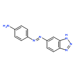
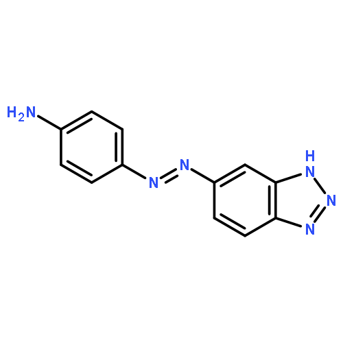
![1H-BENZOTRIAZOL-5-AMINE, 4-[(4-NITRO-1-NAPHTHALENYL)AZO]-](http://img.cochemist.com/ccimg/797100/797043-59-5.png)
![1H-BENZOTRIAZOL-5-AMINE, 4-[(4-NITRO-1-NAPHTHALENYL)AZO]-](http://img.cochemist.com/ccimg/797100/797043-59-5_b.png)
![1H-BENZOTRIAZOL-4-AMINE, 7-[(4-METHOXYPHENYL)AZO]-](http://img.cochemist.com/ccimg/797100/797043-61-9.png)
![1H-BENZOTRIAZOL-4-AMINE, 7-[(4-METHOXYPHENYL)AZO]-](http://img.cochemist.com/ccimg/797100/797043-61-9_b.png)
![2',4,7,7'-Tetrachloro-3',6'-dihydroxy-3-oxo-spiro[isobenzofuran-1(3H),9'(9H)-xanthene]-6-carboxylic acid](/data/chemimg/2265700/155911-14-1.png)
![2',4,7,7'-Tetrachloro-3',6'-dihydroxy-3-oxo-spiro[isobenzofuran-1(3H),9'(9H)-xanthene]-6-carboxylic acid](/data/chemimg/2265700/155911-14-1_b.png)
![1H-BENZOTRIAZOL-5-AMINE, 4-[(2,4-DIMETHOXYPHENYL)AZO]-](http://img.cochemist.com/ccimg/797100/797043-51-7.png)
![1H-BENZOTRIAZOL-5-AMINE, 4-[(2,4-DIMETHOXYPHENYL)AZO]-](http://img.cochemist.com/ccimg/797100/797043-51-7_b.png)
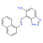
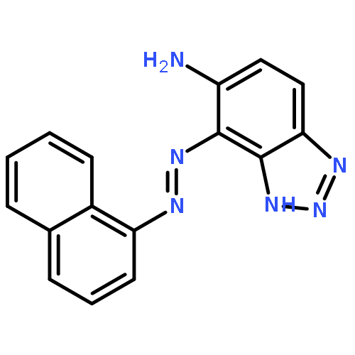
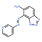
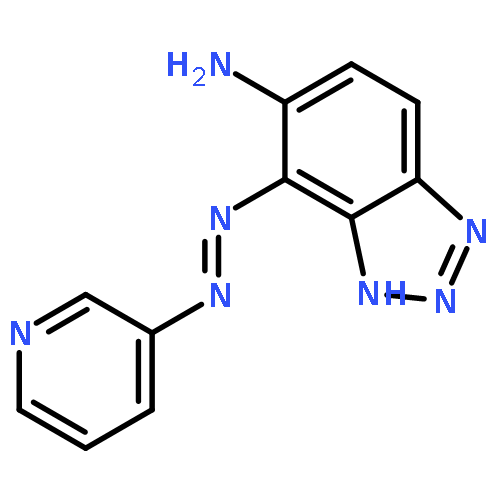
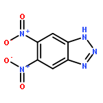
![1H-BENZOTRIAZOL-5-AMINE, 4-[(2,4-DINITROPHENYL)AZO]-](http://img.cochemist.com/ccimg/797100/797043-57-3.png)
![1H-BENZOTRIAZOL-5-AMINE, 4-[(2,4-DINITROPHENYL)AZO]-](http://img.cochemist.com/ccimg/797100/797043-57-3_b.png)

![1H-BENZOTRIAZOL-5-AMINE, 4-[(4-NITROPHENYL)AZO]-](http://img.cochemist.com/ccimg/797100/797043-55-1.png)
![1H-BENZOTRIAZOL-5-AMINE, 4-[(4-NITROPHENYL)AZO]-](http://img.cochemist.com/ccimg/797100/797043-55-1_b.png)
![1H-BENZOTRIAZOL-5-AMINE, 4-[(4-METHOXYPHENYL)AZO]-](http://img.cochemist.com/ccimg/492500/492459-11-7.png)
![1H-BENZOTRIAZOL-5-AMINE, 4-[(4-METHOXYPHENYL)AZO]-](http://img.cochemist.com/ccimg/492500/492459-11-7_b.png)
![Benzoic acid, 4-[[4-(dimethylamino)-1-naphthalenyl]azo]-](http://img.cochemist.com/ccimg/841300/841223-18-5.png)
![Benzoic acid, 4-[[4-(dimethylamino)-1-naphthalenyl]azo]-](http://img.cochemist.com/ccimg/841300/841223-18-5_b.png)
![1-(4-nitro-1H-benzo[d][1,2,3]triazol-1-yl)ethan-1-one](http://img.cochemist.com/ccimg/70800/70740-24-8.png)
![1-(4-nitro-1H-benzo[d][1,2,3]triazol-1-yl)ethan-1-one](http://img.cochemist.com/ccimg/70800/70740-24-8_b.png)


![2-[3,6-bis(dimethylamino)-1h-xanthen-9-yl]-5-(2,5-dioxopyrrol-1-yl)benzoic Acid](http://img.cochemist.com/ccimg/174600/174568-67-3.png)
![2-[3,6-bis(dimethylamino)-1h-xanthen-9-yl]-5-(2,5-dioxopyrrol-1-yl)benzoic Acid](http://img.cochemist.com/ccimg/174600/174568-67-3_b.png)


![3,7-dinitro-1,3,5,7-tetraazabicyclo[3.3.1]nonane](http://img.cochemist.com/ccimg/1000/949-56-4.png)
![3,7-dinitro-1,3,5,7-tetraazabicyclo[3.3.1]nonane](http://img.cochemist.com/ccimg/1000/949-56-4_b.png)
![2(5H)-Furanone, 5-[(4-aminophenyl)imino]-](http://img.cochemist.com/ccimg/635000/634923-11-8.png)
![2(5H)-Furanone, 5-[(4-aminophenyl)imino]-](http://img.cochemist.com/ccimg/635000/634923-11-8_b.png)


![Propanoic acid,3-[2-[2-(2-aminoethoxy)ethoxy]ethoxy]-, 1,1-dimethylethyl ester](http://img.cochemist.com/ccimg/252900/252881-74-6.png)
![Propanoic acid,3-[2-[2-(2-aminoethoxy)ethoxy]ethoxy]-, 1,1-dimethylethyl ester](http://img.cochemist.com/ccimg/252900/252881-74-6_b.png)


![2(5H)-Furanone, 5-[(3-methoxyphenyl)imino]-](http://img.cochemist.com/ccimg/344400/344334-72-1.png)
![2(5H)-Furanone, 5-[(3-methoxyphenyl)imino]-](http://img.cochemist.com/ccimg/344400/344334-72-1_b.png)
![2-Butenoic acid, 4-[(4-aminophenyl)amino]-4-oxo-, (Z)-](http://img.cochemist.com/ccimg/71700/71603-06-0.png)
![2-Butenoic acid, 4-[(4-aminophenyl)amino]-4-oxo-, (Z)-](http://img.cochemist.com/ccimg/71700/71603-06-0_b.png)


![4-Thia-1-azabicyclo[3.2.0]heptane-2-carboxylicacid, 6-[(2,6-dimethoxybenzoyl)amino]-3,3-dimethyl-7-oxo-, (2S,5R,6R)-](http://img.cochemist.com/ccimg/100/61-32-5.png)
![4-Thia-1-azabicyclo[3.2.0]heptane-2-carboxylicacid, 6-[(2,6-dimethoxybenzoyl)amino]-3,3-dimethyl-7-oxo-, (2S,5R,6R)-](http://img.cochemist.com/ccimg/100/61-32-5_b.png)
![5-Quinolinecarboxaldehyde, 8-hydroxy-, 2-[(8-hydroxy-5-quinolinyl)methylene]hydrazone](/data/chemimg/1168300/25350-73-6.png)
![5-Quinolinecarboxaldehyde, 8-hydroxy-, 2-[(8-hydroxy-5-quinolinyl)methylene]hydrazone](/data/chemimg/1168300/25350-73-6_b.png)


![2(5H)-Furanone,5-[(4-methoxyphenyl)imino]-](http://img.cochemist.com/ccimg/20000/19990-24-0.png)
![2(5H)-Furanone,5-[(4-methoxyphenyl)imino]-](http://img.cochemist.com/ccimg/20000/19990-24-0_b.png)


![Methanaminium,N-[4-[[4-(dimethylamino)phenyl]phenylmethylene]-2,5-cyclohexadien-1-ylidene]-N-methyl-](http://img.cochemist.com/ccimg/10400/10309-95-2.png)
![Methanaminium,N-[4-[[4-(dimethylamino)phenyl]phenylmethylene]-2,5-cyclohexadien-1-ylidene]-N-methyl-](http://img.cochemist.com/ccimg/10400/10309-95-2_b.png)


![1H-BENZO[D][1,2,3]TRIAZOLE-5-CARBALDEHYDE](http://img.cochemist.com/ccimg/71000/70938-42-0.png)
![1H-BENZO[D][1,2,3]TRIAZOLE-5-CARBALDEHYDE](http://img.cochemist.com/ccimg/71000/70938-42-0_b.png)


![5-(Trifluoromethyl)-1H-benzo[d][1,2,3]triazole](http://img.cochemist.com/ccimg/1600/1548-67-0.png)
![5-(Trifluoromethyl)-1H-benzo[d][1,2,3]triazole](http://img.cochemist.com/ccimg/1600/1548-67-0_b.png)


![Ferrate(2-), [7,12-diethenyl-3,8,13,17-tetramethyl-21H,23H-porphine-2,18-dipropanoato(4-)-κN21,κN22,κN23,κN24]-, hydrogen (1:2), (SP-4-2)-](/data/chemimg/522800/14875-96-8.png)
![Ferrate(2-), [7,12-diethenyl-3,8,13,17-tetramethyl-21H,23H-porphine-2,18-dipropanoato(4-)-κN21,κN22,κN23,κN24]-, hydrogen (1:2), (SP-4-2)-](/data/chemimg/522800/14875-96-8_b.png)


![N-[9-(2-carboxyphenyl)-6-(dimethylamino)-3H-xanthen-3-ylidene]-N-methylmethanaminium perchlorate](http://img.cochemist.com/ccimg/62700/62669-72-1.png)
![N-[9-(2-carboxyphenyl)-6-(dimethylamino)-3H-xanthen-3-ylidene]-N-methylmethanaminium perchlorate](http://img.cochemist.com/ccimg/62700/62669-72-1_b.png)
![1-[(2,2-DICHLORO-1-ISOTHIOCYANATOVINYL)SULFONYL]-4-METHYLBENZENE](http://img.cochemist.com/ccimg/333800/333767-00-3.png)
![1-[(2,2-DICHLORO-1-ISOTHIOCYANATOVINYL)SULFONYL]-4-METHYLBENZENE](http://img.cochemist.com/ccimg/333800/333767-00-3_b.png)


![3H-Indolium,2-[2-[3-[2-[1,3-dihydro-3,3-dimethyl-1-(4-sulfobutyl)-2H-indol-2-ylidene]ethylidene]-2-[(4-isothiocyanatophenyl)thio]-1-cyclohexen-1-yl]ethenyl]-3,3-dimethyl-1-(4-sulfobutyl)-,inner salt, sodium salt (1:1)](http://img.cochemist.com/ccimg/152200/152111-91-6.png)
![3H-Indolium,2-[2-[3-[2-[1,3-dihydro-3,3-dimethyl-1-(4-sulfobutyl)-2H-indol-2-ylidene]ethylidene]-2-[(4-isothiocyanatophenyl)thio]-1-cyclohexen-1-yl]ethenyl]-3,3-dimethyl-1-(4-sulfobutyl)-,inner salt, sodium salt (1:1)](http://img.cochemist.com/ccimg/152200/152111-91-6_b.png)

![Ethanone,1-(4-pyridinyl)-, 2-[1-(4-pyridinyl)ethylidene]hydrazone](http://img.cochemist.com/ccimg/32100/32064-89-4.png)
![Ethanone,1-(4-pyridinyl)-, 2-[1-(4-pyridinyl)ethylidene]hydrazone](http://img.cochemist.com/ccimg/32100/32064-89-4_b.png)


![2-Butenoic acid,4-[(3-methoxyphenyl)amino]-4-oxo-, (2Z)-](http://img.cochemist.com/ccimg/31500/31460-27-2.png)
![2-Butenoic acid,4-[(3-methoxyphenyl)amino]-4-oxo-, (2Z)-](http://img.cochemist.com/ccimg/31500/31460-27-2_b.png)


![1H-Benzo[d][1,2,3]triazol-5-amine](http://img.cochemist.com/ccimg/3400/3325-11-9.png)
![1H-Benzo[d][1,2,3]triazol-5-amine](http://img.cochemist.com/ccimg/3400/3325-11-9_b.png)
![2',4,4',5',7,7'-Hexachloro-3',6'-dihydroxy-3-oxo-3H-spiro[isobenzofuran-1,9'-xanthene]-6-carboxylic acid](/data/chemimg/106500/155911-16-3.png)
![2',4,4',5',7,7'-Hexachloro-3',6'-dihydroxy-3-oxo-3H-spiro[isobenzofuran-1,9'-xanthene]-6-carboxylic acid](/data/chemimg/106500/155911-16-3_b.png)


![1H,5H,11H,15H-Xantheno[2,3,4-ij:5,6,7-i'j']diquinolizin-18-ium,9-(2,5-dicarboxyphenyl)-2,3,6,7,12,13,16,17-octahydro-, inner salt](/data/chemimg/104900/194785-18-7.png)
![1H,5H,11H,15H-Xantheno[2,3,4-ij:5,6,7-i'j']diquinolizin-18-ium,9-(2,5-dicarboxyphenyl)-2,3,6,7,12,13,16,17-octahydro-, inner salt](/data/chemimg/104900/194785-18-7_b.png)


![Cyclohexanecarboxylic acid, 4-[(2,5-dihydro-2,5-dioxo-1H-pyrrol-1-yl)methyl]-, 2,5-dioxo-3-sulfo-1-pyrrolidinyl ester](/data/chemimg/105000/103708-09-4.png)
![Cyclohexanecarboxylic acid, 4-[(2,5-dihydro-2,5-dioxo-1H-pyrrol-1-yl)methyl]-, 2,5-dioxo-3-sulfo-1-pyrrolidinyl ester](/data/chemimg/105000/103708-09-4_b.png)


![3H-Indolium, 2-[5-[1-[6-[(2,5-dioxo-1-pyrrolidinyl)oxy]-6-oxohexyl]-1,3-dihydro-3,3-dimethyl-5-sulfo-2H-indol-2-ylidene]-1,3-pentadien-1-yl]-1-ethyl-3,3-](/data/chemimg/105700/146368-14-1.png)
![3H-Indolium, 2-[5-[1-[6-[(2,5-dioxo-1-pyrrolidinyl)oxy]-6-oxohexyl]-1,3-dihydro-3,3-dimethyl-5-sulfo-2H-indol-2-ylidene]-1,3-pentadien-1-yl]-1-ethyl-3,3-](/data/chemimg/105700/146368-14-1_b.png)


![Butanoic acid, 4-[(2-furanylmethyl)amino]-4-oxo-](http://img.cochemist.com/ccimg/335300/335216-97-2.png)
![Butanoic acid, 4-[(2-furanylmethyl)amino]-4-oxo-](http://img.cochemist.com/ccimg/335300/335216-97-2_b.png)


![N-[4-(2,5-dioxopyrrol-1-yl)phenyl]acetamide](http://img.cochemist.com/ccimg/5800/5702-75-0.png)
![N-[4-(2,5-dioxopyrrol-1-yl)phenyl]acetamide](http://img.cochemist.com/ccimg/5800/5702-75-0_b.png)


![5-Nitro-1H-benzo[d][1,2,3]triazole](http://img.cochemist.com/ccimg/2400/2338-12-7.png)
![5-Nitro-1H-benzo[d][1,2,3]triazole](http://img.cochemist.com/ccimg/2400/2338-12-7_b.png)


![3',6'-Dihydroxy-3H-spiro[isobenzofuran-1,9'-xanthen]-3-one](http://img.cochemist.com/ccimg/2400/2321-07-5.png)
![3',6'-Dihydroxy-3H-spiro[isobenzofuran-1,9'-xanthen]-3-one](http://img.cochemist.com/ccimg/2400/2321-07-5_b.png)