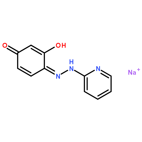
Co-reporter:Yusheng Yuan, Liu Yang, Shaopu Liu, Jidong Yang, Hui Zhang, Jingjing Yan, Xiaoli Hu
Spectrochimica Acta Part A: Molecular and Biomolecular Spectroscopy 2017 Volume 176(Volume 176) pp:
Publication Date(Web):5 April 2017
DOI:10.1016/j.saa.2017.01.014
•This is the first application of biocatalytic synthesis and fluorescence for the determination of warfarin.•The validity was verified by comparing the results with other methods and the detection limit is 0.01 μmol L− 1.•The quenching mechanism has been investigated systematically by the Stern–Volmer equation, fluorescence lifetime, UV-visible absorbance spectra.A sensitive fluorescence sensor for warfarin was proposed via quenching the fluorescence of l-tryptophan due to the interaction between warfarin and l-tryptophan. Warfarin, as one of the most effective anticoagulants, was designed and synthesized via lipase from porcine pancreas (PPL) as a biocatalyst to catalyze the Michael addition of 4-hydroxycoumarin to α, β-unsaturated enones in organic medium in the presence of water. Furthermore, the spectrofluorometry was used to detect the concentration of warfarin with a linear range and detection limit (3σ/k) of 0.04–12.0 μmol L− 1 (R2 = 0.994) and 0.01 μmol L− 1, respectively. Herein, this was the first application of bio-catalytic synthesis and fluorescence for the determination of warfarin. The proposed method was applied to determine warfarin of the drug in tablets with satisfactory results.The fabrication process of warfarin and a specific fluorescence switch sensor for warfarin.Download high-res image (66KB)Download full-size image
Co-reporter:Yusheng Yuan, Junze Jiang, Shaopu Liu, Jidong Yang, Hui zhang, Jingjing Yan, Xiaoli Hu
Sensors and Actuators B: Chemical 2017 Volume 242() pp:545-553
Publication Date(Web):April 2017
DOI:10.1016/j.snb.2016.11.050
•An “AND” logic gate and specific fluorescence switch sensor for sensitively sensing glyphosate is established.•The validity was verified by comparing the results with other methods and the detection limit is 0.6 μmol L−1, which is lower than for most of the recently published methods.•The mechanism of fluorescence resonance energy transfer (FRES) is discussed thoroughly.Herein, a novel method based on fluorescence resonance energy transfer (FRET) between carbon dots (CDs) and glyphosate (Gly) was designed for Gly detection. CDs were synthesized via a facile and one-step hydrothermal method using citric acid and Tris. The CDs possessed strong blue fluorescence and excitation wavelength-dependent emission behavior with the maximum excitation and emission wavelength at 340 nm and 410 nm, respectively. However, the presence of glyphosate could effectively quench the fluorescence intensity of the CDs through FRET and this phenomenon has been exploited to design an “AND” logic gate for sensitively sensing Gly for the first time. Furthermore, the proposed method has been successfully utilized to detect glyphosate in water samples with satisfactory results. The detection limit for glyphosate was 0.6 μmol L−1(3σ/k), with the linear range of 0.02–2.0 μmol L−1. This is a promising approach for rapid screening of glyphosate in environmental water samples without using any costly instruments.An specific fluorescence switch sensor was designed for sensing glyphosate (Gly) based on the fluorescence resonance energy transfer.
Co-reporter:Yusheng Yuan, Yalan Wang, Shaopu Liu, Yuanfang Li, Ruilin Duan, Hui Zhang and Xiaoli Hu
RSC Advances 2016 vol. 6(Issue 57) pp:52255-52263
Publication Date(Web):18 May 2016
DOI:10.1039/C6RA07675K
It is of great significance to develop an eco-friendly and sensitive platform for the detection of 6-mercaptopurine (6-MP) because of its side effects and variable activity. Herein, fluorescence quenching and spectrophotometric methods for the detection of 6-mercaptopurine (6-MP) using carbon dots (CDs) as a fluorescence probe are established. 6-MP not only serves to shelter the CDs effectively from being quenched, but also to reverse the quenching and restore the fluorescence due to its ability to remove Hg2+ from the surface of CDs. After the experimental conditions are optimized, the linear range for the detection of 6-MP is 0.04 to 12 μmol L−1, and the detection limit is 0.01 μmol L−1. When it refers to the spectrophotometric method, the linear range is 0.08 to 12 μmol L−1 and the detection limit is 0.02 μmol L−1. The CDs with 6-MP are systematically characterized with transmission electron microscopy (TEM), ultraviolet-visible (UV-vis) absorption and so on. Furthermore, the proposed methods are not only expected to become a potential tool for the fast response of 6-MP but also possess the potential for practical application to be applied in the determination of 6-MP in human serum samples with satisfactory results.
Co-reporter:Yusheng Yuan, Xin Huang, Shaopu Liu, Jidong Yang, Ruilin Duan and Xiaoli Hu
RSC Advances 2016 vol. 6(Issue 4) pp:3393-3398
Publication Date(Web):21 Dec 2015
DOI:10.1039/C5RA23367D
In neutral conditions, hypochlorite-assisted oxidative conversion of 1-pyrenylboronic acid into 1-hydroxypyrene, which leads to 1-pyrenylboronic acid fluorescence quenching, was used as the signaling tool. Compared with 1-pyrenylboronic acid, the maximum excitation (λex = 347 nm) and emission (λem = 392 nm) wavelength of 1-hydroxypyrene had no obvious change. The surfactant Triton X-100, as a micellar additive, was not only used to enhance the stability of the fluorescence probe, but also to improve its sensitivity. When using Triton X-100, the signaling of 1-pyrenylboronic acid was markedly enhanced. Herein, a spectrofluorimetric method for highly selective and sensitive hypochlorite determination has been performed. It can be noted that the fluorescence intensities positively correlated with the hypochlorite concentration over the range of 0.69–6.0 μmol L−1. The detection limit was 0.21 μmol L−1, which is lower than for most of the recently published methods. The experimental conditions were optimized and the effects of coexisting substances are evaluated. The results showed excellent priority because a certain amount of ions, including SO32−, NH4+, Cu2+ and other acid radicals, would not interfere with the measurement. The accuracy and reliability of the method was further ensured by recovery studies using the standard-addition method. In addition, the quenching mechanism, which was proven to be static quenching, has been investigated systematically by the linear plots at varying temperatures based on the Stern–Volmer equation, fluorescence lifetime, and UV-visible absorbance spectra. This method was finally used to detect hypochlorite in local water samples.
Co-reporter:Jing-Hui Zhu, Xin Zhao, Jidong Yang, Yu-Ting Tan, Lei Zhang, Shao-Pu Liu, Zhong-Fang Liu, Xiao-Li Hu
Spectrochimica Acta Part A: Molecular and Biomolecular Spectroscopy 2016 Volume 159() pp:146-150
Publication Date(Web):15 April 2016
DOI:10.1016/j.saa.2016.01.021
•We performed a fluorescence quenching assay of toxic UO22 + in water solution.•Natural pigment curcumin and Triton X-100 micelles was used as probe.•The LOD was about 9000 times lower than the maximum allowable level given by WHO.•The reaction mechanism was explored.Under pH 4.0 HAc–NaAc buffer medium, curcumin alone possesses extraordinary weak fluorescence emission. Nevertheless, the introduction of Triton X–100 micelles can largely enhance the fluorescence intensity of curcumin. Uranyl ions can complex with micelles–capped curcumin, along with the slight red shift of curcumin fluorescence (about 1–7 nm), a clear decrement of absorbance (424 nm) and fluorescence (507 nm) intensities, and a distinct color change from bright yellow to orange. The fluorescence decrements (ΔF, 507 nm) are positively correlated to the amount of uranyl ions in the concentration range of 3.7 × 10− 6–1.4 × 10− 5 mol L− 1. The detection limit of this fluorescence quenching methods is 3.7 × 10− 6 mol L− 1, which is nearly 9000 times lower than the maximum allowable level in drinking water proposed by World Health Organization. Good selectivity is achieved because of a majority of co–existing substances (such as Ce4 +, La3 +, and Th4 +) do not affect the detection. The content of uranyl ions in tap water samples was determined by the proposed method with satisfactory results.A simple, sensitive and selective assay was established to detecting toxic UO22 + by quenching the enhanced fluorescence of curcumin in Triton X-100 micelles.
Co-reporter:Ruilin Duan, Chunyan Li, Shaopu Liu, Zhongfang Liu, Yuanfang Li, Yusheng Yuan, Xiaoli Hu
Spectrochimica Acta Part A: Molecular and Biomolecular Spectroscopy 2016 Volume 152() pp:272-277
Publication Date(Web):5 January 2016
DOI:10.1016/j.saa.2015.07.003
•The method exhibited highly selective and real time response linear relationship to adenine.•Adenine could be detected at low micromolar levels and was detected in ctDNA.•Fluorescence quenching and recovery mechanism was discussed.A simple and sensitive method for determination of adenine was developed based on fluorescence quenching and recovery of L-Tryptophan (L-Trp). The fluorescence of L-Trp could efficiently quenched by copper ion compared with other common metal ions. Upon addition of adenine (Ade) in L-Trp–Cu(II) system, the fluorescence was reoccurred. Under the optimum conditions, the recovery fluorescence intensity was linearly correlated with the concentration of adenine in the range from 0.34 to 25.0 μmol L−1, with a correlation coefficient (R2) of 0.9994. The detection limit (3σ/k) was 0.046 μmol L−1, indicating that this method could applied to detect trace adenine. In this study, amino acids including L-Trp, D-Trp, L-Tyr, D-Tyr, L-Phe, D-Phe were investigated and only L-Trp could well chelated copper ion. Additionally, the mechanism of quench and recovery also were discussed and the method was successfully applied to detect the adenine in DNA with satisfactory results.The graphical presentation of the fluorescence quenching of L-Tryptophan (L-Trp) by copper ion and the recovery fluorescence occurred upon addition of adenine (Ade) in L-Trp–Cu2+ system.
Co-reporter:Yusheng Yuan, Xin Zhao, Man Qiao, Jinghui Zhu, Shaopu Liu, Jidong Yang, Xiaoli Hu
Spectrochimica Acta Part A: Molecular and Biomolecular Spectroscopy 2016 Volume 167() pp:106-110
Publication Date(Web):5 October 2016
DOI:10.1016/j.saa.2016.05.038
•The CDs were prepared in air and not only highly soluble in water but also uniform in size•A rapid assay of sunset yellow was performed and fluorescence resonance energy transfer process was proved.•A method was designed for sunset yellow from 0.3 to 8.0 μmol L-1, with a detection limit (3 σ/k) of 79.6 nmol L-1.Fluorescent carbon dots was prepared by heating N-(2-hydroxyethyl)ethylene diaminetriacetic acid in air. The carbon dots were not only highly soluble in water but also uniform in size, and possessed strong blue fluorescence and excitation wavelength-dependent emission properties with the maximum excitation and emission wavelength at 366 nm and 423 nm, respectively. Food colorant sunset yellow whose excitation and emission wavelength at 303 nm and 430 nm could selectively quench the fluorescence of carbon dots, efficient fluorescent resonance energy transfer between the carbon dots and sunset yellow is achieved. This was exploited to design a method for the determination of sunset yellow in the concentration range from 0.3 to 8.0 μmol L− 1, with a limit of detection (3 σ/k) of 79.6 nmol L− 1. Furthermore the fluorimetric detection method was established and validated for sunset yellow in soft drinks samples with satisfactory results.Carbon dots with fluorescence properties were prepared and used to sunset yellow determination.
Co-reporter:Yusheng Yuan, Xin Zhao, Shaopu Liu, Yuanfang Li, Ying Shi, Jingjing Yan, Xiaoli Hu
Sensors and Actuators B: Chemical 2016 Volume 236() pp:565-573
Publication Date(Web):29 November 2016
DOI:10.1016/j.snb.2016.06.007
A specific and sensitive fluorometric method based on the Carbon dots (CDs) probe which show unique optical properties is proposed for the determination of D-Penicillamine (D-PA) in human serum samples with satisfactory results. The fluorescence of CDs decreased apparently in the presence of mercury ion (Hg2+), Similarly, the addition of D-Penicillamine (D-PA) resulted in efficient fluorescence recovery of the CDs due to the strong affinity between Hg2+ and D-PA. Herein, this phenomenon has been exploited to design an “AND” logic gate and specific fluorescence switch sensor for sensitively sensing D-PA for the first time. Furthermore, the CDs possesses bright blue fluorescence with the maximum excitation and emission wavelength at 358 nm and 442 nm, respectively. Thus, after the experimental conditions are optimized, the linear range for detection PA is 2.0–24.0 μmol L−1, with correlation coefficient and detection limit is R2 = 0.998, 0.6 μmol L−1, respectively. Moreover, the proposed method is not only expected to be come a potential tool for the fast response of D-PA but also possesses the potential for practical application.An “AND” logic gate and specific fluorescence switch sensor was designed for D-Penicillamine (D-PA).
Co-reporter:Ying Shi, Chunyan Li, Shaopu Liu, Zhongfang Liu, Jinghui Zhu, Jidong Yang and Xiaoli Hu
RSC Advances 2015 vol. 5(Issue 79) pp:64790-64796
Publication Date(Web):22 Jul 2015
DOI:10.1039/C5RA13404H
In the present work, a novel sensing system based on fluorescence resonance energy transfer (FRET) between carbon dots (CDs) and curcumin (Cur) was designed for Cur detection. CDs were synthesized via a facile one-pot pyrolysis treatment using diethylenetriaminepentaacetic acid (DTPA) as the carbon source. The as-prepared CDs possessed strong blue fluorescence and excitation wavelength-dependent emission behavior with the maximum excitation and emission wavelength at 360 nm and 420 nm, respectively. However, the fluorescence of the CDs quenched with the introduction of Cur via FRET and the decreased intensity was linearly proportional to the concentration of Cur in the range of 0.74–5.18 μg mL−1, leading to the quantitative detection of Cur with an excellent detection limit of 44.8 ng mL−1. Furthermore, the CD-based probe can be applied in the determination of Cur in real samples with satisfactory results. The proposed method is thus expected to become a potential tool for the fast response of Cur.
Co-reporter:Yusheng Yuan, Chunyan Li, Jinghui Zhu, Shaopu Liu, Zhongfang Liu, Jidong Yang, Man Qiao, Ying Shi, Ruilin Duan and Xiaoli Hu
Analytical Methods 2015 vol. 7(Issue 21) pp:9347-9353
Publication Date(Web):25 Sep 2015
DOI:10.1039/C5AY01174D
A novel simple sensitive and inexpensive method based on the formation of Turnbull's blue nanoparticles using resonance Rayleigh scattering has been developed for the determination of hydroquinone (HQ). The results show that Fe3+ is deoxidized to Fe2+ by hydroquinone in pH 2.7 HCl (0.002 mol L−1) solution, then Fe2+ reacts with potassium ferricyanide to form a soluble Turnbull's blue (KFe[Fe(CN)6]), and further aggregates to form {KFe[Fe(CN)6]}n nanoparticles. These results induce a significant enhancement of resonance Rayleigh scattering (RRS). The maximum scattering wavelength of the ion-association complex is located at about 310 nm. The increment of scattering intensity (IRRS) is directly proportional to the concentration of HQ in the range of 0.46–50.0 μmol L−1. This method has high sensitivity and the detection limit (3σ/k) for HQ is 0.14 μmol L−1. In this work, the characteristics of absorption and RRS spectra of this reaction have been studied. The optimum reaction conditions and influencing factors have been investigated. Furthermore, the reaction mechanism and the reasons for the RRS enhancement have been explored. Additionally, the method is applied to the determination of HQ in local river water samples with satisfactory results.
Co-reporter:Jing Tian, Qiqi Zhang, Shaopu Liu, Jidong Yang, Ping Teng, Jinghui Zhu, Man Qiao, Ying Shi, Ruilin Duan, Xiaoli Hu
Spectrochimica Acta Part A: Molecular and Biomolecular Spectroscopy 2015 140() pp: 15-20
Publication Date(Web):
DOI:10.1016/j.saa.2014.12.003
Co-reporter:Man Qiao;Yaqiong Wang;Shaopu Liu;Zhongfang Liu;Jidong Yang;Jinghui Zhu
Luminescence 2015 Volume 30( Issue 2) pp:207-215
Publication Date(Web):
DOI:10.1002/bio.2714
ABSTRACT
A new method based on resonance Rayleigh scattering (RRS) was proposed for the determination of quinolones (QNS) at the nanogram level. In pH 3.3–4.4 Britton–Robinson buffer medium, quinolones such as ciprofloxacin, pipemidic acid (PIP), lomefloxacin (LOM), norfloxacin (NOR) and sarafloxacin (SAR) were protonated and reacted with methyl orange (MO) to form an ion-pair complex, which then further formed a six-membered ring chelate with Pd(II). As a result, new RRS spectra appeared and the RRS intensities were enhanced greatly. RRS spectral characteristics of the MO–QNS–Pd(II) systems, the optimum conditions for the reaction, and the influencing factors were investigated. Under optimum conditions, the scattering intensity (∆I) increments were directly proportional to the concentration of QNS with in certain ranges. The method had high sensitivity, and the detection limits (3σ) ranged from 6.8 to 12.6 ng/mL. The proposed method had been successfully applied for the determination of QNS in pharmaceutical formulations and human urine samples. In addition, the mechanism of the reaction system was discussed based on IR, absorption and fluorescence spectral studies. The reasons for the enhancement of scattering spectra were discussed in terms of fluorescence-scattering resonance energy transfer, hydrophobicity and molecular size. Copyright © 2014 John Wiley & Sons, Ltd.
Co-reporter:Man Qiao;Chunyan Li;Ying Shi;Shaopu Liu;Zhongfang Liu
Luminescence 2015 Volume 30( Issue 7) pp:1159-1166
Publication Date(Web):
DOI:10.1002/bio.2876
Abstract
A simple and sensitive resonance Rayleigh scattering (RRS) spectra method was developed for the determination of calf thymus DNA (ctDNA). The enhanced RRS signals were based on the interactions between ctDNA and aminoglycoside antibiotics (AGs) including kanamycin (KANA), tobramycin (TOB), gentamicin (GEN) and neomycin (NEO) in a weakly acidic medium (pH 3.3–5.7). Parameters influencing the method were investigated. Under optimum conditions, increments in the scattering intensity (∆I) were directly proportional to the concentration of ctDNA over certain ranges. The detection limit ranged from 12.2 to 16.9 ng/mL. Spectroscopic methods, including RRS spectra, absorption spectra and circular dichroism (CD) spectroscopy, coupled with thermo-denaturation experiments were used to study the interactions, indicating that the interaction between AGs with ctDNA was electrostatic binding mode. Copyright © 2015 John Wiley & Sons, Ltd.
Co-reporter:Jinghui Zhu, Shaopu Liu, Zhongfang Liu, Yuanfang Li, Man Qiao and Xiaoli Hu
RSC Advances 2014 vol. 4(Issue 12) pp:5990-5994
Publication Date(Web):20 Dec 2013
DOI:10.1039/C3RA46170J
An enhanced spectrofluorimetric method for highly selective and sensitive determination of nmol L−1 of hypochlorite has been performed. In alkaline medium, ferrocyanide was specifically oxidized by hypochlorite to form ferricyanide, which further reacted with non-fluorescent thiamine, resulting in the formation of blue fluorescent thiochrome. Notably, the fluorescence intensities were positively correlated with the concentration of hypochlorite over the range of 0.02–12.5 μmol L−1. The detection limit was 6.1 nmol L−1, which was lower than for most of the recently published methods. The experimental conditions were optimized and effects of coexisting substances were evaluated and the results showed excellent priority since a certain amount of other anions, including ClO3−, BrO3−, IO3−, Cr2O72− and other acid radicals, would not interfere with the measurement.
Co-reporter:Yaqiong Wang, Shaopu Liu, Zhongfang Liu, Jidong Yang, Xiaoli Hu
Journal of Luminescence 2014 Volume 147() pp:107-110
Publication Date(Web):March 2014
DOI:10.1016/j.jlumin.2013.11.006
•Cu2+ reacted with l-Trp to form 1:1 complex, which resulted in the fluorescence quenching.•The quenched fluorescence of l-Trp was recovered upon the addition of some amounts of Met.•The recovery fluorescence intensity was linearly proportional to the concentration of Met.In this paper, a sensitive and simple method for the determination of methionine (Met) was developed based on the fluorescence quenching and recovery of l-tryptophan (l-Trp). In pH 9.7 Na2CO3-NaHCO3 buffer medium, Cu2+ could react with l-Trp to form 1:1 complex because of the strong electrostatic interaction, which resulted in the fluorescence quenching of l-Trp. However, the quenched fluorescence of l-Trp could be recovered upon the addition of Met due to the strong affinity between Cu2+ and thiols. Under the optimized experimental conditions, the recovered fluorescence intensities were linearly proportional to the concentration of Met within the range of 4.8×10⁻⁶–3.0×10⁻⁵ mol L−1. The detection limit was 1.4×10⁻⁶ mol L−1.
Co-reporter:Jing Tian, Chunyan Li, Shaopu Liu, Zhongfang Liu, Jidong Yang, Jinghui Zhu and Xiaoli Hu
Analytical Methods 2014 vol. 6(Issue 14) pp:5221-5226
Publication Date(Web):19 May 2014
DOI:10.1039/C4AY00809J
In this work, the fluorescence of a metal ion (UO22+) was exploited for the determination of meloxicam. The fluorescence quenching values of uranyl acetate (UA) showed an excellent linear relationship with the concentration of meloxicam (MLX) over the range of 0.015–7.5 μg mL−1. The detection limit (3σ) was 4.49 ng mL−1, which is lower than or comparable to most of the previously reported studies. The proposed method was applied satisfactorily to the assay of MLX in tablet and capsule samples. The optimum reaction conditions, influencing factors and effects of coexisting substances were investigated. In addition, the absorption spectrum, quenching constants (Kq) and Stern–Volmer plots confirmed that this was a dynamic-quenching process.
Co-reporter:Jing Tian, Shaopu Liu, Zhongfang Liu, Jidong Yang, Jinghui Zhu, Man Qiao, Xiaoli Hu
Spectrochimica Acta Part A: Molecular and Biomolecular Spectroscopy 2014 120() pp: 7-13
Publication Date(Web):
DOI:10.1016/j.saa.2013.10.014
Co-reporter:Zhi-Ping Cui;Shao-Pu Liu;Zhong-Fang Liu;Hu-Zhi Zheng;Xiao-Li Hu;Jia-Xing Xue;Jing Tian
Luminescence 2014 Volume 29( Issue 7) pp:728-737
Publication Date(Web):
DOI:10.1002/bio.2614
ABSTRACT
In weak acid medium, aluminum(III) can react with chlorophosphonazo III [CPA(III), H8L] to form a 1:1 coordination anion [Al(OH)(H4L)]2-. At the same time, proteins such as bovine serum albumin (BSA), lysozyme (Lyso) and human serum albumin (HSA) existed as large cations with positive charges, which further combined with [Al(OH)(H4L)]2- to form a 1:4 chelate. This resulted in significant enhancement of resonance Rayleigh scattering (RRS), second-order scattering (SOS) and frequency doubling scattering (FDS). In this study, we investigated the interaction between [Al(OH)(H4L)]2- and proteins, optimization of the reaction conditions and the spectral characteristics of RRS, SOS and FDS. The maximum RRS wavelengths of different protein systems were located at 357–370 nm. The maximum SOS and FDS wavelengths were located at 546 and 389 nm, respectively. The scattering intensities (ΔI) of the three methods were proportional to the concentration of the proteins, within certain ranges, and the detection limits of the most sensitive RRS method were 2.6–9.3 ng/mL. Moreover, the chelate reaction mechanism or the reasons for the enhancement of RRS were discussed through absorption spectra, fluorescence spectra and circular dichroism (CD) spectra. Copyright © 2013 John Wiley & Sons, Ltd.
Co-reporter:Jinghui Zhu;Chunyan Li;Shaopu Liu;Zhongfang Liu
European Food Research and Technology 2014 Volume 238( Issue 5) pp:889-894
Publication Date(Web):2014 May
DOI:10.1007/s00217-014-2215-y
A non-diazotization-coupling reaction-based colorimetric method was developed and validated for the determination of nitrite in tap water and milk samples. Under acidic condition, nitrite could selectively react with pyrrole, resulting in the emerging of a new absorption peak at 509 nm and a distinct color change from clear to red, with which the concentration of nitrite can be determined indirectly. Excellent linear range was achieved from 0.72 to 40.0 μmol L−1 with a detection limit of 0.22 μmol L−1. The detection limit was lower than or comparable to most of the recently published methods. The experimental conditions were optimized, and effects of coexisting substances were evaluated, and the results showed excellent priority since a certain amount of other anions, including SO3−, SeO3− and other acid radicals, would not interfere with the measurement. We highlighted the simplicity and efficiency of this method as no diazotization-coupling procedure and toxic aniline were involved. Determination of nitrite in tap water and milk samples was performed.
Co-reporter:Man Qiao;Shaopu Liu;Zhongfang Liu;Jidong Yang;Jinghui Zhu
Food Analytical Methods 2014 Volume 7( Issue 9) pp:1737-1744
Publication Date(Web):2014 October
DOI:10.1007/s12161-014-9812-z
In this work, second order scattering (SOS), frequency double scattering (FDS), the overlapping method of the two (SFOM), and resonance Rayleigh scattering (RRS) method had been developed for sensitive determination of trace 6-benzyladenine (BA) in bean sprout samples. In pH 1.6 HCl-NaAc medium, Pd(II) reacted with BA to form a 1:1 chelate complex, and then, the complex further self-aggregated into nanoparticles [Pd(II)-BA]n. This resulted in a remarkable enhancement of resonance Rayleigh-scattering (RRS), second-order scattering (SOS), and frequency-double scattering (FDS) spectra. The maximum wavelengths were located at 312 nm (RRS), 632 nm (SOS), and 323 nm (FDS), respectively. The increments of scattering intensities ΔI were directly proportional to the concentration of BA in certain ranges. The detection limits were 7.0 nmol L−1 (0.79 μg/kg, RRS), 10.3 nmol L−1 (1.16 μg/kg, SOS), 39.4 nmol L−1 (4.44 μg/kg, FDS), and 8.6 nmol L−1 (0.96 μg/kg, SFOM). In addition, the optimum conditions of the reaction, and the effects of coexisting substances were investigated. The results showed that these methods exhibited a high selectivity. The reaction mechanism and the reasons for the enhancement of scattering were also discussed. Moreover, the feasibility for the SFOM method was illustrated in this paper. The proposed method had been successfully applied to determine a trace amount of BA in bean-sprout samples with the recoveries of 97.0–103.0 %.
Co-reporter:Jinghui Zhu, Shaopu Liu, Zhongfang Liu, Yuanfang Li, Jing Tian, Xiaoli Hu
Spectrochimica Acta Part A: Molecular and Biomolecular Spectroscopy 2014 Volume 124() pp:237-242
Publication Date(Web):24 April 2014
DOI:10.1016/j.saa.2013.12.114
•A rapid assay of doxycycline was performed.•Dual-wavelength overlapping resonance Rayleigh scattering was employed.•The detection limit (1.1 nmol L−1) was lower than or comparable to most of the reported methods.•The generating mechanisms of multi-response RRS signals were proposed.•A semi-empirical principle was established for better design of multi-response RRS probes.A dual-wavelength overlapping resonance Rayleigh scattering (DWO–RRS) method was developed and validated for highly sensitive and selective assay of doxycycline residues in several meat samples. The response signals were dependent on the specific multi-site coordination between lanthanum(III) and doxycycline (DOTC). And La(III)–DOTC complex would further aggregate to form [La(III)–DOTC]n nanoparticles, resulting in the occurrence of two new scattering peaks. Notably, with the addition of DOTC, the increments of both of these two wavelengths were proportional to the concentration of DOTC over the ranges of 3.9–4.0 × 10³ nmol L−1 (1.7–1.8 × 10³ μg/kg). The detection limit of DWO–RRS was 1.1 nmol L−1 (0.5 μg/kg), which was lower than or comparable to most of the published methods. Additionally, the generating mechanisms of multi-response RRS signals were discussed and a semi-empirical principle was established for better design of multi-response RRS probes.Graphical abstractA dual-wavelength overlapping resonance Rayleigh scattering (DWO-RRS) methodology based on the specific multi-site coordination between lanthanum(III) and doxycycline (DOTC) has been successfully designed (rather than accidently encountered) for highly sensitive and selective assay of doxycycline in several meat samples.
Co-reporter:Jing-Hui Zhu, Chun-Yan Li, Shao-Pu Liu, Zhong-Fang Liu, Yuan-Fang Li, Xiao-Li Hu
Sensors and Actuators B: Chemical 2014 198() pp: 255-259
Publication Date(Web):
DOI:10.1016/j.snb.2014.03.026
Co-reporter:Jinghui Zhu, Shaopu Liu, Zhongfang Liu, Yuanfang Li, Man Qiao and Xiaoli Hu
RSC Advances 2013 vol. 3(Issue 44) pp:21570-21575
Publication Date(Web):13 Sep 2013
DOI:10.1039/C3RA43513J
A simple and very selective spectrofluorimetric method for the detection of ng mL−1 levels of inorganic selenium species in infant formulas has been performed. It is based on the oxidation of micelle-capped nile blue A (MCNBA) by sodium selenite under neutral conditions (pH 7.0). A specific quenching of the fluorescence of MCNBA was observed and the response signals were linearly correlated with the concentration of sodium selenite over the ranges of 4.17 × 10⁻⁹ to 4.5 × 10⁻⁶ mol L−1 (0.73–787.1 ng mL−1). The limit of detection was 1.25 nmol L−1 (0.22 ng mL−1). Different parameters such as selectivity, recovery, linearity, limits of detection and quantification of the measurements were evaluated in order to validate the proposed method. Then, the new method was applied to the analysis of several infant formula samples with satisfactory results.
Co-reporter:Xiao-Juan Gan;Shao-Pu Liu;Zhong-Fang Liu;Xiao-Li Hu;Jing Tian ;Jia-Xing Xue
Luminescence 2013 Volume 28( Issue 3) pp:265-269
Publication Date(Web):
DOI:10.1002/bio.2375
ABSTRACT
In Britton-Robinson (BR) buffer medium (pH 3.3), carbazochrome sodium sulfonate (CSS) can react with some aromatic amino acids such as tryptophan (Trp), tyrosine (Tyr) and phenylalanine (Phe) to form a 1:1 complex by electrostatic attraction, aromatic stacking interaction and Van der Waals' force, resulting in fluorescence quenching of these amino acids. Maximum quenching wavelengths were located at 352 nm (CSS-Trp system), 303 nm (CSS-Tyr system) and 284 nm (CSS-Phe system), respectively. The fluorescence quenching value (ΔF) was proportional to the concentration of CSS in a certain range. The fluorescence quenching method for the determination of CSS showed high sensitivity, with detection limits of 31.3 ng/mL (CSS-Trp system), 44.6 ng/mL (CSS-Tyr system) and 315.0 ng/mL (CSS-Phe system), respectively. The optimum conditions of the reaction conditions and the effect of coexisting substances were investigated and results showed that the method had good selectivity. The method was successfully applied for the rapid determination of CSS in blood and urine samples. Based on the bimolecular quenching constant Kq, the effect of temperature and Stern-Volmer plots, this study showed that quenching of fluorescence of amino acids by CSS was a static quenching process. Copyright © 2012 John Wiley & Sons, Ltd.
Co-reporter:Yaqiong Wang;Shaopu Liu;Zhongfang Liu;Jiaxing Xue ;Jing Tian
Luminescence 2013 Volume 28( Issue 5) pp:648-655
Publication Date(Web):
DOI:10.1002/bio.2410
ABSTRACT
In pH 4.0 Britton–Robinson buffer medium, PdCl2 was able to react with enzymes (EZ) such as lysozyme (LYSO) and papain (PAP) to form a coordination complex (EZ–PdCl2), which further reacted with MoO42- to form a ternary complex (MoO42-–EZ–PdCl2). As a result, the absorption and fluorescence spectra changed; new spectra of resonance Rayleigh scattering (RRS), second-order scattering (SOS) and frequency-doubling scattering (FDS) appeared and their intensities were enhanced greatly. The maximum RRS, SOS and FDS wavelengths of two ternary complexes were located at 310, 560 and 350 nm, respectively. The increments of scattering intensity were directly proportional to the concentrations of EZ within certain ranges. The detection limits (3σ) of LYSO and PAP were 4.5 and 14.0 ng/mL (RRS method), 9.6 and 57.8 ng/mL (SOS method), and 5.2 and 106.0 ng/mL (FDS method). Taking the MoO42-–LYSO–PdCl2 system, which was more sensitive, as an example, the effects of coexisting substances were evaluated. The methods showed excellent selectivity. Accordingly, new rapid, convenient, sensitive and selective scattering methods for the determination of LYSO and PAP were proposed and applied to determine LYSO in egg white with satisfactory results. The reaction mechanism and basis of the enhancement of scattering were discussed. Copyright © 2012 John Wiley & Sons, Ltd.
Co-reporter:Zhiping Cui, Shaopu Liu, Zhongfang Liu, Yuanfang Li, Xiaoli Hu, Jing Tian
Spectrochimica Acta Part A: Molecular and Biomolecular Spectroscopy 2013 Volume 114() pp:547-552
Publication Date(Web):October 2013
DOI:10.1016/j.saa.2013.05.080
•A novel fluorescence quenching method for the determination of torasemide is established.•A sensitive, simple and quick method has been developed for determining torasemide.•Some dihalogenated fluorescein dyes are used as fluorescence probes.•The fluorescence quenching of dihalogenated fluorescein dyes by torasemide is a static quenching process.A novel fluorescence quenching method for the determination of torasemide (TOR) with some dihalogenated fluorescein dyes as fluorescence probes was developed. In acidulous medium, TOR could interact with some dihalogenated fluorescein dyes such as dichlorofluorescein (DCF), dibromofluorescein (DBF) and diiodofluorescein (DIF) to form binary complexes, which could lead to fluorescence quenching of above dihalogenated fluorescein dyes. The maximum fluorescence emission wavelengths were located at 532 nm (TOR–DCF), 535 nm (TOR–DBF) and 554 nm (TOR–DIF). The relative fluorescence intensities (ΔF = F0 − F) were proportional to the concentration of TOR in certain ranges. The detection limits were 4.8 ng mL−1 for TOR–DCF system, 9.8 ng mL−1 for TOR–DBF system and 35.1 ng mL−1 for TOR–DIF system. The optimum reaction conditions, influencing factors were studied; and the effect of coexisting substances was investigated owing to the highest sensitivity of TOR–DCF system. In addition, the reaction mechanism, composition and structure of the complex were discussed by quantum chemical calculation and Job’s method. The fluorescence quenching of dihalogenated fluorescein dyes by TOR was a static quenching process judging from the effect of temperature and the Stern–Volmer plots. The method was satisfactorily applied to the determination of TOR in tablets and human urine samples.Fluorescence spectra of TOR–DCF system
Co-reporter:Yaqiong Wang, Shaopu Liu, Zhongfang Liu, Jidong Yang, Xiaoli Hu
Spectrochimica Acta Part A: Molecular and Biomolecular Spectroscopy 2013 Volume 105() pp:612-617
Publication Date(Web):15 March 2013
DOI:10.1016/j.saa.2012.12.094
In 0.1 mol L−1 HCl medium, antiemetic drugs (ATM), such as granisetron hydrochloride (GS) and tropisetron hydrochloride (TS), reacted with H3PW12O40·nH2O and formed 3:1 ion-association complex of [(ATM)3PW12O40], then self-aggregated into nanoparticles-[(ATM)3PW12O40]n with an average size of 100 nm. The reaction resulted in the enhancement of resonance Rayleigh scattering (RRS) and the absorption spectra. The increments of scattering intensity (ΔIRRS) and the change of absorbance (ΔA) were both directly proportional to the concentrations of ATM in certain ranges. Accordingly, two new RRS and spectrophotometric methods were proposed for ATM detection. The detection limits (3σ) of GS and TS were 3.2 ng mL−1 and 4.0 ng mL−1(RRS method), 112.5 ng mL−1 and 100.0 ng mL−1(spectrophotometric method). These two methods were applied to determine GS in orally disintegrating tablets and the results were in good agreement with the official method. The ground-state geometries and electronic structures of GS and TS were optimized by the hybrid density functional theory (DFT) method and the shape of [(ATM)3PW12O40]n was characterized by atomic force microscopy (AFM). Take the RRS method with higher sensitivity as an example, the reaction mechanism and the reasons for enhancement of scattering were discussed.Graphical abstractThe interaction between TPA and ATM was investigated using resonance Rayleigh scattering and UV–vis absorption spectra. And two new methods for the determination of ATM have been developed.Highlights► Two new methods were applied to determine antiemetic drugs. ► Antiemetic drugs could react with H3PW12O40·nH2O to form nanoparticles. ► The detection limits of GS was 3.2 ng mL−1 (RRS method). ► The detection limits of TS was 4.0 ng mL−1 (RRS method).
Co-reporter:Zhiping Cui, Shaopu Liu, Zhongfang Liu and Xiaoli Hu
Analytical Methods 2012 vol. 4(Issue 2) pp:434-438
Publication Date(Web):22 Dec 2011
DOI:10.1039/C2AY05676C
In pH 0–1.0 HCl medium, 12-tungstophosphoric acid (TP) could react with vitamin B1 (VB1) to form a 1:3 ion-association complex. As a result, new spectra of resonance Rayleigh scattering appeared and their intensities were enhanced greatly. The maximum wavelength was located at 335 nm. The scattering intensities were proportional to the concentration of VB1 in the range of 0.25–1.75 μg mL−1. Therefore, a new method for the determination of VB1 was established. The method had high sensitivity and good selectivity, and the detection limit (3σ) was 0.94 ng mL−1. It was applied to the determination of VB1 in urine samples and VB1 tablets, which showed that the results were consistent with those of the China pharmaceutical method. The recovery was between 97.3%–99.0%, and the relative standard deviation (RSD) was between 1.1%–3.4%. Furthermore, the optimum reaction conditions, the RRS spectra characteristics, the reaction mechanism and the reasons for enhancement of scattering were evaluated.
Co-reporter:Xiaojuan Gan, Shaopu Liu, Zhongfang Liu, Xiaoli Hu, Zhiping Cui, Yaqiong Wang
Spectrochimica Acta Part A: Molecular and Biomolecular Spectroscopy 2012 Volume 97() pp:161-166
Publication Date(Web):November 2012
DOI:10.1016/j.saa.2012.05.062
A sensitive, simple and selective spectrofluorimetric method for the reaction of carbazochrome (CBZC) and Eosin Y (EY) or Phloxine B (PB) in acidic medium is developed for the determination of carbazochrome in biological fluids, which gives a highly fluorescent derivative measured at 545 and 565 nm at excitation wavelengths of 301 and 305 nm. The fluorescence quenching extent (ΔF) is proportional to the concentration of CBZC for CBZC–EY and CBZC–PB system at the range of 0.03–1.50 μg/mL and 0.08–1.25 μg/mL, respectively. The detection limit is 9.1 ng/mL for EY system and 22.7 ng/mL for PB system. The intra-day and inter-day reproducibility (RSD values) are less than 8.3% under three concentrations. Moreover, the affecting factors of fluorescence intensity of the product are carefully investigated and optimized, as well as the effect of coexisting substances. Judging from temperature, the Stern–Volmer plots and fluorescence emission decay curves, the quenching of fluorescence of EY and PB by CBZC is a static quenching process, caused by electrostatic attraction and aromatic stacking interaction.Graphical abstractWhen Eosin Y with carbazochrome to combine new complex, their fluorescence spectrum characteristics are almost the same, and fluorescent intensities are significant quenching. Fluorescence quenching values (ΔF) are proportional to the concentration of carbazochrome.Highlights► A new method of fluorescence quenching is used to determination carbazochrome. ► Eosin Y and Phloxine B can react with carbazochrome to form 1:2 complexes. ► The quenching of fluorescence of EY and PB by CBZC is a static quenching process.
Co-reporter:Xiaojuan Gan;Shaopu Liu;Zhongfang Liu
Journal of Fluorescence 2012 Volume 22( Issue 1) pp:129-135
Publication Date(Web):2012 January
DOI:10.1007/s10895-011-0938-8
A novel fluorescence quenching method for the determination of tetracaine hydrochloride (TA·HCl) concentration with some aromatic amino acids as fluorescence probe has been developed. In pH 6.3 acidic medium, tryptophane (Trp), tyrosine (Tyr) or phenylalanine (Phe) can react with tetracaine hydrochloride to form an ion-association complex by electrostatic attraction, aromatic stacking interaction and Van der Waals’ force, which lead to fluorescence quenching of above amino acids. The maximum fluorescence excitation and emission wavelengths of them are located at 278, 274, 258 nm and 354, 306, 285 nm, respectively. The relative fluorescence intensity (F0/F) is proportional to the TA·HCl concentration in certain range. The linear ranges and detection limits are 1.2–5.0 μg/mL and 0.37 μg/mL for Tyr-TA·HCl system, 1.3–6.0 μg/mL and 0.38 μg/mL for Trp-TA·HCl system, and 1.4–6.0 μg/mL and 0.41 μg/mL for Phe-TA·HCl system. The optimum reaction conditions, influencing factors and the effect of coexisting substances are investigated. And the results show the method has a good selectivity. Judging from the effect of temperature, the Stern-Volmer plots and fluorescence lifetime determination, the quenching of fluorescence of amino acids by TA·HCl is a static quenching process.
Co-reporter:Yanqi Song;Shaopu Liu;Zhongfang Liu
Chinese Journal of Chemistry 2011 Volume 29( Issue 12) pp:2803-2808
Publication Date(Web):
DOI:10.1002/cjoc.201180446
Abstract
In pH 4.2–5.2 HOAc-NaOAc buffer solution, Ag+ reacted with dihalogenated fluorescein (DHF) dyes to form a 1:2 anionic complex. This anionic complex could further react with hyoscine butylbromide (HBB) to form 1:1 ion-association complex, which resulted in the significant enhancement of resonance Rayleigh scattering (RRS) intensity. Therefore, a novel method for the determination of HBB by resonance Rayleigh scattering (RRS) coupled with flow injection analysis (FIA) technique has been established. The present method had been applied to determine HBB in capsules and the results were in good agreement with those obtained by the literature method.
Co-reporter:Zhiping Cui, Xiaoli Hu, Shaopu Liu, Zhongfang Liu
Spectrochimica Acta Part A: Molecular and Biomolecular Spectroscopy 2011 Volume 83(Issue 1) pp:1-7
Publication Date(Web):December 2011
DOI:10.1016/j.saa.2011.08.059
A dual-wavelength overlapping resonance Rayleigh scattering (DWO-RRS) method was developed to detect chondroitin sulfate (CS) with nile blue sulfate (NBS). At pH 3.0–4.0 Britton–Robinson (BR) buffer medium, CS interacted with NBS to form an ion-association complex. As a result, the new spectra of resonance Rayleigh scattering (RRS), second order scattering (SOS) and frequence doubling scattering (FDS) appeared and their intensities were enhanced greatly. Their maximum wavelengths were located at 303 nm (RRS), 362 nm (RRS), 588 nm (SOS) and 350 nm (FDS), respectively. The scattering intensities of the three methods were proportional to the concentration of CS in certain ranges. The methods had high sensitivity and the detection limits were between 1.5 and 7.1 ng mL−1. The DWO-RRS method had the highest sensitivity with the detection limit being 1.5 ng mL−1. The characteristics of the spectra and optimal reaction conditions of RRS method were investigated. The effects of coexistent substances on the determination of CS were evaluated. Owing to the high sensitivity, RRS method had been applied to the determination of CS in eye drops with satisfactory results. The recovery range was between 99.4% and 104.6% and the relative standard deviation (RSD) was between 0.4% and 0.8%. In addition, the reasons for RRS enhancement were discussed and the shape of ion-association complex was characterized by atomic force microscopy (AFM).Graphical abstractHighlights► A dual-wavelength overlapping resonance Rayleigh scattering (RRS) method for determination of condroitin sulfate with high sensitivity is established. ► A highly sensitive, simple and quick method has been developed for the determination of condroitin sulfate. ► The possible mechanism for the RRS enhancement of condroitin sulfate nile blue sulfate system is preliminary discussed. ► The method can be applied satisfactorily to the determination of condroitin sulfate in eye drops.
Co-reporter:PeiLi Chen;ShaoPu Liu;ZhongFang Liu;CuiXia Li
Science China Chemistry 2011 Volume 54( Issue 3) pp:506-514
Publication Date(Web):2011 March
DOI:10.1007/s11426-010-4126-5
The interaction between palladium(II)-chlorpromazine hydrochloride and sodium tungstate was investigated by ultraviolet-visible absorption, resonance Rayleigh scattering (RRS), second-order scattering (SOS) and frequency doubling scattering (FDS) spectroscopy. In pH 5.3 Britton-Robinson (BR) buffer medium, chlorpromazine hydrochloride (CPZ) reacted with Pd(II) to form 2:1 cationic chelate, which further reacted with Na2WO4 to form a 1:1 ternary ion-association complex ([Pd(CPZ)2]·WO4). As a result, the signal intensities of RRS, SOS and FDS were enhanced greatly, and the enhancements of scattering were proportional to the CPZ concentration in a certain range. Their maximum wavelengths were located at 310 nm, 570 nm and 391 nm, respectively and the detection limits (3σ) were 1.6 ng/mL (RRS method), 3.2 ng/mL (SOS method) and 5.6 ng/mL (FDS method). The optimum reaction conditions, the influences of coexisting substances and analytical application were mainly investigated by RRS method due to its highest sensitivity. A highly sensitive, simple, rapid and new method had been proposed to determine CPZ in the pharmaceutical form and residue of CPZ in pork. In addition, the Gibbs free energy change (δGf) of ion-association reaction was computed by using B3LYP/3-21g*/LanL2dz method. The formation of ion-association and the reasons for the enhancement of RRS were also discussed.
Co-reporter:Yanqi Song, Shaopu Liu, Zhongfang Liu, Xiaoli Hu
Spectrochimica Acta Part A: Molecular and Biomolecular Spectroscopy 2011 Volume 78(Issue 1) pp:148-152
Publication Date(Web):January 2011
DOI:10.1016/j.saa.2010.09.014
In 0.1 mol L−1 (pH 1.0) HCl medium, 12-tungstophosphoric acid (TP) reacted with malachite green (MG) to form an ion-association complex. As a result, the new spectra of RRS, SOS and FDS appeared and their intensities were enhanced greatly. The maximum wavelengths of RRS, SOS and FDS were located at 334 nm, 586 nm and 330 nm, and the scattering intensities were proportional to the concentration of MG. Based on it a new method for the determination of MG has been established. The detection limits (3σ) of these methods were in the range of 3.7–27 ng mL−1. The RRS, SOS, and FDS characteristics, absorption spectrum characteristics and optimum reaction conditions of the system were discussed. Effects of coexistent substances were tested, and the results demonstrated that this method had good selectivity. It has been applied to the determination of malachite green residues in fish flesh with satisfactory results. The reaction mechanism and reasons of RRS enhancement are discussed.
Co-reporter:Peili Chen, Shaopu Liu, Zhongfang Liu, Xiaoli Hu
Spectrochimica Acta Part A: Molecular and Biomolecular Spectroscopy 2011 Volume 78(Issue 1) pp:518-522
Publication Date(Web):January 2011
DOI:10.1016/j.saa.2010.11.020
The interaction between palladium(II)–aminophylline and fluorescein sodium was investigated by resonance Rayleigh scattering, second-order scattering and frequency doubling scattering spectrum. In pH 4.4 Britton–Robinson (BR) buffer medium, aminophylline (Ami) reacted with palladium(II) to form chelate cation([Pd(Ami)]2+), which further reacted with fluorescein sodium (FS) to form ternary mixed ligand complex [Pd(Ami)(FS)2]. As a result, resonance Rayleigh scattering (RRS), second-order scattering (SOS) and frequency doubling scattering spectrum (FDS) were enhanced. The maximum scattering wavelengths of [Pd(Ami)(FS)2] were located at 300 nm (RRS), 650 nm (SOS) and 304 nm (FDS). The scattering intensities were proportional to the Ami concentration in a certain range and the detection limits were 7.3 ng mL−1 (RRS), 32.9 ng mL−1 (SOS) and 79.1 ng mL−1 (FDS), respectively. Based on it, the new simple, rapid, and sensitive scattering methods have been proposed to determine Ami in urine and serum samples. Moreover, the formation mechanism of [Pd(Ami)(FS)2] and the reasons for enhancement of RRS were fully discussed.
Co-reporter:Peili Chen, Xiaoli Hu, Shaopu Liu, Zhongfang Liu, Yanqi Song
Spectrochimica Acta Part A: Molecular and Biomolecular Spectroscopy 2010 Volume 77(Issue 1) pp:207-212
Publication Date(Web):15 September 2010
DOI:10.1016/j.saa.2010.05.009
In pH 1.0 HCl medium, 12-tungstophosphoric acid (TP) reacted with promethazine hydrochloride (PMZ) and chlorpromazine hydrochloride (CPZ) to form ion-association complexes, which led to a great enhancement of the resonance nonlinear scattering such as second-order scattering (SOS) and frequency doubling scattering (FDS). Their maximum SOS and FDS peaks were located at 585 nm (TP–PMZ), 584 nm (TP–CPZ) and 388 nm (TP–PMZ), 329 nm (TP–CPZ), respectively. These results provided some indication for the determination of PMZ and CPZ by SOS and FDS methods. The linear range of TP–PMZ and TP–CPZ systems were 0.0069–2.5 μg mL−1, 0.102–5.0 μg mL−1 (SOS) and 0.079–6.0 μg mL−1, 0.0133–5.0 μg mL−1 (FDS), respectively. The detection limits (3σ) of PMZ and CPZ were 2.08 ng mL−1, 3.07 ng mL−1 (SOS) and 2.22 ng mL−1, 3.98 ng mL−1 (FDS), respectively. In this work, the optimum reaction conditions, the influences of coexisting substances and ionic strength and analytical application have been investigated. The methods have been successfully applied to the determination of PMZ and CPZ in tablets. In addition, the composition of ion-association complexes and the reaction mechanism are also discussed.
Co-reporter:Mingyou Qin, Shaopu Liu, Zhongfang Liu, Xiaoli Hu
Spectrochimica Acta Part A: Molecular and Biomolecular Spectroscopy 2009 Volume 71(Issue 5) pp:2063-2068
Publication Date(Web):January 2009
DOI:10.1016/j.saa.2008.08.003
The interaction between erythrosine (ET) and tetracaine hydrochloride (TA) was studied by resonance Rayleigh scattering (RRS), frequency doubling scattering (FDS) and second-order scattering (SOS) combining with absorption spectrum. In a weak acidic medium of Britton–Robinson (BR) buffer solution of pH 4.5, erythrosine reacted with tetracaine hydrochloride to form 1:1 ion-association complex. As a result, the new spectra of RRS, SOS and FDS appeared and their intensities enhanced greatly. The maximum peaks of RRS, SOS and FDS were at 342 nm, 680 nm and 380 nm, respectively. The intensities of the three scattering were directly proportional to the concentration of TA in the range of 0.008–4.2 μg mL−1 for RRS, 0.027–4.2 μg mL−1 for SOS and 0.041–4.2 μg mL−1 for FDS. The methods had very high sensitivities and good selectivities, and the detection limits were 0.003 μg mL−1 for RRS, 0.008 μg mL−1 for SOS and 0.012 μg mL−1 for FDS, respectively. Therefore, a new method was developed to determinate trace amounts of TA. The recovery for the determination of TA in blood serum and urine samples was between 97.0% and 103.8%. In this study, mean polarizability was calculated by AM1 quantum chemistry method. In addition, the reasons for the enhancement of scattering spectra and the energy transfer between absorption, fluorescence and RRS were discussed.
Co-reporter:Xiaoli Hu;Dongpo Xu;Shaopu Liu;Zhongfang Liu ;Sa Liu
Luminescence 2009 Volume 24( Issue 2) pp:79-83
Publication Date(Web):
DOI:10.1002/bio.1068
Abstract
A simple, sensitive and rapid flow injection analysis (FIA) method with resonance light scattering (RLS) was described for the determination of propafenone (PPF). The method was based on the ion-association reaction of 12-tungstophosphoric acid (TP) with propafenone. In pH 1.0 acidic medium, TP reacted with PPF to form an ion-associate complex, which resulted in a significant enhancement of RLS intensity. The maximum scattering peak was located at 340 nm, the RLS intensity was proportional to the concentration of PPF in the range 0.003–9.0 µg/mL, and the detection limit (3σ) of 1.0 ng/mL was obtained at a sampling rate of 60 samples/h. The feasible reaction conditions and FIA parameters for the system were optimized. The method proposed in this paper shows satisfactory reproducibility with a relative standard deviation (RSD) of 2.1% for 10 successive determinations of 2.0 µg/mL PPF. The present method had been successfully applied to the determination of PPF in serum samples and pharmaceutical samples. The results obtained were in agreement with the method used in the Chinese Pharmacopoeia. Copyright © 2008 John Wiley & Sons, Ltd.
Co-reporter:Dongpo Xu, Shaopu Liu, Zhongfang Liu, Xiaoli Hu
Analytica Chimica Acta 2007 Volume 588(Issue 1) pp:10-15
Publication Date(Web):4 April 2007
DOI:10.1016/j.aca.2007.01.076
A flow injection analysis (FIA) method coupled to resonance Rayleigh scattering (RRS) detection for the determination of verapamil hydrochloride (VP) was proposed. In pH 1.0 acidic medium, 12-tungstophosphoric acid (TP) reacted with VP to form an ion-association complex, which resulted in a significant enhancement of RRS intensity. The maximum scattering peak was located at 293 nm. RRS intensity was proportional to the concentration of VP in the range of 0.017–13.0 μg mL−1, and the detection limit (3σ) was 5.1 ng mL−1. The proposed method exhibited satisfactory reproducibility with a relative standard derivation (R.S.D.) of 2.1% for 11 successive determinations of 5.0 μg mL−1 VP. Therefore, a novel method for the determination of VP by FIA–RRS was developed. The optimum reaction conditions and the parameters of the FIA operation such as flow rate, injection volume, reactor length, and so on had been optimized in this paper. The present method had been applied to the determination of VP in serum samples and pharmaceuticals with satisfactory results. The maximal sample throughput in the optimized system was 80 h−1.
Co-reporter:
Analytical Methods (2009-Present) 2012 - vol. 4(Issue 2) pp:
Publication Date(Web):
DOI:10.1039/C2AY05676C
In pH 0–1.0 HCl medium, 12-tungstophosphoric acid (TP) could react with vitamin B1 (VB1) to form a 1:3 ion-association complex. As a result, new spectra of resonance Rayleigh scattering appeared and their intensities were enhanced greatly. The maximum wavelength was located at 335 nm. The scattering intensities were proportional to the concentration of VB1 in the range of 0.25–1.75 μg mL−1. Therefore, a new method for the determination of VB1 was established. The method had high sensitivity and good selectivity, and the detection limit (3σ) was 0.94 ng mL−1. It was applied to the determination of VB1 in urine samples and VB1 tablets, which showed that the results were consistent with those of the China pharmaceutical method. The recovery was between 97.3%–99.0%, and the relative standard deviation (RSD) was between 1.1%–3.4%. Furthermore, the optimum reaction conditions, the RRS spectra characteristics, the reaction mechanism and the reasons for enhancement of scattering were evaluated.
Co-reporter:
Analytical Methods (2009-Present) 2014 - vol. 6(Issue 14) pp:
Publication Date(Web):
DOI:10.1039/C4AY00809J
In this work, the fluorescence of a metal ion (UO22+) was exploited for the determination of meloxicam. The fluorescence quenching values of uranyl acetate (UA) showed an excellent linear relationship with the concentration of meloxicam (MLX) over the range of 0.015–7.5 μg mL−1. The detection limit (3σ) was 4.49 ng mL−1, which is lower than or comparable to most of the previously reported studies. The proposed method was applied satisfactorily to the assay of MLX in tablet and capsule samples. The optimum reaction conditions, influencing factors and effects of coexisting substances were investigated. In addition, the absorption spectrum, quenching constants (Kq) and Stern–Volmer plots confirmed that this was a dynamic-quenching process.
Co-reporter:
Analytical Methods (2009-Present) 2015 - vol. 7(Issue 21) pp:NaN9353-9353
Publication Date(Web):2015/09/25
DOI:10.1039/C5AY01174D
A novel simple sensitive and inexpensive method based on the formation of Turnbull's blue nanoparticles using resonance Rayleigh scattering has been developed for the determination of hydroquinone (HQ). The results show that Fe3+ is deoxidized to Fe2+ by hydroquinone in pH 2.7 HCl (0.002 mol L−1) solution, then Fe2+ reacts with potassium ferricyanide to form a soluble Turnbull's blue (KFe[Fe(CN)6]), and further aggregates to form {KFe[Fe(CN)6]}n nanoparticles. These results induce a significant enhancement of resonance Rayleigh scattering (RRS). The maximum scattering wavelength of the ion-association complex is located at about 310 nm. The increment of scattering intensity (IRRS) is directly proportional to the concentration of HQ in the range of 0.46–50.0 μmol L−1. This method has high sensitivity and the detection limit (3σ/k) for HQ is 0.14 μmol L−1. In this work, the characteristics of absorption and RRS spectra of this reaction have been studied. The optimum reaction conditions and influencing factors have been investigated. Furthermore, the reaction mechanism and the reasons for the RRS enhancement have been explored. Additionally, the method is applied to the determination of HQ in local river water samples with satisfactory results.


![Methanaminium,N-[4-[[4-(dimethylamino)phenyl][4-(methylphenylamino)-1-naphthalenyl]methylene]-2,5-cyclohexadien-1-ylidene]-N-methyl-,chloride (1:1)](http://img.cochemist.com/ccimg/2200/2185-87-7.png)
![Methanaminium,N-[4-[[4-(dimethylamino)phenyl][4-(methylphenylamino)-1-naphthalenyl]methylene]-2,5-cyclohexadien-1-ylidene]-N-methyl-,chloride (1:1)](http://img.cochemist.com/ccimg/2200/2185-87-7_b.png)


![Spiro[isobenzofuran-1(3H),9'-[9H]xanthen]-3-one,4,5,6,7-tetrachloro-3',6'-dihydroxy-2',4',5',7'-tetraiodo-, potassium salt(1:2)](http://img.cochemist.com/ccimg/700/632-68-8.png)
![Spiro[isobenzofuran-1(3H),9'-[9H]xanthen]-3-one,4,5,6,7-tetrachloro-3',6'-dihydroxy-2',4',5',7'-tetraiodo-, potassium salt(1:2)](http://img.cochemist.com/ccimg/700/632-68-8_b.png)


![Bleomycinamide,N1-[3-(dimethylsulfonio)propyl]-](http://img.cochemist.com/ccimg/11200/11116-31-7.png)
![Bleomycinamide,N1-[3-(dimethylsulfonio)propyl]-](http://img.cochemist.com/ccimg/11200/11116-31-7_b.png)


![Methanaminium,N-[4-[[4-(dimethylamino)phenyl]phenylmethylene]-2,5-cyclohexadien-1-ylidene]-N-methyl-](http://img.cochemist.com/ccimg/10400/10309-95-2.png)
![Methanaminium,N-[4-[[4-(dimethylamino)phenyl]phenylmethylene]-2,5-cyclohexadien-1-ylidene]-N-methyl-](http://img.cochemist.com/ccimg/10400/10309-95-2_b.png)


![3',6'-Dihydroxy-3H-spiro[isobenzofuran-1,9'-xanthen]-3-one](http://img.cochemist.com/ccimg/2400/2321-07-5.png)
![3',6'-Dihydroxy-3H-spiro[isobenzofuran-1,9'-xanthen]-3-one](http://img.cochemist.com/ccimg/2400/2321-07-5_b.png)


![N1-[3-[(4-aminobutyl)amino]propyl]-](http://img.cochemist.com/ccimg/11200/11116-32-8.png)
![N1-[3-[(4-aminobutyl)amino]propyl]-](http://img.cochemist.com/ccimg/11200/11116-32-8_b.png)
![Tungstate(3-),tetracosa-m-oxododecaoxo[m12-[phosphato(3-)-kO:kO:kO:kO':kO':kO':kO'':kO'':kO'':kO''':kO''':kO''']]dodeca-,hydrogen (1:3)](http://img.cochemist.com/ccimg/1400/1343-93-7.png)
![Tungstate(3-),tetracosa-m-oxododecaoxo[m12-[phosphato(3-)-kO:kO:kO:kO':kO':kO':kO'':kO'':kO'':kO''':kO''':kO''']]dodeca-,hydrogen (1:3)](http://img.cochemist.com/ccimg/1400/1343-93-7_b.png)