Co-reporter:Qingling Yang, Jian Liu, Banglin Li, Xiaoli Hu, Shaopu Liu, Gangcai Chen
Spectrochimica Acta Part A: Molecular and Biomolecular Spectroscopy 2016 Volume 167() pp:19-25
Publication Date(Web):5 October 2016
DOI:10.1016/j.saa.2016.05.012
•The formation of (TP)2(HgI4) ion-association nanoparticles was discussed.•Ion-association nanoparticles contribute to the development of RRS technique.•Trace determination of Hg2 + was achieved using a novel RRS method.•The developed method was used to environmental analysis with great results.In this paper, Hg2 + ions are demonstrated to form anionic [HgI4]2 − complexes after interacting with massive amount of I− ions. Subsequently, the addition of tetradecyl pyridyl bromide (TPB) can make [HgI4]2 − anionic complexes react with univalent tetradecyl pyridyl cationic ions (TP+), forming dispersed ion-association complexes (TP)2(HgI4). Due to the extrusion action of water and Van der Waals force, the hydrophobic ion-association complexes aggregate together, forming dispersed nanoparticles with an average size of about 8.5 nm. Meanwhile, resonance Rayleigh scattering (RRS) intensity is apparently enhanced due to the formation of (TP)2(HgI4) ion-association nanoparticles, contributing to a novel technique for Hg2 + detection. The wavelength of 365 nm is chosen as a detection wavelength and several conditions affecting the RRS responses of Hg2 + are optimized. Under the optimum condition, the developed method is used for the determination of Hg2 + in aqueous solution and the detection limit is estimated to be 0.8 ng mL− 1. Finally, the practical application of the developed method can be confirmed through the detections of Hg2 + in waste and river water samples with satisfactory results.A novel assay for the detection of Hg2 + in environmental sample based on the enhanced resonance Rayleigh scattering spectrum.
Co-reporter:Lunfu Tian, Xiaoli Hu, Zhongfang Liu, Shaopu Liu
Spectrochimica Acta Part A: Molecular and Biomolecular Spectroscopy 2016 Volume 154() pp:27-32
Publication Date(Web):5 February 2016
DOI:10.1016/j.saa.2015.09.026
•A resonance Rayleigh scattering method was established for determination of heparin.•Interaction between heparin with lysozyme was investigated by spectroscopic techniques.•The conformational and topography change of lysozyme was explored before and after complexing with heparin.The interaction between heparin (Hep) and lysozyme (Lyso) in vitro was studied by fluorescence, UV–vis, circular dichroism (CD), resonance Rayleigh scattering (RRS) spectroscopy and atomic force microscopy (AFM) under normal physiological conditions. UV–vis spectra of Lyso showed the absorbance was significantly increased with the addition of Hep. Fluorescence studies revealed that the emission quenching of Lyso with Hep was initiated by static quenching mechanism. CD spectral studies showed that Hep induced conformational changes in the secondary structure of Lyso. RRS spectra of Lyso showed the intensity of scattering was significantly increased with the addition of Hep and the enhanced RRS intensities were proportional to the concentration of Hep in a certain range. Thus, a new RRS method using Lyso as a probe could be used for the determination of Hep. The detection limit for Hep was 3.9 ng mL− 1. In addition, the shape of the complex was characterized by AFM. The possible reaction mechanism and the reasons for the enhancement of RRS intensity had been discussed through experimental results.The interaction between heparin and lysozyme was studied by fluorescence, UV–vis, circular dichroism, resonance Rayleigh scattering spectroscopy and atomic force microscopy.
Co-reporter:Yusheng Yuan, Shenghui Fu, Qianying Xu, Jidong Yang, Xiaoli Hu, Shaopu Liu
Spectrochimica Acta Part A: Molecular and Biomolecular Spectroscopy 2016 Volume 162() pp:93-97
Publication Date(Web):5 June 2016
DOI:10.1016/j.saa.2016.03.001
•A simple, fast method for the determination of cefoperazone is performed.•The mechanism aspect concerning the remarkable fluorescence response was discussed.•The validity was verified by comparing the results with other methods.In weak acidic medium of pH 3.5–5.6, Ce(IV) can be reduced by cefoperazone (CPZ) to be Ce(III), which further combined with CPZ to form complex Ce(OH)3CPZ. This complex not only has higher fluorescence than Ce(III), but also results in significant increase of resonance Rayleigh scattering (RRS), second order scattering (SOS) and frequency doubling scattering (FDS). The wavelengths of maximum fluorescence exciting and emission are located at 356 nm/349 nm, while the maximum wavelengths of RRS, SOS and FDS are at 312 nm, 550 nm and 390 nm, respectively. The intensity of fluorescence and scattering are all linear with the concentration of CPZ in certain conditions. The detection limit of most sensitive RRS method for CPZ is 2.1 ng mL− 1. The optimum conditions for detecting CPZ using RRS method are investigated. The effect of co-existing substances shows that the method has excellent selectivity, especially since other cephalosporins don't have similar reactions. Therefore, it can be achieved to determine CPZ in cephalosporins selectively. The paper also focuses on the reaction mechanism, the consistent and contracture of the resultant. The reasons for enhanced intensity are presumed in the meantime.Florescence spectra. 1: Ce(IV); 2: Ce(IV)–CPZ; cCe(IV) = 3.0 × 10− 5 mol L− 1; c(CPZ) = 2.0 μg mL− 1.
Co-reporter:Jian Wang, Ling Kong, Wei Shen, Xiaoli Hu, Yizhong Shen and Shaopu Liu
Analytical Methods 2014 vol. 6(Issue 12) pp:4343-4352
Publication Date(Web):14 May 2014
DOI:10.1039/C4AY00508B
In weakly acidic and neutral media, palladium(II) or sodium dodecyl benzene sulfonate (SDBS) can separately quench the fluorescence of quinolone antibiotics (FLQs) in varying degrees. When Pd(II) reacts with SDBS and FLQs to form ternary complexes, enhanced fluorescence quenching of FLQs can be observed. This synergistic fluorescence quenching effect has high sensitivity for Pd(II) detection, and the detection limit could reach 0.13 ng mL−1. Based on this, a rapid, simple and reliable method for the determination of Pd(II) in aqueous samples was established. The optimum reaction conditions of the method were tested. The reaction information of the FLQs–SDBS–Pd(II) system was investigated by absorption spectra and fluorescence spectra, and quantum chemical calculations using density functional theory B3LYP under a polarizable continuum model (PCM). The results showed that the pefloxacin (PEF) molecule exists as a zwitter-ion of HL±, reacting with Pd(II) to form plane bis(PEF)Pd chelates, which further bind with two SDBS molecules to form ternary complexes. The composition of Pd(II)–PEF–SDBS complex was found to be 1:2:2. The ternary complexes resulted in a higher fluorescence quenching efficiency and enhanced the sensitivity for the determination of Pd(II).
Co-reporter:Ling Kong;Zhongfang Liu;Shaopu Liu;Limin Wu ;Fengling Tian
Luminescence 2014 Volume 29( Issue 1) pp:29-35
Publication Date(Web):
DOI:10.1002/bio.2471
ABSTRACT
At pH 1.3–1.6, tungstate WO42–, can be converted to hexatungstate W6O192–, which can react with positively charged polymyxin B sulfate (PMB) to result in enhancement of resonance Rayleigh scattering (RRS) and resonance non-linear scattering, including second order scattering and frequency doubling scattering. Linear relationships can be established between enhanced scattering intensity and PMB concentration. The detection limits (3σ) were 5.5 ng/mL (RRS), 10.1 ng/mL (second order scattering) and 34.6 ng/mL (frequency doubling scattering). The optimum reaction conditions, influencing factors and related analytical properties were tested. The interaction mechanism was investigated via absorption spectrum, circular dichroism spectra and atomic force microscopy imaging. The basis of scattering enhancement is discussed. PMB in eardrops, human serum and urine, were quantified satisfactorily by RRS. Copyright © 2013 John Wiley & Sons, Ltd.
Co-reporter:Ling Kong, Zhongfang Liu, Xiaoli Hu, Shaopu Liu, Jiedan Meng
Journal of Luminescence 2013 Volume 137() pp:186-190
Publication Date(Web):May 2013
DOI:10.1016/j.jlumin.2012.12.041
In weak acidic medium, bovine hemoglobin would bind with salmon DNA (sDNA) to form a complex. This resulted in changes of absorption and circular dichroism spectra, fluorescence quenching of hemoglobin and remarkable enhancement of resonance Rayleigh scattering (RRS), as well as the appearance of a new RRS spectrum. The spectral characteristics of these spectra were investigated. The type of fluorescence quenching was discussed via the fluorescence lifetime of hemoglobin before and after the reaction as well as effects of temperature on fluorescence intensity. The conformational change of bovine hemoglobin was explored through circular dichroism spectra. The reasons of RRS enhancement were also discussed. In addition, it was found that the fluorescence quenching and RRS methods using bovine hemoglobin as a probe could be used to the determination of DNA. The detection limits (3σ) for sDNA were 5.5 ng mL−1 (RRS method) and 202.3 ng mL−1 (fluorescence quenching method). The optimum reaction conditions of the two methods were tested. The selectivity of RRS method was examined owing to its higher sensitivity. The RRS method was used for the determination of DNA in synthetic samples with satisfactory results.Highlights► Interaction between bovine hemoglobin and sDNA was discussed. ► The conformational change of bovine hemoglobin was explored. ► A fluorescence quenching and a RRS method were established for determination of DNA.
Co-reporter:Qianying Xu, Zhongfang Liu, Xiaoli Hu, Ling Kong and Shaopu Liu
Analyst 2012 vol. 137(Issue 4) pp:868-874
Publication Date(Web):06 Jan 2012
DOI:10.1039/C2AN15981C
In pH 6.6–7.2 Tris-HCl buffer, Cu2+ could react with adenine (A) to form a 1:1 coordination cation [CuA]2+, which only resulted in minor change of the absorption spectrum. However, when this cation further combined with WO42− to form a 1:1 ternary ion-association complex [CuA]WO4, the absorption spectrum changed a lot, and the resonance Rayleigh scattering (RRS), second-order scattering (SOS) and frequency doubling scattering (FDS) enhanced significantly. The maximum wavelengths of RRS, SOS and FDS were located at 310, 592 and 395 nm, respectively. The enhanced intensities of the three methods were proportional to the concentration of adenine in certain ranges, and the detection limit of the most sensitive RRS method was 7.4 × 10⁻⁹ mol L−1 (1.0 ng mL−1), indicating that this method could detect trace adenine. In this work, the optimum reaction conditions and the influencing factors have been studied, some potential interferences and the composition of the ion-association complex have been investigated. Meanwhile, the construction of the product and the reaction mechanism have been investigated by atomic force microscopy, transmission electron microscope and quantum chemical calculation. Accordingly, a novel RRS method for determination of adenine has been proposed and applied to detect adenine in real samples with satisfactory results.
Co-reporter:Lun Fu Tian, Zhong Fang Liu, Ling Kong, Xiao Li Hu, Shao Pu Liu
Chinese Chemical Letters 2012 Volume 23(Issue 11) pp:1283-1286
Publication Date(Web):November 2012
DOI:10.1016/j.cclet.2012.09.018
The interaction between [Hg(SCN)4]2− and hemoglobin (Hb) under conditions that simulate a physiological environment was investigated by UV–vis spectroscopy, fluorescence spectroscopy, resonance Rayleigh scattering (RRS) spectroscopy and circular dichroism (CD) spectroscopy. The results obtained from the change of UV–vis and CD spectra, the quenching of Hb fluorescence and the enhancement of RRS intensity proved that a 10:1 type complex was formed between [Hg(SCN)4]2− and Hb. The possible mechanism suggested for the interaction was that ten Hg(SCN)4]2− anions entered the four subunits of a Hb molecule to react with some residues to form an adduct by coordination and electrostatic forces. The coordination of [Hg(SCN)4]2− with Trp was the major cause of the fluorescence quenching of Hb.
Co-reporter:Ling Kong, Zhongfang Liu, Shaopu Liu and Defeng Wang
Analytical Methods 2012 vol. 4(Issue 12) pp:4346-4352
Publication Date(Web):05 Nov 2012
DOI:10.1039/C2AY26050F
Assays were developed for DNA based on enhanced resonance Rayleigh scattering and resonance nonlinear scattering (including second order scattering and frequency doubling scattering) that result from the interaction of vancomycin with DNA. Vancomycin possesses the unusual property of promoting the aggregation of DNA, which was concluded from the results obtained with RRS and absorption spectroscopy and from atomic force microscopy. The types of interaction and reasons behind RRS enhancement are discussed. Linear relationships exist over a wide range between the intensity of enhanced scattering and the concentration of DNA. The detection limit (3σ) is in the range of 6.8 to 11.1 ng mL−1.
Co-reporter:Ling Kong;Zhongfang Liu;Xiaoli Hu;Shaopu Liu;Wenqiang Li
Microchimica Acta 2012 Volume 179( Issue 3-4) pp:307-313
Publication Date(Web):2012 November
DOI:10.1007/s00604-012-0891-5
The interaction of insulin with calf thymus deoxyribonucleic acid (ctDNA) leads to a complex that displays remarkably enhanced resonance Rayleigh scattering (RRS). The complex and its formation were investigated by atomic force microscopy and by absorption, fluorescence and circular dichroism spectroscopies. We show that the Tyr B16, Tyr B26 and Phe B24 amino acids near the active center (Phe B25) were influenced by the interaction, whereas Tyr A14, Tyr A19 and Phe B1 (which are located far away from the active center) were less influenced. The interaction provide a way in the quantitation of both ctDNA and insulin with high sensitivity. When ctDNA is used as a probe to quantify insulin, the detection limit (3σ) is 6.0 ng mL-1. If, inversely, insulin is used as a probe to quantify ctDNA, the detection limit (3σ) is 7.2 ng mL-1. The analysis of synthetic DNA samples and an insulin infection sample provided satisfactory results.
Co-reporter:Qianying Xu, Zhongfang Liu, Xiaoli Hu, Ling Kong, Shaopu Liu
Analytica Chimica Acta 2011 Volume 707(1–2) pp:114-120
Publication Date(Web):30 November 2011
DOI:10.1016/j.aca.2011.08.051
In pH 1.7–3.5 acid medium, palladium chloride could react with adenine (A) to form a ternary complex of [PdCl2·A], which would self-aggregate to form uniformly dispersed nanoparticles-[PdCl2·A]n with an average size of 42 nm through the squeezing effect of aqueous phase and van der Waals force. This resulted in an enhancement of resonance Rayleigh scattering (RRS), second-order scattering (SOS) and frequency doubling scattering (FDS). The maximum wavelengths were located at 311 nm, 611 nm and 395 nm, respectively. The scattering intensities of the three methods were proportional to the concentration of adenine in certain ranges, and the detection limit of the most sensitive RRS method was 5.4 × 10⁻⁹ mol L−1 (0.73 ng mL−1). The experimental conditions were optimized and effects of coexisting substances were evaluated. The method showed excellent selectivity because a certain amount of other nucleobase, nucleoside or nucleotide would not influence the measurement. Accordingly, a novel rapid, convenient, sensitive and selective RRS method for determination of adenine was proposed and applied to detect adenine in tablet and hydrolyzates of ctDNA samples with satisfactory results. The shape of nanoparticles was characterized by atomic force microscopy. The reaction mechanism and the reasons for enhancement of scattering were discussed by infrared spectra, quantum chemical calculations and absorption spectroscopy.Graphical abstractPalladium chloride could react with adenine to form a 1:1 chelate, which led to the remarkable enhancement of resonance Rayleigh scattering (RRS) with the peak at 311 nm. The enhanced intensities present a good linear relationship with the concentration of adenine, which suggests that the system can be used for the quantitative determination of adenine.Highlights► This method is sensitive, the detection limit for adenine is 5.4 × 10⁻⁹ mol L−1. ► Other nucleobases have no similar reaction, the method has high selectivity. ► The interaction are characterized by many methods.
Co-reporter:Ling Kong;Zhongfang Liu;Xiaoli Hu ;Shaopu Liu
Chinese Journal of Chemistry 2011 Volume 29( Issue 4) pp:822-828
Publication Date(Web):
DOI:10.1002/cjoc.201190164
Abstract
In weak acidic medium, interaction between papain and calf thymus DNA (ctDNA) resulted in absorption spectral change, fluorescence quenching of papain and remarkable enhancement of resonance Rayleigh scattering (RRS). The interaction types and binding modes were discussed by characteristics of RRS, absorption, fluorescence and circular dichroism spectra combining thermodynamic data. Four interaction types include electrostatic attraction, hydrophobic force, hydrogen bonding and aromatic stacking interaction. Papain interacted with the major groove of ctDNA. Aromatic stacking interaction is the main reason of change of absorption spectrum and fluorescence quenching of papain. Surface enhanced scattering effect, resonance energy transfer effect, increase of molecular volume and conformational change make contribution to RRS enhancement. The enhanced RRS intensity (ΔI) is directly proportional to the concentration of ctDNA or papain. The detection limit (3σ) is 5.2 ng·mL−1 for ctDNA and 5.6 ng·mL−1 for papain. This creates conditions for determination of papain and ctDNA.
Co-reporter:LiHong Yu;ZhongFang Liu;XiaoLi Hu;Ling Kong;ShaoPu Liu
Science Bulletin 2010 Volume 55( Issue 8) pp:693-700
Publication Date(Web):2010 March
DOI:10.1007/s11434-009-0692-3
In a pH 4.2–6.0 HAc-NaAc buffer medium, the resonance Rayleigh scattering intensities of separate basic triphenylmethane dyes, such as methyl violet (MV), ethyl violet (EV), crystal violet (CV), methyl green (MeG), iodine green (IG), malachite green (MG), light green (LG), and tungstate ion solution, are very weak, but when these cationic dyes react with the tungstate ion (WO42−) to form ion-association complexes, their RRS are enhanced sharply and new RRS spectra appear. The reaction products of methyl violet, ethyl violet, and crystal violet with WO42− have similar spectral characteristics and there are two strong RRS peaks at 330 nm and 560 nm. The reaction products of methyl green, iodine green, malachite green, light green with WO42− have similar spectral characteristics and there are three RRS peaks at about 335 nm, 450 nm and 580 nm. The sensitivities of seven kinds of reaction products show a sequence of malachite green>light green>crystal violet>ethyl violet>methyl green>iodine green>methyl violet. The linear ranges and detection limits of the above seven systems are 0.06–2.78 μg mL−1 and 18.1 ng mL−1 (MG), 0.08–4.83 μg mL−1 and 22.6 ng mL−1 (BG), 0.10–4.90 μg mL−1 and 31.4 ng mL−1 (CV), 0.14–2.95 μg mL−1 and 40.5 ng mL−1 (EV), 0.17–3.70 μg mL−1 and 50.2 ng mL−1 (MeG), 0.22–5.70 μg mL−1 and 67.1 ng mL−1 (IG), and 0.25–7.30 μg mL−1 and 76.3 ng mL−1 (MV). A sensitive, simple and fast RRS method for the determination of basic triphenylmethane dyes using the tungstate ion as the probe has been developed. The method is applicable to the determination of malachite green residues in fish flesh.
Co-reporter:Lihong Yu;Zhongfang Liu;Xiaoli Hu;Ling Kong;Shaopu Liu
Journal of Fluorescence 2010 Volume 20( Issue 3) pp:733-738
Publication Date(Web):2010 May
DOI:10.1007/s10895-010-0615-3
In pH 1.8 ∼ 2.8 weak acid medium, polyvinylpyrrolidone (PVP) and Eosin Y reacted to form complex that could result in Eosin Y (EY) fluorescence quenching. The maximum quenching wavelength was at 542 nm. The fluorescence quenching (ΔF) was proportional to the concentration of polyvinylpyrrolidone in a certain range. The linear range, the correlation coefficient and the detection limit were 0.33 ∼ 2.0 μg•mL−1, 0.9994 and 99.6 ng•mL−1, respectively. The influences of the coexistence substances were tested and the results showed that the method had good selectivity. Therefore, a new method based on fluorescence quenching of eosin Y by PVP for the determination of trace PVP was developed. The method was sensitive, simple and rapid, which was applied to the determination of trace PVP in the beer with satisfactory results. The reaction mechanism was also discussed.
Co-reporter:Lihong YU;Zhongfang LIU;Shaopu LIU;Xiaoli HU ;Linfeng LIU
Chinese Journal of Chemistry 2009 Volume 27( Issue 8) pp:1505-1509
Publication Date(Web):
DOI:10.1002/cjoc.200990253
Abstract
In weak acid medium, eosin Y (EY) has a strong absorption band in the visible light region, and the maximum absorption wavelength (λmax) is at 517 nm. There is no light absorption for polyvinylpyrrolidone (PVP) in 250–700 nm. When EY was reacted with PVP to form a binding product, a fading reaction of EY appeared and the maximum fading wavelength was still located at 517 nm. At the same time, a smaller absorption peak was observed at 545 nm. The extent of the fading (ΔA) is directly proportional to the concentration of PVP in the range of 0.40–3.20 mg· L−1. The fading reaction has high sensitivity. The molar absorptivity (ε) is 6.4×10⁶ L·mol−1·cm−1 and the detection limit for PVP is 0.12 mg·L−1. The influencing factors for the reaction have been studied, and the results show that the selectivity is good. Based on this fact, a new fading spectrophotometric method for the determination of PVP with EY has been developed, which is simple and rapid, and it can be applied to the quantitative determination of PVP in beer samples.
Co-reporter:Jia-Gang Luo;Zhong-Fang Liu;Ling Kong;Xiao-Li Hu ;Shao-Pu Liu
Luminescence 2009 Volume 24( Issue 6) pp:429-437
Publication Date(Web):
DOI:10.1002/bio.1131
Abstract
In an acid medium solution, proteins such as bovine serum albumin, human serum albumin, ovalbumin, hemoglobin, lysozyme, γ-globulin, α-chymotrypsin and papain could react with [PdI4]2− by virtue of electrostatic attraction and hydrophobic force to form ion-association complexes. As a result, the resonance Rayleigh scattering (RRS) and resonance nonlinear scattering such as second-order scattering (SOS) and frequency doubling scattering (FDS) intensities were enhanced greatly and new scattering spectra appeared. The maximum scattering peaks of RRS, SOS and FDS were at 367, 720 and 370 nm, respectively. The enhanced RRS, SOS and FDS intensities were directly proportional to the concentrations of proteins. The detection limits for the different proteins were 2.4–11.8 ng/mL for RRS method, 9.5–47.9 ng/mL for SOS method and 4.6–18.5 ng/mL for FDS method. In this work, the influences of the interaction of [PdI4]2− with proteins on spectral characteristics of RRS, SOS and FDS were investigated and the optimum conditions were tested. Meanwhile, the effects of coexisting substances were tested and the results showed that the method exhibited a good selectivity. Based on the above research, a highly sensitive, simple and rapid method for the determination of trace amounts of proteins by resonance light scattering technique has been developed. It can be applied to the determination of proteins in tablet, human serum and urine samples. Copyright © 2009 John Wiley & Sons, Ltd.
Co-reporter:TaiShan Li;ShaoPu Liu;ZhongFang Liu;XiaoLi Hu
Science China Chemistry 2009 Volume 52( Issue 1) pp:76-85
Publication Date(Web):2009 January
DOI:10.1007/s11426-008-0108-2
CdTe nanocrystals (CdTe NCs) were achieved by reaction of CdCl2 with KHTe solution and were capped with sodium mercaptoacetate. The product was detected by transmission electron microscopy (TEM), high-resolution transmission electron microscopy (HRTEM), energy dispersive spectroscopy (EDS), fluorescence spectra, ultraviolet-visible spectra and X-ray diffraction (XRD). The CdTe NCs are of cubic structure and the average size is about 5 nm. The fluorescence quantum yield of CdTe NCs aqueous solution increased from 37% to 97% after 20 d under room light. The maximum λem of fluorescence changed from 543 nm to 510 nm and the blue shift was 33 nm. CdTe NCs aqueous solution can be steady for at least 10 months at 4 in° a refrigerator. The resonance Rayleigh scattering (RRS) of CdTe NCs in the aqueous solution was investigated. The maximum scattering peak was located at about 554 nm. The interactions of CdTe NCs with amikacin sulfate (AS) and micronomicin sulfate (MS) were investigated respectively. The effects of AS and MS on fluorescence and RRS of CdTe NCs were analyzed. It was found that AS and MS quenched the photoluminescence of CdTe NCs and enhanced RRS of CdTe NCs. Under optimum conditions, there are linear relationships between quenching intensity (F0-F), intensity of RRS (I-I0) and concentration of AS and MS. The detection limits (3б) of AS and MS are respectively 3.4 ng·mL−1 and 2.6 ng·mL−1 by the fluorescence quenching method, and 15.2 ng·mL−1 and 14.0 ng·mL−1 by the RRS method. The methods have high sensitivity, thus CdTe NCs may be used as fluorescence probes and RRS probes for the detection of aminoglycoside antibiotics.
Co-reporter:Aoer Yi;Zhongfang Liu;Shaopu Liu;Xiaoli Hu
Luminescence 2009 Volume 24( Issue 1) pp:23-29
Publication Date(Web):
DOI:10.1002/bio.1057
Abstract
In pH 5.0–5.4 HAc–NaAc buffer solution, lincomycin (Linco) reacted with Pd(II) to form 1:1 cationic chelate, which could further react with erythrosine (Ery) to form 1:1 ion-association complexes (Pd–Linco)Ery. As a result, not only were the absorption and fluorescence spectra changed, but also the resonance Rayleigh scattering (RRS) intensity was greatly enhanced. These phenomena offered useful means for the determination of Linco by spectrophotometry, fluorescence and RRS methods. The linear range and detection limit of Linco were 0.20–3.00 µg/mL and 0.057 µg/mL, 0.20–4.80 µg/mL and 0.061 µg/mL, 0.05–2.70 µg/mL and 0.015 µg/mL for the spectrophotometric, fluorescence quenching and RRS methods, respectively. Among these, the RRS method obtained the highest sensitivity. Therefore, the optimum reaction conditions and the influences of coexisting substances were investigated using the RRS method. A simple, sensitive and rapid method has been developed for the determination of Linco in either the pharmaceutical form or human body fluids, and the reasons for RRS enhancement are discussed. Copyright © 2008 John Wiley & Sons, Ltd.
Co-reporter:Cunxian Xi, Zhongfang Liu, Ling Kong, Xiaoli Hu, Shaopu Liu
Analytica Chimica Acta 2008 Volume 613(Issue 1) pp:83-90
Publication Date(Web):14 April 2008
DOI:10.1016/j.aca.2008.02.019
In pH 4.2–4.8 HAc–NaAc buffer solution, folic acid (FA) could react with uranium (VI) to form a 2:1 anionic chelate which further reacted with some basic triphenylmethane dyes (BTPMD) such as Ethyl Violet (EV), Methyl Violet (MV) and Crystal Violet (CV) to form 1:2 ion-association complexes. As a result, not only the absorption spectra were changed, but also the intensities of resonance Rayleigh scattering (RRS) were enhanced greatly and the new RRS spectra were observed. The maximum RRS wavelengths were located at 328 nm for EV system, 325 nm for MV system and 328 nm for CV system. The fading degree (ΔA) and RRS intensities (ΔI) of three systems were different. Under given conditions, the ΔA and ΔI were all directly proportional to the concentration of FA. The linear ranges and the detection limits of RRS methods were 0.0039–5.0 μg mL−1 and 1.2 ng mL−1 for EV system, 0.0073–4.0 μg mL−1 and 2.2 ng mL−1 for MV system, 0.014–3.5 μg mL−1 and 4.7 ng mL−1 for CV system. The RRS methods exhibited higher sensitivity, so they are more suitable for the determination of trace FA. The optimum conditions, the influencing factors and the effects of coexisting substances on the reaction were investigated. The method can be applied to the determination of FA in serum and urine samples with satisfactory results. The structure of the ternary ion-association complex and the reaction mechanism were discussed in this work.
Co-reporter:Hui DUAN;Zhong-Fang LIU;Shao-Pu LIU;Ling KONG
Chinese Journal of Chemistry 2008 Volume 26( Issue 2) pp:295-301
Publication Date(Web):
DOI:10.1002/cjoc.200890058
Abstract
Heated in a boiling water bath, penicillin antibiotics such as amoxicillin, ampicillin, sodium cloxacillin, sodium carbenicillin and sodium benzylpenicillin could react with K3[Fe(CN)6] to form combined products in a dilute HCl medium. As a result, resonance Rayleigh scattering (RRS) intensity was enhanced greatly and new RRS spectra appeared. The maximum scattering wavelengths of the five combined products are all located at 330 nm. The scattered intensity increments (ΔI) of the combined products are directly proportional to the concentrations of the antibiotics in a certain range. The methods exhibit high sensitivity, and the detection limits for the five penicillin antibiotics are between 4.61 and 5.62 ng·mL−1. The spectral characteristics of RRS and the optimum reaction conditions were investigated. The mechanism of reaction and the reasons for the enhancement of resonance light scattering were discussed. The effects of coexisting substances have been examined, and the results indicated that the method had a good selectivity. It can be applied to the determination of penicillin antibiotics in capsule, tablet, human serum and urine samples.
Co-reporter:Ji-Dong YANG;Tuan-Wu CAO;Zhong-Fang LIU;Ling KONG;Shao-Pu LIU
Chinese Journal of Chemistry 2008 Volume 26( Issue 5) pp:893-897
Publication Date(Web):
DOI:10.1002/cjoc.200890164
Abstract
In pH 4.5–4.8 Britton-Robinson buffer solution, rifamycin SV (i.e. rifamycin sodium) can react with serum albumin such as human serum albumin (HSA) and bovine serum albumin (BSA) to form macromolecular complexes by electrostatic attraction and hydrophobic force. As a result, the resonance Rayleigh scattering (RRS) of the drug was enhanced remarkably and the RRS peaks were at 374 and 552 nm. The enhancement of RRS (ΔI) is directly proportional to the concentration of HSA or BSA. The linear ranges and the detection limits are 0.03–6.0 µg/mL and 9.0 ng/mL for HSA, and 0.01–8.0 µg/mL and 2.0 ng/mL for BSA, respectively. In this work, a sensitive, selective, simple and fast method for the determination of trace amounts of serum albumin by RRS technique has been developed, which was applied to the determination of serum albumin in the synthesized samples and human urine samples with satisfactory results.
Co-reporter:Tai-Shan LI;Shao-Pu LIU;Zhi-Min CUI ;Zhong-Fang LIU
Chinese Journal of Chemistry 2008 Volume 26( Issue 6) pp:1085-1088
Publication Date(Web):
DOI:10.1002/cjoc.200890192
Abstract
Eu2O3nanorods were synthesized and characterized. The crystallites of Eu2O(CO3)2·H2O nanorods and Eu2O3 nanorods were obtained by means of surfactant assistance, with aqueous butanol solution as the solvent and hexamethylene tetramine as the base. The characteristics of the nanorods were analyzed by transmission electron microscopy, high-resolution transmission electron microscopy, scanning electron microscopy and X-ray diffraction. The Eu2O3 nanorod is about 80–300 nm in diameter and 1–5 µm in length. The formation mechanism of the 1D products was also proposed.
Co-reporter:Cun-Xian XI;Zhong-Fang LIU;Xiao-Li HU;Shao-Pu LIU;Hui DUAN
Chinese Journal of Chemistry 2008 Volume 26( Issue 9) pp:1619-1624
Publication Date(Web):
DOI:10.1002/cjoc.200890292
Abstract
In pH 4.9 Britton-Robinson buffer solution, methotrexate (MTX) reacted with thallium(III) to form a 3:1 chelate. This resulted in great enhancement of second-order scattering (SOS) spectra and frequency doubling scattering (FDS) spectra and appearance of new SOS and FDS spectra. Their maximum wavelengths were located at 520 and 390 nm, respectively. The increments of scattering intensities (ΔI) were directly proportional to the concentrations of MTX in the ranges of 0.022–2.0 µg·mL−1 (SOS method) and 0.008–2.5 µg·mL−1 (FDS method). The methods exhibited high sensitivities. The detection limits for MTX were 7.4 ng·mL−1 (SOS method) and 2.3 ng·mL−1 (FDS method), respectively. The optimum conditions of the reaction, the influencing factors and the effects of coexisting substances were investigated. A highly sensitive, simple and fast method for the determination of MTX has been developed. The method can be applied satisfactorily to the determination of MTX in human serum samples. In this work, the charge distribution of MTX was calculated by a CNDO quantum chemistry method. In addition, the reaction mechanism was discussed.
Co-reporter:Ao-Er YI;Zhong-Fang LIU;Shao-Pu LIU;Ling KONG
Chinese Journal of Chemistry 2008 Volume 26( Issue 10) pp:1879-1886
Publication Date(Web):
DOI:10.1002/cjoc.200890338
Abstract
In pH 5.0–5.4 HOAc-NaOAc buffer solution, clindamycin (Clin) could react with Pd(II) to form a 1:1 cationic chelate, which could further react with halofluorescein dyes such as diiodofluorescein (DIF), erythrosine (Ery) and eosin Y (EY) to form 1:1 ion-association complexes. As a result, not only the absorption and fluorescence spectra were changed, but also the resonance Rayleigh scattering (RRS) intensities enhanced greatly and new RRS spectra appeared. The three reaction products had characteristic RRS spectra and their maximum RRS wavelengths were located at 285 (DIF system), 287 (Ery system) and 321 (EY system) nm, respectively. The scattering intensities were proportional to the concentration of Clin in certain range which could be applied to determine Clin. The linear ranges and detection limits of Clin were 0.025–2.1 µg·mL−1 and 7.8 ng·mL−1 for the DIF system, 0.053–2.4 µg·mL−1 and 16.0 ng·mL−1 for the Ery system, 0.038–2.4 µg·mL−1 and 11.0 ng·mL−1 for the EY system, respectively. In this work, the optimum reaction conditions and the foreign substances were investigated. A simple, sensitive and fast method was developed for the determination of Clin either in the pharmaceutical form or in the human body fluid. Moreover, the composition, structure, reaction mechanism of the ternary complexes and their effects on the absorption, fluorescence and RRS spectra as well as the reasons of RRS enhancement were discussed.
Co-reporter:Jiangtao Liu;Zhongfang Liu;Xiaoli Hu;Ling Kong;Shaopu Liu
Luminescence 2008 Volume 23( Issue 1) pp:1-6
Publication Date(Web):
DOI:10.1002/bio.1007
Abstract
In weak acidic medium, the anticancer antibiotics bleomycin A5 (BLMA5) and bleomycin A2 (BLMA2) bind with halofluorescein dyes, such as erythrosin (Ery), eosin Y (EY) and eosin B (EB), to form ion-association complexes, which causes fluorescence quenching of halofluorescein dyes. The quenching values (ΔF) are directly in proportional to the concentrations of bleomycins over the range 0.09–2.5 µg/mL. Based on this, a fluorescence quenching method for the determination of BLMA5 and BLMA2 has been developed. The dynamic range is 0.12–2.5 µg/mL for the determination of BLMA5 and 0.09–2.0 µg/mL for BLMA2, with detection limits (3σ) of 0.04 µg/mL for BLMA5, 0.03 µg/mL for BLMA2, respectively. It has been applied to determine the two antibiotics in human serum, urine and rabbit serum samples. The recovery is in the range 90–102%. In this work, the optimum reaction conditions and the spectral characteristics of the fluorescence are investigated. The reasons for fluorescence quenching are discussed, based on the fluorescence theory. Copyright © 2007 John Wiley & Sons, Ltd.
Co-reporter:Hui Duan, Zhongfang Liu, Shaopu Liu, Aoer Yi
Talanta 2008 Volume 75(Issue 5) pp:1253-1259
Publication Date(Web):15 June 2008
DOI:10.1016/j.talanta.2008.01.028
Under the HCl solution and heating condition, penicillin antibiotics such as amoxicillin (AMO), ampicillin (AMP), sodium cloxacillin (CLO), sodium carbenicillin (CAR) and sodium benzylpenicillin (BEN) could react with Fe(III) to produce Fe(II) which further reacted with Fe(CN)63− to form a Fe3[Fe(CN)6]2 complex. By virtue of hydrophobic force and Van der Waals force, the complex aggregated to form Fe3[Fe(CN)6]2 nanoparticles with an average diameter of 45 nm. This resulted in a significant enhancement of resonance Rayleigh scattering (RRS) and non-linear scattering such as second-order scattering (SOS) and frequency doubling scattering (FDS). The increments of scattering intensity (ΔI) were directly proportional to the concentrations of the antibiotics in a certain range. The detection limits for the five penicillin antibiotics were 2.9–6.1 ng ml−1 for RRS method, 4.0–6.8 ng ml−1 for SOS method and 7.4–16.2 ng ml−1 for FDS method, respectively. Among them, the RRS method exhibited the highest sensitivity and the AMO system was more sensitive than other antibiotics systems. Based on the above researches, a new highly sensitive and simple method for the indirect determination of penicillin antibiotics has been developed. It can be applied to the determination of penicillin antibiotics in capsule, tablet, human serum and urine samples. In this work, the spectral characteristics of absorption, RRS, SOS and FDS spectra, the optimum conditions of the reaction and the influencing factors were investigated. In addition, the reaction mechanism was discussed.
Co-reporter:Shenghui Fu, Zhongfang Liu, Shaopu Liu, Aoer Yi
Talanta 2008 Volume 75(Issue 2) pp:528-535
Publication Date(Web):15 April 2008
DOI:10.1016/j.talanta.2007.11.046
In pH 1.8–2.9 Britton–Robinson (BR) buffer medium, ceftriaxone (CTRX) can react with palladium(II) (Pd(II)) to form 1:2 cationic chelate, which can further react with anionic surfactants (AS) such as sodium lauryl sulfonate (SLS), sodium dodecyl sulfate (SDS) and sodium dodecylbenzene sulfonate (SDBS) to form 1:3 ion-association complexes. As a result, the resonance Rayleigh scattering (RRS), second-order scattering (SOS) and frequency doubling scattering (FDS) were enhanced greatly. The maximum RRS, SOS and FDS wavelengths of three ion-association complexes were located at 335 nm, 560 nm and 390 nm, respectively. The increments of scattering intensity (ΔI) were directly proportional to the concentrations of CTRX in certain ranges. The detection limits (3σ) of CTRX for SLS, SDBS and SDS systems were 1.8 ng ml−1, 2.3 ng ml−1 and 2.3 ng ml−1 (RRS method), 4.9 ng ml−1, 7.4 ng ml−1 and 4.7 ng ml−1 (SOS method) and 6.8 ng ml−1, 7.3 ng ml−1 and 9.1 ng ml−1 (FDS method), separately. The sensitivity of RRS method was higher than those of SOS and FDS methods. The optimum conditions of RRS method and the influence factors were investigated, and the composition of ion-association complexes and the reaction mechanism were discussed also. The effects of foreign substances were tested and it showed that the method has a good selectivity. Based on the ion-association reaction, the sensitive, simple and rapid methods for the determination of CTRX have been developed.
Co-reporter:Jian Wang;ZhongFang Liu;ShaoPu Liu;JiangTao Liu
Science China Chemistry 2008 Volume 51( Issue 1) pp:31-40
Publication Date(Web):2008 January
DOI:10.1007/s11426-007-0122-9
In pH 4.2–5.0 Britton-Robinson buffer solution medium, fluoroquinolone antibiotics (FLQs), such as ciprofloxacin (CIP), norfloxacin (NOR), ofloxacin (OF), levofloxacin (LEV), lomefloxacin (LOM), and sparfloxacin (SPA), react with Cu (II) to form chelate cations, which further bind with erythrosine to form the ion association complexes. They can result in the changes of the absorption spectra. Simultaneously, erythrosine fades obviously and the maximum fading wavelength is located at 526 nm. The fading reactions have high sensitivities. Thus, new spectrophotometries of determination for these drugs are developed. The ion-association reactions result in the quenching of fluorescence, which also have high sensitivities. The detection limits for six antibiotics are in the range of 7.1–12.2 μg·L−1. Furthermore, the reactions can result in the enhancement of resonance Rayleigh scattering (RRS). The maximum scattering peaks of six ion-association complexes are located at 566 nm, and there are two small RRS peaks at 333 nm and 287 nm. The detection limits for fluoroquinolone antibiotics are in the range of 1.70–3.10 μg·L−1 for RRS method. Among the above three methods, the RRS method has the highest sensitivity. In this work, we investigated the spectral characteristics of the absorption, fluorescence and RRS, the optimum conditions of the reactions, and the properties of the analytical chemistry. In addition, the mechanism of reactions were discussed by density function theory (DFT) and AM1 methods.
Co-reporter:Jian Wang, Zhongfang Liu, Jiangtao Liu, Shaopu Liu, Wei Shen
Spectrochimica Acta Part A: Molecular and Biomolecular Spectroscopy 2008 Volume 69(Issue 3) pp:956-963
Publication Date(Web):March 2008
DOI:10.1016/j.saa.2007.05.057
In pH 4.4–4.5 Britton–Robinson (BR) buffer solution, fluoroquinolone antibiotics (FLQs) including ciprofloxacin (CIP), norfloxacin (NOR), levofloxacin (LEV) and lomefloxacin (LOM) could react with erythrosine (Ery) to form 1:1 ion-association complexes, which not only resulted in the changes of the absorption spectra and the quenching of fluorescence, but also resulted in the great enhancement of resonance Rayleigh scattering (RRS). These offered some indications of the determination of fluoroquinolone antibiotics by spectrophotometric, fluorescence and resonance Rayleigh scattering methods. The detection limits for fluoroquinolone antibiotics were in the range of 0.097–0.265 μg/mL for absorption methods, 0.022–0.100 μg/mL for fluorophotometry and 0.014–0.027 μg/mL for RRS method, respectively. Among them, the RRS method had the highest sensitivity. In this work, the spectral characteristics of the absorption, fluorescence and RRS, the optimum conditions of the reactions and the properties of the analytical chemistry were investigated. The methods have been successfully applied to determination of some fluoroquinolone antibiotics in human urine samples and tablets. Taking CIP–Ery system as an example, the charge distribution, the enthalpy of formation and the mean polarizability were calculated by density function theory (DFT) method. In addition, the reasons for the enhancement of scattering spectra were discussed.
Co-reporter:Shenghui Fu, Zhongfang Liu, Shaopu Liu, Jangtao Liu, Aoer Yi
Analytica Chimica Acta 2007 Volume 599(Issue 2) pp:271-278
Publication Date(Web):19 September 2007
DOI:10.1016/j.aca.2007.07.071
In pH 2.8–3.8 BR buffer medium, the third generation cephalosporin antibiotics (TGCs) such as ceftazidime (CZD), ceftriaxone (CTRX), cefoperazone (CPZ), and cefotaxime (CFTM) react with palladium(II) (Pd(II)) to form 1:2 yellowish-brown cationic chelates, which further react with 4, 5-dibromofluorescein (DBF) to form 1:3 brown ion-association complexes. As a result, not only the spectra of absorption and fluorescence are changed, but also the resonance Rayleigh scattering (RRS) is enhanced greatly and the new RRS spectra are observed. The four TGCs products have similar spectral characteristics and their maximum RRS wavelengths are all located at 291 nm. The quantitative determination ranges and the detection limits of the four TGCs are 0.0065–1.0 μg mL−1 and 2.0 ng mL−1 for CZD, 0.0070–1.1 μg mL−1 and 2.2 ng mL−1 for CTRX, 0.0090–1.6 μg mL−1 and 2.7 ng mL−1 for CPZ, and 0.014–2.2 μg mL−1 and 4.2 ng mL−1 for CFTM, respectively. The optimum conditions of the reactions and the effects of foreign substances are investigated, and the composition of ion-association complexes is discussed also. Based on the ion-association reaction, a highly sensitive, simple and rapid method has been proposed to the determination of TGCs.
Co-reporter:Shaopu Liu, Fen Wang, Zhongfang Liu, Xiaoli Hu, Aoer Yi, Hui Duan
Analytica Chimica Acta 2007 Volume 601(Issue 1) pp:101-107
Publication Date(Web):3 October 2007
DOI:10.1016/j.aca.2007.08.043
In pH 5.8 acidic medium, the anionic surfactants such as sodium dodecyl sulfate (SDS), sodium dodecyl benzene sulfonate (SDBS) or sodium dodecyl sulfonate (SLS) can react with anthracycline antibiotics such as epirubicin (EPI), daunorubicin (DNR) or mitoxantrone (MXT) to form ion-association complexes, which lead to a great enhancement of resonance Rayleigh scattering (RRS) intensity and appearances of new RRS spectra. The maximum RRS peaks are situated at 313 nm for SDS–DNR and SDS–EPI system, 296 nm for SDS–MXT system. The linear ranges and detection limits for EPI, DNR and MXT are 0.26–20.0, 0.25–20.0, 0.14–10.0 and 0.074, 0.078, 0.042 μg mL−1, respectively. In this paper, the characteristics of the absorption, fluorescence and RRS spectra of the reaction products are studied as well as the optimum reaction conditions and analytical chemistry properties. A sensitive, simple and rapid RRS method for the determination of anthracycline anticancer antibiotics has been developed.
Co-reporter:Tai Shan Li, Shao Pu Liu, Zhao Xia Lu, Zhong Fang Liu
Chinese Chemical Letters 2007 Volume 18(Issue 5) pp:617-620
Publication Date(Web):May 2007
DOI:10.1016/j.cclet.2007.03.023
A simple method has been developed to prepare cuprous selenide nanocrystals by the reaction of copper nitrate trihydrate with selenium and sodium mercaptoacetate in aqueous ammonia system. Cu2Se nanocrystals were characterized by transmission electron microscopy (TEM), high resolution transmission electron microscopy (HRTEM), X-ray diffraction (XRD), electron diffraction (ED), fluorescence spectrum and ultraviolet–visible absorption spectrum. Cu2Se nanocrystals showed berzelianite structure with 20–40 nm in length and 10–20 nm in width. A possible mechanism is also discussed.
Co-reporter:Jiang-Tao Liu;Zhong-Fang Liu;Xiao-Li Hu;Ling Kong;Shao-Pu Liu
Chinese Journal of Chemistry 2007 Volume 25(Issue 8) pp:
Publication Date(Web):15 AUG 2007
DOI:10.1002/cjoc.200790216
In weak acidic medium, anticancer antibiotics bleomycin A5 (BLMA5) and bleomycin A2 (BLMA2) can react with halofluorescein dyes such as erythrosin (Ery), eosin Y (EY), eosin B (EB) and rose bengal (RB) by virtue of electrostatic attraction and hydrophobic force to form the ion-association complexes, which can result in the fading reactions of four halofluorescein dyes. The maximum fading wavelengths of these four dyes were located at 527 nm for Ery, 515 nm for EY, 517 nm for EB and 546 nm for RB, respectively. The decrements of absorbance ((A) were directly proportional to the concentrations of bleomycin in a certain range. A new method for the determination of bleomycins anticancer drugs based on fading reactions of halofluorescein dyes has been developed. The method was not only highly sensitive but also simple and rapid. The molar absorptivities (ε) ranged from 1.5×10⁵ to 7.5×10⁵ L·mol−1·cm−1. It was applied to determination of the bleomycins in human serum, urine and rabbit serum samples. In this work, the spectral properties and the optimum reaction conditions were investigated. The structure of ion-association complexes and the reaction mechanism were discussed.
Co-reporter:Yong-Li Li;Zhong-Fang Liu;Li-Sha Xie;Ling Kong;Shao-Pu Liu
Chinese Journal of Chemistry 2007 Volume 25(Issue 7) pp:
Publication Date(Web):16 JUL 2007
DOI:10.1002/cjoc.200790184
In pH 1.0 acidic medium, double-charged triaminotriphenylmethane dyes such as methyl green (MeG) and iodine green (IG) react with potassium ferrocyanide to form 2:1 ion-association complexes by virtue of electrostatic forces and hydrophobic interaction. It results in the change of absorption and the great enhancement of resonance Rayleigh scattering (RRS) and the appearance of new RRS spectra. Two systems have similar spectral characteristics and their maximum RRS wavelengths are all located at 276 nm and smaller peaks are located at 332 and 457 nm, respectively. The intensity of RRS is directly proportional to the concentration of [Fe(CN)6]4− in the range of 0.03–5.7 µg·mL−1 (MeG system) or 0.04–5.9 µg·mL−1 (IG system). The RRS method has high sensitivity and the detection limit (3σ) for potassium ferrocyanide is 9.3 ng·mL−1 (MeG system) or 11.2 ng·mL−1 (IG system). The optimum conditions, influencing factors and effects of foreign substances are investigated. The method also has a good selectivity. A sensitive, rapid and simple RRS method for the determination of potassium ferrocyanide in salinized food and table salt has been developed.
Co-reporter:Ji-Dong Yang;Shi-Xing Deng;Zhong-Fang Liu;Shao-Pu Liu;Ling Kong
Luminescence 2007 Volume 22(Issue 6) pp:559-566
Publication Date(Web):3 SEP 2007
DOI:10.1002/bio.1001
In neutral medium, rifamycin antibiotics such as rifapentin (RFPT), rifampicin (RFP), rifandin (RFD) and rifamycin SV (RFSV) can bind with human serum albumin (HSA) and bovine serum albumin (BSA) to form complexes, resulting in the quenching of the intrinsic fluorescence (λex/λem = 285/355 nm) of the BSA and HSA. The quenching intensity (ΔF) is directly proportional to the concentration of the rifamycin antibiotics. Therefore, a new analytical method was established to determine trace rifamycin antibiotics. The method had fairly high sensitivity and the detecting limits (3σ) for RFPT, RFP, RFD and RFSV were 0.85, 0.98, 1.83, 1.89 ng/mL, respectively, for the HSA system and 0.76, 0.89, 1.55, 1.77 ng/mL, respectively, for the BSA system. All relative standard deviations (RSDs) were <3.8%. In this work, the characteristics of the fluorescence spectra were studied and the optimum reaction conditions and influencing factors were investigated. The influence of coexisting substances was tested and the results showed that the method had good selectivity and could be applied to determine trace rifamycin antibiotics in medicine capsules and urine samples. Taking the RFSV–serum albumin system as an example, the reaction mechanisms, such as binding constants, binding sites, binding distance and the type of fluorescence quenching, were investigated. Copyright © 2007 John Wiley & Sons, Ltd.
Co-reporter:Shan Li;Zhongfang Liu;Shaopu Liu;Ling Kong
Frontiers of Chemistry in China 2007 Volume 2( Issue 1) pp:79-84
Publication Date(Web):2007 March
DOI:10.1007/s11458-007-0016-6
When trypsin reacts with Herring sperm DNA (hsDNA), Salmon sperm DNA (sDNA), and Calf thymus DNA (ctDNA) to form a complex, the resonance Rayleigh scattering (RRS) was remarkably enhanced and new RRS spectra appear. These new spectra have similar characteristics of RRS spectra. The maximum RRS peaks are at 307 nm (hsDNA, sDNA) and 290 nm (ctDNA), and other peaks are at 350 nm. The scattering intensity is proportional to the concentration of DNA or trypsin; so this intereaction can be used to determine trypsin using DNA or DNA using trypsin. In the determination of DNA using trypsin, the linear ranges for hsDNA, sDNA, and ctDNA are 0–2.3, 0–2.5, and 0–1.9 μg·mL−1, and the detection limits are 0.4, 0.7, and 1.1 ng·mL−1, respectively. In the determination of trypsin using hsDNA, the linear range is 0–30.0 μg·mL−1, and the detection limit is 39.0 ng·mL−1. In this paper, the intereaction conditions were optimized. The affecting factors, chemical properties of the complex, and the composition ratio of trypsin with DNA were investigated. Using trypsin as RRS probe, a sensitive method for the determination of trace amounts of DNA was developed.
Co-reporter:XiaoLing Tang;ZhongFang Liu;ShaoPu Liu;XiaoLi Hu
Science China Chemistry 2007 Volume 50( Issue 1) pp:54-62
Publication Date(Web):2007 February
DOI:10.1007/s11426-007-2023-3
In pH 4.5 Britton-Robinson (BR) buffer solution, erythrosin (ET) can react with diphenhydramine (DP) to form a 1:1 ion-association complex, which not only results in the change of the absorption spectra, but also results in the great enhancement of resonance Rayleigh scattering (RRS) and the quenching of fluorescence. Furthermore, a new RRS spectrum will appear, and the maximum RRS wavelength was located at about 580 nm. In this work, the spectral characteristics of the absorption, fluorescence and RRS, the optimum conditions of the reaction and the properties of an analytical chemistry were investigated. A sensitive, simple and new method for the determination of DP by using erythrosin as a probe has been developed. The detection limits for DP were 0.0020 μg/mL for RRS method, 0.088 μg/mL for absorption method and 0.094 μg/mL for fluorophotometry. There was a linear relationship between the absorbance, RRS and fluorescence intensities and the drug concentration in the range of 0.0067–2.0, 0.29–6.4 and 0.31–3.2 μg/mL, respectively. The effects of the interaction of diphenhydramine and erythrosin on the absorption, fluorescence and resonance Rayleigh scattering spectra were discussed. In light polarization experiment, the polarization of RRS at maximum wavelength was measured to be P = 0.9779, and it revealed that the RRS spectrum of DP-ET complex consists mostly of resonance scattering and few resonance fluorescence. In this study, enthalpy of formation and mean polarizability were calculated by AM1 quantum chemistry method. In addition, the reaction mechanism and the reasons for the enhancement of scattering spectra and the energy transfer between absorption, fluorescence and RRS were discussed.
Co-reporter:JiangTao Liu;ZhongFang Liu;ShaoPu Liu
Science China Chemistry 2007 Volume 50( Issue 5) pp:629-637
Publication Date(Web):2007 October
DOI:10.1007/s11426-007-0046-4
The interaction of bleomycinA5 with nucleic acids has been investigated by using resonance Rayleigh scattering (RRS), molecular absorption and fluorescence spectra. The result shows that in near pH 2.2 buffer medium and absence of any metal ions, nucleic acids are capable of binding with bleomycinA5 (BLMA5) to form complexes which can remarkably enhance the RRS intensity and result in bathochromic and hyperchromic molecular absorption of nucleic acids and fluorescence quenching of bleomycinA5. The RRS spectral characteristics for the binding products of bleomycinA5 with various DNA and RNA are similar, and the maximum RRS peaks are at 301 nm for ctDNA and sDNA, 370 nm for hsDNA, 310 nm for RNAtypeVI and RNAtypeIII, respectively. The increments of RRS intensity are greatly different in which DNA enhances greatly and RNA enhances lightly. In this work, the optimum conditions of the interaction and some influencing factors have been investigated. The reaction mechanism and a binding model for the interaction of BLMA5 with the nucleic acids are discussed. In addition, a highly sensitive, simple and rapid new method for the determination of DNA has been developed. The detection limits (3σ) are 5.7 ng/mL for ctDNA, 7.4 ng/mL for sDNA and 9.2 ng/mL for hsDNA, respectively. The method can be applied to determination of trace amounts of DNA.
Co-reporter:Shao Pu Liu, Zhuo Yang, Zhong Fang Liu, Jiang Tao Liu, Yan Shi
Analytica Chimica Acta 2006 Volume 572(Issue 2) pp:283-289
Publication Date(Web):21 July 2006
DOI:10.1016/j.aca.2006.05.058
The interaction of gold nanoparticles with berberine hydrochloride has been studied by using resonance Rayleigh scattering (RRS) spectra. In pH 3.8–5.5 aqueous solution, citrate acid ([H2L2−]) self-assembled on the surface of positively charged gold nanoparticles (average diameter is about 12.0 nm) to form a supermolecular complex with negative charges. By virtue of electrostatic attraction, hydrophobic force and charge transfer, the complex bound with berberine to form complex, which had bigger diameter (35 nm) than gold nanoparticles. The formation of the binding production not only resulted in the red shift of absorption of gold nanoparticles from 518 to 672 nm, but also led to the greatly enhancement of RRS intensity. At the same time, the intensities of second-order scattering (SOS) and frequency-doubling scattering (FDS) were also increased. Under definite condition, the increment of the RRS (ΔI) were proportional to the concentration of berberine. A sensitive and simple method for the determination of berberine based on the RRS technique has been developed. The detection limit (3σ) for berberine was 0.40 ng mL−1 and the quantitative determination range was 1.33–240 ng mL−1. In this work, the optimum conditions of reaction, the effect of foreign substances and the analytical application had been investigated.
Co-reporter:Ling Kong, Zhongfang Liu, Shaopu Liu and Defeng Wang
Analytical Methods (2009-Present) 2012 - vol. 4(Issue 12) pp:NaN4352-4352
Publication Date(Web):2012/11/05
DOI:10.1039/C2AY26050F
Assays were developed for DNA based on enhanced resonance Rayleigh scattering and resonance nonlinear scattering (including second order scattering and frequency doubling scattering) that result from the interaction of vancomycin with DNA. Vancomycin possesses the unusual property of promoting the aggregation of DNA, which was concluded from the results obtained with RRS and absorption spectroscopy and from atomic force microscopy. The types of interaction and reasons behind RRS enhancement are discussed. Linear relationships exist over a wide range between the intensity of enhanced scattering and the concentration of DNA. The detection limit (3σ) is in the range of 6.8 to 11.1 ng mL−1.
Co-reporter:
Analytical Methods (2009-Present) 2014 - vol. 6(Issue 12) pp:
Publication Date(Web):
DOI:10.1039/C4AY00508B
In weakly acidic and neutral media, palladium(II) or sodium dodecyl benzene sulfonate (SDBS) can separately quench the fluorescence of quinolone antibiotics (FLQs) in varying degrees. When Pd(II) reacts with SDBS and FLQs to form ternary complexes, enhanced fluorescence quenching of FLQs can be observed. This synergistic fluorescence quenching effect has high sensitivity for Pd(II) detection, and the detection limit could reach 0.13 ng mL−1. Based on this, a rapid, simple and reliable method for the determination of Pd(II) in aqueous samples was established. The optimum reaction conditions of the method were tested. The reaction information of the FLQs–SDBS–Pd(II) system was investigated by absorption spectra and fluorescence spectra, and quantum chemical calculations using density functional theory B3LYP under a polarizable continuum model (PCM). The results showed that the pefloxacin (PEF) molecule exists as a zwitter-ion of HL±, reacting with Pd(II) to form plane bis(PEF)Pd chelates, which further bind with two SDBS molecules to form ternary complexes. The composition of Pd(II)–PEF–SDBS complex was found to be 1:2:2. The ternary complexes resulted in a higher fluorescence quenching efficiency and enhanced the sensitivity for the determination of Pd(II).

![Sodium 1,6,6-trimethyl-10,11-dioxo-6,7,8,9,10,11-hexahydrophenanthro[1,2-b]furan-2-sulfonate](http://img.cochemist.com/ccimg/69700/69659-80-9.png)
![Sodium 1,6,6-trimethyl-10,11-dioxo-6,7,8,9,10,11-hexahydrophenanthro[1,2-b]furan-2-sulfonate](http://img.cochemist.com/ccimg/69700/69659-80-9_b.png)


![Benzoic acid,3-[(1,8-dihydroxy-3,6-disulfo-2-naphthalenyl)azo]-2-hydroxy-5-sulfo-](http://img.cochemist.com/ccimg/63900/63823-40-5.png)
![Benzoic acid,3-[(1,8-dihydroxy-3,6-disulfo-2-naphthalenyl)azo]-2-hydroxy-5-sulfo-](http://img.cochemist.com/ccimg/63900/63823-40-5_b.png)


![L-Aspartic acid, N-[(2-hydroxyphenyl)methylene]-](http://img.cochemist.com/ccimg/51100/51044-28-1.png)
![L-Aspartic acid, N-[(2-hydroxyphenyl)methylene]-](http://img.cochemist.com/ccimg/51100/51044-28-1_b.png)
![1,2-dimethoxy-12-methyl[1,3]benzodioxolo[5,6-c]phenanthridinium](http://img.cochemist.com/ccimg/34400/34316-15-9.png)
![1,2-dimethoxy-12-methyl[1,3]benzodioxolo[5,6-c]phenanthridinium](http://img.cochemist.com/ccimg/34400/34316-15-9_b.png)
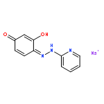
![1,3-Benzenediamine,4-[2-(5-chloro-2-pyridinyl)diazenyl]-](http://img.cochemist.com/ccimg/33100/33006-91-6.png)
![1,3-Benzenediamine,4-[2-(5-chloro-2-pyridinyl)diazenyl]-](http://img.cochemist.com/ccimg/33100/33006-91-6_b.png)


![Benzoic acid, 2-[(1,8-dihydroxy-3,6-disulfo-2-naphthalenyl)azo]-](http://img.cochemist.com/ccimg/3500/3440-26-4.png)
![Benzoic acid, 2-[(1,8-dihydroxy-3,6-disulfo-2-naphthalenyl)azo]-](http://img.cochemist.com/ccimg/3500/3440-26-4_b.png)


![Spiro[isobenzofuran-1(3H),9'-[9H]xanthen]-3-one,4,5,6,7-tetrachloro-3',6'-dihydroxy-2',4',5',7'-tetraiodo-, potassium salt(1:2)](http://img.cochemist.com/ccimg/700/632-68-8.png)
![Spiro[isobenzofuran-1(3H),9'-[9H]xanthen]-3-one,4,5,6,7-tetrachloro-3',6'-dihydroxy-2',4',5',7'-tetraiodo-, potassium salt(1:2)](http://img.cochemist.com/ccimg/700/632-68-8_b.png)
![2,7-Naphthalenedisulfonicacid,4,4'-[(3-sulfo[1,1'-biphenyl]-4,4'-diyl)bis(2,1-diazenediyl)]bis[3-amino-,sodium salt (1:5)](http://img.cochemist.com/ccimg/600/574-64-1.png)
![2,7-Naphthalenedisulfonicacid,4,4'-[(3-sulfo[1,1'-biphenyl]-4,4'-diyl)bis(2,1-diazenediyl)]bis[3-amino-,sodium salt (1:5)](http://img.cochemist.com/ccimg/600/574-64-1_b.png)

![Benzenesulfonic acid,4-[2-[4-(dimethylamino)phenyl]diazenyl]-](http://img.cochemist.com/ccimg/600/502-02-3.png)
![Benzenesulfonic acid,4-[2-[4-(dimethylamino)phenyl]diazenyl]-](http://img.cochemist.com/ccimg/600/502-02-3_b.png)


![4,9-DIETHOXY-7-METHYLFURO[3,2-G]CHROMEN-5-ONE](http://img.cochemist.com/ccimg/54400/54327-10-5.png)
![4,9-DIETHOXY-7-METHYLFURO[3,2-G]CHROMEN-5-ONE](http://img.cochemist.com/ccimg/54400/54327-10-5_b.png)


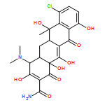
![Bleomycinamide,N1-[3-(dimethylsulfonio)propyl]-](http://img.cochemist.com/ccimg/11200/11116-31-7.png)
![Bleomycinamide,N1-[3-(dimethylsulfonio)propyl]-](http://img.cochemist.com/ccimg/11200/11116-31-7_b.png)


![Methanaminium,N-[4-[[4-(dimethylamino)phenyl]phenylmethylene]-2,5-cyclohexadien-1-ylidene]-N-methyl-](http://img.cochemist.com/ccimg/10400/10309-95-2.png)
![Methanaminium,N-[4-[[4-(dimethylamino)phenyl]phenylmethylene]-2,5-cyclohexadien-1-ylidene]-N-methyl-](http://img.cochemist.com/ccimg/10400/10309-95-2_b.png)


![3',6'-Dihydroxy-3H-spiro[isobenzofuran-1,9'-xanthen]-3-one](http://img.cochemist.com/ccimg/2400/2321-07-5.png)
![3',6'-Dihydroxy-3H-spiro[isobenzofuran-1,9'-xanthen]-3-one](http://img.cochemist.com/ccimg/2400/2321-07-5_b.png)


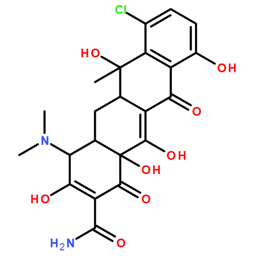
![N1-[3-[(4-aminobutyl)amino]propyl]-](http://img.cochemist.com/ccimg/11200/11116-32-8.png)
![N1-[3-[(4-aminobutyl)amino]propyl]-](http://img.cochemist.com/ccimg/11200/11116-32-8_b.png)


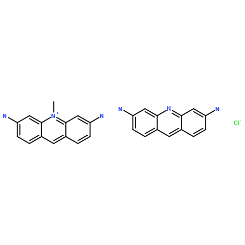
![Methanaminium,N-[4-[[4-(dimethylamino)phenyl][4-(methylphenylamino)-1-naphthalenyl]methylene]-2,5-cyclohexadien-1-ylidene]-N-methyl-,chloride (1:1)](http://img.cochemist.com/ccimg/2200/2185-87-7.png)
![Methanaminium,N-[4-[[4-(dimethylamino)phenyl][4-(methylphenylamino)-1-naphthalenyl]methylene]-2,5-cyclohexadien-1-ylidene]-N-methyl-,chloride (1:1)](http://img.cochemist.com/ccimg/2200/2185-87-7_b.png)
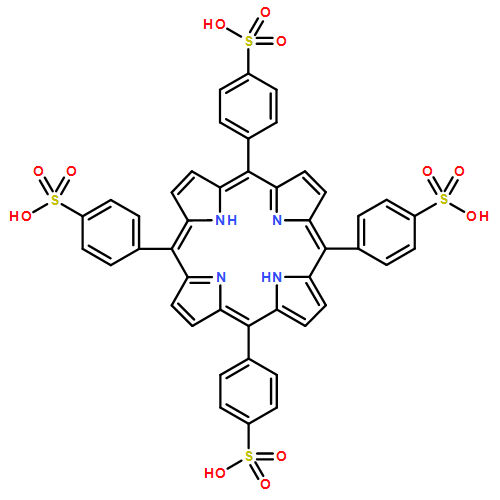
![Tungstate(3-),tetracosa-m-oxododecaoxo[m12-[phosphato(3-)-kO:kO:kO:kO':kO':kO':kO'':kO'':kO'':kO''':kO''':kO''']]dodeca-,hydrogen (1:3)](http://img.cochemist.com/ccimg/1400/1343-93-7.png)
![Tungstate(3-),tetracosa-m-oxododecaoxo[m12-[phosphato(3-)-kO:kO:kO:kO':kO':kO':kO'':kO'':kO'':kO''':kO''':kO''']]dodeca-,hydrogen (1:3)](http://img.cochemist.com/ccimg/1400/1343-93-7_b.png)

