Co-reporter:Quankun Gao, Wenzhu Zhang, Bo Song, Run Zhang, Weihua Guo, and Jingli Yuan
Analytical Chemistry April 18, 2017 Volume 89(Issue 8) pp:4517-4517
Publication Date(Web):March 21, 2017
DOI:10.1021/acs.analchem.6b04925
Considering the important roles of biothiols in lysosomes of live organisms, and unique photophysical/photochemical properties of ruthenium(II) complexes, a novel ruthenium(II) complex, Ru-2, has been developed as a molecular probe for phosphorescence and time-gated luminescence assay of biothiols in human sera, live cells, and in vivo. Ru-2 is weakly luminescent due to the effective photoinduced electron transfer (PET) from Ru(II) luminophore to electron acceptor, 2,4-dinitrobenzene-sulfonyl (DNBS). In the presence of biothiols, such as glutathione (GSH), cysteine (Cys), and homocysteine (Hcy), the emission of Ru-2 solution was switched ON, as a result of the cleavage of quencher to form the product, Ru-1. Ru-2 showed high selectivity and sensitivity for the detection of biothiols under physiological conditions, with detection limits of 62 nM, 146 nM, and 115 nM for GSH, Cys, and Hcy, respectively. The emission lifetimes of Ru-1 and Ru-2 were measured to be 405 and 474 ns, respectively, which enabled them to be used for the background-free time-gated luminescence detection even in the presence of strongly fluorescent dye, rhodamine B. On the basis of this mode, the quantification of biothiols in human serum samples was achieved without interference of background autofluorescence. A morpholine moiety was introduced into the complex to ensure Ru-2 molecules to be driven into lysosomes of live cells. Ru-2 showed low cytotoxicity and excellent membrane permeability toward live cells. Using Ru-2 as an imaging agent, visualizations of biothiols in lysosomes of live cells and in Daphnia magna were successfully demonstrated. The results suggested the potential of Ru-2 for the biomedical diagnosis of biothiol-related human diseases.
Co-reporter:Hua Ma, Bo Song, Yuanxiu Wang, Chaolong Liu, Xin Wang, Jingli Yuan
Dyes and Pigments 2017 Volume 140(Volume 140) pp:
Publication Date(Web):1 May 2017
DOI:10.1016/j.dyepig.2017.01.062
•Two novel Eu3+ complex-based luminescence probes for detection of HClO were developed.•The probes exhibited fast luminescence responses towards HClO with good selectivity and high sensitivity.•The probes displayed highly specific localizations in mitochondria and lysosomes, respectively.•The probes were successfully used for imaging of HClO in living cells and laboratory animals.Hypochlorous acid (HClO) plays a vital role in the immune system and is involved in various human diseases. To fully understand its biological functions in cellular signaling pathways, apoptosis and human diseases, effective chemical tools for directly tracing HClO at subcellular levels are greatly demanded. Herein, two mitochondria- and lysosome-targetable luminescent β-diketonate–Eu3+ complexes, Mito-BHHBCB-Eu3+ and Lyso-BHHBCB-Eu3+, were developed as probes for the time-gated luminescence detection of HClO inside mitochondria and lysosomes of living cells, respectively. The probes were designed by incorporating a mitochondria-anchoring (triphenylphosphonium) motif or a lysosome-anchoring (morpholine) motif with a strongly luminescent HOCl-responsive β-diketonate–Eu3+ complex, BHHBCB-Eu3+, to ensure the probe molecules to be driven into mitochondria or lysosomes for responding to HOCl therein. Upon exposure to HClO, the probes exhibited a fast luminescence response (within 5 s) towards HClO with good selectivity and high sensitivity (<15 nM). In live cell experiments, both probes, Mito-BHHBCB-Eu3+ and Lyso-BHHBCB-Eu3+, were successfully located in the corresponding organelles as expected, which enabled exogenous and endogenous HClO to be imaged at subcellular levels. Taking advantages of time-gated luminescence bioimaging technique, the uptake of exogenous HClO by Daphnia magna was also successfully imaged by time-gated luminescence microscopy. The results reveal that Mito-BHHBCB-Eu3+ and Lyso-BHHBCB-Eu3+ could serve as useful tools for real-time imaging of HClO at subcellular levels and in vivo with high specificity and contrast.Two novel Eu3+ complex-based probes were developed for time-gated luminescence imaging of HClO in mitochondria and lysosomes of living cells and laboratory animals.Download high-res image (223KB)Download full-size image
Co-reporter:Xiangli Liu;Zhixin Tang;Bo Song;Hua Ma;Jingli Yuan
Journal of Materials Chemistry B 2017 vol. 5(Issue 15) pp:2849-2855
Publication Date(Web):2017/04/12
DOI:10.1039/C6TB02991D
Bioresponsive luminescence probes based on lanthanide complexes have shown great utility in a variety of time-gated luminescence bioassays. However, functional lanthanide complexes that can target individual organelles for probing biospecies therein have rarely been investigated. In this work, a unique Eu3+ complex, Mito-NPSTTA-Eu3+, was designed and synthesized as a probe for the time-gated luminescence sensing of HOCl inside the mitochondria of living cells. The probe showed a fast, highly sensitive and selective luminescence response to HOCl with a wide available pH range (pH 3–10), and highly specific mitochondria-localization performance. Taking advantage of time-gated luminescence bioimaging and the excellent properties of the Eu3+ complex, the generation of endogenous HOCl in RAW 264.7 cells and the uptake of exogenous HOCl by zebrafish were successfully imaged, respectively. The results demonstrated the feasibility of Mito-NPSTTA-Eu3+ for the imaging of mitochondrial HOCl, and validated the potential of our strategy for the design of lanthanide complex-based organelle-targeting bioresponsive probes.
Co-reporter:Zhichao Dai;Lu Tian;Bo Song;Xiangli Liu;Jingli Yuan
Chemical Science (2010-Present) 2017 vol. 8(Issue 3) pp:1969-1976
Publication Date(Web):2017/02/28
DOI:10.1039/C6SC03667H
Rapid, multiplexed, sensitive and specific identification and quantitative detection of nitric oxide (NO) are in great demand in biomedical science. Herein, a novel multifunctional probe based on the intramolecular LRET (luminescence resonance energy transfer) strategy, TRP-NO, was designed for the highly sensitive and selective ratiometric and luminescence lifetime detection of lysosomal NO. Before reaction with NO, the emission of the rhodamine moiety in TRP-NO is switched off, which prevents the LRET process, so that the probe emits only the long-lived Tb3+ luminescence. However, upon reaction with NO, accompanied by the turn-on of rhodamine emission, the LRET from the Tb3+-complex moiety to rhodamine moiety occurs, which results in a remarkable increase of the rhodamine emission and decrease of the Tb3+ emission. After the reaction, the intensity ratio of the rhodamine emission to the Tb3+ emission, I565/I540, was found to be 28.8-fold increased, and the dose-dependent enhancement of the I565/I540 value showed a good linearity upon the increase of NO concentration. In addition, a dose-dependent luminescence lifetime decrease was distinctly observed between the average luminescence lifetime of the probe and NO concentration, which provides a ∼10-fold contrast window for the detection of NO. These unique properties allowed TRP-NO to be conveniently used as a time-gated luminescence probe for the quantitative detection of NO using both luminescence intensity ratio and luminescence lifetime as signals. The applicability of TRP-NO for ratiometric time-gated luminescence imaging of NO in living cells was investigated. Meanwhile, dye co-localization studies confirmed a quite precise distribution of TRP-NO in lysosomes by confocal microscopy imaging. Furthermore, the practical applicability of TRP-NO was demonstrated by the visualization of NO in Daphnia magna. All of the results demonstrated that TRP-NO could serve as a useful tool for exploiting and elucidating the function of NO at sub-cellular levels with high specificity, accuracy and contrast.
Co-reporter:Hua Ma;Bo Song;Yuanxiu Wang;Deyuan Cong;Yufei Jiang;Jingli Yuan
Chemical Science (2010-Present) 2017 vol. 8(Issue 1) pp:150-159
Publication Date(Web):2016/12/19
DOI:10.1039/C6SC02243J
We have developed a ratiometric time-gated luminescence sensory system for in vivo imaging of hypochlorous acid (HClO) by preparing a dual-emissive nanoarchitecture of europium- and terbium-complex-modified silica nanoparticles. The design of this nanoarchitecture is based on our new finding that the strong, long-lived luminescence of the β-diketonate–Eu3+ complex can be rapidly and selectively quenched by HClO. Therefore, the β-diketonate–Eu3+ complex was decorated on the surface of the silica nanoparticles for responding to HClO, while a HClO-insensitive luminescent terbium complex was immobilized in the inner solid core of the nanoparticles to serve as an internal standard. This nanosensing probe combines the advantages of both ratiometric and time-gated detection modes to afford high accuracy and sensitivity. Upon exposure to HClO, the nanoprobe displayed a remarkable luminescence color change from red to green, and the intensity ratio of the green over the red luminescence (I539/I607) showed a rapid, sensitive and selective response to HClO. Additionally, the feasibility of using the nanoprobe for intracellular detection of exogenous and endogenous HClO and for real-time mapping of HClO in small laboratory animals has been demonstrated via ratiometric time-gated luminescence imaging microscopy. The results reveal that the constructed nanoarchitecture cloud is a favorable and useful sensing probe for the real-time imaging of HClO in vivo with high specificity and contrast.
Co-reporter:Lu Tian, Zhichao Dai, Xiangli Liu, Bo Song, Zhiqiang Ye, and Jingli Yuan
Analytical Chemistry 2015 Volume 87(Issue 21) pp:10878
Publication Date(Web):October 13, 2015
DOI:10.1021/acs.analchem.5b02347
Using apoferritin (AFt) as a carrier, a novel ratiometric luminescence probe based on luminescence resonance energy transfer (LRET) between a Tb3+ complex (PTTA-Tb3+) and a rhodamine derivative (Rh-NO), PTTA-Tb3+@AFt-Rh-NO, has been designed and prepared for the specific recognition and time-gated luminescence detection of nitric oxide (NO) in living samples. In this LRET probe, PTTA-Tb3+ encapsulated in the core of AFt is the energy donor, and Rh-NO, a NO-responsive rhodamine derivative, bound on the surface of AFt is the energy acceptor. The probe only emits strong Tb3+ luminescence because the emission of rhodamine is switched off in the absence of NO. Upon reaction with NO, accompanied by the turn-on of rhodamine emission, the LRET from Tb3+ complex to rhodamine occurs, which results in the remarkable increase and decrease of the long-lived emissions of rhodamine and PTTA-Tb3+, respectively. After the reaction, the intensity ratio of rhodamine emission to Tb3+ emission, I565/I539, is ∼24.5-fold increased, and the dose-dependent enhancement of I565/I539 shows a good linearity in a wide concentration range of NO. This unique luminescence response allowed PTTA-Tb3+@AFt-Rh-NO to be conveniently used as a ratiometric probe for the time-gated luminescence detection of NO with I565/I539 as a signal. Taking advantages of high specificity and sensitivity of the probe as well as its good water-solubility, biocompatibility, and cell membrane permeability, PTTA-Tb3+@AFt-Rh-NO was successfully used for the luminescent imaging of NO in living cells and Daphnia magna. The results demonstrated the efficacy of the probe and highlighted it’s advantages for the ratiometric time-gated luminescence bioimaging application.
Co-reporter:Jingyan Sun; Bo Song; Zhiqiang Ye;Jingli Yuan
Inorganic Chemistry 2015 Volume 54(Issue 24) pp:11660-11668
Publication Date(Web):December 4, 2015
DOI:10.1021/acs.inorgchem.5b02458
Singlet oxygen (1O2) plays a key role in the photodynamic therapy (PDT) technique of neoplastic diseases. In this work, by using a 9,10-dimethyl-2-anthryl-containing β-diketone, 1,1,1,2,2-pentafluoro-5-(9′,10′-dimethyl-2′-anthryl)-3,5-pentanedione (Hpfdap), as a 1O2-recognition ligand, a novel β-diketonate–europium(III) complex that can act as a luminescence probe for 1O2, [Eu(pfdap)3(tpy)] (tpy = 2,2′,2″-terpyridine), has been designed and synthesized for the time-gated luminescence detection of 1O2 in living cells. The complex is weakly luminescent due to the quenching effect of 9,10-dimethyl-2-anthryl groups. After reaction with 1O2, accompanied by the formation of endoperoxides of 9,10-dimethyl-2-anthryl groups, the luminescence quenching disappears, so that the long-lived luminescence of the europium(III) complex is switched on. The complex showed highly selective luminescence response to 1O2 with a remarkable luminescence enhancement. Combined with the time-gated luminescence imaging technique, the complex was successfully used as a luminescent probe for the monitoring of the time-dependent generation of 1O2 in 5-aminolevulinic acid (a PDT drug) loaded HepG2 cells during the photodynamic process. In addition, by coloading the complex and a mitochondrial indicator, Mito-Tracker Green, into HepG2 cells, the specific localization of [Eu(pfdap)3(tpy)] molecules in mitochondria of HepG2 cells was demonstrated by confocal fluorescence imaging measurements.
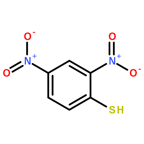
Co-reporter:Zhiqiang Ye, Quankun Gao, Xin An, Bo Song and Jingli Yuan
Dalton Transactions 2015 vol. 44(Issue 17) pp:8278-8283
Publication Date(Web):24 Mar 2015
DOI:10.1039/C5DT00290G
A unique ruthenium(II) complex, [Ru(bpy)2(DNS-bpy)](PF6)2 [bpy: 2,2′-bipyridine, DNS-bpy: 4-(2,4-dinitrophenylthio)-2,2′-bipyridine], that can act as a probe for the recognition and luminescence sensing of biothiols has been designed and synthesized. Due to the presence of effective photo-induced electron transfer (PET) from the potent electron donor (Ru-bpy centre) to the strong electron acceptor (2,4-dinitrophenyl moiety), the Ru(II) complex itself is weakly luminescent. Reaction of [Ru(bpy)2(DNS-bpy)](PF6)2 with biothiols leads to the replacement of the 2,4-dinitrophenyl moiety by biothiols, which results in the loss of PET within the complex, to allow recovery of the MLCT-based emission of the Ru(II) complex with an 80-fold increase in luminescence intensity. Taking advantage of the high specificity and sensitivity, and the excellent photophysical properties of Ru(II) complexes, [Ru(bpy)2(DNS-bpy)](PF6)2 was successfully applied to the luminescence imaging of biothiols in living Daphnia magna. The results demonstrated the practical applicability of [Ru(bpy)2(DNS-bpy)](PF6)2 as a luminescent probe for the monitoring of biothiols in living bodies.
Co-reporter:Yan Liu, Qiu-Ling Shi, Jing-Li Yuan
Chinese Chemical Letters 2015 Volume 26(Issue 12) pp:1485-1489
Publication Date(Web):December 2015
DOI:10.1016/j.cclet.2015.10.021
Lanthanide complex-based luminescent probes/chemosensors have shown great utilities in various biological and environmental assays with time-resolved detection mode to eliminate background noises. In this work, by conjugating di(2-picolyl)amine (DPA) with a tetradentate β-diketone 1,2-bis[4′-(1′′,1′′,1′′,2′′,2′′-pentafluoro-3′′,5′′-pentanedion-5′′-yl)benzyl]-4-chlorosulfo-benzene (BPPBCB), a novel dual-functional ligand that can coordinate to Eu3+ for responding to Cu2+ and S2− ions in aqueous media, DPA–BPPBCB, has been designed and synthesized. The β-diketone moiety of DPA–BPPBCB can form a strongly luminescent complex with Eu3+. Upon reaction with Cu2+, accompanied by the formation of heterobimetallic complex Cu2+–DPA–BPPBCB–Eu3+, the Eu3+ luminescence was quenched. While in the presence of S2−, owing to the high affinity of S2− to Cu2+, stable CuS was formed, which resulted in the release of Cu2+ from Cu2+–DPA–BPPBCB–Eu3+, to restore the luminescence of the Eu3+ complex. This unique “on-off-on” luminescence response of the Eu3+ complex enabled Cu2+ and S2− ions in aqueous media to be detected with time-resolved luminescence detection mode.A dual-functional ligand that can coordinate to Eu3+, DPA–BPPBCB, has been synthesized for responding to Cu2+ and S2− ions in aqueous media.
Co-reporter:Zhichao Dai, Lu Tian, Bo Song, Zhiqiang Ye, Xiangli Liu, and Jingli Yuan
Analytical Chemistry 2014 Volume 86(Issue 23) pp:11883
Publication Date(Web):November 12, 2014
DOI:10.1021/ac503611f
Developments of ratiometric bioprobes are highly appealing due to the superiority of their self-calibration capability for the quantitative biotracking. In this work, we designed and synthesized a novel lanthanide complex-based ratiometric luminescence probe, [4′-(2,4-dinitrophenyloxy)-2,2′:6′,2″-terpyridine-6,6″-diyl]bis(methylenenitrilo) tetrakis(acetate)-Eu3+/Tb3+ (NPTTA-Eu3+/Tb3+), for the specific recognition and quantitative time-gated luminescence detection of hydrogen sulfide (H2S) in aqueous and living cell samples. Due to the presence of the photoinduced electron transfer (PET) process from the terpyridine-Eu3+/Tb3+ moiety to 2,4-dinitrophenyl (DNP), the probe itself is weakly luminescent. In physiological pH aqueous media, the reaction of NPTTA-Eu3+/Tb3+ with H2S leads to the cleavage of DNP moiety from the probe molecule, which affords the deprotonated (4′-hydroxy-2,2′:6′,2″-terpyridine-6,6″-diyl)bis(methylenenitrilo) tetrakis(acetate)-Eu3+/Tb3+ and terminates the PET process. Meanwhile, the intensity of Tb3+ emission at 540 nm is remarkably increased, while that of the Eu3+ emission at 610 nm is slightly decreased. After the reaction, the intensity ratio of Tb3+ emission to Eu3+ emission, I540/I610, was ∼220-fold increased, and the dose-dependent enhancement of I540/I610 showed a good linearity upon the increase of H2S concentration with a detection limit of 3.5 nM. This unique luminescence response allowed NPTTA-Eu3+/Tb3+ to be conveniently used as a ratiometric probe for the time-gated luminescence detection of H2S with I540/I610 as a signal. In addition, the applicability of the probe for the quantitative time-gated luminescence imaging of intracellular H2S in living cells was investigated. The results demonstrated the efficacy and advantage of the new probe for the time-gated luminescence cell imaging application.
Co-reporter:Hua Ma, Bo Song, Yuanxiu Wang, Deyuan Cong, Yufei Jiang and Jingli Yuan
Chemical Science (2010-Present) 2017 - vol. 8(Issue 1) pp:NaN159-159
Publication Date(Web):2016/07/29
DOI:10.1039/C6SC02243J
We have developed a ratiometric time-gated luminescence sensory system for in vivo imaging of hypochlorous acid (HClO) by preparing a dual-emissive nanoarchitecture of europium- and terbium-complex-modified silica nanoparticles. The design of this nanoarchitecture is based on our new finding that the strong, long-lived luminescence of the β-diketonate–Eu3+ complex can be rapidly and selectively quenched by HClO. Therefore, the β-diketonate–Eu3+ complex was decorated on the surface of the silica nanoparticles for responding to HClO, while a HClO-insensitive luminescent terbium complex was immobilized in the inner solid core of the nanoparticles to serve as an internal standard. This nanosensing probe combines the advantages of both ratiometric and time-gated detection modes to afford high accuracy and sensitivity. Upon exposure to HClO, the nanoprobe displayed a remarkable luminescence color change from red to green, and the intensity ratio of the green over the red luminescence (I539/I607) showed a rapid, sensitive and selective response to HClO. Additionally, the feasibility of using the nanoprobe for intracellular detection of exogenous and endogenous HClO and for real-time mapping of HClO in small laboratory animals has been demonstrated via ratiometric time-gated luminescence imaging microscopy. The results reveal that the constructed nanoarchitecture cloud is a favorable and useful sensing probe for the real-time imaging of HClO in vivo with high specificity and contrast.
Co-reporter:Zhichao Dai, Lu Tian, Bo Song, Xiangli Liu and Jingli Yuan
Chemical Science (2010-Present) 2017 - vol. 8(Issue 3) pp:NaN1976-1976
Publication Date(Web):2016/11/23
DOI:10.1039/C6SC03667H
Rapid, multiplexed, sensitive and specific identification and quantitative detection of nitric oxide (NO) are in great demand in biomedical science. Herein, a novel multifunctional probe based on the intramolecular LRET (luminescence resonance energy transfer) strategy, TRP-NO, was designed for the highly sensitive and selective ratiometric and luminescence lifetime detection of lysosomal NO. Before reaction with NO, the emission of the rhodamine moiety in TRP-NO is switched off, which prevents the LRET process, so that the probe emits only the long-lived Tb3+ luminescence. However, upon reaction with NO, accompanied by the turn-on of rhodamine emission, the LRET from the Tb3+-complex moiety to rhodamine moiety occurs, which results in a remarkable increase of the rhodamine emission and decrease of the Tb3+ emission. After the reaction, the intensity ratio of the rhodamine emission to the Tb3+ emission, I565/I540, was found to be 28.8-fold increased, and the dose-dependent enhancement of the I565/I540 value showed a good linearity upon the increase of NO concentration. In addition, a dose-dependent luminescence lifetime decrease was distinctly observed between the average luminescence lifetime of the probe and NO concentration, which provides a ∼10-fold contrast window for the detection of NO. These unique properties allowed TRP-NO to be conveniently used as a time-gated luminescence probe for the quantitative detection of NO using both luminescence intensity ratio and luminescence lifetime as signals. The applicability of TRP-NO for ratiometric time-gated luminescence imaging of NO in living cells was investigated. Meanwhile, dye co-localization studies confirmed a quite precise distribution of TRP-NO in lysosomes by confocal microscopy imaging. Furthermore, the practical applicability of TRP-NO was demonstrated by the visualization of NO in Daphnia magna. All of the results demonstrated that TRP-NO could serve as a useful tool for exploiting and elucidating the function of NO at sub-cellular levels with high specificity, accuracy and contrast.
Co-reporter:Zhiqiang Ye, Quankun Gao, Xin An, Bo Song and Jingli Yuan
Dalton Transactions 2015 - vol. 44(Issue 17) pp:NaN8283-8283
Publication Date(Web):2015/03/24
DOI:10.1039/C5DT00290G
A unique ruthenium(II) complex, [Ru(bpy)2(DNS-bpy)](PF6)2 [bpy: 2,2′-bipyridine, DNS-bpy: 4-(2,4-dinitrophenylthio)-2,2′-bipyridine], that can act as a probe for the recognition and luminescence sensing of biothiols has been designed and synthesized. Due to the presence of effective photo-induced electron transfer (PET) from the potent electron donor (Ru-bpy centre) to the strong electron acceptor (2,4-dinitrophenyl moiety), the Ru(II) complex itself is weakly luminescent. Reaction of [Ru(bpy)2(DNS-bpy)](PF6)2 with biothiols leads to the replacement of the 2,4-dinitrophenyl moiety by biothiols, which results in the loss of PET within the complex, to allow recovery of the MLCT-based emission of the Ru(II) complex with an 80-fold increase in luminescence intensity. Taking advantage of the high specificity and sensitivity, and the excellent photophysical properties of Ru(II) complexes, [Ru(bpy)2(DNS-bpy)](PF6)2 was successfully applied to the luminescence imaging of biothiols in living Daphnia magna. The results demonstrated the practical applicability of [Ru(bpy)2(DNS-bpy)](PF6)2 as a luminescent probe for the monitoring of biothiols in living bodies.
Co-reporter:Xiangli Liu, Zhixin Tang, Bo Song, Hua Ma and Jingli Yuan
Journal of Materials Chemistry A 2017 - vol. 5(Issue 15) pp:NaN2855-2855
Publication Date(Web):2017/03/17
DOI:10.1039/C6TB02991D
Bioresponsive luminescence probes based on lanthanide complexes have shown great utility in a variety of time-gated luminescence bioassays. However, functional lanthanide complexes that can target individual organelles for probing biospecies therein have rarely been investigated. In this work, a unique Eu3+ complex, Mito-NPSTTA-Eu3+, was designed and synthesized as a probe for the time-gated luminescence sensing of HOCl inside the mitochondria of living cells. The probe showed a fast, highly sensitive and selective luminescence response to HOCl with a wide available pH range (pH 3–10), and highly specific mitochondria-localization performance. Taking advantage of time-gated luminescence bioimaging and the excellent properties of the Eu3+ complex, the generation of endogenous HOCl in RAW 264.7 cells and the uptake of exogenous HOCl by zebrafish were successfully imaged, respectively. The results demonstrated the feasibility of Mito-NPSTTA-Eu3+ for the imaging of mitochondrial HOCl, and validated the potential of our strategy for the design of lanthanide complex-based organelle-targeting bioresponsive probes.


![[2,2'-Bipyridine]-4-carboxylic acid, 4'-methyl-](/data/chemimg/117200/103946-54-9.png)
![[2,2'-Bipyridine]-4-carboxylic acid, 4'-methyl-](/data/chemimg/117200/103946-54-9_b.png)