Co-reporter:Lijuan Jiao, Yaxin Li, Yanqing Zhang, Junjun Liu, Junbo Xie, Kunsheng Zhang, and Aimin Zhou
Journal of Agricultural and Food Chemistry June 7, 2017 Volume 65(Issue 22) pp:4449-4449
Publication Date(Web):May 17, 2017
DOI:10.1021/acs.jafc.7b01486
6‴-p-Coumaroylspinosin (P-CS), a bioactive flavonoid, is typically extracted from Semen Ziziphi Spinosae (SZS). In this study, a high-performance liquid chromatography–tandem mass spectrometry (HPLC–MS/MS) method was developed to determine P-CS for investigating the degradation characteristics of P-CS incubated with rat feces. The results showed that P-CS degraded rapidly and the degradation speeds varied depending upon the P-CS concentrations (3, 15, and 30 μg/mL). The degradation of P-CS processes follow first-order kinetics. On the basis of the mass spectrometry (MS) spectrum mode of the product ions, two main metabolites of P-CS were identified. Swertisin was the main metabolite at 3 and 15 μg/mL, while spinosin was produced when the P-CS concentration was 30 μg/mL. Spinosin and swertisin could improve mRNA transcription levels of glutamate receptor K1, K2, and K3 (GluK1, GluK2, and GluK3) subunits in rat hippocampal neurons. In addition, they showed an obvious synergistic effect in this respect. Collectively, the results can be used to explain the metabolic and pharmacological mechanisms of P-CS.Keywords: 6‴-p-coumaroylspinosin; degradation; HPLC−MS/MS; intestinal flora; Semen Ziziphi Spinosae;
Co-reporter:Lihui Wei;Xingya Wang;Dingqiang Lu;Yang Li;Guangchang Pang
Food Analytical Methods 2017 Volume 10( Issue 4) pp:892-899
Publication Date(Web):2017 April
DOI:10.1007/s12161-016-0632-1
A novel and green electrochemical immunosensor for detection of Staphylococcal enterotoxin Q (SEQ), a toxic superantigen that can induce severe food poisoning and even fatal conditions, was developed by fixing double-layer gold nanoparticles (AuNPs), horseradish peroxidase, and thionin-chitosan composite membrane on the glassy carbon electrode surface. Under optimal conditions, the developed immunosensor showed a wide linear range from 0.1 to 100 pg mL−1 (R2 = 0.992) for SEQ with a low detection limit of 0.046 pg mL−1 (S/N = 3). The immunosensor had good specificity (no significant cross reaction with lipopolysaccharides, bovine IgG, Escherichia coli, or other common biological components), and remained fairly stable (over 87 % of the original signal response after stored for 20 days at 4 °C). In addition, the immunosensor was successfully applied to milk sample detection and demonstrated with high recoveries (from 91 to 113 %). In conclusion, the developed electrochemical immunosensor can supply a green and feasible tool for detection of SEQ in food.
Co-reporter:Dingqiang Lu;Guangchang Pang
Biomedical Microdevices 2017 Volume 19( Issue 1) pp:
Publication Date(Web):2017 March
DOI:10.1007/s10544-017-0149-4
In the current study, a novel double-layer gold nanoparticles- electrochemical immunosensor electrode (DGN-EIE) immobilized with Salmonella plasmid virulence C (SpvC) antibody was developed. To increase the fixed quantity of antibodies and electrochemical signal, an electrochemical biosensing signal amplification system was utilized with gold nanoparticles-thionine-chitosan absorbing horseradish peroxidase (HRP). In addition, the SpvC monoclonal antibodies (derived from Balb/c mice) were prepared and screened with a high affinity to SpvC. To evaluate the quality of DGN-EIE, the amperometric I-t curve method was applied to determine Salmonella in PBS. The results showed that the response current had a good linear correlation with the bacterial quantity ranged from 1.0 × 101–5.0 × 10⁴ cfu/mL. The lowest detection limit was found at 5 cfu/mL. Furthermore, the proposed immunosensor has been demonstrated with high sensitivity, good selectivity and reproducibility. Apparently, DGN-EIE may be a very useful tool for monitoring the bacteria.
Co-reporter:Longdong Qiao, Yan Liu, Xiaoyan Chen, Junbo Xie, Yanqing Zhang, Ke Yang, Hongjian Zhou, Yayun Duan, Wei Zheng, Wenlin Xie
Journal of Pharmaceutical and Biomedical Analysis 2016 Volume 121() pp:77-83
Publication Date(Web):20 March 2016
DOI:10.1016/j.jpba.2016.01.005
•A HPLC-MS/MS method was developed for assaying 6′′′-feruloylspinosin in rats.•Its pharmacokinetics and tissues distribution were studied for the first time.•Its sedative effect is through enhancing the mRNA expression of GABA receptors.A sensitive, reliable and accurate HPLC-MS/MS method was developed and validated for the quantification of 6′′′-feruloylspinosin in rat plasma and tissues with puerarin as the internal standard. The separation was performed on a Proshell 120 EC-C18 column (4.6 × 150 mm, 2.7 μm) with a mobile phase consisting of acetonitrile and 0.1% formic acid (20:80, v/v) at 0.3 mL/min. The quantification was performed by MRM with m/z [M–H]− 783.3 → 427.2 for 6′′′-feruloylspinosin and m/z [M–H]− 415.4 → 295.4 for the internal standard, respectively. The calibration curves covered over a concentration range of 20–2000 ng/mL in plasma and various tissues samples (heart, liver, spleen, lung, kidney, stomach, intestine, muscle, cerebrum and cerebellum) with good linearity (r2 ≥ 0.9914). Both the intra- and inter-day precisions were less than 14.70%, and the accuracy (RE%) ranged from −5.80% to 4.93%. The extraction recoveries were within 75.21–92.96%, and the matrix effect ranged from 87.21% to 113.44%. Compared with spinosin, 6′′′-feruloylspinosin was distributed in rats faster whereas more slowly eliminated from the plasma. 6′′′-Feruloylspinosin could be distributed rapidly and widely in various tissues, and transfer across the blood–brain barrier. In addition, both 6′′′-feruloylspinosin and spinosin could enhance the expression of GABAAα1, GABAAα5, GABABR1 mRNA in rat hippocampal neurons significantly, indicating the bioactivity mechanism of 6′′′-feruloylspinosin was involved in the GABA receptors.
Co-reporter:Lixin Qiao, Lihua Jiao, Guangchang Pang, Junbo Xie
Biosensors and Bioelectronics 2015 Volume 68() pp:454-461
Publication Date(Web):15 June 2015
DOI:10.1016/j.bios.2015.01.032
•.The biosensor reflects taste bud sensing process and simulates the taste nerve signal.•The biosensor measures the neural signaling of rats to pungency for the first time.•The biosensor can precisely simulate the ligand–receptor interaction environment.A novel taste biosensor based on ligand–receptor interaction was developed through fixing taste-bud tissues of SD rats to a glassy carbon electrode. Using the sodium alginate-starch gel as a fixing agent, taste-bud tissues of SD rats were fixed between two nuclear microporous membranes to make a sandwich-type sensing membrane. With the taste biosensor, the response current induced by capsaicin and gingerol stimulating the corresponding receptors was measured. The results showed that the lowest limit of detection of this biosensor to capsaicin was 1×10⁻¹³ mol/L and the change rate of response current was the highest at the concentration of 9×10⁻¹³ mol/L, indicating that the capsaicin receptor was saturated at this point. The lowest limit of detection of this biosensor to gingerol was 1×10⁻¹² mol/L, and the gingerol receptor was saturated when the concentration of gingerol was 3×10⁻¹¹ mol/L. It was demonstrated that the interaction curves of capsaicin and gingerol with their respective receptors exhibited high correlation (R2: 0.9841 and 0.9904). The binding constant and dissociation constant of gingerol with its receptor were 1.564×10⁻¹¹ and 1.815×10⁻¹¹ respectively, which were all higher than those of capsaicin with its receptor (1.249×10⁻¹² and 2.078×10⁻¹²). This study, for the first time, made it possible to quantitatively determine the interaction of the taste receptor and pungent substances with a new biosensor, thus providing a simple approach for monitoring pungent substances and investigating the mechanism of ligand–receptor interaction.
Co-reporter:Yaxin Li, Yanqing Zhang, Tan Yang, Hui Li, Jiang Guo, Qiqing Zhao, Junbo Xie
Journal of Chromatography B 2015 Volume 991() pp:13-20
Publication Date(Web):1 June 2015
DOI:10.1016/j.jchromb.2015.04.003
•A new HPLC-MS/MS was developed to assay isovitexin in rat plasma and various tissues.•Pharmacokinetic of isovitexin in rat after i.v. as a single compound was investigated.•Tissue distribution of isovitexin in rat was investigated for the first time.A sensitive and credible high performance liquid chromatography-tandem mass spectrometry (HPLC-MS/MS) method was established and validated for the determination of isovitexin in rat plasma and various tissues (including heart, liver, lung, kidney, stomach, intestine, muscle, brain and cerebellum). The samples were prepared with methanol by liquid–liquid extraction, and puerarin was used as the internal standard. The chromatographic separation was carried out on an Agilent Poroshell 120 EC-C18 column (4.6 mm × 50 mm, 2.7 μm) with a mobile phase consisting of acetonitrile and 0.1% formic acid (21:79, v/v). The MS analysis was performed by multiple reaction monitoring (MRM) with electronic spray ionization source (ESI−) for quantitative response of isovitexin (431.0→311.0) and puerarin (415.1→295.0). The linearity of isovitexin in all the biosamples was good, with correlation coefficients greater than 0.9912 within the corresponding concentration range. The intra- and inter-day precisions in plasma and various tissues were less than 11.80%, and the accuracy (RE %) ranged from −4.89% to 4.78%. The extraction recoveries were in the range of 72.70%–90.81%. The present method was successfully applied to pharmacokinetics and tissue distribution of isovitexin in rats after tail vein injection with 2.0 mg/kg of the compound. The pharmacokinetic parameters were demonstrated as followed: the half-life (t1/2) was 1.05 ± 0.325 h, the apparent volume of mean residual time (MRT) was 1.229 ± 0.429 h, and the area under the curve (AUC) was 11.39 ± 5.05 μg/mL/h. The results of tissue distribution showed that the main tissue depots for isovitexin in rats were kidney, intestine and liver. The results provided a meaningful insight for the further pharmacological investigation of isovitexin.
Co-reporter:Lihui Wei, Lixin Qiao, Guangchang Pang, Junbo Xie
Biosensors and Bioelectronics (15 June 2017) Volume 92() pp:
Publication Date(Web):15 June 2017
DOI:10.1016/j.bios.2017.01.064
•The split tissue keeps the original receptor cells and biological micro-environment.•The activation constant was used to describe the relation of receptor and ligand.•Performed signal amplification factor of cell and number of receptors per cell.•Established causal relation of input and output variables of signal cascade of cell.At present, developing an efficient assay method for truly reflecting the real feelings of gustatory tissues is of great importance. In this study, a novel biosensor was fabricated to investigate the kinetic characteristics of the receptors in taste bud tissues sensing bitter substances for the first time. Porcine taste bud tissues were used as the sensing elements, and the sandwich-type sensing membrane was fixed onto a glassy carbon electrode for assembling the biosensor. With the developed sensor, the response currents induced by sucrose octaacetate, denatonium benzoate, and quercetin stimulating corresponding receptors were determined. The results demonstrated that the interaction between the analyst with their receptors were fitting to hyperbola (R2=0.9776, 0.9980 and 0.9601), and the activation constants were 8.748×10⁻¹⁵ mol/L, 1.429×10⁻¹² mol/L, 6.613×10⁻¹⁴ mol/L, respectively. The average number of receptors per cell was calculated as 1.75, 28.58, and 13.23, while the signal amplification factors were 1.08×10⁴, 2.89×10³ and 9.76×10⁴. These suggest that the sensor can be used to quantitatively describe the interaction characteristics of cells or tissue receptors with their ligands, the role of cellular signaling cascade, the number of receptors, and the signal transmission pathways.
Co-reporter:Xi-xi Wang, Gu-ijie Ma, Jun-bo Xie, Guang-chang Pang
Journal of Ethnopharmacology (15 January 2015) Volume 159() pp:215-223
Publication Date(Web):15 January 2015
DOI:10.1016/j.jep.2014.11.012
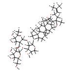
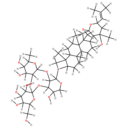
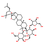
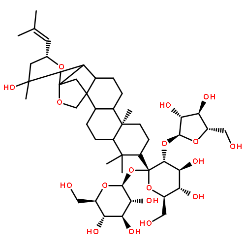
Ethnopharmacological relevanceJujuboside A (JuA) is a main active ingredient of semen ziziphi spinosae, which can significantly reduce spontaneous activity in mammals, increase the speed of falling asleep, prolong the sleeping time as well as improve the sleeping efficiency. In this study, the mechanism and the pathway of the sedative and hypnotic effect of JuA were investigated.Materials and methodsAfter being treated with JuA (in vitro), the rat׳s small intestine tissues cultures were used to stimulate the brain tissues. Then 27 cytokine levels were detected in the two kinds of tissue culture via liquid protein chip technology; In addition, the cultured hippocampal neurons of rat were treated with JuA, and γ-aminobutyric acid (GABA) receptor subunits (GABAAα1, GABAAα5, GABAAβ1 and GABABR1) mRNAs were evaluated by Real-time PCR.ResultsThe levels of IL-1α, MIP-1α, IL-1β and IL-2 were reduced significantly after 3 h of treating the small intestine tissues with JuA (200 µl/ml), and the concentration change rates, in order, were −59.3%, −3.59%, −50.1% and −49.4%; these cytokines were transmitted to brain tissues 2 h later, which could lead to significant levels of reduction of IL-1α, IFN-γ, IP-10 and TNF-α; the concentration change rates were −62.4%, −25.7%, −55.2% and −38.5%, respectively. Further, the intercellular communication network diagram was mapped out, which could suggest the mechanism and the pathway of the sedative and hypnotic effect of JuA. The results also indicated that JuA (50 µl/ml) increased significantly GABAAα1 receptor mRNAs and reduced GABABR1, mRNAs in hippocampal neurons after 24 h of stimulation; however, all the mRNA transcription levels of GABAAα1,GABAAα5, GABAAβ1 and GABABR1 receptors increased significantly after 48 h.ConclusionJuA performed its specific sedative and hypnotic effect through not only adjusting GABA receptors subunit mRNAs expression, but also down-regulating the secretion of relevant inflammation cytokines on the intestinal mucosal system to affect the intercellular cytokine network between nerve cells in the brain. This mechanism is similar to that of melatonin.Download high-res image (140KB)Download full-size image
Co-reporter:Xi-xi Wang, Gu-ijie Ma, Jun-bo Xie, Guang-chang Pang
Journal of Ethnopharmacology (15 January 2015) Volume 159() pp:215-223
Publication Date(Web):15 January 2015
DOI:10.1016/j.jep.2014.11.012
Ethnopharmacological relevanceJujuboside A (JuA) is a main active ingredient of semen ziziphi spinosae, which can significantly reduce spontaneous activity in mammals, increase the speed of falling asleep, prolong the sleeping time as well as improve the sleeping efficiency. In this study, the mechanism and the pathway of the sedative and hypnotic effect of JuA were investigated.Materials and methodsAfter being treated with JuA (in vitro), the rat׳s small intestine tissues cultures were used to stimulate the brain tissues. Then 27 cytokine levels were detected in the two kinds of tissue culture via liquid protein chip technology; In addition, the cultured hippocampal neurons of rat were treated with JuA, and γ-aminobutyric acid (GABA) receptor subunits (GABAAα1, GABAAα5, GABAAβ1 and GABABR1) mRNAs were evaluated by Real-time PCR.ResultsThe levels of IL-1α, MIP-1α, IL-1β and IL-2 were reduced significantly after 3 h of treating the small intestine tissues with JuA (200 µl/ml), and the concentration change rates, in order, were −59.3%, −3.59%, −50.1% and −49.4%; these cytokines were transmitted to brain tissues 2 h later, which could lead to significant levels of reduction of IL-1α, IFN-γ, IP-10 and TNF-α; the concentration change rates were −62.4%, −25.7%, −55.2% and −38.5%, respectively. Further, the intercellular communication network diagram was mapped out, which could suggest the mechanism and the pathway of the sedative and hypnotic effect of JuA. The results also indicated that JuA (50 µl/ml) increased significantly GABAAα1 receptor mRNAs and reduced GABABR1, mRNAs in hippocampal neurons after 24 h of stimulation; however, all the mRNA transcription levels of GABAAα1,GABAAα5, GABAAβ1 and GABABR1 receptors increased significantly after 48 h.ConclusionJuA performed its specific sedative and hypnotic effect through not only adjusting GABA receptors subunit mRNAs expression, but also down-regulating the secretion of relevant inflammation cytokines on the intestinal mucosal system to affect the intercellular cytokine network between nerve cells in the brain. This mechanism is similar to that of melatonin.Download high-res image (140KB)Download full-size image
Co-reporter:Xiaotong Zhao, Junjun Liu, Zhiyou Wen, Yanqing Zhang, Mingxin Yu, Bingcheng Pan, Jun Zeng, Junbo Xie
Journal of Chromatography B (1 March 2017) Volume 1046() pp:18-25
Publication Date(Web):1 March 2017
DOI:10.1016/j.jchromb.2017.01.030


![1-[4-(3-METHYL-BUTOXY)-PHENYL]-ETHANONE](http://img.cochemist.com/ccimg/30300/30237-26-4.png)
![1-[4-(3-METHYL-BUTOXY)-PHENYL]-ETHANONE](http://img.cochemist.com/ccimg/30300/30237-26-4_b.png)