Co-reporter:Bin Liu, Xiaoli Gou, Xupeng Bai, Xiangyu Hou, Dongshun Li, Guoping Zhong, Jing Jin, Min Huang
Journal of Pharmaceutical and Biomedical Analysis 2015 Volume 107() pp:346-354
Publication Date(Web):25 March 2015
DOI:10.1016/j.jpba.2015.01.001
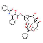
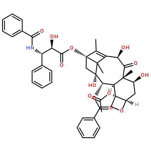
•A novel UPLC–MS/MS for the determination of seven taxoids in rat plasma was established.•The validated method has been successfully applied to a pharmacokinetic study.•First report on the pharmacokinetics of taxoids after administration of T. yunnanensis extract.A rapid, sensitive and reliable method has been developed and validated for the simultaneous determination of seven taxoids including 10-deacetylbaccatin III (10-DAB III), baccatin III, 5-epi-canadensene, taxinine M, 10-deacetyltaxol (10-DAT), cephalomannine and paclitaxel in rat plasma using docetaxel as the internal standard (IS). The plasma samples were pretreated by liquid–liquid extraction with methyl tert-butyl ether. The chromatographic separation was achieved on a C18 column (50 mm × 2.1 mm, 1.8 μm, Waters, USA) with a gradient elution program consisting of methanol and water (containing 0.1% formic acid) at a flow rate of 0.2 mL/min. Detection was performed under the selected reaction monitoring (SRM) scan using an electrospray ionization (ESI) in the positive ion mode. The mass transitions were as follows: m/z 567.4 → 444.9 for 10-DAB III, m/z 609.0 → 549.3 for baccatin III, m/z 617.4 → 496.9 for 5-epi-canadensene, m/z 709.6 → 649.3 for taxinine M, m/z 834.8 → 307.9 for 10-DAT, m/z 854.5 → 285.4 for cephalomannine, m/z 876.8 → 307.3 for paclitaxel and m/z 830.8 → 549.6 for IS, respectively. All calibration curves exhibited good linearity (r2 > 0.99) over a wide concentration range for all components. The intra-day and inter-day precisions at three different levels were both less than 14.3% in terms of relative standard deviation (RSD) and the accuracies ranged from −8.3% to 14.8% in terms of relative error (RE). The extraction recoveries of the seven compounds ranged from 62.5% to 100.5%. The developed method was successfully applied to the pharmacokinetic study of the seven taxoids in rat plasma after oral administration of the crude extract of the twigs and leaves of Taxus yunnanensis.
Co-reporter:Bao Yang, Tao Yang, Qinglong Tan, Jinping Zhu, Lian Zhou, Tianqin Xiong, Zhongxiang Zhao, Jing Jin
Phytochemistry Letters 2015 13() pp: 302-307
Publication Date(Web):
DOI:10.1016/j.phytol.2015.07.008
Co-reporter:Bao Yang, Tao Yang, Qinglong Tan, Jinping Zhu, Lian Zhou, Tianqin Xiong, Zhongxiang Zhao, Jing Jin
Phytochemistry Letters (September 2015) Volume 13() pp:302-307
Publication Date(Web):1 September 2015
DOI:10.1016/j.phytol.2015.07.008
•Two new ursane-type and thirteen known triterpene saponins were identified from the leaves of Ilex kudingcha.•All the compounds were screened for antiplatelet aggregation activity induced by ADP (5 μM) in vitro.•Six compounds showed significant inhibition of platelet aggregation.Two new ursane-type triterpene saponins, 3-O-β-d-glucopyranosyl(1 → 3)-[α-l-rhamnopyranosyl(1 → 2)]-α-l-arabinopyranosylurs-12,19(29)-dien-28-oic acid 28-O-α-l-rhamnopyranosyl(1 → 2)-β-d-glucopyranosyl ester (1) and 3-O-β-d-glucopyranosyl(1 → 3)-[α-l-rhamnopyranosyl(1 → 2)]-α-l-arabinopyranosyl-19α,20α-dihydroxyurs-12-en-28-oic acid 28-O-α-l-rhamnopyranosyl(1 → 2)-β-d-glucopyranosyl ester (2), along with thirteen known triterpene saponins were isolated from the n-BuOH part of the MeOH extraction of the leaves of Ilex kudingcha C.J. Tseng (also called “Ku-Ding-Cha”). The structures of new compounds were elucidated on the basis of detailed spectroscopic analysis, including HR-ESI-TOF-MS, 1D and 2D-NMR experiments, and by acid hydrolysis. All the compounds were screened for antiplatelet aggregation activity in vitro, and compounds 1, 2, 3, 7, 12 and 15 showed significant inhibition of platelet aggregation induced by ADP (5 μM) with IC50 values of 14.7 ± 3.7, 11.3 ± 2.5, 17.4 ± 4.6, 20.5 ± 3.1, 8.1 ± 1.5 and 18.9 ± 4.2 μM, respectively.Download full-size image
Co-reporter:Xinmeng Chen, Zhongxiang Zhao, Yibei Chen, Xiaoli Gou, Ziyi Zhou, Guoping Zhong, Yefeng Cai, Min Huang, Jing Jin
Journal of Ethnopharmacology (4 November 2016) Volume 192() pp:362-369
Publication Date(Web):4 November 2016
DOI:10.1016/j.jep.2016.07.066
Ethnopharmacological relevanceThe Dengzhan Shengmai capsule (DZSM) is known in China for its remarkable curative effect as a treatment of cardiovascular diseases, such as coronary heart disease and ischemic stroke. DZSM is a Chinese herbal compound preparation that consists of four ingredients, including Erigeron breviscapus (Vaniot) Hand.-Mazz., Panax ginseng C.A. Mey, Ophiopogon japonicas (Thunb.) Ker-Gawl. and Schisandra chinensis (Turcz.) Baill., and was indexed in the Chinese Pharmacopoeia 2010. DZSM and clopidogrel are often co-prescribed in the clinic to prevent the recurrence of stroke or other cardiovascular and cerebrovascular diseases. However, the effect of DZSM on the pharmacokinetics of clopidogrel remains unclear.Aim of the studyThe purpose of the study is to explore the pharmacokinetics and potential interaction between DZSM and clopidogrel and the underlying mechanism.Materials and methodsRats were used to investigate the effect of DZSM on the pharmacokinetics of clopidogrel and its active metabolite in vivo. The plasma concentrations were simultaneously determined using LC–MS/MS. The effects of DZSM on the P-gp-mediated efflux transport and CYP450-mediated metabolism of clopidogrel were investigated using MDCKII-MDR1 cells and rat liver microsomes, respectively.ResultsAfter pretreatment with DZSM, the Cmax and AUC0–∞ of clopidogrel increased from 0.4±0.1 to 1.7±0.6 ng/mL and 0.9±0.4 to 2.0±0.2 ng/mL h, respectively. The Cmax and AUC0–∞ of the derivatized active metabolite of clopidogrel decreased from 8.2±1.2 to 2.8±0.5 ng/mL and 18.2±5.6 to 6.4±3.7 ng h/mL, respectively. In MDCKII-MDR1 cells, the P-gp-mediated efflux transport of clopidogrel was significantly inhibited by the DZSM extract. In rat liver microsomes, DZSM inhibited clopidogrel metabolism with an IC50 of 0.02 mg/mL.ConclusionsDZSM significantly affects the pharmacokinetics of clopidogrel and its active metabolite by inhibiting the P-gp-mediated efflux transport and CYP450-mediated metabolism of clopidogrel. Thus, caution is needed when DZSM is co-administered with clopidogrel in the clinic because the interaction of these drugs may result in altered plasma concentrations of clopidogrel and its active metabolite.Download high-res image (210KB)Download full-size image
Co-reporter:Xupeng Bai, Weipeng Hong, Peiheng Cai, Yibei Chen, Chuncao Xu, Di Cao, Weibang Yu, Zhongxiang Zhao, Min Huang, Jing Jin
Toxicology and Applied Pharmacology (1 June 2017) Volume 324() pp:12-25
Publication Date(Web):1 June 2017
DOI:10.1016/j.taap.2017.03.022
•• VPA induced hepatic steatosis and modulated genes associated with lipid metabolism.•• CD36-mediated fatty acid uptake contributed to VPA-induced lipid accumulation.•• VPA increased the hepatic level of DGAT2 through inhibiting MEK-ERK pathway and enhanced triglyceride synthesis.•• SREBP-1c-mediated fatty acid synthesis was not involved in VPA-induced hepatic steatosis.Steatosis is the characteristic type of VPA-induced hepatotoxicity and may result in life-threatening hepatic lesion. Approximately 61% of patients treated with VPA have been diagnosed with hepatic steatosis through ultrasound examination. However, the mechanisms underlying VPA-induced intracellular fat accumulation are not yet fully understood. Here we demonstrated the involvement of fatty acid uptake and lipogenesis in VPA-induced hepatic steatosis in vitro and in vivo by using quantitative real-time PCR (qRT-PCR) analysis, western blotting analysis, fatty acid uptake assays, Nile Red staining assays, and Oil Red O staining assays. Specifically, we found that the expression of cluster of differentiation 36 (CD36), an important fatty acid transport, and diacylglycerol acyltransferase 2 (DGAT2) were significantly up-regulated in HepG2 cells and livers of C57B/6J mice after treatment with VPA. Furthermore, VPA treatment remarkably enhanced the efficiency of fatty acid uptake mediated by CD36, while this effect was abolished by the interference with CD36-specific siRNA. Also, VPA treatment significantly increased DGAT2 expression as a result of the inhibition of mitogen-activated protein kinase kinase (MEK) – extracellular regulated kinase (ERK) pathway; however, DGAT2 knockdown significantly alleviated VPA-induced intracellular lipid accumulation. Additionally, we also found that sterol regulatory element binding protein-1c (SREBP-1c)-mediated fatty acid synthesis may be not involved in VPA-induced hepatic steatosis. Overall, VPA-triggered over-regulation of CD36 and DGAT2 could be helpful for a better understanding of the mechanisms underlying VPA-induced hepatic steatosis and may offer novel therapeutic strategies to combat VPA-induced hepatotoxicity.Download high-res image (190KB)Download full-size image
Co-reporter:Wenwen Wang, Cuiwen Guan, Xiaozhe Sun, Zhongxiang Zhao, Jia Li, Xinlu Fu, Yuwen Qiu, Min Huang, Jing Jin, Zhiying Huang
Phytomedicine (1 June 2016) Volume 23(Issue 6) pp:589-596
Publication Date(Web):1 June 2016
DOI:10.1016/j.phymed.2016.02.022
BackgroundTanshinone IIA (Tan), the main active component of Salvia miltiorrhiza, has been demonstrated to have antioxidant activity. Acetaminophen (APAP), a widely used antipyretic and analgesic, can cause severe hepatotoxicity and liver failure when taken overdose. Oxidative stress has been reported to be involved in APAP-induced liver failure.PurposeThis study aimed to investigate the effect of Tan on APAP-induced hepatotoxicity and the underlying mechanisms involved.Study DesignC57BL/6 J mice were divided into six groups: (1) control, (2) APAP group, (3) APAP + Tan (30 mg/kg) group, (4) Tan (30 mg/kg) group, (5) APAP + Tan (10 mg/kg) group, (6) Tan (10 mg/kg) group. Mice in group 3 and 5 were pre-treated with specified dose of Tan by gavage and subsequently injected with an overdose of APAP intraperitoneally (i.p., 300 mg/kg). The effect of Tan on Nrf2 pathway was investigated in HepG2 cells and mice.MethodsPlasma aspartate transaminase (ALT), aspartate transaminase (AST), lactate dehydrogenase (LDH), liver glutathione (GSH), glutathione transferase (GST), glutathione peroxidase (GPx), superoxide dismutase (SOD) and catalase (CAT) levels were determined after mice were sacrificed. Lipid peroxidation and histological examination were performed. The effect of Tan on the Nrf2 pathway was detected by western blotting and qRT-PCR.ResultsTan pretreatment reduced APAP-induced liver injury. Tan was able to activate Nrf2 and increase the expression levels of Nrf2 target genes, including glutamate-cysteine ligase catalytic subunit (GCLC), NAD(P)H:quinine oxidoreductase 1 (NQO1) and hemeoxygenase-1 (HO-1), in a dose-dependent manner in HepG2 cells. Consistent with our observations in HepG2 cells, Tan increased nuclear Nrf2 accumulation and upregulated mRNA and protein levels of the Nrf2 target genes GCLC, NQO1 and HO-1 in C57BL/6 J mice compared with mice treated with APAP alone.ConclusionsOur results demonstrate that Tan pretreatment could protect the liver from APAP-induced hepatic injury by activating the Nrf2 pathway. Tan may provide a new strategy for the protection against APAP-induced liver injury.Download high-res image (109KB)Download full-size image


![2-Propenoic acid,3-phenyl-,(1S,3aR,4R,5R,5aR,7S,9S,9aR,10R,11S,11aR)-5,9,10,11-tetrakis(acetyloxy)-9a-[(benzoyloxy)methyl]tetradecahydro-11a-hydroxy-1,3a-dimethyl-6-methylene-13-oxo-1,4-ethanobenzo[5,6]cycloocta[1,2-c]furan-7-ylester, (2E)-](http://img.cochemist.com/ccimg/117300/117229-54-6.png)
![2-Propenoic acid,3-phenyl-,(1S,3aR,4R,5R,5aR,7S,9S,9aR,10R,11S,11aR)-5,9,10,11-tetrakis(acetyloxy)-9a-[(benzoyloxy)methyl]tetradecahydro-11a-hydroxy-1,3a-dimethyl-6-methylene-13-oxo-1,4-ethanobenzo[5,6]cycloocta[1,2-c]furan-7-ylester, (2E)-](http://img.cochemist.com/ccimg/117300/117229-54-6_b.png)


![Benzenepropanoic acid, a-hydroxy-b-[(1-oxohexyl)amino]-,(2aR,4S,4aS,6R,9S,11S,12S,12aR,12bS)-12b-(acetyloxy)-12-(benzoyloxy)-2a,3,4,4a,5,6,9,10,11,12,12a,12b-dodecahydro-6,11-dihydroxy-4a,8,13,13-tetramethyl-5-oxo-4-(b-D-xylopyranosyloxy)-7,11-methano-1H-cyclodeca[3,4]benz[1,2-b]oxet-9-ylester, (aR,bS)-](http://img.cochemist.com/ccimg/90400/90332-65-3.png)
![Benzenepropanoic acid, a-hydroxy-b-[(1-oxohexyl)amino]-,(2aR,4S,4aS,6R,9S,11S,12S,12aR,12bS)-12b-(acetyloxy)-12-(benzoyloxy)-2a,3,4,4a,5,6,9,10,11,12,12a,12b-dodecahydro-6,11-dihydroxy-4a,8,13,13-tetramethyl-5-oxo-4-(b-D-xylopyranosyloxy)-7,11-methano-1H-cyclodeca[3,4]benz[1,2-b]oxet-9-ylester, (aR,bS)-](http://img.cochemist.com/ccimg/90400/90332-65-3_b.png)


![Thieno[3,2-c]pyridine-5(4H)-aceticacid, a-(2-chlorophenyl)-6,7-dihydro-,methyl ester](http://img.cochemist.com/ccimg/90100/90055-48-4.png)
![Thieno[3,2-c]pyridine-5(4H)-aceticacid, a-(2-chlorophenyl)-6,7-dihydro-,methyl ester](http://img.cochemist.com/ccimg/90100/90055-48-4_b.png)


![5-(2-Chlorobenzyl)-4,5,6,7-tetrahydrothieno[3,2-c]pyridine](http://img.cochemist.com/ccimg/55200/55142-85-3.png)
![5-(2-Chlorobenzyl)-4,5,6,7-tetrahydrothieno[3,2-c]pyridine](http://img.cochemist.com/ccimg/55200/55142-85-3_b.png)
![5-Heptenoic acid,7-[(2R,3S,4S)-tetrahydro-4,6-dihydroxy-2-[(1E,3S)-3-hydroxy-1-octen-1-yl]-2H-pyran-3-yl]-,(5Z)-](http://img.cochemist.com/ccimg/54400/54397-85-2.png)
![5-Heptenoic acid,7-[(2R,3S,4S)-tetrahydro-4,6-dihydroxy-2-[(1E,3S)-3-hydroxy-1-octen-1-yl]-2H-pyran-3-yl]-,(5Z)-](http://img.cochemist.com/ccimg/54400/54397-85-2_b.png)


![Benzene, 1-[2-(3,5-dimethoxyphenyl)ethenyl]-2,4-dimethoxy-](http://img.cochemist.com/ccimg/20600/20578-92-1.png)
![Benzene, 1-[2-(3,5-dimethoxyphenyl)ethenyl]-2,4-dimethoxy-](http://img.cochemist.com/ccimg/20600/20578-92-1_b.png)