Co-reporter:Marieke Poß, Robert J. Tower, Joanna Napp, Lia Christina Appold, Twan Lammers, Frauke Alves, Claus-Christian Glüer, Susann Boretius, and Claus Feldmann
Chemistry of Materials April 25, 2017 Volume 29(Issue 8) pp:3547-3547
Publication Date(Web):March 30, 2017
DOI:10.1021/acs.chemmater.6b05406
Multimodal contrast agents with high biocompatibility and biodegradability, as well as low material complexity, are in great demand for clinical diagnostics at different scales of resolution and/or for translating preclinical diagnosis into intraoperative imaging. Multimodality, however, often results in multicomponent and multistructured materials with complexity becoming a severe restriction for synthesis, approval, and use in routine clinical practice. Here, we present sulfonate-based saline [GdO]+[ICG]− (ICG, indocyanine green) inorganic-organic hybrid nanoparticles (IOH-NPs with an inorganic [GdO]+ cation and an organic [ICG]− anion) as a novel, multimodality contrast agent for optical, photoacoustic, and magnetic resonance imaging (OI, PAI, MRI). [GdO]+[ICG]− IOH-NPs have a plain composition based on clinically used constituents and are prepared as an insoluble saline compound in water. The high [ICG]− content (81 wt %) ensures intense near-infrared emission (780–840 nm) and a strong photoacoustic signal. First, in vitro studies demonstrate longer detectability and greater emission intensity for [GdO]+[ICG]− IOH-NP suspensions than for ICG solutions, as well as a reduced toxicity compared to that of Gd-DTPA, a standard MRI contrast agent. Conceptual in vivo studies confirm the utility of the [GdO]+[ICG]− IOH-NPs for optical and magnetic resonance imaging with a T1 relaxivity better than that of Gd-DTPA. Taken together, [GdO]+[ICG]− represents a new compound and nanomaterial that can be highly interesting as a multimodal contrast agent.
Co-reporter:J. Jung-König;M. Sanhaji;R. Popescu;C. Seidl;E. Zittel;U. Schepers;D. Gerthsen;I. Hilger;C. Feldmann
Nanoscale (2009-Present) 2017 vol. 9(Issue 24) pp:8362-8372
Publication Date(Web):2017/06/22
DOI:10.1039/C7NR01784G
Gadolinium carbonate (Gd2(CO3)3) hollow nanospheres and their suitability for drug transport and magnetothermally-induced drug release are presented. The hollow nanospheres are prepared via a microemulsion-based synthesis using tris(tetramethylcyclopentadienyl)gadolinium(III) and CO2 as the starting materials. Size, structure and composition of the as-prepared Gd2(CO3)3 hollow nanospheres are comprehensively validated by several independent analytical methods (HRTEM, HAADF-STEM, DLS, EDXS, XRD, FT-IR, DTA-TG). Accordingly, they exhibit an outer diameter of 26 ± 4 nm, an inner cavity of 7 ± 2 nm, and a wall thickness of 9 ± 3 nm. As a conceptual study, the nanocontainer-functionality of the Gd2(CO3)3 hollow nanospheres is validated upon filling with the anti-cancerogenic agent doxorubicin (DOX), which is straightforward via the microemulsion (ME) strategy. The resulting DOX@Gd2(CO3)3 nanocontainers provide the option of multimodal imaging including optical and magnetic resonance imaging (OI, MRI) as well as magnetothermal heating and drug release. As a proof-of-concept, we could already prove the intrinsic DOX-based fluorescence, a low systemic toxicity according to in vitro studies as well as the magnetothermal effect and a magnetothermally-induced DOX release. In particular, the latter is new for Gd-containing nanoparticles and highly promising in view of theranostic nanocontainers and synergistic physical and chemical tumor treatment.
Co-reporter:David Hausmann;Ralf Köppe;Silke Wolf;Peter W. Roesky
Dalton Transactions 2017 vol. 46(Issue 16) pp:5457-5457
Publication Date(Web):2017/04/19
DOI:10.1039/C7DT90063E
Correction for ‘Ionic-liquid-assisted synthesis of the phosphorus interhalides [PBr4][IBr2] and [PBr4][I5Br7]’ by David Hausmann et al., Dalton Trans., 2016, 45, 16526–16532.
Co-reporter:Fabian Gyger, Pascal Bockstaller, Dagmar Gerthsen, and Claus Feldmann
Chemistry of Materials 2016 Volume 28(Issue 21) pp:7816
Publication Date(Web):October 5, 2016
DOI:10.1021/acs.chemmater.6b03219
Liquid-crystalline phases in liquid ammonia were used to obtain meso- and microporous Si3N4, TiN, and VN. The liquid-crystalline phase was established at −50 °C with liquid ammonia as the polar phase, heptane as the nonpolar dispersant phase, dimethyldioctylammonium iodide (DDAI) as the surfactant, and heptylamine as the cosurfactant. Silicon(IV) iodide, tetrakis(dimethylamino)titan, and vanadium(IV) chloride were added and ammonolyzed in the liquid-crystalline phase. After the mixture contents were separated and washed, ammonolysis was completed by slow heating to 600 °C (TiN, VN) and 800 °C (Si3N4) in vacuum or forming gas (N2/H2). The obtained high-surface nitrides are characterized by high purity (e.g., Si3N4 with carbon content <3 at %), great specific surface area (Si3N4: 610 m2/g; TiN: 203 m2/g; VN: 63 m2/g), and great total pore volume (Si3N4: 1.5 cm3/g; TiN: 0.3 cm3/g; VN: 0.4 cm3/g). Si3N4 and TiN show considerable meso- and microporosity, whereas the less-stable VN only shows mesoporosity. Electron energy loss spectroscopy (EELS) proves low oxygen contents. As a proof-of-concept, uniform one-pot modification of Si3N4 with Pd nanoparticles (Pd@Si3N4) as well as the reversible hydrogen sorption of differently treated Si3N4 were studied, which results in a maximum H2 uptake of 2.5 wt % (at 25 °C, 40 bar).
Co-reporter:Ying-Chu Chen, Yan-Gu Lin, Liang-Ching Hsu, Alexander Tarasov, Po-Tuan Chen, Michitoshi Hayashi, Jan Ungelenk, Yu-Kuei Hsu, and Claus Feldmann
ACS Catalysis 2016 Volume 6(Issue 4) pp:2357
Publication Date(Web):February 23, 2016
DOI:10.1021/acscatal.5b02444
A distinct morphology of β-SnWO4 with hierarchically multiarmed architecture and overall hexahedral symmetry – entitled as spikecube – is fabricated for the first time via a polyol-mediated synthesis. The growth of the β-SnWO4 spikecubes is investigated and attributed to thermodynamic and kinetic control. In a sequential reaction, crystalline cubes of β-SnWO4 enclosed by {100} facets grow in a first Ostwald ripening-based step. A kinetically controlled growth process to spikecubes follows under formation of multiarmed spikes on the facets of the cubic seeds. Such a growth process differs significantly from the literature concerning highly branched crystals. The synergistic effect of morphological modification (i.e., introducing more surface reaction sites) and textural alteration (i.e., incorporation of the p-block Sn2+ into simple tungsten oxide to reframe its band structure) leads to an enhanced photocatalytic activity of the β-SnWO4 spikecubes being 150% higher in comparison to benchmark WO3 photocatalysts.Keywords: hierarchical structure; multibranch; photocatalyst; polyol; tin tungstate
Co-reporter:Christian Schöttle, Dmitry E. Doronkin, Radian Popescu, Dagmar Gerthsen, Jan-Dierk Grunwaldt and Claus Feldmann
Chemical Communications 2016 vol. 52(Issue 37) pp:6316-6319
Publication Date(Web):29 Mar 2016
DOI:10.1039/C6CC01957A
Metallic titanium (Ti0) nanoparticles, 1.5 ± 0.4 nm in diameter, are obtained via lithium naphthalenide ([LiNaph])-driven reduction of TiCl4 × 2THF in tetrahydrofuran (THF). HRTEM, fast Fourier transformation (FFT), optical spectra and X-ray absorption near edge structure (XANES) confirm their chemical composition. Besides their pyrophoric properties, their high reactivity is validated by direct transformation of Ti0 into TiC maintaining the size.
Co-reporter:David Hausmann and Claus Feldmann
Inorganic Chemistry 2016 Volume 55(Issue 12) pp:6141-6147
Publication Date(Web):June 9, 2016
DOI:10.1021/acs.inorgchem.6b00663
The bromine-rich zinc bromides Zn6Br12(18-crown-6)2×(Br2)5 (1), Zn4Br8(18-crown-6)2×(Br2)3 (2), and Zn6Br12(18-crown-6)2×(Br2)2 (3) are prepared by reaction of ZnBr2, 18-crown-6, and elemental bromine in the ionic liquid [MeBu3N][N(Tf)2] (N(Tf)2 = bis(trifluoromethylsulfonyl)amide). Zn6Br12(18-crown-6)2×(Br2)5 (1) is formed instantaneously by the reaction. Even at room temperature, compound 1 releases bromine, which was confirmed by thermogravimetry (TG) and mass spectrometry (MS). The release of Br2 can also be directly followed by the color and density of the title compounds. With controlled conditions (2 weeks, 25 °C, absence of excess Br2) Zn6Br12(18-crown-6)2×(Br2)5 (1) slowly releases bromine with conconcurrent generation of Zn4Br8(18-crown-6)2×(Br2)3 (2) (in ionic liquid) and Zn6Br12(18-crown-6)2×(Br2)2 (3) (in inert oil). All bromine-rich zinc bromides contain voluminous uncharged (e.g., Zn3Br6(18-crown-6), Zn2Br4(18-crown-6)) or ionic (e.g., [Zn2Br3(18-crown-6)]+, [(Zn2Br6)×(Br2)2]2–) building units with dibromine molecules between the Zn oligomers and partially interconnecting the Zn-containing building units. Due to the structural similarity, the bromine release is possible via crystal-to-crystal transformation with retention of the crystal shape.
Co-reporter:Joachim G. Heck, Claus Feldmann
Journal of Colloid and Interface Science 2016 Volume 481() pp:69-74
Publication Date(Web):1 November 2016
DOI:10.1016/j.jcis.2016.07.030
•Synthesis of [ZrO]2+[AAP]2− (AAP: Acetaminophenphosphate) inorganic-organic hybrid nanoparticles presented.•Water-based synthesis results in particle diameter of 37 nm and excellent colloidal stability.•Nanoparticles exhibit extraordinary high prodrug load of 68 wt.% AAP.•[ZrO]2+[AAP]2− shows continuous release of analgetic acetaminophen on a timescale of 48 h.Drug release belongs to the most challenging aspects of nanoparticles addressing molecular biology and medicine. Besides targeted delivery, obvious challenges are related to high drug load and continuous slow drug release. Based on our recently developed concept of inorganic-organic hybrid nanoparticles (IOH-NP), we here present [ZrO]2+[AAP]2− IOH-NPs containing the analgetic phosphate prodrug acetaminophen phosphate for drug release. [ZrO]2+[AAP]2− combines an uncomplex synthesis in water with a high prodrug load of 68 wt.%. [ZrO]2+[AAP]2− nanoparticles exhibit a diameter of 37(11) nm and can be readily obtained as colloidally highly stable suspension in water. The chemical composition is studied in detail based on infrared spectroscopy, energy-dispersive X-ray analysis, thermogravimetry and elemental analysis. Moreover, the release of acetaminophen from [ZrO]2+[AAP]2− is studied by means of model experiments indicating the carbon content of the nanoparticles and, in alternative, the fluorescence of labeled nanoparticles. Both data show a continuous release of 80 wt.% of the analgetic acetaminophen on a time scale up to 48 h.
Co-reporter:David Hausmann, Ana Kuzmanoski and Claus Feldmann
Dalton Transactions 2016 vol. 45(Issue 15) pp:6541-6547
Publication Date(Web):24 Feb 2016
DOI:10.1039/C6DT00458J
The reaction of manganese(II) bromide and the crown ether 18-crown-6 in the ionic liquid [(n-Bu)3MeN][N(Tf)2] under mild conditions (80–130 °C) resulted in the formation of three different coordination compounds: MnBr2(18-crown-6) (1), Mn3Br6(18-crown-6)2 (2) and Mn3Br6(18-crown-6) (3). In general, the local coordination and the crystal structure of all compounds are driven by the mismatch between the small radius of the Mn2+ cation (83 pm) and the ring opening of 18-crown-6 as a chelating ligand (about 300 pm). This improper situation leads to different types of coordination and bonding. MnBr2(18-crown-6) represents a molecular compound with Mn2+ coordinated by two bromine atoms and only five oxygen atoms of 18-crown-6. Mn3Br6(18-crown-6)2 falls into a [MnBr(18-crown-6)]+ cation – with Mn2+ coordinated by six oxygen atoms and Br – and a [MnBr(18-crown-6)MnBr4]− anion. In this anion, Mn2+ is coordinated by five oxygen atoms of the crown ether as well as by two bromine atoms, one of them bridging to an isolated (MnBr4) tetrahedron. Mn3Br6(18-crown-6), finally, forms an infinite, non-charged 1∞[Mn2(18-crown-6)(MnBr6)] chain. Herein, 18-crown-6 is exocyclically coordinated by two Mn2+ cations. All compounds show intense luminescence in the yellow to red spectral range and exhibit remarkable quantum yields of 70% (Mn3Br6(18-crown-6)) and 98% (Mn3Br6(18-crown-6)2). The excellent quantum yield of Mn3Br6(18-crown-6)2 and its differentiation from MnBr2(18-crown-6) and Mn3Br6(18-crown-6) can be directly correlated to the local coordination.
Co-reporter:Ana Kuzmanoski, Vladimir Pankratov, Claus Feldmann
Journal of Luminescence 2016 Volume 179() pp:555-561
Publication Date(Web):November 2016
DOI:10.1016/j.jlumin.2016.07.040
CaF2:Pr (1 mol%), CaF2:Mn (5 mol%) and CaF2:Pr,Mn (1 mol%, 5 mol%) nanoparticles are prepared via a microwave-mediated synthesis in ionic liquids. The nanoparticles are highly crystalline and exhibit particle diameters <50 nm.In contrast to bulk-CaF2:Pr,Mn,energy transfer between Pr3+and Mn2+under 1S0→1I6 relaxation on Pr3+ and 4G(4T1g)→6S(A1g) emission of Mn2+ is observed for the first time. Such energy transfer represents the essential first step of the quantum-cutting cascade via the Pr3+–Mn2+ couple, which is most interesting as both expected photons – 3P0→3H4 emission of Pr3+and 4G(4T1g)→6S(A1g) emission of Mn2+ – are emitted in the green spectral range. While bulk crystals were said not to show energy transfer due to prohibiting selection rules, vacuum ultraviolet (VUV) spectroscopy of CaF2:Pr, Mn nanoparticles firstly proves efficient Pr3+→Mn2+ energy transfer, which can be ascribed to the reduced site symmetry and considerable spin–orbit interaction in the nanocrystals.
Co-reporter:Sara Simonato, Jens Möllmer, Marcus Lange, Roger Gläser, Reiner Staudt and Claus Feldmann
RSC Advances 2016 vol. 6(Issue 15) pp:12446-12452
Publication Date(Web):22 Jan 2016
DOI:10.1039/C5RA24657A
Magnesium 2-aminoethylphosphonate (Mg2O(2AEP) × 4H2O) nanoparticles (particle diameter: 20–30 nm; specific surface area: 360 m2 g−1) are presented for selective separation of CO2 and CH4. Due to the base amino function, the nanoparticles can reversibly absorb CO2 with a maximal uptake of 153 mg g−1. Absorption and desorption are studied by infrared spectroscopy as well as by gravimetric sorption analysis. Furthermore, Mg2O(2AEP) × 4H2O shows reversible selective separation of CO2 from CH4. Here, pure and mixed gas adsorption isotherms (25 °C, 25 bar) of CO2 and CH4 show maximal uptakes of 153 mg g−1 (CO2) and 15 mg g−1 (CH4). Especially, data of mixed gas isotherms are comparably rare, but highly relevant for material characterization. Experimental isotherms were fitted by a dual-site Langmuir isotherm model (CO2) and a Tòth model (CH4). Mixed adsorption isotherms were modelled by volumetric-chromatographic methods resulting in a selectivity of α = 8 to 20.
Co-reporter:Christian Schöttle, Claus Feldmann
Solid State Sciences 2016 Volume 55() pp:125-129
Publication Date(Web):May 2016
DOI:10.1016/j.solidstatesciences.2016.03.002
•Sodium naphthalenide-driven reduction of ZnCl2 to Zn0 nanoparticles.•Laux-type, nitrobenzene-driven oxidation of Zn0 nanoparticles to ZnO hollow nanospheres.•ZnO hollow nanospheres exhibit an outer diameter of 10 nm and a wall thickness of 3.0 nm.•Laux oxidation performed in the liquid phase without separation of the nanoparticles.•Quantum-size effect with blue-shifted bandgap observed for ZnO hollow nanospheres.Zinc oxide hollow nanospheres were obtained via a Laux-like oxidation of zinc nanoparticles using nitrobenzene as oxidizing agent. The ZnO hollow nanospheres exhibit an outer diameter of 10.4 ± 1.3 nm and a well crystallized sphere wall with a thickness of 2.9 ± 0.4 nm. Laux-like oxidation and formation of the ZnO hollow nanospheres were performed instantaneously after sodium naphthalenide ([NaNaph]) driven reduction of ZnCl2 to Zn0 nanoparticles in the liquid phase without any separation of the intermediate Zn0 nanoparticles. The diameter of the resulting ZnO hollow nanospheres (10.4 ± 1.3 nm) reflects the diameter of the intermediate Zn0 nanoparticles (10.1 ± 2.3 nm). In accordance with the small diameter of the ZnO sphere wall, quantum-size effects occur with a band gap that is blue-shifted by 0.2 eV in comparison to bulk-ZnO.
Co-reporter:Carmen Seidl, Jan Ungelenk, Eva Zittel, Thomas Bergfeldt, Jonathan P. Sleeman, Ute Schepers, and Claus Feldmann
ACS Nano 2016 Volume 10(Issue 3) pp:3149
Publication Date(Web):February 19, 2016
DOI:10.1021/acsnano.5b03060
The nanoparticulate inorganic photosensitizer β-SnWO4 is suggested for photodynamic therapy (PDT) of near-surface tumors via reiterated 5 min blue-light LED illumination. β-SnWO4 nanoparticles are obtained via water-based synthesis and comprise excellent colloidal stability under physiological conditions and high biocompatibility at low material complexity. Antitumor and antimetastatic effects were investigated with a spontaneously metastasizing (4T1 cells) orthotopic breast cancer BALB/c mouse model. Besides protamine-functionalized β-SnWO4 (23 mg/kg of body weight, in PBS buffer), chemotherapeutic doxorubicin was used as positive control (2.5 mg/kg of body weight, in PBS buffer) and physiological saline (DPBS) as a negative control. After 21 days, treatment with β-SnWO4 resulted in a clearly inhibited growth of the primary tumor (all tumor volumes below 3 cm3) as compared to the doxorubicin and DPBS control groups (volumes up to 6 cm3). Histological evaluations of lymph nodes and lungs as well as the volume of ipsilateral lymph nodes show a remarkable antimetastatic effect being similar to chemotherapeutic doxorubicin but—according to blood counts—at significantly reduced side effects. On the basis of low material complexity, high cytotoxicity under blue-light LED illumination at low dark and long-term toxicity, β-SnWO4 can be an interesting addition to PDT and the treatment of near-surface tumors, including skin cancer, esophageal/gastric/colon tumors as well as certain types of breast cancer.Keywords: antimetastatic; nanoparticle; photodynamic therapy; tin tungstate; tumor
Co-reporter:Joachim G. Heck; Joanna Napp; Sara Simonato; Jens Möllmer; Marcus Lange; Holger M. Reichardt; Reiner Staudt; Frauke Alves
Journal of the American Chemical Society 2015 Volume 137(Issue 23) pp:7329-7336
Publication Date(Web):May 27, 2015
DOI:10.1021/jacs.5b01172
Phosphate-based inorganic–organic hybrid nanoparticles (IOH-NPs) with the general composition [M]2+[Rfunction(O)PO3]2– (M = ZrO, Mg2O; R = functional organic group) show multipurpose and multifunctional properties. If [Rfunction(O)PO3]2– is a fluorescent dye anion ([RdyeOPO3]2–), the IOH-NPs show blue, green, red, and near-infrared fluorescence. This is shown for [ZrO]2+[PUP]2–, [ZrO]2+[MFP]2–, [ZrO]2+[RRP]2–, and [ZrO]2+[DUT]2– (PUP = phenylumbelliferon phosphate, MFP = methylfluorescein phosphate, RRP = resorufin phosphate, DUT = Dyomics-647 uridine triphosphate). With pharmaceutical agents as functional anions ([RdrugOPO3]2–), drug transport and release of anti-inflammatory ([ZrO]2+[BMP]2–) and antitumor agents ([ZrO]2+[FdUMP]2–) with an up to 80% load of active drug is possible (BMP = betamethason phosphate, FdUMP = 5′-fluoro-2′-deoxyuridine 5′-monophosphate). A combination of fluorescent dye and drug anions is possible as well and shown for [ZrO]2+[BMP]2–0.996[DUT]2–0.004. Merging of functional anions, in general, results in [ZrO]2+([RdrugOPO3]1–x[RdyeOPO3]x)2– nanoparticles and is highly relevant for theranostics. Amine-based functional anions in [MgO]2+[RaminePO3]2– IOH-NPs, finally, show CO2 sorption (up to 180 mg g–1) and can be used for CO2/N2 separation (selectivity up to α = 23). This includes aminomethyl phosphonate [AMP]2–, 1-aminoethyl phosphonate [1AEP]2–, 2-aminoethyl phosphonate [2AEP]2–, aminopropyl phosphonate [APP]2–, and aminobutyl phosphonate [ABP]2–. All [M]2+[Rfunction(O)PO3]2– IOH-NPs are prepared via noncomplex synthesis in water, which facilitates practical handling and which is optimal for biomedical application. In sum, all IOH-NPs have very similar chemical compositions but can address a variety of different functions, including fluorescence, drug delivery, and CO2 sorption.
Co-reporter:H. Dong, Y.-C. Chen and C. Feldmann
Green Chemistry 2015 vol. 17(Issue 8) pp:4107-4132
Publication Date(Web):07 Jul 2015
DOI:10.1039/C5GC00943J
Since the first description by Fievet, Lagier and Figlarz in 1989, the synthesis of nanoparticles in high-boiling, multivalent alcohols – so-called polyols – has been developed into a widely applied strategy, and nowadays belongs to the standard repertoire for preparing high-quality nanomaterials. The polyols take advantage of several features such as: (i) water-comparable solubility of simple metal-salt precursors; (ii) high boiling points (up to 320 °C); (iii) reducing properties for the instantaneous synthesis of metals; (iv) coordinating properties for surface functionalization and colloidal stabilisation of nanoparticles; (v) wide adaptability of the polyols ranging from low-weight ethylene glycol (EG) to high-weight polyethylene glycols (PEGs). This review summarises the status and perspectives on nanoscaled elemental metals, metal oxides, metal chalcogenides, and non-metal elements that were prepared via the polyol synthesis. Moreover, we summarize our results and concepts to expand the limits of the polyol synthesis. This includes strategies for less-noble metal synthesis, phase transfer reactions, photochemical reduction, NMR-based characterisation of polyol-functionalised nanoparticles, realisation of phase-pure and readily crystalline metal tungstates, stabilisation of low-melting elements, and controlled thermal decomposition of polyols to obtain high-quality, lanthanide-modified carbon dots (C-dots).
Co-reporter:Marieke Poß, Joanna Napp, Oliver Niehaus, Rainer Pöttgen, Frauke Alves and Claus Feldmann
Journal of Materials Chemistry A 2015 vol. 3(Issue 16) pp:3860-3868
Publication Date(Web):18 Mar 2015
DOI:10.1039/C5TC00413F
Lanthanum and gadolinium amaranth-red hybrid nanoparticles consist of an inorganic cation M3+ (M = La, Gd) and the fluorescent organic dye anion [AMA]3− (AMA: amaranth red, C20H11N2O10S3) that is systematically named (4E)-3-oxo-4-[(4-sulfonatonaphth-1-yl)hydrazinyliden]naphthalin-2,7-disulfonate (as well named E123, C.I. 16185, Acid Red 27, C-Red 46, Echtrot D, or Food Red 9). M3+[AMA]3− (M = La, Gd) nanoparticles are prepared via aqueous synthesis as highly stable colloidal suspensions with a mean particle diameter of 47 nm. The chemical composition is validated by infrared spectroscopy (FT-IR), energy-dispersive X-ray analysis (EDX), thermogravimetry (TG) and elemental analysis (EA). M3+[AMA]3− (M = La, Gd) shows intense red emission (λmax = 700 nm) upon excitation at 400–650 nm. Even after 15 hours of UV irradiation (310 nm), the nanoparticles do not show any significant photobleaching. Based on its red fluorescence and its Gd3+-based magnetism, especially, Gd3+[AMA]3− nanoparticles can be interesting as a multimodal contrast agent for biomedical applications or as a magneto-optical marker in polymers. This holds even more in view of biocompatibility, high dye load (79 wt%), excellent photostability, and water-based synthesis of the M3+[AMA]3− (M = La, Gd) inorganic–organic hybrid nanoparticles.
Co-reporter:Silke Wolf, Kevin Reiter, Florian Weigend, Wim Klopper, and Claus Feldmann
Inorganic Chemistry 2015 Volume 54(Issue 8) pp:3989-3994
Publication Date(Web):April 1, 2015
DOI:10.1021/acs.inorgchem.5b00170
[BMIm]2[{PbMn(CO)5)}6I8] (BMIm: 1-butyl-3-methylimidazolium) is obtained by ionic liquid mediated reaction of PbI2 and Mn2(CO)10. Central is a cubelike (Pb6I8) unit containing a nonfilled Pb6 octahedron. Each Pb of this (Pb6I8) unit is terminated on its outside by Mn(CO)5, exhibiting Pb–Mn metal-to-metal bonding (280 pm). Structurally, the (Pb6I8) unit is similar to the well-known octahedral (M6Xn) cluster-type family (M = Zr, Nb, Ta, Mo, W; X = Cl, Br, I). In contrast to most similar cluster compounds, such as W6Br12 ([W6Br8]Br2/1Br4/2, according to Niggli notation) or the carbonyl cluster [Sn6{Cr(CO5)6}]2–, however, the nonfilled central Pb6 octahedron in [{PbMn(CO)5)}6I8]2– does not exhibit any metal-to-metal bonding. Structure and bonding of the title compound are validated by single-crystal structure analysis, energy-dispersive X-ray analysis (EDX), infrared spectroscopy (FT-IR), and density functional theory (DFT) calculations. Based on the isolobal principle, electronegativity considerations, bond lengths, and DFT calculations including Mulliken population analysis and natural population analysis (NPA), in sum, the charge distribution of Pb is best reflected by an oxidation state of +1.
Co-reporter:Ana Kuzmanoski, Vladimir Pankratov, Claus Feldmann
Solid State Sciences 2015 Volume 41() pp:56-62
Publication Date(Web):March 2015
DOI:10.1016/j.solidstatesciences.2015.02.005
•Ionic-liquid-based synthesis of fluorescent CaMoO4:RE3+ (RE = Tb, Sm, Eu) and Y2Mo4O15:Eu nanoparticles.•Microwave heating used for fast crystallization at low degree of agglomeration.•Full-color emission with CaMoO4 (blue), CaMoO4:Tb (green), CaMoO4:Sm (orange), CaMoO4:Eu (red) and Y2Mo4O15:Eu (red).•High quantum yields achieved for CaMoO4:Tb (52%), CaMoO4:Eu (82%), and Y2Mo4O15:Eu (67%).Fluorescent CaMoO4:RE3+ (RE = Tb, Sm, Eu) nanoparticles, 50–70 nm in diameter, were prepared via a microwave-assisted synthesis in ionic liquids. Herein, the ionic liquid allows heating to high temperatures in the liquid phase (200 °C), which guarantees for an optimal crystallization of the nanoparticles. All nanoparticles were indeed readily crystalline without the need of any additional powder sintering. Especially, CaMoO4:Tb and CaMoO4:Eu exhibit high quantum yields of 52% and 82% under UV-excitation (300–320 nm). All compounds were characterized by electron microscopy (SEM), dynamic light scattering (DLS), infrared spectroscopy (FT-IR), energy-dispersive X-ray analysis (EDX), X-ray diffraction (XRD), and fluorescence spectroscopy (FL). In order to shift the excitation to even higher wavelengths, Y2Mo4O15:Eu was firstly realized as a nanomaterial, again, using the microwave-assisted synthesis in ionic liquids. Y2Mo4O15:Eu exhibits a particle size of 25–30 nm, and shows a high quantum yield of 67%, too. As this nanomaterial can be excited up to 400 nm, it represents one of the first efficient red-emitting, Eu3+-doped nanomaterials for near-UV excitation (>350 nm) with a simple, low-cost UV-LED. This can be relevant for all kinds of thin-film applications as well as for optical imaging.
Co-reporter:Jan Ungelenk, Sabine Roming, Peter Adler, Walter Schnelle, Jürgen Winterlik, Claudia Felser, Claus Feldmann
Solid State Sciences 2015 Volume 46() pp:89-94
Publication Date(Web):August 2015
DOI:10.1016/j.solidstatesciences.2015.06.004
•Polyol synthesis of magnetic MnWO4 nanoparticles.•Prolate ellipsoidal particle shape and an unprecedented high specific surface area of 166 m2 g−1.•Readily crystalline nanoparticles with 10 nm in size after liquid-phase synthesis.•Nanoparticles can be manipulated with bar magnet (room temperature) and a single magnetic phase transition (6 K).Ultrafine nanoparticles of MnWO4, a compound showing low-temperature multiferroicity in the bulk, were synthesized by the polyol method. Studies using powder X-ray diffraction, scanning and transmission electron microscopy, dynamic light scattering, differential sedimentation and sorption techniques show the formation of a single-phase material, which is composed of MnWO4 nanoparticles with a prolate ellipsoidal shape (short axis of 4–5 nm, long axis of 11–12 nm) and an unprecedented high specific surface area of 166 m2 g−1. The as-prepared MnWO4 nanoparticles are readily crystalline after the liquid-phase synthesis. Temperature and field dependent magnetization measurements indicate antiferromagnetic behavior with a single magnetic phase transition near TN ≈ 6 K. In contrast, three successive transitions below 14 K were reported for multiferroic bulk-MnWO4. Above TN, the nanoparticles show Curie–Weiss-type paramagnetic behavior. Due to the large paramagnetic moment of Mn2+ (μeff ≈ 6.2 μB), the nanoparticles can be easily manipulated by a bar magnet at ambient temperature.
Co-reporter:Dipl.-Chem. Christian Schöttle;Dr. Pascal Bockstaller;Dr. Radian Popescu;Dr. Dagmar Gerthsen;Dr. Claus Feldmann
Angewandte Chemie International Edition 2015 Volume 54( Issue 34) pp:9866-9870
Publication Date(Web):
DOI:10.1002/anie.201503269
Abstract
Mo0, W0, Fe0, Ru0, Re0, and Zn0 nanoparticles—essentially base metals—are prepared as a general strategy by a sodium naphthalenide ([NaNaph])-driven reduction of simple metal chlorides in ethers (1,2-dimethoxyethane (DME), tetrahydrofuran (THF)). All the nanoparticles have diameters ≤10 nm, and they can be obtained either as powder samples or long-term stable suspensions. Direct follow-up reactions (e.g., Mo0+S8, FeCl3+AsCl3, ReCl5+MoCl5), moreover, allow the preparation of MoS2, FeAs2, or Re4Mo nanoparticles of similar size as the pristine metals (≤10 nm).
Co-reporter:Dipl.-Chem. Christian Schöttle;Dr. Pascal Bockstaller;Dr. Radian Popescu;Dr. Dagmar Gerthsen;Dr. Claus Feldmann
Angewandte Chemie 2015 Volume 127( Issue 34) pp:10004-10008
Publication Date(Web):
DOI:10.1002/ange.201503269
Abstract
Mo0, W0, Fe0, Ru0, Re0, and Zn0 nanoparticles—essentially base metals—are prepared as a general strategy by a sodium naphthalenide ([NaNaph])-driven reduction of simple metal chlorides in ethers (1,2-dimethoxyethane (DME), tetrahydrofuran (THF)). All the nanoparticles have diameters ≤10 nm, and they can be obtained either as powder samples or long-term stable suspensions. Direct follow-up reactions (e.g., Mo0+S8, FeCl3+AsCl3, ReCl5+MoCl5), moreover, allow the preparation of MoS2, FeAs2, or Re4Mo nanoparticles of similar size as the pristine metals (≤10 nm).
Co-reporter:Raquel Gomes, Sabine Roming, Andreas Przybilla, Michael A. R. Meier and Claus Feldmann
Journal of Materials Chemistry A 2014 vol. 2(Issue 8) pp:1513-1518
Publication Date(Web):06 Dec 2013
DOI:10.1039/C3TC32273D
Barium peroxide (BaO2) nanoparticles are prepared via microemulsion techniques, using concentrated hydrogen peroxide (perhydrol, 30%) as a polar micelle phase. The as-prepared BaO2 nanoparticles are characterized by SEM, STEM, XRD, and FT-IR to validate the particle size (40 nm in diameter) and phase purity (e.g., the absence of BaO, Ba(OH)2, and BaCO3). BaO2 nanoparticles are strong oxidizing agents due to in situ release of O2 as is proven by de-N-acetylation of Ampliflu Red and the strong red fluorescence of resorufin. Moreover, they can be used for luminol-driven chemiluminescence detection of Fe2+/Fe3+ (<0.05 mmol) as an alternative to H2O2. In comparison with conventional aqueous H2O2, BaO2 nanoparticles show a high storage stability (without autocatalytic decomposition) and a 5-times more intense chemiluminescence with luminol. Via fluorescence transfer, the chemiluminescence can also be shifted from blue (luminol) to green (fluorescein), which is at optimal eye sensitivity and therefore facilitates naked-eye detection. The newly prepared BaO2 nanoparticles as an active peroxide and strong oxidizing agent can be of potential interest for organic synthesis, CO2 adsorption, disinfection, wastewater treatment as well as for luminol-driven chemiluminescence detection of iron in analytical chemistry and criminology.
Co-reporter:Hailong Dong, Ana Kuzmanoski, Dorothee M. Gößl, Radian Popescu, Dagmar Gerthsen and Claus Feldmann
Chemical Communications 2014 vol. 50(Issue 56) pp:7503-7506
Publication Date(Web):19 May 2014
DOI:10.1039/C4CC01715C
C-dots (3–5 nm in diameter) obtained by most simple heating of polyols (glycerol, diethylene glycol and PEG 400) show intense blue and green emission (50% quantum yield). Upon modification with TbCl3/EuCl3, energy transfer from the C-dots to the rare-earth metal results in line-type Tb3+ (green)/Eu3+ (red) emission with quantum yields up to 85%.
Co-reporter:Jan Ungelenk, Carmen Seidl, Eva Zittel, Sabine Roming, Ute Schepers and Claus Feldmann
Chemical Communications 2014 vol. 50(Issue 50) pp:6600-6603
Publication Date(Web):05 May 2014
DOI:10.1039/C4CC00308J
The nanoparticulate photocatalyst β-SnWO4 is transfected into human liver carcinoma cells (HepG2). CLSM images and MTT assays validate a high phototoxicity under artificial daylight illumination at low systemic cytotoxicity and without long-term effects. β-SnWO4 offers multifunctionality, including high colloidal stability and intrinsic fluorescence.
Co-reporter:Christian Schöttle, Pascal Bockstaller, Dagmar Gerthsen and Claus Feldmann
Chemical Communications 2014 vol. 50(Issue 35) pp:4547-4550
Publication Date(Web):03 Mar 2014
DOI:10.1039/C3CC49854A
Tungsten nanoparticles were obtained from liquid-ammonia-based synthesis via reduction of WCl6 with dissolved sodium. The W0 nanoparticles exhibit a diameter of 1–2 nm and can be dispersed in alkanes, showing a grayish-orange color due to red-shifted plasmon resonance absorption.
Co-reporter:Fabian Gyger, Pascal Bockstaller, Henriette Gröger, Dagmar Gerthsen and Claus Feldmann
Chemical Communications 2014 vol. 50(Issue 22) pp:2939-2942
Publication Date(Web):28 Jan 2014
DOI:10.1039/C4CC00180J
GaN nanoparticles, 3–4 nm in size, are synthesized in a microemulsion using liquid ammonia as the polar droplet phase. Surprisingly, GaN is readily crystalline although prepared at −40 °C. The nanoparticles show a band gap of 4.4 eV as well as light emission with its maximum at 336 nm. Both confirm the expected quantum-confinement effect.
Co-reporter:Dominic Freudenmann and Claus Feldmann
Dalton Transactions 2014 vol. 43(Issue 37) pp:14109-14113
Publication Date(Web):30 Jul 2014
DOI:10.1039/C4DT01100G
[BMIm]4[AgMo10Cl35] is prepared by reaction of MoCl5 and elemental silver in the ionic liquid [BMIm][AlCl4] ([BMIm+]: 1-butyl-4-methylimidazolium). Surprisingly, elemental silver is oxidized under these conditions. The title compound contains a new wheel-shaped [Mo10Cl35]5− chlorido molybdenum(III) species with five pairs of Mo–Mo bonds. The Mo–Mo distances are found to be 263 pm on average. The [Mo10Cl35]5− wheels exhibit a maximum opening of 558 pm in diameter. They are interlinked via Ag+ to form infinite 1∞[AgMo10Cl35]4− chains. The title compound is characterized by single crystal structure analysis, EDX, FT-IR and UV-Vis spectroscopy. The wheel-type structure and Ag+ linkage to infinite chains are a new aspect of halogenido metalates and low-valence molybdenum compounds.
Co-reporter:Hailong Dong, Aina Quintilla, Marco Cemernjak, Radian Popescu, Dagmar Gerthsen, Erik Ahlswede, Claus Feldmann
Journal of Colloid and Interface Science 2014 Volume 415() pp:103-110
Publication Date(Web):1 February 2014
DOI:10.1016/j.jcis.2013.10.001
•Se@CuSe core@shell nanostructure established.•Phase transition of selenium shifted from <31 °C to >100 °C.•Use of Se@CuSe as selenium precursor in CIS solar cells.•CIS solar cells with 3% efficiency realized.Selenium nanoparticles with diameters of 100–400 nm are prepared via hydrazine-driven reduction of selenious acid. The as-prepared amorphous, red selenium (a-Se) particles were neither a stable phase nor were they colloidally stable. Due to phase transition to crystalline (trigonal), grey selenium (t-Se) at or even below room temperature, the particles merged rapidly and recrystallized as micronsized crystal needles. As a consequence, such Se particles were not suited for layer deposition and as a precursor to manufacture thin-film CIS (copper indium selenide/CuInSe2) solar cells. To overcome this restriction, Se@CuSe core@shell particles are presented here. For these Se@CuSe core@shell nanoparticles, the phase transition a-Se → t-Se is shifted to temperatures higher than 100 °C. Moreover, a spherical shape of the particles is retained even after phase transition. Composition and structure of the Se@CuSe core@shell nanostructure are evidenced by electron microscopy (SEM/STEM), DLS, XRD, FT-IR and line-scan EDXS. As a conceptual study, the newly formed Se@CuSe core@shell nanostructures with CuSe acting as a protecting layer to increase the phase-transition temperature and to improve the colloidal stability were used as a selenium precursor for manufacturing of thin-film CIS solar cells and already lead to conversion efficiencies up to 3%.
Co-reporter:Hailong Dong, Thomas Schnabel, Erik Ahlswede, Claus Feldmann
Solid State Sciences 2014 Volume 29() pp:52-57
Publication Date(Web):March 2014
DOI:10.1016/j.solidstatesciences.2014.01.006
•Kesterite nanoparticles synthesized via polyol-mediated synthesis.•Particle size, chemical composition and band gap determined.•Detailed characterization of chemical composition prior/after layer-deposition and selenization.•Thin-film CZTSSe solar cell manufactured with 2.2% efficiency.Cu2ZnSnS4 kesterite nanoparticles (CZTS) with a particle diameter of 10–20 nm are prepared by a polyol-mediated synthesis with diethylene glycol as the liquid phase. The polyol – a high-boiling multidentate alcohol − allows controlling the particle size and agglomeration as well as preparing readily crystalline nanoparticles. The as-prepared kesterite nanoparticles exhibit an overall composition of Cu1.56Zn1.29Sn1.16S4.59 and a band gap of 1.37 eV. As a first test, thin-film solar cells are manufactured after layer deposition of the as-prepared CZTS nanoparticles and conversion to Cu2ZnSn(S,Se)4 (CZTSSe) via gas-phase selenization. The volume increase of about 15% due to the CZTS-to-CZTSSe conversion supports the formation of a dense layer, reduces the interparticulate surfaces and leads to a reduction of the band gap to 1.14 eV. The chemical composition of the as-prepared CZTS nanoparticles and of the deposited CZTSSe thin film prior and after selenization are studied in detail by energy-dispersive X-ray spectroscopy, Raman spectroscopy and X-ray fluorescence analysis. All these methods confirm the intended copper-poor and zinc-/tin-rich CZTS/CZTSSe composition. The resulting thin-film solar cells show an open-circuit voltage of 247.3 mV, a short-circuit current density of 21.3 mA/cm2, a fill factor of 41.1% and a power-conversion efficiency of 2.2%.
Co-reporter:Jan Ungelenk, Manfred Speldrich, Richard Dronskowski, Claus Feldmann
Solid State Sciences 2014 Volume 31() pp:62-69
Publication Date(Web):May 2014
DOI:10.1016/j.solidstatesciences.2014.02.020
•Polyol-mediated synthesis of wolframite-type tungstate nanoparticles.•Full series of compositions MWO4 accessible (M = Mn, Fe, Co, Ni, Cu, Zn).•Readily crystalline as-prepared nanoparticles without thermal post-treatment.•Small particle size (<20 nm) and high specific surface (up to 200 m2 g−1).•Fully functional, including superparamagnetism (CoWO4), intrinsic photoluminescence (ZnWO4), photocatalytic activity (CuWO4).A polyol-mediated synthesis is presented as a general access to nanoscaled transition-metal tungstates MWO4 (M = Mn, Fe, Co, Ni, Cu, Zn). Using simple inorganic salts as starting materials, uniform and readily crystalline nanoparticles are prepared under mild conditions (T < 220 °C). The nanoparticles are of high quality in terms of small diameter (<20 nm), high surface area (up to 200 m2 g−1), phase purity and yield (>85%). Size, morphology and composition can be adjusted by precise variation of the reaction parameters, including type of starting material, duration and temperature of reaction. The transition-metal tungstate nanoparticles are fully functional, exhibiting typical properties of this class of materials, for instance, superparamagnetism (CoWO4), luminescence (ZnWO4) and photocatalytic activity (CuWO4).
Co-reporter:Michael Wolff, Claus Feldmann
Inorganica Chimica Acta 2014 Volume 415() pp:1-6
Publication Date(Web):1 May 2014
DOI:10.1016/j.ica.2014.02.019
•Ionic liquid based synthesis of [M(18-crown-6)X][N(Tf)2](M: Zn, Co; X: I, N(Tf)2).•[M(18-crown-6)X]+ cation (M: Zn, Co; X: I, N(Tf)2).•Sevenfold coordination of Zn2+/Co2+.•Unusually twisted 18-crown-6 molecule as a ligand.[M(18-crown-6)X][N(Tf)2] (M: Zn, Co; X: I, N(Tf)2) is obtained by reacting equimolar amounts of MI2 and 18-crown-6 with an excess of I2 in the ionic liquid [NMe(n-Bu)3]2[N(Tf)2] at 100 °C. The cationic complexes [Zn(18-crown-6)I]+ and [Co(18-crown-6)N(Tf)2]+ exhibit an unusual sevenfold coordination of Zn2+/Co2+ and an unusually twisted 18-crown-6 molecule as a ligand. The structural arrangement is ascribed to the small radius of Zn2+/Co2+, the weakly coordinating features of the ionic liquid and the stabilization of the [Zn(18-crown-6)I]+ and [Co(18-crown-6)N(Tf)2]+ cations via hydrogen-bridge bonding.Graphical abstract[M(18-crown-6)X][N(Tf)2] (M: Zn, Co; X: I, N(Tf)2) show an unusual coordination and twisted conformation of 18-crown-6.
Co-reporter:Peter Leidinger, Radian Popescu, Dagmar Gerthsen, and Claus Feldmann
Chemistry of Materials 2013 Volume 25(Issue 21) pp:4173
Publication Date(Web):October 30, 2013
DOI:10.1021/cm401668g
Nanoparticle superlattices are built up with Ag2S hollow spheres (outer diameter, 37 nm; wall thickness, 10 nm; inner cavity size, 17 nm) and Ag2S nanodiscs (diameter, 20 nm; thickness, 7 nm) as building blocks. Both types of Ag2S superstructures are formed via microemulsion-based synthesis, followed by a phase-separation reaction. The nanoparticle superlattices exhibit dimensions of 5–20 μm. Herein, the Ag2S hollow spheres are arranged like a closest packing of hard spheres, whereas the Ag2S nanodiscs are stacked in parallel rows. Both as-prepared building blocks crystallize with the α-Ag2S/acanthite type of structure. The here described nanoparticle superlattices with nanoscale Ag2S hollow spheres are some of the first examples employing nanoscale hollow spheres (diameter <50 nm) as building blocks. The individual building blocks—hollow spheres and nanodiscs—and the nanoparticle superlattices are characterized by advanced electron microscopy (scanning electron microscopy, scanning transmission electron microscopy, and high-resolution transmission electron microscopy), dynamic light scattering, energy-dispersive X-rays, and X-ray diffraction.Keywords: hollow sphere; nanodisc; nanoparticle superlattice; self-assembly; silver sulfide;
Co-reporter:Andreas Luz, Alicia Malek-Luz, and Claus Feldmann
Chemistry of Materials 2013 Volume 25(Issue 2) pp:202
Publication Date(Web):December 20, 2012
DOI:10.1021/cm303264j
Particulate main-group elements (As0, Sb0, Bi0, Pb0, Se0, Te0) and compounds (Bi4Te3, SbxBi1–x with 0 ≤ x ≤ 1) are obtained via photoinitiated reduction under UV irradiation. The synthesis of Bi0 and Se0 is exemplarily studied in detail. Here, meso- to micrometer-scaled particles are obtained with mean diameters of 81(11) nm (Bi0) and 1.15(18) μm (Se0) in the absence of specific stabilizers that allow controlling the particle growth. In contrast, the particle diameter is significantly reduced in the presence of specific stabilizers (e.g., polyvinylpyrrolidone/PVP for Bi0, 2-mercaptoacetid acid/MAA for Se0). Now, even the nanoregime is reached with mean diameters of 4(2) nm (Bi0) and 290(39) nm (Se0). The photochemical synthesis is easy to perform (i.e., aqueous solution/suspension, room temperature, conventional chlorides/oxides as starting materials) and leads to a homogeneous particle nucleation, only initiated by UV irradiation as an external physical trigger. The resulting particulate main group elements and compounds are characterized by electron microscopy (SEM), dynamic light scattering (DLS), X-ray powder diffraction (XRD), and energy-dispersive X-ray (EDX) analysis. The mechanism of the light-initiated reaction can be clarified by polymerization experiments to involve radicals as intermediate species.Keywords: main-group elements; particles; photochemistry; thermoelectric materials;
Co-reporter:David Hausmann and Claus Feldmann
Dalton Transactions 2013 vol. 42(Issue 37) pp:13487-13494
Publication Date(Web):11 Jul 2013
DOI:10.1039/C3DT51130H
The chain-like polynuclear coordination compounds (ZnBr2)n(18-crown-6)2 (n = 4, 6, 8, 10) and [Zn5Br9][N(Tf)2] are obtained by reacting ZnBr2, SnBr4 and 18-crown-6 in the ionic liquid [(n-Bu)3MeN][N(Tf)2] (N(Tf)2: bis(trifluoromethylsulfonyl)imide). Structurally, chain-like anionic building units with corner- and edge-sharing ZnBr4/Zn(Br,O)4 tetrahedra of increasing lengths are obtained for (ZnBr2)n(18-crown-6)2. In contrast, [Zn5Br9][N(Tf)2] exhibits a cationic [Zn5Br9(18-crown-6)2]+ building unit with distorted tetrahedral, trigonal-bipyramidal and octahedral coordination of Zn2+. Besides the coordination of Zn2+ to Br−, Zn2+ is partially coordinated by 18-crown-6 with unusual folding of the crown-ether molecule. In sum, the polynuclear Zn–Br chains can be considered as intermediates between the finite [ZnBr4]2− anion and the infinite solid . The addition of the Lewis-acid SnBr4 turned out to be essential for product formation and results in a Br− subtraction from ZnBr2. The coordination compounds are characterized based on structure analysis, thermogravimetry and energy-dispersive X-ray analysis.
Co-reporter:Andreas Luz, Jonas Conradt, Michael Wolff, Heinz Kalt, Claus Feldmann
Solid State Sciences 2013 Volume 19() pp:172-177
Publication Date(Web):May 2013
DOI:10.1016/j.solidstatesciences.2013.02.021
BiOCl and BiOBr nanodiscs (100–150 nm in diameter, 15–25 nm in thickness) are prepared via water-based nucleation and purified by a phase-transfer reaction, including oleylamine-induced transfer of the as-prepared nanodiscs from the polar water phase to the non-polar toluene phase. The oleylamine-capping is then removed by hydrazine treatment, and the BiOCl/BiOBr nanodiscs are redispersed in 2-propanol. The as-prepared nanodiscs are finally deposited as a porous, p-type semiconductor layer to obtain dye-sensitized solar cells (DSSCs). Herein, coumarin 343 is applied as sensitizer together with BiOCl as p-type semiconductor and a KI–I2 electrolyte. In addition, eosin Y is applied as sensitizer together with BiOBr as p-type semiconductor and a [C4MPyr]2[Br20] polybromide electrolyte (C4MPyr: N-butyl-N-methylpyrrolidinium). Such polybromide electrolyte is firstly applied in a DSSC and allows for a higher redox potential. Both here established p-DSSCs show the characteristic features and function of a solar cell (BiOCl/coumarin 343/KI–I2: Voc = 120 mV, Jsc = 57 μA cm−2, FF = 40.6%, η = 0.003; BiOBr/eosin Y/[C4MPyr]2[Br20]: Voc = 78 mV, Jsc = 3.1 μA cm−2, FF = 28.6%, η = 0.0005) as a result of this conceptual study.
Co-reporter:Dr. Claus Feldmann
Angewandte Chemie International Edition 2013 Volume 52( Issue 30) pp:7610-7611
Publication Date(Web):
DOI:10.1002/anie.201303750
Co-reporter:Dr. Claus Feldmann
Angewandte Chemie 2013 Volume 125( Issue 30) pp:7762-7763
Publication Date(Web):
DOI:10.1002/ange.201303750
Co-reporter:Dr. Fabian Gyger;Dipl.-Phys. Pascal Bockstaller;Dr. Dagmar Gerthsen;Dr. Claus Feldmann
Angewandte Chemie 2013 Volume 125( Issue 47) pp:12671-12675
Publication Date(Web):
DOI:10.1002/ange.201305289
Co-reporter:Dr. Fabian Gyger;Dipl.-Phys. Pascal Bockstaller;Dr. Dagmar Gerthsen;Dr. Claus Feldmann
Angewandte Chemie International Edition 2013 Volume 52( Issue 47) pp:12443-12447
Publication Date(Web):
DOI:10.1002/anie.201305289
Co-reporter:Peter Leidinger, Nico Dingenouts, Radian Popescu, Dagmar Gerthsen and Claus Feldmann
Journal of Materials Chemistry A 2012 vol. 22(Issue 29) pp:14551-14558
Publication Date(Web):29 May 2012
DOI:10.1039/C2JM31609A
Nanoscale ZnO hollow spheres are prepared via a microemulsion approach. The as-prepared hollow spheres exhibit an outer diameter of 7 nm, a wall thickness of about 2 nm and an inner cavity of 3 nm. The presence of ZnO hollow spheres and their inner cavities is evidenced by electron microscopy (i.e., SEM, STEM, HRTEM). In addition, small-angle X-ray scattering (SAXS) is involved for the first time to reliably prove the shape and structure of the hollow spheres. Moreover, the size and composition of the as-prepared ZnO hollow spheres are characterized by DLS, XRD, FT-IR, UV-Vis and fluorescence spectroscopy. Thiourea (TU) is introduced as a case study to investigate encapsulation/release of molecules in/from nanoscale hollow spheres. Encapsulation of TU is instantaneously performed with the microemulsion-based synthesis and confirmed by FT-IR spectra. Controlled release is possible via addition of acid as well as by ultrasonic treatment (320 W, 35 kHz, 30 min) and validated by characteristic reaction of TU with Bi3+, Cd2+ and Cu2+ to form deeply colored Bi[TU]Cl3, CuS and CdS.
Co-reporter:Christian Kind, André Weber and Claus Feldmann
Journal of Materials Chemistry A 2012 vol. 22(Issue 3) pp:987-993
Publication Date(Web):16 Nov 2011
DOI:10.1039/C1JM12779A
Cu0
nanoparticles with a diameter of 20 nm and a narrow size distribution are obtained by NaBH4-induced reduction of CuCl2·2H2O in diethylene glycol. The course of the reaction essentially involves an intermediate formation of copper citrate (Cu3(citrate)2·nH2O) nanoparticles to control the nucleation of almost monodisperse and non-agglomerated Cu0 nanoparticles. The as-prepared and citrate-capped Cu0 nanoparticles turn out as surprisingly stable against air oxidation. When left in contact with air for 14 months, the Cu0 powder samples and thin films do only show minor oxide impurities. Even short-time heating to 120 °C in air is tolerable. Via simple solvent evaporation, porous Cu0 thin-films are prepared on corundum substrates that exhibit a very low resistivity of 3 × 10⁻⁴ Ω cm after sintering at 220 °C in forming gas (N2:H2 = 90:10). With these features the as-prepared, citrate-capped Cu0 nanoparticles become highly relevant to thin-film electronics, thin-film sensors and high-power batteries.
Co-reporter:Jan Ungelenk and Claus Feldmann
Chemical Communications 2012 vol. 48(Issue 63) pp:7838-7840
Publication Date(Web):27 Jun 2012
DOI:10.1039/C2CC33224H
Highly crystalline β-SnWO4 truncated rhombic dodecahedrons with sharp edges and smooth faces are prepared via a wet-chemical route. The compound exhibits a much higher photocatalytic activity than spherical β-SnWO4 nanoparticles or conventionally prepared bulk β-SnWO4 as well as faceted microcrystals of m-BiVO4 and Ag3PO4.
Co-reporter:Peter Leidinger, Sara Simonato and Claus Feldmann
Chemical Communications 2012 vol. 48(Issue 56) pp:7046-7048
Publication Date(Web):22 May 2012
DOI:10.1039/C2CC32691D
The inorganic–organic hybrid magnesium aminoethyl phosphonate (Mg(AEP)(H2O), particle diameter: 20 nm; specific surface area: 322(10) m2 g−1; pore volume: 0.9(1) cm3 g−1) shows reversible CO2 sorption (152(5) mg g−1) at high pressure (≤110 bar). In contrast, N2 uptake remains below 1.0(1) mg g−1. Based on this selectivity (∼100%) Mg(AEP)(H2O) expands the range of materials available for CO2 capture.
Co-reporter:Sara Simonato, Henriette Gröger, Jens Möllmer, Reiner Staudt, Angela Puls, Frieder Dreisbach and Claus Feldmann
Chemical Communications 2012 vol. 48(Issue 6) pp:844-846
Publication Date(Web):01 Nov 2011
DOI:10.1039/C1CC15140A
The CO2 uptake on nanoscale AlO(OH) hollow spheres (260 mg g−1) as a new material is comparable to that on many metal–organic frameworks although their specific surface area is much lower (530 m2 g¬1versus 1500–6000 m2g¬1). Suited temperature–pressure cycles allow for reversible storage and separation of CO2 while the CO2 uptake is 4.3-times higher as compared to N2.
Co-reporter:Silke Wolf, Florian Winter, Rainer Pöttgen, Nils Middendorf, Wim Klopper and Claus Feldmann
Dalton Transactions 2012 vol. 41(Issue 35) pp:10605-10611
Publication Date(Web):12 Jul 2012
DOI:10.1039/C2DT31253K
By reacting Fe(CO)5 and SnI4 in the ionic liquids [XIm][NTf2] (XIm: 1-ethyl-3-methylimidazolium/EMIm, 1-ethyl-imidazolium/EHIm, 1-propyl-3-methylimidazolium/PMIm; NTf2: bistrifluoridomethansulfonimide), the compounds [XIm][FeI(CO)3(SnI3)2] are obtained as transparent, dark red crystals. According to single-crystal structure analysis, the title compounds crystallize monoclinically and contain the anionic carbonyl complex [FeI(CO)3(SnI3)2]− as well as [EMIm]+, [EHIm]+ or [PMIm]+ cations. The anionic carbonyl is composed of a Sn–Fe–Sn barbell-shaped building unit with Fe–Sn distances of 252.0(1) pm. Herein, tin is coordinated distorted tetrahedrally by iodine; iron is coordinated pseudo-octahedrally by three carbonyl ligands, one iodine atom and two tin atoms. Bonding situation and valence state are investigated in detail for [EMIm][FeI(CO)3(SnI3)2] based on bond-lengths considerations, infrared spectroscopy, Mössbauer spectroscopy, density functional theory and DFT-based Mulliken population analysis. Hence, the formal oxidation state of the metal atoms can be concluded to Fe±0 and Sn3+.
Co-reporter:Silke Wolf and Claus Feldmann
Dalton Transactions 2012 vol. 41(Issue 27) pp:8455-8459
Publication Date(Web):16 May 2012
DOI:10.1039/C2DT30411B
Dark red transparent crystals of [Co{1,4-C6H4(CN)2}2{NTf2}2][SnI{Co(CO)4}3]2 are obtained by reacting SnI4, Co2(CO)8 and 1,4-C6H4(CN)2 in the ionic liquid [EMIm][NTf2] (EMIm: 1-ethyl-3-methylimidazolium; NTf2: bis(trifluoromethylsulfonyl)imide). According to X-ray structure analysis based on single crystals, the title compound crystallizes in a triclinic manner and contains the novel 2∞[Co{1,4-C6H4(CN)2}2{NTf2}2] coordination network. This infinite 2D network is composed of Co2+ ions that are planarily interlinked by four 1,4-dicyanobenzene ligands. As a non-charged 2D network, Co2+ is furthermore coordinated by two [NTf2]− anions. The 2∞[Co{1,4-C6H4(CN)2}2{NTf2}2] layers are stacked on top of each other with SnI[Co(CO)4]3 molecules intercalated in distorted cubic gaps between the layers. The title compound is furthermore characterized by energy dispersive X-ray (EDX) analysis, thermogravimetry (TG), infrared spectroscopy (FT-IR) and optical spectroscopy (UV-Vis).
Co-reporter:Christian Kind, Radian Popescu, Reinhard Schneider, Erich Müller, Dagmar Gerthsen and Claus Feldmann
RSC Advances 2012 vol. 2(Issue 25) pp:9473-9487
Publication Date(Web):03 Aug 2012
DOI:10.1039/C2RA21659K
Bimetallic nanomaterials and nanostructures constituted of the coinage metals (Cu, Ag, Au) and indium with elaborate compositions and structures are realized via a microemulsion-based approach. In detail, this comprises Cu11In9@CuIn@In core@shell-A@shell-B nanoparticles, In-Ag Janushead-like nanoparticles, Ag0 hollow spheres, Ag3In@In core@shell nanoparticles, Au@AuIn2@In core@shell-A@shell-B nanoparticles and AuIn2 nanoparticles. To obtain these advanced architectures, two approaches are applied: (1) In0 nanoparticles—pre-synthesized in a microemulsion—were reacted in a follow-up reaction with CuCl2·2H2O, AgNO3 or KAuCl4; (2) simultaneous co-reduction of InCl3·4H2O together with CuCl2·2H2O, AgNO3 or KAuCl4 in a microemulsion. Characterization of the resulting advanced structures and compositions requires elaborate electron microscopy techniques, combined with energy dispersive X-ray spectroscopy for chemical analyses of single nanoparticles as well as X-ray powder diffraction and optical spectroscopy. The versatility of the experimental approach toward complex nanoparticle architectures is related to a precise control and fine-tuning of the experimental conditions. The resulting tool kit of In–Cu/In–Ag/In–Au-based bimetallic and intermetallic nanomaterials and, in general, of nanostructured metal architectures with such variability and complexity have not yet been described.
Co-reporter:Dr. Silke Wolf;Dipl.-Chem. Florian Winter; Dr. Rainer Pöttgen;Dipl.-Chem. Nils Middendorf; Dr. Wim Klopper; Dr. Claus Feldmann
Chemistry - A European Journal 2012 Volume 18( Issue 43) pp:13600-13604
Publication Date(Web):
DOI:10.1002/chem.201202683
Co-reporter:Peter Schmitt;Nadine Brem;Stephan Schunk
Advanced Functional Materials 2011 Volume 21( Issue 16) pp:3037-3046
Publication Date(Web):
DOI:10.1002/adfm.201100655
Abstract
The nanometer-scale molybdates/tungstates Fe2(MoO4)3, CoMoO4, NiMoO4, CuMoO4, In2(MoO4)3, and CuWO4 are synthesized via a polyol-mediated approach. All nanomaterials are obtained as spherical particles with mean diameters between 25 and 75 nm. Detailed characterization is carried out based on DLS, SEM, XRD, EDX, UV–visible, and luminescence spectroscopy. The as-prepared, non-crystalline nanoparticles already exhibit interesting material properties. CoMoO4, CuWO4, and Fe2(MoO4)3 show brilliant blue, green, and orange-yellow colors. Moreover, a surprisingly high photocatalytic activity is observed for as-prepared, non-crystalline CuWO4 and In2(MoO4)3. Comparing the degradation of methylene blue in neutral water, CuWO4 and In2(MoO4)3 are found to be 4.2 and 7.4 times faster in daylight than standard TiO2 nanoparticles (i.e., Degussa P25). On short-timeframe sintering the nanomaterials become crystalline with an almost preserved particle size. Sintered In2(MoO4)3 (450 °C, 0.5 h) doped with europium (5 mol%) shows intense red luminescence with a quantum yield of 37%−39% upon UV excitation. Finally, sintered CuMoO4 (600 °C, 1 h) supported on commercial porous SiO2 substrates turns out to be well-suited for the oxidation of o-xylene to o-tolyl aldehyde by air, with a selectivity of 64%.
Co-reporter:Christian Kind and Claus Feldmann
Chemistry of Materials 2011 Volume 23(Issue 22) pp:4982
Publication Date(Web):October 19, 2011
DOI:10.1021/cm202256t
In0 nanoparticles with tunable size are obtained via NaBH4-induced reduction of InCl3·4H2O in diethylene glycol. Citrate-capping allows nucleating almost monodisperse and non-agglomerated In0 nanoparticles. Effective size tuning is possible in a wide range (10–100 nm) just by varying the concentration of NaBH4, resulting in mean diameters of 8, 55, and 105 nm. The citrate-capped In0 nanoparticles, moreover, turn out as surprisingly stable against air oxidation. According to XRD and SEM analysis, the 8 nm-sized In0 particles are molten at room temperature. Size-dependent evolution of the plasmon resonance is observed and results in a brownish-red color and a distinct absorption in the case of the smallest In0 particles.Keywords: citrate; indium; nanomaterial; oxidation stability; particle size; polyol;
Co-reporter:Christian Kind, Claus Feldmann, Aina Quintilla, and Erik Ahlswede
Chemistry of Materials 2011 Volume 23(Issue 23) pp:5269
Publication Date(Web):October 31, 2011
DOI:10.1021/cm2024668
Intermetallic Cu11In9 nanoparticles with diameters of 10–30 nm were prepared via a facile, easy-to-scale-up polyol-mediated synthesis. Citrate is used as surface-capping and guarantees for efficient stabilization of the Cu11In9 nanoparticles against oxidation in suspension and of powder samples in contact to air. Moreover, the citrate-capping suppresses particle-to-particle agglomeration and allows to prepare high-quality suspensions and even to redisperse Cu11In9 powder samples. The latter is essential to obtain stable inks with precise element composition that can be directly used for thin-film deposition via doctor blading. Based on as-deposited thin-films, high-quality CuInSe2 (CIS) solar cells with power-conversion efficiencies up to 7% were produced by a simple and low-cost, vacuum-free selenization process without the need of additional reducing or sintering processes. Cu11In9 nanoparticles and CIS thin-films as well as the completed solar cells were characterized by various independent analytical tools, including electron microscopy (SEM/STEM), DLS, FT-IR spectroscopy, EDX, XFA, XRD, and SIMS/SNMS.Keywords: CIS; Cu11In9; efficiency; nanoparticles; solar cell; thin-film deposition;
Co-reporter:Peter Leidinger, Radian Popescu, Dagmar Gerthsen, Heinrich Lünsdorf and Claus Feldmann
Nanoscale 2011 vol. 3(Issue 6) pp:2544-2551
Publication Date(Web):09 May 2011
DOI:10.1039/C1NR10076A
Covellite (CuS), digenite (Cu1.8S) and chalcocite (Cu2S) are prepared as nanoscaled hollow spheres by reaction at the liquid-to-liquid phase boundary of a w/o-microemulsion. According to electron microscopy (SEM, STEM, TEM, HRTEM) the hollow spheres exhibit an outer diameter of 32–36 nm, a wall thickness of 8–12 nm and an inner cavity of 8–16 nm in diameter. The phase composition is determined based on HRTEM, electron-energy loss spectroscopy, X-ray powder diffraction and thermal analysis. In face of the advanced morphology of the hollow spheres, precise control of its phase composition is nevertheless possible by adjusting the experimental conditions (i.e. type and concentration of the copper precursor, concentration of ammonia inside of the micelle). Such phase-engineering of nanoscale hollow spheres is firstly observed and might allow adjusting even further compositions/structures as well as tailoring of phase-specific properties in the future.
Co-reporter:Claus Feldmann
Nanoscale 2011 vol. 3(Issue 5) pp:1947-1948
Publication Date(Web):26 Apr 2011
DOI:10.1039/C1NR90008K
A graphical abstract is available for this content
Co-reporter:Michael Wolff ; Alexander Okrut
Inorganic Chemistry 2011 Volume 50(Issue 22) pp:11683-11694
Publication Date(Web):October 25, 2011
DOI:10.1021/ic201291k
The five polyhalides [(Ph)3PBr][Br7], [(Bz)(Ph)3P]2[Br8], [(n-Bu)3MeN]2[Br20], [C4MPyr]2[Br20] ([C4MPyr] = N-butyl-N-methylpyrrolidinium), and [(Ph)3PCl]2[Cl2I14] were prepared by the reaction of dibromine and iodine monochloride in ionic liquids. The compounds [(Ph)3PBr][Br7] and [(Bz)(Ph)3P]2[Br8] contain discrete pyramidal [Br7]− and Z-shaped [Br8]2– polybromide anions. [(n-Bu)3MeN]2[Br20] and [C4MPyr]2[Br20] exhibit new infinite two- and three-dimensional polybromide networks and contain the highest percentage of dibromine ever observed in a compound. [(Ph)3PCl]2[Cl2I14] also consists of a three-dimensional network and is the first example of an infinite polyiodine chloride. All compounds were obtained from ionic liquids as the solvent that, on the one hand, guarantees for a high stability against strongly oxidizing Br2 and ICl and that, on the other hand, reduces the high volatility of the molecular halogens.
Co-reporter:Dominic Freudenmann, Claus Feldmann
Inorganica Chimica Acta 2011 Volume 375(Issue 1) pp:311-313
Publication Date(Web):1 September 2011
DOI:10.1016/j.ica.2011.05.006
The reaction of equimolar amounts of AgI and the ligand bis(2-(diphenylphosphino)phenyl)ether (DPEphos) in the ionic liquid [NMe(n-Bu)3]2[N(Tf)2] yields the dinuclear complex Ag2I2(DPEphos)2. Herein, each silver atom is coordinated by two iodide anions and two DPEphos ligands, resulting in a distorted tetrahedral coordination. Moreover, Ag–Ag interaction (293.7 pm) is observed and represents the shortest bonding observed for dinuclear silver phosphine complexes.Graphical abstractThe dinuclear complex Ag2I2(DPEphos)2 was prepared by reaction in ionic liquids and reveals short Ag–Ag interactions.Highlights► The dinuclear complex Ag2I2(DPEphos)2 was prepared by reaction of AgI and DPEphos. ► Ionic liquids serve as the solvent and reaction medium. ► The crystal structure of the compound with 294 pm reveals a short Ag–Ag interaction.
Co-reporter:Marcus Roming, Claus Feldmann
Solid State Sciences 2011 Volume 13(Issue 3) pp:508-512
Publication Date(Web):March 2011
DOI:10.1016/j.solidstatesciences.2010.12.010
Zirconium umbelliferonephosphate (ZrO(UFP)) is prepared by nucleation in the ionic liquid [MeBu3N][NTf2]. According to electron microscopy the resulting nanoparticles exhibit mean particle diameters of about 50 nm. The organic–inorganic hybrid material ZrO(UFP) shows blue emission upon UV-excitation. Luminescence originates from the organic dye and is highly intense due to the molar amount of luminescent centers per nanoparticle. The as-prepared material turns out to be non-crystalline. Therefore, its chemical composition is validated by infrared spectroscopy, thermogravimetry, energy-dispersive X-ray analysis and elemental analysis. The results (i.e., thermal decomposition, Zr:P ratio, C-/H-concentration) are in accordance to the composition of ZrO(UFP). Upon addition of acid phosphatase the luminescence intensity of ZrO(UFP) is significantly increased due to enzymatic hydrolysis accompanied by a release of non-bound umbelliferone. Both aspects – the increase in luminescence intensity as well as the release of umbelliferone – might be of future interest regarding biomedical application of ZrO(UFP) nanoparticles.
Co-reporter:C. Zurmühl, R. Popescu, D. Gerthsen, C. Feldmann
Solid State Sciences 2011 Volume 13(Issue 8) pp:1505-1509
Publication Date(Web):August 2011
DOI:10.1016/j.solidstatesciences.2011.05.011
Nanoscale TiO2 hollow spheres are prepared based on gelatine-filled reversed microemulsions. The resulting nanomaterial exhibits an outer diameter of 25–35 nm, a thickness of the sphere wall of 4–6 nm and an inner cavity of 15–20 nm in diameter. The as-prepared hollow spheres are characterized based on different electron microscopic techniques, infrared spectroscopy and optical spectroscopy. Thermogravimetry, X-ray powder diffraction and sorption measurements according to the Brunauer–Emmett–Teller analysis are used to elucidate the thermal properties as well as the specific surface of the hollow spheres. Finally, the photocatalytic properties of as-prepared TiO2 hollow spheres are studied.
Co-reporter:Andreas Luz, Claus Feldmann
Solid State Sciences 2011 Volume 13(Issue 5) pp:1017-1021
Publication Date(Web):May 2011
DOI:10.1016/j.solidstatesciences.2011.01.028
Nanoscale BiOI is prepared via a phase-transfer assisted reaction. To this concern, reaction of Bi(NO3)3·5H2O and KI in water/2-propanol leads to the nucleation of BiOI. These nanoparticles are separated by an oleylamine-induced phase transfer to toluene. Colloidal stabilization of BiOI in the non-polar phase can be reversed by hydrazine-initiated removal of oleylamine. With glycine as a colloidal stabilizer the particles are redispersable in a polar phase such as water or alcohol. As-prepared BiOI is platelet-shaped with sizes of 200·200·18 nm and a specific surface of 16.0 m2g−1. Due to one-dimensional size confinement, the color of the BiOI nanoplatelets – in contrast to carmine red bulk-BiOI (Eg: 1.83 eV) – is shifted to yellow (Eg: 2.00 eV). Based on its adaptable dispersibility and its color, BiOI nanoplatelets might be relevant as a color pigment as well as a semiconductor in solar cells.
Co-reporter:Michael Wolff;Jens Meyer ;Dr. Claus Feldmann
Angewandte Chemie 2011 Volume 123( Issue 21) pp:5073-5077
Publication Date(Web):
DOI:10.1002/ange.201004804
Co-reporter:Dipl.-Chem. Dominic Freudenmann;Dipl.-Chem. Silke Wolf;Dr. Michael Wolff ;Dr. Claus Feldmann
Angewandte Chemie 2011 Volume 123( Issue 47) pp:11244-11255
Publication Date(Web):
DOI:10.1002/ange.201100904
Abstract
Ionischen Flüssigkeiten wird eine Reihe an ungewöhnlichen Eigenschaften zugeschrieben. Hierzu zählen geringer Dampfdruck, weiter flüssiger Existenzbereich, schwach koordinierende Eigenschaften, hohe thermisch-chemische Stabilität – Eigenschaften, die ohne Frage für die anorganische Synthese und die Herstellung neuartiger anorganischer Verbindungen von großem Interesse sind. Anderseits ist das Syntheserepertoire für anorganische Verbindungen seit jeher breit und reicht von Synthesen in Lösungen und Schmelzen bis zu Festkörperreaktionen und von der Kristallzüchtung aus der Gasphase bis zu Hochdrucksynthesen. Was also können ionische Flüssigkeiten für die Synthese anorganischer Verbindungen Neues bringen? Dieser Kurzaufsatz zeigt an ersten Beispielen, dass der Einsatz ionischer Flüssigkeiten tatsächlich einen Zugang zu ungewöhnlichen anorganischen Verbindungen eröffnet.
Co-reporter:Dominic Freudenmann;Silke Wolf;Dr. Michael Wolff ;Dr. Claus Feldmann
Angewandte Chemie International Edition 2011 Volume 50( Issue 47) pp:11050-11060
Publication Date(Web):
DOI:10.1002/anie.201100904
Abstract
Ionic liquids are credited with a number of unusual properties. These include a low vapor pressure, a wide liquid-phase range, weakly coordinating properties, and a high thermal/chemical stability. These properties are certainly of great interest for inorganic synthesis and the creation of novel inorganic compounds. On the other hand, the synthesis repertoire for preparing inorganic compounds has always been broad, ranging from syntheses in solutions and melts to solid-state reactions, and from crystal growth in the gas phase to high-pressure syntheses. What new aspects can ionic liquids then add to the synthesis of inorganic compounds? This Minireview uses some early examples to show that the use of ionic liquids indeed provides access to unusual inorganic compounds.
Co-reporter:Michael Wolff;Jens Meyer ;Dr. Claus Feldmann
Angewandte Chemie International Edition 2011 Volume 50( Issue 21) pp:4970-4973
Publication Date(Web):
DOI:10.1002/anie.201004804
Co-reporter:Fabian Gyger, Michael Hübner, Claus Feldmann, Nicolae Barsan and Udo Weimar
Chemistry of Materials 2010 Volume 22(Issue 16) pp:4821
Publication Date(Web):July 22, 2010
DOI:10.1021/cm1011235
The use of nanoscale SnO2 hollow spheres as a redox-active sensor is investigated. The underlying hollow spheres are prepared via a microemulsion approach and exhibit an outer diameter of about 15−25 nm, a highly crystalline shell with a thickness of 3−5 nm and an inner cavity of 10−20 nm in diameter. Subsequent to materials characterization based on SEM, STEM, TEM, IR, TG, BET, and XRD the applicability of as-prepared hollow spheres as highly porous layers in sensor operation is tested. Accordingly, SnO2 hollow spheres deposited on common sensor substrates show a good response to CO in a concentration range of 50 to 300 ppm. Moreover, the material turned out to be useful as a model system to study the conduction model of a porous layer with small grains.
Co-reporter:Christian Kind, Radian Popescu, Erich Müller, Dagmar Gerthsen and Claus Feldmann
Nanoscale 2010 vol. 2(Issue 10) pp:2223-2229
Publication Date(Web):06 Aug 2010
DOI:10.1039/C0NR00291G
Nanoscale silver hollow spheres are first prepared via a microemulsion approach with 15–20 nm as the outer diameter, 3–5 nm as the wall thickness, and 10–15 nm as the diameter of the inner cavity. The presence of hollow spheres is confirmed by electron microscopy (SEM, BF-/HAADF-STEM, HRTEM) as well as by X-ray diffraction with a line-shape analysis to characterize the microcrystalline properties. In addition to the hollow spheres, massive silver nanoparticles of similar size (outer diameter of 15–20 nm) are gained via microemulsions. Based on the similarity of experimental conditions and the resulting particle size, as-prepared silver hollow spheres and massive nanoparticles are used to compare their optical properties and surface-plasmon resonance. In contrast to reducing the diameter of massive particles, “hollowing” of silver nanoparticles leads to a red-shift of the plasmon resonance. With a red shift of about 33 nm in the case of the hollow spheres, a quantum-size effect is indeed observed and in accordance with the thin sphere wall.
Co-reporter:Silke Wolf and Claus Feldmann
Journal of Materials Chemistry A 2010 vol. 20(Issue 36) pp:7694-7699
Publication Date(Web):10 Aug 2010
DOI:10.1039/C0JM00681E
Uniform Cu2Cl(OH)3 and Cu2(NO3)(OH)3 nanoparticles are firstly prepared and characterized based on various analytical tools (i.e. DLS, SEM, XRD, FT-IR, UV-Vis, and DTA-TG). Accordingly, Cu2Cl(OH)3 and Cu2(NO3)(OH)3 exhibit mean diameters of 51 nm and 37 nm, respectively. Both nanomaterials show a bright green colour with the maxima of absorption at 505 nm (Cu2Cl(OH)3) and 503 nm (Cu2(NO3)(OH)3). Thermally Cu2Cl(OH)3 and Cu2(NO3)(OH)3 decompose below 600 °C and 240 °C, respectively, to form CuO. In addition, as-prepared Cu2Cl(OH)3 and Cu2(NO3)(OH)3 nanoparticles can be reduced to form copper metal at room temperature. While depositing/printing suspensions of the as-prepared nanoparticles in ethanol on silicon wafers, glass plates or paper, NaBH4-driven reduction leads to highly conductive copper thin-films. Namely these thin-films exhibit sheet resistances of 3–10 Ω□ and specific resistances of 1.3–4.3 × 10⁻³ Ω cm, which matches with the resistivity of bulk copper metal (1.7 × 10⁻³ Ω cm).
Co-reporter:Nure Alam, Claus Feldmann
Solid State Sciences 2010 Volume 12(Issue 4) pp:471-475
Publication Date(Web):April 2010
DOI:10.1016/j.solidstatesciences.2009.12.010
By reaction of (NH4)6Mo7O24·4H2O, Cu(NO3)2·2.5H2O and 1-methylimidazole (mim) under hydrothermal conditions the novel copper molybdate [Cu(mim)4]2[α-Mo8O26] is obtained in the form of blue, rectangular-shaped crystals. The title compound crystallizes with monoclinic lattice symmetry in the space group P21/n. The predominant structural feature of the title compound is a two-dimensional framework that is constituted by [α-Mo8O26]4−octamolybdate units as framework nods and the copper complex [Cu(mim)4]2+ as a linker. In addition to single-crystal structure analysis [Cu(mim)4]2[α-Mo8O26] is characterized by powder diffraction as well as by FT-IR and UV–vis spectroscopy.
Co-reporter:Marcus Roming;Heinrich Lünsdorf;KurtE.J. Dittmar Dr.
Angewandte Chemie International Edition 2010 Volume 49( Issue 3) pp:632-637
Publication Date(Web):
DOI:10.1002/anie.200902893
Co-reporter:Marcus Roming;Heinrich Lünsdorf;KurtE.J. Dittmar Dr.
Angewandte Chemie 2010 Volume 122( Issue 3) pp:642-647
Publication Date(Web):
DOI:10.1002/ange.200902893
Co-reporter:Helmut Goesmann Dr. Dr.
Angewandte Chemie 2010 Volume 122( Issue 8) pp:1402-1437
Publication Date(Web):
DOI:10.1002/ange.200903053
Abstract
Nanopartikuläre Funktionsmaterialien bieten vielfältige Perspektiven für die fortschreitende Miniaturisierung und die zunehmende Komplexität der technischen Entwicklung. Ebenso tragen Nanopartikel maßgeblich zum ressourcenschonenden Einsatz von Stoffen bei. Neben diesen naheliegenden Aspekten beruht die Bedeutung der Nanopartikel jedoch auf ihren grundsätzlich neuartigen Eigenschaften und Funktionen. Diese reichen von photonischen Kristallen und effizienten Leuchtstoffen, Einzelpartikeln und Dünnschichten für elektronische Speicher und Schaltelemente, magnetischen Flüssigkeiten und hochselektiven Katalysatoren, vielfältigen Möglichkeiten zur Veredlung von Oberflächen, neuartigen Materialien und Konzepten zur Energieumwandlung und -speicherung, Kontrastmitteln für die molekularbiologische und medizinische Diagnostik bis hin zu grundsätzlich neuartigen Materialformen und -strukturen wie Nanocontainern und Superkristallen. Die Realisierung hochwertiger Nanopartikel erfordert dabei bereits während der Materialsynthese die Berücksichtigung vieler Einflussgrößen, die Partikelkern, Partikeloberflächen, kolloidale Eigenschaften und Partikelabscheidung betreffen. Eine sinnvolle Eigenschaftscharakterisierung und -bewertung bedingt dabei die Einbindung vielfältiger Kompetenzen aus unterschiedlichsten Fachgebieten. Dieses Umfeld macht insgesamt Anspruch als auch Reiz für den “Nanowissenschaftler” aus.
Co-reporter:Helmut Goesmann Dr. Dr.
Angewandte Chemie 2010 Volume 122( Issue 8) pp:
Publication Date(Web):
DOI:10.1002/ange.200907226
Co-reporter:Helmut Goesmann Dr. Dr.
Angewandte Chemie International Edition 2010 Volume 49( Issue 8) pp:1362-1395
Publication Date(Web):
DOI:10.1002/anie.200903053
Abstract
Nanoparticulate functional materials offer manifold perspectives for the increasing miniaturization and complexity of technical developments. Nanoparticles also make a major contribution to utilization of materials that is sparing of natural resources. Besides these obvious aspects, however, the importance of nanoparticles is due to their fundamentally novel properties and functions. These include photonic crystals and efficient luminophors, single particles and thin films for electronic storage media and switching elements, magnetic fluids and highly selective catalysts, a wide variety of possibilities for surface treatments, novel materials and concepts for energy conversion and storage, contrast agents for molecular biology and medical diagnosis, and fundamentally novel forms and structures of materials, such as nanocontainers and supercrystals. Creating high-quality nanoparticles requires that numerous parameters, involving the particle core and surface, colloidal properties, and particle deposition, are taken into consideration during synthesis of the material. An appropriate characterization and evaluation of the properties requires the incorporation of a wide range of expertise from widely differing areas. These circumstances are what challenges and appeals to the nanoscientist.
Co-reporter:Helmut Goesmann Dr. Dr.
Angewandte Chemie International Edition 2010 Volume 49( Issue 8) pp:
Publication Date(Web):
DOI:10.1002/anie.200907226
Co-reporter:Christian Kind, André Weber and Claus Feldmann
Journal of Materials Chemistry A 2012 - vol. 22(Issue 3) pp:NaN993-993
Publication Date(Web):2011/11/16
DOI:10.1039/C1JM12779A
Cu0
nanoparticles with a diameter of 20 nm and a narrow size distribution are obtained by NaBH4-induced reduction of CuCl2·2H2O in diethylene glycol. The course of the reaction essentially involves an intermediate formation of copper citrate (Cu3(citrate)2·nH2O) nanoparticles to control the nucleation of almost monodisperse and non-agglomerated Cu0 nanoparticles. The as-prepared and citrate-capped Cu0 nanoparticles turn out as surprisingly stable against air oxidation. When left in contact with air for 14 months, the Cu0 powder samples and thin films do only show minor oxide impurities. Even short-time heating to 120 °C in air is tolerable. Via simple solvent evaporation, porous Cu0 thin-films are prepared on corundum substrates that exhibit a very low resistivity of 3 × 10⁻⁴ Ω cm after sintering at 220 °C in forming gas (N2:H2 = 90:10). With these features the as-prepared, citrate-capped Cu0 nanoparticles become highly relevant to thin-film electronics, thin-film sensors and high-power batteries.
Co-reporter:Dominic Freudenmann and Claus Feldmann
Dalton Transactions 2011 - vol. 40(Issue 2) pp:NaN456-456
Publication Date(Web):2010/11/03
DOI:10.1039/C0DT00985G
By reaction of elemental bismuth, sulfur, bismuth(III) chloride and gallium(III) chloride in the ionic liquid (BMIm)Cl (BMIm: 1-butyl-3-methylimidazolium), [Bi3GaS5]2[Ga3Cl10]2[GaCl4]2·S8 is obtained as red transparent crystals. According to X-ray structure analysis based on single crystals, the title compound crystallizes with triclinic lattice symmetry and is composed of heterocubane-type [Bi3GaS5]2+ cations, trimeric star-shaped [Ga3Cl10]− anions with three (GaCl4) tetrahedra sharing a single central chlorine atom, monomeric [GaCl4]− tetrahedra and neutral, crown-shaped S8-rings. Here, the heterocubane [Bi3GaS5]2+ as well as the star-shaped [Ga3Cl10]− are observed as building units for the first time. [Bi3GaS5]2[Ga3Cl10]2[GaCl4]2·S8 is further characterized by X-ray powder diffraction as well as by thermogravimetry/differential thermal analysis.
Co-reporter:Silke Wolf and Claus Feldmann
Journal of Materials Chemistry A 2010 - vol. 20(Issue 36) pp:NaN7699-7699
Publication Date(Web):2010/08/10
DOI:10.1039/C0JM00681E
Uniform Cu2Cl(OH)3 and Cu2(NO3)(OH)3 nanoparticles are firstly prepared and characterized based on various analytical tools (i.e. DLS, SEM, XRD, FT-IR, UV-Vis, and DTA-TG). Accordingly, Cu2Cl(OH)3 and Cu2(NO3)(OH)3 exhibit mean diameters of 51 nm and 37 nm, respectively. Both nanomaterials show a bright green colour with the maxima of absorption at 505 nm (Cu2Cl(OH)3) and 503 nm (Cu2(NO3)(OH)3). Thermally Cu2Cl(OH)3 and Cu2(NO3)(OH)3 decompose below 600 °C and 240 °C, respectively, to form CuO. In addition, as-prepared Cu2Cl(OH)3 and Cu2(NO3)(OH)3 nanoparticles can be reduced to form copper metal at room temperature. While depositing/printing suspensions of the as-prepared nanoparticles in ethanol on silicon wafers, glass plates or paper, NaBH4-driven reduction leads to highly conductive copper thin-films. Namely these thin-films exhibit sheet resistances of 3–10 Ω□ and specific resistances of 1.3–4.3 × 10⁻³ Ω cm, which matches with the resistivity of bulk copper metal (1.7 × 10⁻³ Ω cm).
Co-reporter:Raquel Gomes, Sabine Roming, Andreas Przybilla, Michael A. R. Meier and Claus Feldmann
Journal of Materials Chemistry A 2014 - vol. 2(Issue 8) pp:NaN1518-1518
Publication Date(Web):2013/12/06
DOI:10.1039/C3TC32273D
Barium peroxide (BaO2) nanoparticles are prepared via microemulsion techniques, using concentrated hydrogen peroxide (perhydrol, 30%) as a polar micelle phase. The as-prepared BaO2 nanoparticles are characterized by SEM, STEM, XRD, and FT-IR to validate the particle size (40 nm in diameter) and phase purity (e.g., the absence of BaO, Ba(OH)2, and BaCO3). BaO2 nanoparticles are strong oxidizing agents due to in situ release of O2 as is proven by de-N-acetylation of Ampliflu Red and the strong red fluorescence of resorufin. Moreover, they can be used for luminol-driven chemiluminescence detection of Fe2+/Fe3+ (<0.05 mmol) as an alternative to H2O2. In comparison with conventional aqueous H2O2, BaO2 nanoparticles show a high storage stability (without autocatalytic decomposition) and a 5-times more intense chemiluminescence with luminol. Via fluorescence transfer, the chemiluminescence can also be shifted from blue (luminol) to green (fluorescein), which is at optimal eye sensitivity and therefore facilitates naked-eye detection. The newly prepared BaO2 nanoparticles as an active peroxide and strong oxidizing agent can be of potential interest for organic synthesis, CO2 adsorption, disinfection, wastewater treatment as well as for luminol-driven chemiluminescence detection of iron in analytical chemistry and criminology.
Co-reporter:Silke Wolf and Claus Feldmann
Dalton Transactions 2012 - vol. 41(Issue 27) pp:NaN8459-8459
Publication Date(Web):2012/05/16
DOI:10.1039/C2DT30411B
Dark red transparent crystals of [Co{1,4-C6H4(CN)2}2{NTf2}2][SnI{Co(CO)4}3]2 are obtained by reacting SnI4, Co2(CO)8 and 1,4-C6H4(CN)2 in the ionic liquid [EMIm][NTf2] (EMIm: 1-ethyl-3-methylimidazolium; NTf2: bis(trifluoromethylsulfonyl)imide). According to X-ray structure analysis based on single crystals, the title compound crystallizes in a triclinic manner and contains the novel 2∞[Co{1,4-C6H4(CN)2}2{NTf2}2] coordination network. This infinite 2D network is composed of Co2+ ions that are planarily interlinked by four 1,4-dicyanobenzene ligands. As a non-charged 2D network, Co2+ is furthermore coordinated by two [NTf2]− anions. The 2∞[Co{1,4-C6H4(CN)2}2{NTf2}2] layers are stacked on top of each other with SnI[Co(CO)4]3 molecules intercalated in distorted cubic gaps between the layers. The title compound is furthermore characterized by energy dispersive X-ray (EDX) analysis, thermogravimetry (TG), infrared spectroscopy (FT-IR) and optical spectroscopy (UV-Vis).
Co-reporter:David Hausmann and Claus Feldmann
Dalton Transactions 2013 - vol. 42(Issue 37) pp:NaN13494-13494
Publication Date(Web):2013/07/11
DOI:10.1039/C3DT51130H
The chain-like polynuclear coordination compounds (ZnBr2)n(18-crown-6)2 (n = 4, 6, 8, 10) and [Zn5Br9][N(Tf)2] are obtained by reacting ZnBr2, SnBr4 and 18-crown-6 in the ionic liquid [(n-Bu)3MeN][N(Tf)2] (N(Tf)2: bis(trifluoromethylsulfonyl)imide). Structurally, chain-like anionic building units with corner- and edge-sharing ZnBr4/Zn(Br,O)4 tetrahedra of increasing lengths are obtained for (ZnBr2)n(18-crown-6)2. In contrast, [Zn5Br9][N(Tf)2] exhibits a cationic [Zn5Br9(18-crown-6)2]+ building unit with distorted tetrahedral, trigonal-bipyramidal and octahedral coordination of Zn2+. Besides the coordination of Zn2+ to Br−, Zn2+ is partially coordinated by 18-crown-6 with unusual folding of the crown-ether molecule. In sum, the polynuclear Zn–Br chains can be considered as intermediates between the finite [ZnBr4]2− anion and the infinite solid . The addition of the Lewis-acid SnBr4 turned out to be essential for product formation and results in a Br− subtraction from ZnBr2. The coordination compounds are characterized based on structure analysis, thermogravimetry and energy-dispersive X-ray analysis.
Co-reporter:David Hausmann, Ana Kuzmanoski and Claus Feldmann
Dalton Transactions 2016 - vol. 45(Issue 15) pp:NaN6547-6547
Publication Date(Web):2016/02/24
DOI:10.1039/C6DT00458J
The reaction of manganese(II) bromide and the crown ether 18-crown-6 in the ionic liquid [(n-Bu)3MeN][N(Tf)2] under mild conditions (80–130 °C) resulted in the formation of three different coordination compounds: MnBr2(18-crown-6) (1), Mn3Br6(18-crown-6)2 (2) and Mn3Br6(18-crown-6) (3). In general, the local coordination and the crystal structure of all compounds are driven by the mismatch between the small radius of the Mn2+ cation (83 pm) and the ring opening of 18-crown-6 as a chelating ligand (about 300 pm). This improper situation leads to different types of coordination and bonding. MnBr2(18-crown-6) represents a molecular compound with Mn2+ coordinated by two bromine atoms and only five oxygen atoms of 18-crown-6. Mn3Br6(18-crown-6)2 falls into a [MnBr(18-crown-6)]+ cation – with Mn2+ coordinated by six oxygen atoms and Br – and a [MnBr(18-crown-6)MnBr4]− anion. In this anion, Mn2+ is coordinated by five oxygen atoms of the crown ether as well as by two bromine atoms, one of them bridging to an isolated (MnBr4) tetrahedron. Mn3Br6(18-crown-6), finally, forms an infinite, non-charged 1∞[Mn2(18-crown-6)(MnBr6)] chain. Herein, 18-crown-6 is exocyclically coordinated by two Mn2+ cations. All compounds show intense luminescence in the yellow to red spectral range and exhibit remarkable quantum yields of 70% (Mn3Br6(18-crown-6)) and 98% (Mn3Br6(18-crown-6)2). The excellent quantum yield of Mn3Br6(18-crown-6)2 and its differentiation from MnBr2(18-crown-6) and Mn3Br6(18-crown-6) can be directly correlated to the local coordination.
Co-reporter:Sara Simonato, Henriette Gröger, Jens Möllmer, Reiner Staudt, Angela Puls, Frieder Dreisbach and Claus Feldmann
Chemical Communications 2012 - vol. 48(Issue 6) pp:NaN846-846
Publication Date(Web):2011/11/01
DOI:10.1039/C1CC15140A
The CO2 uptake on nanoscale AlO(OH) hollow spheres (260 mg g−1) as a new material is comparable to that on many metal–organic frameworks although their specific surface area is much lower (530 m2 g¬1versus 1500–6000 m2g¬1). Suited temperature–pressure cycles allow for reversible storage and separation of CO2 while the CO2 uptake is 4.3-times higher as compared to N2.
Co-reporter:Christian Schöttle, Dmitry E. Doronkin, Radian Popescu, Dagmar Gerthsen, Jan-Dierk Grunwaldt and Claus Feldmann
Chemical Communications 2016 - vol. 52(Issue 37) pp:NaN6319-6319
Publication Date(Web):2016/03/29
DOI:10.1039/C6CC01957A
Metallic titanium (Ti0) nanoparticles, 1.5 ± 0.4 nm in diameter, are obtained via lithium naphthalenide ([LiNaph])-driven reduction of TiCl4 × 2THF in tetrahydrofuran (THF). HRTEM, fast Fourier transformation (FFT), optical spectra and X-ray absorption near edge structure (XANES) confirm their chemical composition. Besides their pyrophoric properties, their high reactivity is validated by direct transformation of Ti0 into TiC maintaining the size.
Co-reporter:David Hausmann, Ralf Köppe, Silke Wolf, Peter W. Roesky and Claus Feldmann
Dalton Transactions 2016 - vol. 45(Issue 41) pp:NaN16532-16532
Publication Date(Web):2016/09/23
DOI:10.1039/C6DT03303B
The phosphorus interhalides [PBr4][IBr2] (1) and [PBr4]2[I5Br7] (2) were prepared by reaction of PBr5 and the interhalogen IBr in the ionic liquid [MeBu3N][N(Tf)2] (N(Tf)2: bis(trifluoromethylsulfonyl)amide). [PBr4][IBr2] (1) consists of tetrahedral [PBr4]+ cations and linear [IBr2]− anions. [PBr4]2[I5Br7] (2) also contains [PBr4]+ cations as well as the iodine bromide anion [I5Br7]2−. The latter represents the yet largest known polyiodinebromide. Moreover, (2) shows remarkable halogen release (IBr and Br2) of 96.8 wt% below 300 °C. For the ternary system P–Br–I, (1) and (2) are the first compounds that were characterized by crystal structure analysis. Composition, bonding situation and properties were further validated by energy dispersive X-ray (EDX) analysis, thermogravimetry (TG) and Raman spectroscopy.
Co-reporter:David Hausmann, Ralf Köppe, Silke Wolf, Peter W. Roesky and Claus Feldmann
Dalton Transactions 2017 - vol. 46(Issue 16) pp:NaN5457-5457
Publication Date(Web):2017/04/11
DOI:10.1039/C7DT90063E
Correction for ‘Ionic-liquid-assisted synthesis of the phosphorus interhalides [PBr4][IBr2] and [PBr4][I5Br7]’ by David Hausmann et al., Dalton Trans., 2016, 45, 16526–16532.
Co-reporter:Christian Schöttle, Pascal Bockstaller, Dagmar Gerthsen and Claus Feldmann
Chemical Communications 2014 - vol. 50(Issue 35) pp:NaN4550-4550
Publication Date(Web):2014/03/03
DOI:10.1039/C3CC49854A
Tungsten nanoparticles were obtained from liquid-ammonia-based synthesis via reduction of WCl6 with dissolved sodium. The W0 nanoparticles exhibit a diameter of 1–2 nm and can be dispersed in alkanes, showing a grayish-orange color due to red-shifted plasmon resonance absorption.
Co-reporter:Fabian Gyger, Pascal Bockstaller, Henriette Gröger, Dagmar Gerthsen and Claus Feldmann
Chemical Communications 2014 - vol. 50(Issue 22) pp:NaN2942-2942
Publication Date(Web):2014/01/28
DOI:10.1039/C4CC00180J
GaN nanoparticles, 3–4 nm in size, are synthesized in a microemulsion using liquid ammonia as the polar droplet phase. Surprisingly, GaN is readily crystalline although prepared at −40 °C. The nanoparticles show a band gap of 4.4 eV as well as light emission with its maximum at 336 nm. Both confirm the expected quantum-confinement effect.
Co-reporter:Jan Ungelenk, Carmen Seidl, Eva Zittel, Sabine Roming, Ute Schepers and Claus Feldmann
Chemical Communications 2014 - vol. 50(Issue 50) pp:NaN6603-6603
Publication Date(Web):2014/05/05
DOI:10.1039/C4CC00308J
The nanoparticulate photocatalyst β-SnWO4 is transfected into human liver carcinoma cells (HepG2). CLSM images and MTT assays validate a high phototoxicity under artificial daylight illumination at low systemic cytotoxicity and without long-term effects. β-SnWO4 offers multifunctionality, including high colloidal stability and intrinsic fluorescence.
Co-reporter:Hailong Dong, Ana Kuzmanoski, Dorothee M. Gößl, Radian Popescu, Dagmar Gerthsen and Claus Feldmann
Chemical Communications 2014 - vol. 50(Issue 56) pp:NaN7506-7506
Publication Date(Web):2014/05/19
DOI:10.1039/C4CC01715C
C-dots (3–5 nm in diameter) obtained by most simple heating of polyols (glycerol, diethylene glycol and PEG 400) show intense blue and green emission (50% quantum yield). Upon modification with TbCl3/EuCl3, energy transfer from the C-dots to the rare-earth metal results in line-type Tb3+ (green)/Eu3+ (red) emission with quantum yields up to 85%.
Co-reporter:Jan Ungelenk and Claus Feldmann
Chemical Communications 2012 - vol. 48(Issue 63) pp:NaN7840-7840
Publication Date(Web):2012/06/27
DOI:10.1039/C2CC33224H
Highly crystalline β-SnWO4 truncated rhombic dodecahedrons with sharp edges and smooth faces are prepared via a wet-chemical route. The compound exhibits a much higher photocatalytic activity than spherical β-SnWO4 nanoparticles or conventionally prepared bulk β-SnWO4 as well as faceted microcrystals of m-BiVO4 and Ag3PO4.
Co-reporter:Peter Leidinger, Sara Simonato and Claus Feldmann
Chemical Communications 2012 - vol. 48(Issue 56) pp:NaN7048-7048
Publication Date(Web):2012/05/22
DOI:10.1039/C2CC32691D
The inorganic–organic hybrid magnesium aminoethyl phosphonate (Mg(AEP)(H2O), particle diameter: 20 nm; specific surface area: 322(10) m2 g−1; pore volume: 0.9(1) cm3 g−1) shows reversible CO2 sorption (152(5) mg g−1) at high pressure (≤110 bar). In contrast, N2 uptake remains below 1.0(1) mg g−1. Based on this selectivity (∼100%) Mg(AEP)(H2O) expands the range of materials available for CO2 capture.
Co-reporter:Dominic Freudenmann and Claus Feldmann
Dalton Transactions 2014 - vol. 43(Issue 37) pp:NaN14113-14113
Publication Date(Web):2014/07/30
DOI:10.1039/C4DT01100G
[BMIm]4[AgMo10Cl35] is prepared by reaction of MoCl5 and elemental silver in the ionic liquid [BMIm][AlCl4] ([BMIm+]: 1-butyl-4-methylimidazolium). Surprisingly, elemental silver is oxidized under these conditions. The title compound contains a new wheel-shaped [Mo10Cl35]5− chlorido molybdenum(III) species with five pairs of Mo–Mo bonds. The Mo–Mo distances are found to be 263 pm on average. The [Mo10Cl35]5− wheels exhibit a maximum opening of 558 pm in diameter. They are interlinked via Ag+ to form infinite 1∞[AgMo10Cl35]4− chains. The title compound is characterized by single crystal structure analysis, EDX, FT-IR and UV-Vis spectroscopy. The wheel-type structure and Ag+ linkage to infinite chains are a new aspect of halogenido metalates and low-valence molybdenum compounds.
Co-reporter:Silke Wolf, Florian Winter, Rainer Pöttgen, Nils Middendorf, Wim Klopper and Claus Feldmann
Dalton Transactions 2012 - vol. 41(Issue 35) pp:NaN10611-10611
Publication Date(Web):2012/07/12
DOI:10.1039/C2DT31253K
By reacting Fe(CO)5 and SnI4 in the ionic liquids [XIm][NTf2] (XIm: 1-ethyl-3-methylimidazolium/EMIm, 1-ethyl-imidazolium/EHIm, 1-propyl-3-methylimidazolium/PMIm; NTf2: bistrifluoridomethansulfonimide), the compounds [XIm][FeI(CO)3(SnI3)2] are obtained as transparent, dark red crystals. According to single-crystal structure analysis, the title compounds crystallize monoclinically and contain the anionic carbonyl complex [FeI(CO)3(SnI3)2]− as well as [EMIm]+, [EHIm]+ or [PMIm]+ cations. The anionic carbonyl is composed of a Sn–Fe–Sn barbell-shaped building unit with Fe–Sn distances of 252.0(1) pm. Herein, tin is coordinated distorted tetrahedrally by iodine; iron is coordinated pseudo-octahedrally by three carbonyl ligands, one iodine atom and two tin atoms. Bonding situation and valence state are investigated in detail for [EMIm][FeI(CO)3(SnI3)2] based on bond-lengths considerations, infrared spectroscopy, Mössbauer spectroscopy, density functional theory and DFT-based Mulliken population analysis. Hence, the formal oxidation state of the metal atoms can be concluded to Fe±0 and Sn3+.
Co-reporter:Marieke Poß, Joanna Napp, Oliver Niehaus, Rainer Pöttgen, Frauke Alves and Claus Feldmann
Journal of Materials Chemistry A 2015 - vol. 3(Issue 16) pp:NaN3868-3868
Publication Date(Web):2015/03/18
DOI:10.1039/C5TC00413F
Lanthanum and gadolinium amaranth-red hybrid nanoparticles consist of an inorganic cation M3+ (M = La, Gd) and the fluorescent organic dye anion [AMA]3− (AMA: amaranth red, C20H11N2O10S3) that is systematically named (4E)-3-oxo-4-[(4-sulfonatonaphth-1-yl)hydrazinyliden]naphthalin-2,7-disulfonate (as well named E123, C.I. 16185, Acid Red 27, C-Red 46, Echtrot D, or Food Red 9). M3+[AMA]3− (M = La, Gd) nanoparticles are prepared via aqueous synthesis as highly stable colloidal suspensions with a mean particle diameter of 47 nm. The chemical composition is validated by infrared spectroscopy (FT-IR), energy-dispersive X-ray analysis (EDX), thermogravimetry (TG) and elemental analysis (EA). M3+[AMA]3− (M = La, Gd) shows intense red emission (λmax = 700 nm) upon excitation at 400–650 nm. Even after 15 hours of UV irradiation (310 nm), the nanoparticles do not show any significant photobleaching. Based on its red fluorescence and its Gd3+-based magnetism, especially, Gd3+[AMA]3− nanoparticles can be interesting as a multimodal contrast agent for biomedical applications or as a magneto-optical marker in polymers. This holds even more in view of biocompatibility, high dye load (79 wt%), excellent photostability, and water-based synthesis of the M3+[AMA]3− (M = La, Gd) inorganic–organic hybrid nanoparticles.
Co-reporter:Peter Leidinger, Nico Dingenouts, Radian Popescu, Dagmar Gerthsen and Claus Feldmann
Journal of Materials Chemistry A 2012 - vol. 22(Issue 29) pp:NaN14558-14558
Publication Date(Web):2012/05/29
DOI:10.1039/C2JM31609A
Nanoscale ZnO hollow spheres are prepared via a microemulsion approach. The as-prepared hollow spheres exhibit an outer diameter of 7 nm, a wall thickness of about 2 nm and an inner cavity of 3 nm. The presence of ZnO hollow spheres and their inner cavities is evidenced by electron microscopy (i.e., SEM, STEM, HRTEM). In addition, small-angle X-ray scattering (SAXS) is involved for the first time to reliably prove the shape and structure of the hollow spheres. Moreover, the size and composition of the as-prepared ZnO hollow spheres are characterized by DLS, XRD, FT-IR, UV-Vis and fluorescence spectroscopy. Thiourea (TU) is introduced as a case study to investigate encapsulation/release of molecules in/from nanoscale hollow spheres. Encapsulation of TU is instantaneously performed with the microemulsion-based synthesis and confirmed by FT-IR spectra. Controlled release is possible via addition of acid as well as by ultrasonic treatment (320 W, 35 kHz, 30 min) and validated by characteristic reaction of TU with Bi3+, Cd2+ and Cu2+ to form deeply colored Bi[TU]Cl3, CuS and CdS.


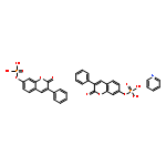
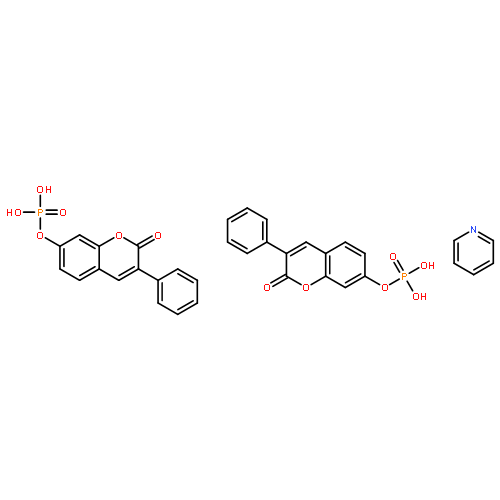
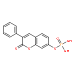
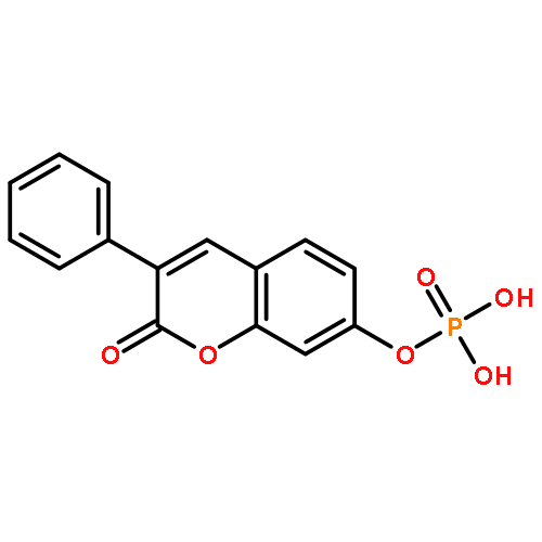


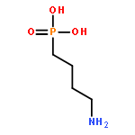
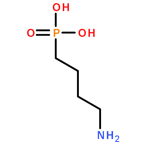
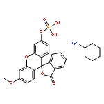
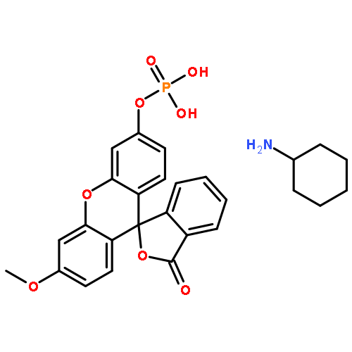
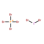

![Zinc,[1,2,3-propanetriolato(2-)-kO1,kO2]-](http://img.cochemist.com/ccimg/16800/16754-68-0.png)
![Zinc,[1,2,3-propanetriolato(2-)-kO1,kO2]-](http://img.cochemist.com/ccimg/16800/16754-68-0_b.png)


![3',6'-Dihydroxy-3H-spiro[isobenzofuran-1,9'-xanthen]-3-one](http://img.cochemist.com/ccimg/2400/2321-07-5.png)
![3',6'-Dihydroxy-3H-spiro[isobenzofuran-1,9'-xanthen]-3-one](http://img.cochemist.com/ccimg/2400/2321-07-5_b.png)