Co-reporter:Na Ye ; Chuan-Huizi Chen ; TianTian Chen ; Zilan Song ; Jin-Xue He ; Xia-Juan Huan ; Shan-Shan Song ; Qiufeng Liu ; Yi Chen ; Jian Ding ; Yechun Xu ; Ze-Hong Miao ;Ao Zhang
Journal of Medicinal Chemistry 2013 Volume 56(Issue 7) pp:2885-2903
Publication Date(Web):March 8, 2013
DOI:10.1021/jm301825t
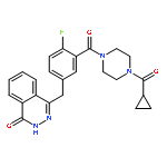
A series of benzo[de][1,7]naphthyridin-7(8H)-ones possessing a functionalized long-chain appendage have been designed and evaluated as novel PARP1 inhibitors. The initial effort led to the first-generation PARP1 inhibitor 26 bearing a terminal phthalazin-1(2H)-one framework and showing remarkably high PARP1 inhibitory activity (0.31 nM) but only moderate potency in the cell. Further effort generated the second-generation lead 41, showing high potency against both the PARP1 enzyme and BRCA-deficient cells, especially for the BRCA1-deficient MDA-MB-436 cells (CC50 < 0.26 nM). Mechanistic studies revealed that the new PARP1 inhibitors significantly inhibited H2O2-triggered PARylation in SKOV3 cells, induced cellular accumulation of DNA double-strand breaks, and impaired cell-cycle progression in BRCA2-deficient cells. Significant potentiation on the cytotoxicity of Temozolomide was also observed. The unique structural character and exceptionally high potency of 41 made it stand out as a promising drug candidate worthy for further evaluation.
Co-reporter:Zhao-Li Zhou, Ya-Xi Yang, Jian Ding, Yuan-Chao Li and Ze-Hong Miao
Natural Product Reports 2012 vol. 29(Issue 4) pp:457-475
Publication Date(Web):23 Jan 2012
DOI:10.1039/C2NP00088A
Covering: 1972 to 2011
Triptolide, a principal bioactive ingredient of Tripterygium wilfordii Hook F, has attracted extensive exploration due to its unique structure of a diterpenoid triepoxide and multiple biological activities. This review will focus on the structural modifications, structure–activity relationships, pharmacology, and clinical development of triptolide in the last forty years.
Co-reporter:Jin-xue He;Ying-qing Wang;Jian-ming Feng;Jia-xin Li;Lei Xu;Xiao-hua Li;Wei Wang;Xia-juan Huan;Yi Jiang;Bing Yu;Guang Chen;Ze-hong Miao
Acta Pharmacologica Sinica 2012 33(3) pp:426-428
Publication Date(Web):2012-01-23
DOI:10.1038/aps.2012.1
Receptor-interacting protein 3 (RIP3) is a serine/threonine protein kinase, which has extensive substrates including its cognate kinase RIP1 and multiple metabolic enzymes involving oxidative phosphorylation1, 2. RIP3 has been shown to be essential for development, immunity and some physiological or pathophysiological responses to exogenous and endogenous stimuli3, 4, 5. In 2009, three groups independently reported that RIP3 acted as a molecular switch between apoptosis and necrosis (also called as necroptosis) 6, 7, 8. Specifically, RIP3 could turn tumor necrosis factor (TNF)-induced cell death from apoptosis to necrosis6. Most of small-molecule anticancer drugs elicit their anticancer effects via apoptotic induction9. However, it is unclear whether RIP3 affects the cellular sensitivity to small-molecule anticancer drugs.We treated A cells (NIH 3T3 cells, murine fibroblasts, RIP3−/−; typically undergoing apoptosis in response to TNF stimulation) and N cells (NIH 3T3 cells, murine fibroblasts, RIP3+/+; undergoing necroptosis in response to TNF stimulation)6 with small-molecule anticancer drugs of different mechanisms of action. Detection by sulforhodamine B assays showed that A cells and N cells displayed differential sensitivity only to the examined topoisomerase I (Top1) inhibitors camptothecins. RIP3-deficient A cells revealed 32.6- and 40.2-fold higher sensitivity than RIP3-proficient N cells to SN38 and chimmitecan10, respectively (Figure 1A). Such differential sensitivity was reflected as higher apoptosis rates (Figure 1B) and more rapid G2/M arrest (Figure 1C) in A cells than in N cells treated with chimmitecan. Consistently, exposure to chimmitecan caused faster reduction of the target protein Top1 in A cells than in N cells (Figure 1D); and the treatment with chimmitecan or SN38 drove more γ-H2AX formation (a molecular marker for DNA double-strand breaks; Figure 1E) and produced more foci of phosphorylated ataxia telangiectasia mutated kinase (p-ATM) at Ser 1981 (sensing DNA double-strand breaks; Figures 1F and 1G) in A cells than in N cells. These results collectively indicate that the differential sensitivity of the RIP3-deficient A cells and the RIP3-proficient N cells to the examined Top1 inhibitors in all the tested events from inducing DNA double-strand breaks through sensing the damage signals to eliciting the final biological effects including cell cycle arrest, apoptosis and proliferation/growth inhibition.Both A cells and N cells were kindly gifted from Prof Jiahuai HAN, who used them to successfully demonstrate the role of RIP3 in regulating TNF-inducing cell death6. The differential sensitivity of the RIP3-deficient and proficient murine fibroblasts to the Top1 inhibitors suggests a role of RIP3 in determining the cellular sensitivity to those agents. In this case, however, RIP3 does not seem to directly affect the choice of cell death between apoptosis and necrosis, because no significant difference of the cellular sensitivity to the other examined anticancer drugs except camptothecins was detectable (data not shown) and because RIP3-based differential responses of A cells and N cells to the Top1 inhibitors took place actually at the levels of the target protein Top1 and DNA double-strand breaks, not only at the level of cell death. In contrast, our data seem to imply that RIP3 is involved in the DNA damage induced by the Top1 inhibitors and/or subsequent repair. As a clinically important class of anticancer drugs, Top1 inhibitors are extensively used to treat various solid tumors such as colon and lung cancers. Occurrence of drug resistance to those inhibitors is a serious obstacle to successful therapy in the clinic11. The differential sensitivity of A cells and N cells to SN38 and chimmitecan also suggests that RIP3 could contribute to cellular resistance to Top1 inhibitors. Actually, we detected and found that RIP3 was expressed at high levels in cancer cells originated from different human tissues including blood (K562), liver (SMMC-7402 and SMMC-7721), colon (HCT116), stomach (MKN45), lung (A549), breast (MDA-MB-468, MDA-MB-231, T47D, and BT549), cervix (HeLa) and bone (Rh30) (data not shown). Inhibition of RIP3 might be an alternative approach to circumventing drug resistance. However, the exact molecular mechanisms remain to be further clarified.Taken together, our results demonstrate for the first time the differential sensitivity of the RIP3-deficient A cells and the RIP3-proficient N cells to Top1 inhibitors, suggesting a potential new role of RIP3 in Top1 inhibitor-induced DNA damage/repair and cellular resistance to Top1 inhibitors, probably independently of its regulation in the choice of cell death modes.We thank Prof Jia-huai HAN (Xiamen University, China) for his kind gifts of RIP3-deficient A cells and RIP3-proficient N cells. This work was supported by grants from the National Natural Science Foundation of China (No 81025020 and No 81021062), the National Basic Research Program of China (No 2012CB932502), the National Science & Technology Major Project of China (No 2012ZX09301-001-002) and the State Key Laboratory of Drug Research of China (No SIMM1105KF-02).
Co-reporter:Jin-xue He, Ying-qing Wang, Jian-ming Feng, Jia-xin Li, Lei Xu, Xiao-hua Li, Wei Wang, Xia-juan Huan, Yi Jiang, Bing Yu, Guang Chen and Ze-hong Miao
Acta Pharmacologica Sinica 2012 33(3) pp:
Publication Date(Web):January 23, 2012
DOI:10.1038/aps.2012.1
Co-reporter:Ze-hong Miao;Jian-ming Feng;Jian Ding
Acta Pharmacologica Sinica 2012 33(9) pp:1103-1111
Publication Date(Web):2012-08-27
DOI:10.1038/aps.2012.97
In the past decade, the success of angiogenesis inhibitors in clinical contexts has established the antiangiogenic strategy as an important part of cancer therapy. During that time period, we have discovered and reported 17 compounds that exert potent inhibition on angiogenesis. These compounds exhibit tremendous diversity in their sources, structures, targets and mechanisms. These studies have generated new models for further modification and optimization of inhibitory compounds, new information for mechanistic studies and a new drug candidate for clinical development. In particular, through studies on the antiangiogenic mechanism of pseudolaric acid B, we discovered a novel mechanism by which the stability of hypoxia-inducible factor 1α is regulated by the transcription factor c-Jun. We also completed a preclinical study of AL3810, a compound with the potential to circumvent tumor drug resistance to a certain extent. All of these findings will be briefly reviewed in this article.
Co-reporter:Bing Yu;Mei-Hong Li;Wei Wang;Ying-Qing Wang;Yi Jiang
Journal of Molecular Medicine 2012 Volume 90( Issue 8) pp:971-981
Publication Date(Web):2012 August
DOI:10.1007/s00109-012-0865-4
We have recently discovered that c-Jun executes a non-transcriptional function to stabilize hypoxia inducible factor 1α (HIF-1α) and that pseudolaric acid B (PAB) accelerates HIF-1α degradation and phosphorylates c-Jun at Ser63/73. In this study, PAB was used as a probe to investigate whether and how the Ser63/73 phosphorylation of c-Jun regulates its functions. The PAB-induced reduction of HIF-1α protein was rescued through supplying additional non-phosphorylated c-Jun. However, c-Jun siRNA, which reduced both the PAB-driven phosphorylated c-Jun and the total c-Jun protein, did not prevent the PAB-induced decrease in HIF-1α. HIF-1α was revealed to be co-immunoprecipitated only with the non-phosphorylated c-Jun. PAB increased the phosphorylated c-Jun while reducing the non-phosphorylated c-Jun at Ser63/73, which impaired its function in stabilizing HIF-1α. Consequently, PAB led to the degradation of HIF-1α, thus resulting in the decreased HIF-1α-dependent expression of mdr-1 and VEGF. We accordingly propose a function-converter model of c-Jun: the Ser63/73 phosphorylation serves as a function converter to convert c-Jun from its non-transcriptional function to its transcriptional function.
Co-reporter:Zhixiang Zhang;Tao Meng;Jingxue He;Ming Li;Lin-Jiang Tong
Investigational New Drugs 2010 Volume 28( Issue 6) pp:715-728
Publication Date(Web):2010 December
DOI:10.1007/s10637-009-9303-z
Targeting cellular mitosis is an attractive antitumor strategy. Here, we reported MT7, a novel compound from the 6H-Pyrido[2′,1′:2,3]imidazo [4,5-c]isoquinolin- 5(6H)-one library generated by using the multi-component reaction strategy, as a new mitotic inhibitor. MT7 elicited apparent inhibition of cell proliferation by arresting mitosis specifically and reversibly in various tumor cell lines originating from different human tissues. Detailed mechanistic studies revealed that MT7 induced typical gene expression profiles related to mitotic arrest shown by cDNA microarray assays. Connectivity Map was used to analyze the microarray data and suggested that MT7 was possibly a tubulin inhibitor due to its similar gene expression profiles to those of the known tubulin inhibitors demecolcine, celastrol and paclitaxel. Further analyses demonstrated that MT7 inhibited the polymerization of cellular microtubules although it was not detectable to bind to purified tubulin. The inhibition of cellular tubulin polymerization by MT7 subsequently resulted in the disruption of mitotic spindle formation, activated the spindle assembly checkpoint and consequently arrested the cells at mitosis. The persistent mitotic arrest by the treatment with MT7 led the tested tumor cells to apoptosis. Our data indicate that MT7 could act as a promising lead for further optimization, in hopes of developing new anticancer therapeutics and being used to probe the biology of mitosis, specifically, the mode of interference with microtubules.
Co-reporter:Jin-xue He;Chun-hao Yang;Ze-hong Miao
Acta Pharmacologica Sinica 2010 31(9) pp:1172-1180
Publication Date(Web):2010-08-02
DOI:10.1038/aps.2010.103
The year of 2005 was a watershed in the history of poly(ADP-ribose) polymerase (PARP) inhibitors due to the important findings of selective killing in BRCA-deficient cancers by PARP inhibition. The findings made PARP inhibition one of the most promising new therapeutic approaches to cancers, especially to those with specific defects. With AZD2281 and BSI-201 entering phase III clinical trials, the final application of PARP inhibitors in clinic would come true soon. This current paper will review the major advances in targeting PARP for cancer therapy and discuss the existing questions, the answers to which may influence the future of PARP inhibitors as cancer therapeutics.
Co-reporter:Hong Zhu, Ze-Hong Miao, Min Huang, Jian-Ming Feng, ... Jian Ding
Neoplasia (November 2009) Volume 11(Issue 11) pp:1226-1234
Publication Date(Web):1 November 2009
DOI:10.1593/neo.09986
Naphthalimides, particularly amonafide and 2-(2-dimethylamino)-6-thia-2-aza-benzo[def]chrysene-1,3-diones (R16), have been identified to possess anticancer activities and to induce G2-M arrest through inhibiting topoisomerase II accompanied by Chk1 degradation. The current study was designed to precisely dissect the signaling pathway(s) responsible for the naphthalimide-induced cell cycle arrest in human colon carcinoma HCT116 cells. Using phosphorylated histone H3 and mitotic protein monoclonal 2 as mitosis markers, we first specified the G2 arrest elicited by the R16 and amonafide. Then, R16 and amonafide were revealed to induce phosphorylation of the DNA damage sensor ataxia telangiectasia-mutated (ATM) responding to DNA double-strand breaks (DSBs). Inhibition of ATM by both the pharmacological inhibitor caffeine and the specific small interference RNA (siRNA) rescued the G2 arrest elicited by R16, indicating its ATM-dependent characteristic. Furthermore, depletion of Chk2, but not Chk1 with their corresponding siRNA, statistically significantly reversed the R16- and amonafide-triggered G2 arrest. Moreover, the naphthalimides phosphorylated Chk2 in an ATM-dependent manner but induced Chk1 degradation. These data indicate that R16 and amonafide preferentially used Chk2 as evidenced by the differential ATM-executed phosphorylation of Chk1 and Chk2. Thus, a clear signaling pathway can be established, in which ATM relays the DNA DSBs signaling triggered by the naphthalimides to the checkpoint kinases, predominantly to Chk2,which finally elicits G2 arrest. The mechanistic elucidation not only favors the development of the naphthalimides as anticancer agents but also provides an alternative strategy of Chk2 inhibition to potentiate the anticancer activities of these agents.


![Bis[(pentamethylcyclopentadienyl)dichloro-rhodium]](http://img.cochemist.com/ccimg/12400/12354-85-7.png)
![Bis[(pentamethylcyclopentadienyl)dichloro-rhodium]](http://img.cochemist.com/ccimg/12400/12354-85-7_b.png)
![4-tert-butyl-1-[(2,2-dichlorocyclopropyl)methyl]-2-nitrobenzene](http://img.cochemist.com/ccimg/5300/5216-30-8.png)
![4-tert-butyl-1-[(2,2-dichlorocyclopropyl)methyl]-2-nitrobenzene](http://img.cochemist.com/ccimg/5300/5216-30-8_b.png)