Co-reporter:Chi Lu;Seongjun Park;Thomas J. Richner;Alexander Derry;Imogen Brown;Chong Hou;Siyuan Rao;Jeewoo Kang;Chet T. Moritz;Yoel Fink
Science Advances 2017 Vol 3( Iss 3) pp:
Publication Date(Web):29 Mar 2017
DOI: 10.1126/sciadv.1600955
Stretchable optoelectronic fibers interrogate spinal cord circuits during free behavior.
Co-reporter:Simone Schuerle, Jaideep S. Dudani, Michael G. Christiansen, Polina Anikeeva, and Sangeeta N. Bhatia
Nano Letters 2016 Volume 16(Issue 10) pp:6303-6310
Publication Date(Web):September 13, 2016
DOI:10.1021/acs.nanolett.6b02670
Targeted cancer therapies require a precise determination of the underlying biological processes driving tumorigenesis within the complex tumor microenvironment. Therefore, new diagnostic tools that capture the molecular activity at the disease site in vivo are needed to better understand tumor behavior and ultimately maximize therapeutic responses. Matrix metalloproteinases (MMPs) drive multiple aspects of tumorigenesis, and their activity can be monitored using engineered peptide substrates as protease-specific probes. To identify tumor specific activity profiles, local sampling of the tumor microenvironment is necessary, such as through remote control of probes, which are only activated at the tumor site. Alternating magnetic fields (AMFs) provide an attractive option to remotely apply local triggering signals because they penetrate deep into the body and are not likely to interfere with biological processes due to the weak magnetic properties of tissue. Here, we report the design and evaluation of a protease-activity nanosensor that can be remotely activated at the site of disease via an AMF at 515 kHz and 15 kA/m. Our nanosensor was composed of thermosensitive liposomes containing functionalized protease substrates that were unveiled at the target site by remotely triggered heat dissipation of coencapsulated magnetic nanoparticles (MNPs). This nanosensor was combined with a unique detection assay to quantify the amount of cleaved substrates in the urine. We applied this spatiotemporally controlled system to determine tumor protease activity in vivo and identified differences in substrate cleavage profiles between two mouse models of human colorectal cancer.Keywords: activity-based biomarkers; hysteresis-loss heating; magnetic nanoparticles; nanosensors; proteases; Thermoliposomes;
Co-reporter:Ritchie Chen, Michael G. Christiansen, Alexandra Sourakov, Alan Mohr, Yuri Matsumoto, Satoshi Okada, Alan Jasanoff, and Polina Anikeeva
Nano Letters 2016 Volume 16(Issue 2) pp:1345-1351
Publication Date(Web):January 12, 2016
DOI:10.1021/acs.nanolett.5b04761
From magnetic resonance imaging to cancer hyperthermia and wireless control of cell signaling, ferrite nanoparticles produced by thermal decomposition methods are ubiquitous across biomedical applications. While well-established synthetic protocols allow for precise control over the size and shape of the magnetic nanoparticles, structural defects within seemingly single-crystalline materials contribute to variability in the reported magnetic properties. We found that stabilization of metastable wüstite in commonly used hydrocarbon solvents contributed to significant cation disorder, leading to nanoparticles with poor hyperthermic efficiencies and transverse relaxivities. By introducing aromatic ethers that undergo radical decomposition upon thermolysis, the electrochemical potential of the solvent environment was tuned to favor the ferrimagnetic phase. Structural and magnetic characterization identified hallmark features of nearly defect-free ferrite nanoparticles that could not be demonstrated through postsynthesis oxidation with nearly 500% increase in the specific loss powers and transverse relaxivity times compared to similarly sized nanoparticles containing defects. The improved crystallinity of the nanoparticles enabled rapid wireless control of intracellular calcium. Our work demonstrates that redox tuning during solvent thermolysis can generate potent theranostic agents through selective phase control in ferrites and can be extended to other transition metal oxides relevant to memory and electrochemical storage devices.
Co-reporter:Ryan A. Koppes, Seongjun Park, Tiffany Hood, Xiaoting Jia, Negin Abdolrahim Poorheravi, Anilkumar Harapanahalli Achyuta, Yoel Fink, Polina Anikeeva
Biomaterials 2016 81() pp: 27-35
Publication Date(Web):March 2016
DOI:10.1016/j.biomaterials.2015.11.063
Synthetic neural scaffolds hold promise to eventually replace nerve autografts for tissue repair following peripheral nerve injury. Despite substantial evidence for the influence of scaffold geometry and dimensions on the rate of axonal growth, systematic evaluation of these parameters remains a challenge due to limitations in materials processing. We have employed fiber drawing to engineer a wide spectrum of polymer-based neural scaffolds with varied geometries and core sizes. Using isolated whole dorsal root ganglia as an in vitro model system we have identified key features enhancing nerve growth within these fiber scaffolds. Our approach enabled straightforward integration of microscopic topography at the scale of nerve fascicles within the scaffold cores, which led to accelerated Schwann cell migration, as well as neurite growth and alignment. Our findings indicate that fiber drawing provides a scalable and versatile strategy for producing nerve guidance channels capable of controlling direction and accelerating the rate of axonal growth.
Co-reporter:Colleen N. Loynachan;Gabriela Romero;Michael G. Christiansen;Ritchie Chen;Rachel Ellison;Tiernan T. O'Malley;Ulrich P. Froriep;Dominic M. Walsh
Advanced Healthcare Materials 2015 Volume 4( Issue 14) pp:2100-2109
Publication Date(Web):
DOI:10.1002/adhm.201500487
Remotely triggered hysteretic heat dissipation by magnetic nanoparticles (MNPs) selectively attached to targeted proteins can be used to break up self-assembled aggregates. This magnetothermal approach is applied to the amyloid-β (Aβ) protein, which forms dense, insoluble plaques characteristic of Alzheimer's disease. Specific targeting of dilute MNPs to Aβ aggregates is confirmed via transmission electron microscopy (TEM) and is found to be consistent with a statistical model of MNP distribution on the Aβ substrates. MNP composition and size are selected to achieve efficient hysteretic power dissipation at physiologically safe alternating magnetic field (AMF) conditions. Dynamic light scattering, fluorescence spectroscopy, and TEM are used to characterize the morphology and size distribution of aggregates before and after exposure to AMF. A dramatic reduction in aggregate size from microns to tens of nanometers is observed, suggesting that exposure to an AMF effectively destabilizes Aβ deposits decorated with targeted MNPs. Experiments in primary hippocampal neuronal cultures indicate that the magnetothermal disruption of aggregates reduces Aβ cytotoxicity, which may enable future applications of this approach for studies of protein disaggregation in physiological environments.
Co-reporter:Gabriela Romero;Ritchie Chen;Alan Mohr;Michael G. Christiansen
Science 2015 Volume 347(Issue 6229) pp:
Publication Date(Web):
DOI:10.1126/science.1261821
Exciting nerve cells deep inside the brain
Current techniques to stimulate regions inside the brain need a permanently implanted wire or an optical fiber. Working in mice, Chen et al. developed a method to overcome this problem (see the Perspective by Temel and Jahanshahi). They introduced heat-sensitive capsaicin receptors into nerve cells and then injected magnetic nanoparticles into specific brain regions. The nanoparticles could be heated by external alternating magnetic fields, which activated the ion channel–expressing neurons. Thus, cellular signaling deep inside the brain can be controlled remotely without permanent implants.
Science, this issue p. 1477; see also p. 1418
Co-reporter:Chi Lu;Ulrich P. Froriep;Ryan A. Koppes;Andres Canales;Vittorio Caggiano;Jennifer Selvidge;Emilio Bizzi
Advanced Functional Materials 2014 Volume 24( Issue 42) pp:6594-6600
Publication Date(Web):
DOI:10.1002/adfm.201401266
Restoration of motor and sensory functions in paralyzed patients requires the development of tools for simultaneous recording and stimulation of neural activity in the spinal cord. In addition to its complex neurophysiology, the spinal cord presents technical challenges stemming from its flexible fibrous structure and repeated elastic deformation during normal motion. To address these engineering constraints, we developed highly flexible fiber probes, consisting entirely of polymers, for combined optical stimulation and recording of neural activity. The fabricated fiber probes exhibit low-loss light transmission even under repeated extreme bending deformations. Using our fiber probes, we demonstrate simultaneous recording and optogenetic stimulation of neural activity in the spinal cord of transgenic mice expressing the light sensitive protein channelrhodopsin 2 (ChR2). Furthermore, optical stimulation of the spinal cord with the polymer fiber probes induces on-demand limb movements that correlate with electromyographical (EMG) activity.
Co-reporter:Ritchie Chen, Michael G. Christiansen, and Polina Anikeeva
ACS Nano 2013 Volume 7(Issue 10) pp:8990
Publication Date(Web):September 9, 2013
DOI:10.1021/nn4035266
This article develops a set of design guidelines for maximizing heat dissipation characteristics of magnetic ferrite MFe2O4 (M = Mn, Fe, Co) nanoparticles in alternating magnetic fields. Using magnetic and structural nanoparticle characterization, we identify key synthetic parameters in the thermal decomposition of organometallic precursors that yield optimized magnetic nanoparticles over a wide range of sizes and compositions. The developed synthetic procedures allow for gram-scale production of magnetic nanoparticles stable in physiological buffer for several months. Our magnetic nanoparticles display some of the highest heat dissipation rates, which are in qualitative agreement with the trends predicted by a dynamic hysteresis model of coherent magnetization reversal in single domain magnetic particles. By combining physical simulations with robust scalable synthesis and materials characterization techniques, this work provides a pathway to a model-driven design of magnetic nanoparticles tailored to a variety of biomedical applications ranging from cancer hyperthermia to remote control of gene expression.Keywords: anisotropy energy; large-scale synthesis; magnetic hyperthermia; magnetic nanoparticles; organometallic decomposition; water-soluble nanoparticles
Co-reporter:Polina Anikeeva and Karl Deisseroth
ACS Nano 2012 Volume 6(Issue 9) pp:7548
Publication Date(Web):September 6, 2012
DOI:10.1021/nn3039287
Optical methods for manipulation of cellular function have enabled deconstruction of genetic and neural circuits in vitro and in vivo. Plasmonic gold nanomaterials provide an alternative platform for external optical manipulation of genetic circuits. The tunable absorption of gold nanoparticles in the infrared spectral region and straightforward surface functionalization has led to applications in intracellular delivery and photorelease of short RNAs, recently enabling bidirectional photothermal modulation of specific genes via RNA interference (RNAi). We discuss recent advances in optical gene circuit engineering and plasmonic nanomaterials, as well as future research opportunities and challenges in photothermal gene manipulation.
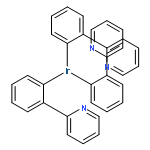
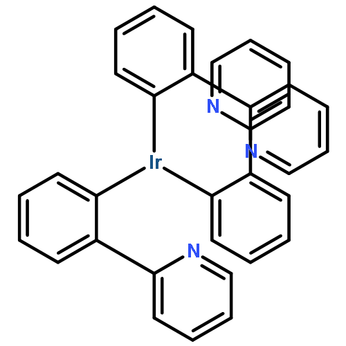
Co-reporter:P.O. Anikeeva, C.F. Madigan, S.A. Coe-Sullivan, J.S. Steckel, M.G. Bawendi, V. Bulović
Chemical Physics Letters 2006 Volume 424(1–3) pp:120-125
Publication Date(Web):12 June 2006
DOI:10.1016/j.cplett.2006.04.009
We demonstrate exciton energy transfer from a thin film of phosphorescent dye fac tris(2-phenylpyridine) iridium (Ir(ppy)3) to a monolayer of colloidal CdSe/ZnS core/shell quantum dots (QDs). The energy transfer is manifested in time-resolved photoluminescence (PL) measurements as elongation of the QD PL time constant from 40 to 400 ns, and a concomitant 55% increase of time-integrated QD PL intensity. The observed PL dynamics are shown to be dominated by exciton diffusion within the Ir(ppy)3 film to the QD layer.We demonstrate exciton energy transfer from a thin film of phosphorescent dye fac tris(2-phenylpyridine) iridium (Ir(ppy)3) to a monolayer of colloidal CdSe/ZnS core/shell quantum dots (QDs) using time-resolved photoluminescence (PL) measurements. We also show that observed PL dynamics are dominated by exciton diffusion within the Ir(ppy)3 film to the QD layer.
.jpg)
![3,5,8-Trioxa-4-phosphahexacosan-1-aminium,4-hydroxy-N,N,N-trimethyl-9-oxo-7-[[(1-oxotetradecyl)oxy]methyl]-, inner salt,4-oxide, (7R)-](http://img.cochemist.com/ccimg/76400/76343-22-1.png)
![3,5,8-Trioxa-4-phosphahexacosan-1-aminium,4-hydroxy-N,N,N-trimethyl-9-oxo-7-[[(1-oxotetradecyl)oxy]methyl]-, inner salt,4-oxide, (7R)-](http://img.cochemist.com/ccimg/76400/76343-22-1_b.png)
![N2,N7-Diphenyl-N2,N7-di-m-tolyl-9,9'-spirobi[fluorene]-2,7-diamine](http://img.cochemist.com/ccimg/1033100/1033035-83-4.png)
![N2,N7-Diphenyl-N2,N7-di-m-tolyl-9,9'-spirobi[fluorene]-2,7-diamine](http://img.cochemist.com/ccimg/1033100/1033035-83-4_b.png)


![Poly[(chloro-1,4-phenylene)-1,2-ethanediyl]](http://img.cochemist.com/ccimg/9100/9052-19-1.png)
![Poly[(chloro-1,4-phenylene)-1,2-ethanediyl]](http://img.cochemist.com/ccimg/9100/9052-19-1_b.png)