Co-reporter:Satyabrata Pany, Anjoy Majhi, and Joydip Das
Biochemistry 2016 Volume 55(Issue 14) pp:2135-2143
Publication Date(Web):March 17, 2016
DOI:10.1021/acs.biochem.6b00057
Members of the protein kinase C (PKC) family of serine/threonine kinases regulate various cellular functions, including cell growth, differentiation, metabolism, and apoptosis. Modulation of isoform-selective activity of PKC by curcumin (1), the active constituent of Curcuma L., is poorly understood, and the literature data are inconsistent and obscure. The effect of curcumin (1) and its analogues, 4-[(2Z,6E)-3-hydroxy-7-(4-hydroxy-3-methoxyphenyl)-5-oxohepta-2,6-dien-1-yl]-2-methoxyphenyl oleate (2), (9Z,12Z)-4-[(2Z,6E)-3-hydroxy-7-(4-hydroxy-3-methoxyphenyl)-5-oxohepta-2,6-dien-1-yl]-2-methoxyphenyl octadeca-9,12-dienoate (3), (9Z,12Z,15Z)-4-[(2Z,6E)-3-hydroxy-7-(4-hydroxy-3-methoxyphenyl)-5-oxohepta-2,6-dien-1-yl]-2-methoxyphenyl octadeca-9,12,15-trienoate (4), and (1E,6E)-1-[4-(hexadecyloxy)-3-methoxyphenyl]-7-(4-hydroxy-3-methoxyphenyl)hepta-1,6-diene-3,5-dione (5), and didemethylcurcumin (6) on the membrane translocation of PKCα, a conventional PKC, and PKCε, a novel PKC, has been studied in CHO-K1 cells, in which these PKC isoforms are endogenously expressed. Translocation of PKC from the cytosol to the membrane was measured using immunoblotting and confocal microscopy. 1 and 6 inhibited the TPA-induced membrane translocation of PKCα but not of PKCε. Modification of the hydroxyl group of curcumin with a long aliphatic chain containing unsaturated double bonds in 2–4 completely abolished this inhibition property. Instead, 2–4 showed significant translocation of PKCα but not of PKCε to the membrane. No membrane translocation was observed with 1, 6, or the analogue 5 having a saturated long chain for either PKCα or PKCε. 1 and 6 inhibited TPA-induced activation of ERK1/2, and 2–4 activated it. ERK1/2 is the downstream readout of PKC. These results show that the hydroxyl group of curcumin is important for PKC activity and the curcumin template can be useful in developing isoform specific PKC modulators for regulating a particular disease state.
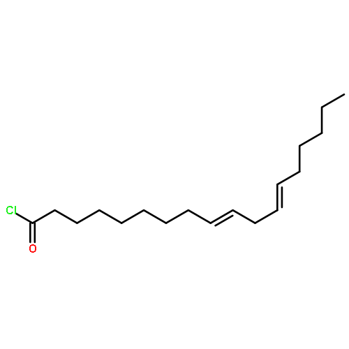
Co-reporter:Satyabrata Pany, Youngki You, and Joydip Das
Biochemistry 2016 Volume 55(Issue 45) pp:6327
Publication Date(Web):October 24, 2016
DOI:10.1021/acs.biochem.6b00932
Curcumin is a polyphenolic nutraceutical that acts on multiple biological targets, including protein kinase C (PKC). PKC is a family of serine/threonine kinases central to intracellular signal transduction. We have recently shown that curcumin selectively inhibits PKCα, but not PKCε, in CHO-K1 cells [Pany, S. (2016) Biochemistry 55, 2135–2143]. To understand which domain(s) of PKCα is responsible for curcumin binding and inhibitory activity, we made several domain-swapped mutants in which the C1 (combination of C1A and C1B) and C2 domains are swapped between PKCα and PKCε. Phorbol ester-induced membrane translocation studies using confocal microscopy and immunoblotting revealed that curcumin inhibited phorbol ester-induced membrane translocation of PKCε mutants, in which the εC1 domain was replaced with αC1, but not the PKCα mutant in which αC1 was replaced with the εC1 domain, suggesting that αC1 is a determinant for curcumin’s inhibitory effect. In addition, curcumin inhibited membrane translocation of PKCε mutants, in which the εC1A and εC1B domains were replaced with the αC1A and αC1B domains, respectively, indicating the role of both αC1A and αC1B domains in curcumin’s inhibitory effects. Phorbol 13-acetate inhibited the binding of curcumin to αC1A and αC1B with IC50 values of 6.27 and 4.47 μM, respectively. Molecular docking and molecular dynamics studies also supported the higher affinity of curcumin for αC1B than for αC1A. The C2 domain-swapped mutants were inactive in phorbol ester-induced membrane translocation. These results indicate that curcumin binds to the C1 domain of PKCα and highlight the importance of this domain in achieving PKC isoform selectivity.
Co-reporter:Joydip Das and Ghazi M. Rahman
Chemical Reviews 2014 Volume 114(Issue 24) pp:12108
Publication Date(Web):November 6, 2014
DOI:10.1021/cr300481j
Co-reporter:Joydip Das;Shiyu Xu;Satyabrata Pany;Ashley Guillory;Vrutant Shah;Gregg W. Roman
Journal of Neurochemistry 2013 Volume 126( Issue 6) pp:715-726
Publication Date(Web):
DOI:10.1111/jnc.12315
Co-reporter:Joydip Das
Chemical Reviews 2011 Volume 111(Issue 8) pp:4405
Publication Date(Web):April 5, 2011
DOI:10.1021/cr1002722
Co-reporter:Joydip Das, Satyabrata Pany, Anjoy Majhi
Bioorganic & Medicinal Chemistry 2011 Volume 19(Issue 18) pp:5321-5333
Publication Date(Web):15 September 2011
DOI:10.1016/j.bmc.2011.08.008
Resveratrol (1) is a naturally occurring phytoalexin that affects a variety of human disease models, including cardio- and neuroprotection, immune regulation, and cancer chemoprevention. One of the possible mechanisms by which resveratrol affects these disease states is by affecting the cellular signaling network involving protein kinase C alpha (PKCα). PKCα is a member of the family of serine/threonine kinases, whose activity is inhibited by resveratrol. To study the structure–activity relationship, several monoalkoxy, dialkoxy and hydroxy analogs of resveratrol have been synthesized, tested for their cytotoxic effects on HEK293 cells, measured their effects on the membrane translocation properties of PKCα in the presence and absence of the PKC activator TPA, and studied their binding with the activator binding domain of PKCα. The analogs showed less cytotoxic effects on HEK293 cells and caused higher membrane translocation (activation) than that of resveratrol. Among all the analogs, 3, 16 and 25 showed significantly higher activation than resveratrol. Resveratrol analogs, however, inhibited phorbol ester-induced membrane translocation, and the inhibition was less than that of resveratrol. Binding studies using steady state fluorescence spectroscopy indicated that resveratrol and the analogs bind to the second cysteine-rich domain of PKCα. The molecular docking studies indicated that resveratrol and the analogs interact with the protein by forming hydrogen bonds through its hydroxyl groups. These results signify that molecules developed on a resveratrol scaffold can attenuate PKCα activity and this strategy can be used to regulate various disease states involving PKCα.
Co-reporter:Joydip Das, Satyabrata Pany, Shyam Panchal, Anjoy Majhi, Ghazi M. Rahman
Bioorganic & Medicinal Chemistry 2011 Volume 19(Issue 21) pp:6196-6202
Publication Date(Web):1 November 2011
DOI:10.1016/j.bmc.2011.09.011
The protein kinase C (PKC) family of serine/threonine kinases is an attractive drug target because of its involvement in the regulation of various cellular functions, including cell growth, differentiation, metabolism, and apoptosis. The endogenous PKC activator diacylglycerol contains two long carbon chains, which are attached to the glycerol moiety via ester linkage. Natural product curcumin (1), the active constituent of Curcuma L., contains two carbonyl and two hydroxyl groups. It modulates PKC activity and binds to the activator binding site (Majhi et al., Bioorg. Med. Chem.2010, 18, 1591). To investigate the role of the carbonyl and hydroxyl groups of curcumin in PKC binding and to develop curcumin derivatives as effective PKC modulators, we synthesized several isoxazole and pyrazole derivatives of curcumin (2–6), characterized their absorption and fluorescence properties, and studied their interaction with the activator-binding second cysteine-rich C1B subdomain of PKCδ, PKCε and PKCθ. The EC50s of the curcumin derivatives for protein fluorescence quenching varied in the range of 3–25 μM. All the derivatives showed higher binding with the PKCθC1B compared with PKCδC1B and PKCεC1B. Fluorescence emission maxima of 2–5 were blue shifted in the presence of the C1B domains, confirming their binding to the protein. Molecular docking revealed that hydroxyl, carbonyl and pyrazole ring of curcumin (1), pyrazole (2), and isoxazole (4) derivatives form hydrogen bonds with the protein residues. The present result shows that isoxazole and pyrazole derivatives bind to the activator binding site of novel PKCs and both carbonyl and hydroxy groups of curcumin play roles in the binding process, depending on the nature of curcumin derivative and the PKC isotype used.Binding of isoxazole and pyrazole derivatives of curcumin with PKCC1B.
Co-reporter:Anjoy Majhi, Ghazi M. Rahman, Shyam Panchal, Joydip Das
Bioorganic & Medicinal Chemistry 2010 Volume 18(Issue 4) pp:1591-1598
Publication Date(Web):15 February 2010
DOI:10.1016/j.bmc.2009.12.075
Protein kinase C (PKC) is a family of serine/threonine kinases that play a central role in cellular signal transduction. The second messenger diacylglycerol having two long carbon chains acts as the endogenous ligand for the PKCs. Polyphenol curcumin, the active constituent of Curcuma longa is an anti-cancer agent and modulates PKC activity. To develop curcumin derivatives as effective PKC activators, we synthesized several long chain derivatives of curcumin, characterized their absorption and fluorescence properties and studied their interaction with the activator binding second cysteine-rich C1B subdomain of PKCδ, PKCε and PKCθ. Curcumin (1) and its C16 long chain analog (4) quenched the intrinsic fluorescence of PKCδC1B, PKCεC1B and PKCθC1B in a manner similar to that of PKC activator 12-O-tetradecanoylphorbol 13-acetate (TPA). The EC50s of the curcumin derivatives for fluorescence quenching varied in the range of 4–11 μM, whereas, EC50s for TPA varied in the range of 3–6 μM. Fluorescence emission maxima of 1 and 4 were blue shifted and the fluorescence anisotropy values were increased in the presence of the C1B domains in a manner similar to that shown by the fluorescent analog of TPA, sapintoxin-D, confirming that they were bound to the proteins. Molecular docking of 1 and 4 with novel PKC C1B revealed that both the molecules form hydrogen bonds with the protein residues. The present result shows that curcumin and its long chain derivatives bind to the C1B subdomain of novel PKCs and can be further modified structurally to improve its binding and activity.Binding of curcumin and PKC C1B.
Co-reporter:Joydip Das
Journal of Photochemistry and Photobiology B: Biology 2009 Volume 95(Issue 3) pp:185-188
Publication Date(Web):3 June 2009
DOI:10.1016/j.jphotobiol.2009.03.004
Protein kinase C (PKC) is a signal transducing protein that has been implicated in binding alcohol and anesthetics. The alcohol and anesthetic binding of protein kinase C delta C1B domain has been determined previously by photolabeling and mass spectrometry [J. Das, G.H. Addona, W.S. Sandberg, S.S. Husain, T. Stehle, K.W. Miller, Identiffcation of a general anesthetic binding site in the diacylglycerol-binding domain of protein kinase C delta, J. Biol. Chem. 279 (2004) 37964-37972]. Here we studied photoincorporation of 3-azioctanol, a photoactive analog of octanol into PKC delta C1B in two buffer systems containing tris and hepes. The extent of photoincorporation was higher in hepes compared to tris as determined by high performance liquid chromatography and mass spectrometric analysis. The results are explained on the basis of the presence of number of primary hydroxyl and amino groups in tris and hepes molecules that could affect the binding of alcohol molecules to protein. This observation will be useful in selecting buffer system for biochemical studies on PKC.
Co-reporter:Joydip Das, Rashmi Ramani, M. Olufemi Suraju
Biochimica et Biophysica Acta (BBA) - General Subjects (October 2016) Volume 1860(Issue 10) pp:2107-2121
Publication Date(Web):October 2016
DOI:10.1016/j.bbagen.2016.06.022