Co-reporter:Tomonori Tamura, Zhining Song, Kazuma Amaike, Shin Lee, Sifei Yin, Shigeki Kiyonaka, and Itaru Hamachi
Journal of the American Chemical Society October 11, 2017 Volume 139(Issue 40) pp:14181-14181
Publication Date(Web):September 15, 2017
DOI:10.1021/jacs.7b07339
Catalyst-mediated protein modification is a powerful approach for the imaging and engineering of natural proteins. We have previously developed affinity-guided 4-dimethylaminopyridine (AGD) chemistry as an efficient protein modification method using a catalytic acyl transfer reaction. However, because of the high electrophilicity of the thioester acyl donor molecule, AGD chemistry suffers from nonspecific reactions to proteins other than the target protein in crude biological environments, such as cell lysates, live cells, and tissue samples. To overcome this shortcoming, we here report a new acyl donor/organocatalyst system that allows more specific and efficient protein modification. In this method, a highly nucleophilic pyridinium oxime (PyOx) catalyst is conjugated to a ligand specific to the target protein. The ligand-tethered PyOx selectively binds to the target protein and facilitates the acyl transfer reaction of a mild electrophilic N-acyl-N-alkylsulfonamide acyl donor on the protein surface. We demonstrated that the new catalytic system, called AGOX (affinity-guided oxime) chemistry, can modify target proteins, both in test tubes and cell lysates, more selectively and efficiently than AGD chemistry. Low-background fluorescence labeling of the endogenous cell-membrane proteins, carbonic anhydrase XII and the folate receptor, in live cells allowed for the precise quantification of diffusion coefficients in the protein’s native environment. Furthermore, the excellent biocompatibility and bioorthogonality of AGOX chemistry were demonstrated by the selective labeling of an endogenous neurotransmitter receptor in mouse brain slices, which are highly complicated tissue samples.
Co-reporter:Kazuma Amaike;Tomonori Tamura
Chemical Communications 2017 vol. 53(Issue 88) pp:11972-11983
Publication Date(Web):2017/11/02
DOI:10.1039/C7CC07177A
Endogenous protein labeling is one of the most invaluable methods for studying the bona fide functions of proteins in live cells. However, multi-molecular crowding conditions, such as those that occur in live cells, hamper the highly selective chemical labeling of a protein of interest (POI). We herein describe how the efficient coupling of molecular recognition with a chemical reaction is crucial for selective protein labeling. Recognition-driven protein labeling is carried out by a synthetic labeling reagent containing a protein (recognition) ligand, a reporter tag, and a reactive moiety. The molecular recognition of a POI can be used to greatly enhance the reaction kinetics and protein selectivity, even under live cell conditions. In this review, we also briefly discuss how such selective chemical labeling of an endogenous protein can have a variety of applications at the interface of chemistry and biology.
Co-reporter:Yuki Yasueda; Tomonori Tamura; Alma Fujisawa; Keiko Kuwata; Shinya Tsukiji; Shigeki Kiyonaka
Journal of the American Chemical Society 2016 Volume 138(Issue 24) pp:7592-7602
Publication Date(Web):May 26, 2016
DOI:10.1021/jacs.6b02254
Protein functions are tightly regulated by their subcellular localization in live cells, and quantitative evaluation of dynamically altered proteomes in each organelle should provide valuable information. Here, we describe a novel method for organelle-focused chemical proteomics using spatially limited reactions. In this work, mitochondria-localizable reactive molecules (MRMs) were designed that penetrate biomembranes and spontaneously concentrate in mitochondria, where protein labeling is facilitated by the condensation effect. The combination of this selective labeling and liquid chromatography–mass spectrometry (LC–MS) based proteomics technology facilitated identification of mitochondrial proteomes and the profile of the intrinsic reactivity of amino acids tethered to proteins expressed in live cultured cells, primary neurons and brain slices. Furthermore, quantitative profiling of mitochondrial proteins whose expression levels change significantly during an oxidant-induced apoptotic process was performed by combination of this MRMs-based method with a standard quantitative MS technique (SILAC: stable isotope labeling by amino acids in cell culture). The use of a set of MRMs represents a powerful tool for chemical proteomics to elucidate mitochondria-associated biological events and diseases.
Co-reporter:Ryou Kubota and Itaru Hamachi
Chemical Society Reviews 2015 vol. 44(Issue 13) pp:4454-4471
Publication Date(Web):25 Mar 2015
DOI:10.1039/C4CS00381K
Chemical sensing of amino acids, peptides, and proteins provides fruitful information to understand their biological functions, as well as to develop the medical and technological applications. To detect amino acids, peptides, and proteins in vitro and in vivo, vast kinds of chemical sensors including small synthetic binders/sensors, genetically-encoded fluorescent proteins and protein-based semisynthetic biosensors have been intensely investigated. This review deals with concepts, strategies, and applications of protein recognition and sensing using small synthetic binders/sensors, which are now actively studied but still in the early stage of investigation. The recognition strategies for peptides and proteins can be divided into three categories: (i) recognition of protein substructures, (ii) protein surface recognition, and (iii) protein sensing through protein–ligand interaction. Here, we overview representative examples of protein recognition and sensing, and discuss biological or diagnostic applications such as potent inhibitors/modulators of protein–protein interactions.
Co-reporter:Takahiro Hayashi; Yuki Yasueda; Tomonori Tamura; Yousuke Takaoka
Journal of the American Chemical Society 2015 Volume 137(Issue 16) pp:5372-5380
Publication Date(Web):April 8, 2015
DOI:10.1021/jacs.5b02867
A general technique for introducing biophysical probes into selected receptors in their native environment is valuable for the study of their structure, dynamics, function, and molecular interactions. A number of such techniques rely on genetic engineering, which is not applicable for the study of endogenous proteins, and such approaches often suffer from artifacts due to the overexpression and bulky size of the probes/protein tags used. Here we designed novel catalyst-antibody conjugates capable of introducing small chemical probes into receptor proteins such as epidermal growth factor receptor (EGFR) and human epidermal growth factor receptor 2 (HER2) in a selective manner on the surface of living cells. Because of the selectivity and efficiency of this labeling technique, we were able to monitor the cellular dynamics and lifetime of HER2 endogenously expressed on cancer cells. More significantly, the current labeling technique comprises a stable covalent bond, which combined with a peptide mass fingerprinting analysis allowed epitope mapping of antibodies on living cells and identification of potential binding sites of anti-EGFR affibody. Although as yet unreported in the literature, the binding sites predicted by our labeling method were consistently supported by the subsequent mutation and binding assay experiments. In addition, this covalent labeling method provided experimental evidence that HER2 exhibits a more dynamic structure than expected on the basis of crystallographic analysis alone. Our novel catalyst-antibody conjugates are expected to provide a general tool for investigating the protein trafficking, fluctuation, and molecular interactions of an important class of cell-surface receptors on live cell surfaces.
Co-reporter:Tatsuyuki Yoshii; Shoji Onogi; Hajime Shigemitsu
Journal of the American Chemical Society 2015 Volume 137(Issue 9) pp:3360-3365
Publication Date(Web):February 13, 2015
DOI:10.1021/ja5131534
Multicomponent supramolecular hydrogels are constructed for sensitive, naked-eye detection of small-molecule biomarkers. A dendritic self-immolative molecule and the corresponding enzyme as a signal amplification system were stably embedded in a hydrogen peroxide (H2O2)-responsive supramolecular hydrogel (BPmoc-F3), together with other enzymes. The nanostructure and mechanical strength of the hybrid BPmoc-F3 gel were not substantially diminished by incorporation of these multiple components in the absence of target biomarkers, but could be destroyed by addition of the biomarker through the multiple enzymatic and chemical cascade reactions operating in combination within the gel matrix. The sensitivity to biomarkers such as H2O2, glucose, and uric acid, detected by gel–sol transition, was significantly enhanced by the signal amplification system. An array chip consisting of these multicomponent hydrogels enabled the detection of the level of hyperuricemia disease in human plasma samples.
Co-reporter:Yousuke Takaoka, Yuki Nishikawa, Yuki Hashimoto, Kenta Sasaki and Itaru Hamachi
Chemical Science 2015 vol. 6(Issue 5) pp:3217-3224
Publication Date(Web):23 Mar 2015
DOI:10.1039/C5SC00190K
A rapid and selective ligand-directed chemical reaction was developed for the acylation of proteins in living cells on the basis of ligand-directed chemistry. By fine tuning the reactivity and stability of the phenyl ester derivatives, we successfully identified ortho-dibromophenyl benzoate as the optimal reactive motif. It was sufficiently stable in an aqueous buffer, hydrolyzing less than 10% after 13 h of incubation, but reactive enough for efficient and selective protein labeling in living mammalian cells, as well as in vitro (referred to as ligand-directed dibromophenyl benzoate (LDBB) chemistry). Using this chemistry, various fluorophores can be tethered to the target protein directly, which allows fluorescence visualization of the labeled protein in live cells using different colored fluorophore groups (including coumarin, fluorescein and rhodamine). Furthermore, this labeling is applicable to not only an overexpressed protein (E. coli dihydrofolate reductase) but also endogenous human carbonic anhydrase II and XII under living cell conditions. LDBB chemistry is a new entry of ligand-directed protein labeling methods, and should be particularly useful for the imaging of natural proteins in living cells.
Co-reporter:Dr. Hajime Shigemitsu;Dr. Itaru Hamachi
Chemistry – An Asian Journal 2015 Volume 10( Issue 10) pp:2026-2038
Publication Date(Web):
DOI:10.1002/asia.201500563
Abstract
Stimuli-responsive supramolecular assemblies consisting of small molecules are attractive functional materials for biological applications such as drug delivery, medical diagnosis, enzyme immobilization, and tissue engineering. By use of their dynamic and reversible properties, many supramolecular assemblies responsive to a variety of biomolecules have been designed and synthesized. This review focuses on promising strategies for the construction of such dynamic supramolecular assemblies and their functions. While studies of biomolecule-responsive supramolecular assemblies have mainly been performed in vitro, it has recently been demonstrated that some of them can work in live cells. Supramolecular assemblies now open up new avenues in chemical biology and biofunctional materials.
Co-reporter:Tatsuyuki Yoshii ; Keigo Mizusawa ; Yousuke Takaoka
Journal of the American Chemical Society 2014 Volume 136(Issue 47) pp:16635-16642
Publication Date(Web):October 31, 2014
DOI:10.1021/ja508955y
Supramolecular nanomaterials responsive to specific intracellular proteins should be greatly promising for protein sensing and imaging, controlled drug release or dynamic regulation of cellular processes. However, valid design strategies to create useful probes are poorly developed, particularly for proteins inside living cells as targets. We recently reported a unique supramolecular strategy for specific protein detection using self-assembling fluorescent probes consisting of a protein ligand and a fluorophore on the live cell surface, as well as in test tube settings. Herein, we discovered that our self-assembled supramolecular probes having a rhodamine derivative (tetramethylrhodamine or rhodamine-green) can incorporate and stay as less-fluorescent aggregates inside the living cells, so as to sense the protein activity in a reversible manner. Using the overexpressed model protein (dihydrofolate reductase), we demonstrated that this turn-on/off mode is controlled by selective ligand–protein recognition inside the live cells. Not only such a model protein, but also endogenous human carbonic anhydrase and heat shock protein 90 were specifically visualized in living mammalian cells, by use of the similar ligand-tethered supramolecular probes. Furthermore, such reversibility allowed us to intracellularly construct a unique system to evaluate the inhibitors affinity toward specific endogenous proteins in live cells, highlighting the potential of dynamic supramolecules as novel intelligent biomaterials.
Co-reporter:Rika Ochi, Takashi Nishida, Masato Ikeda and Itaru Hamachi
Journal of Materials Chemistry A 2014 vol. 2(Issue 11) pp:1464-1469
Publication Date(Web):22 Jan 2014
DOI:10.1039/C3TB21680B
Supramolecular hydrogels have attracted much attention as smart soft materials. To install various stimuli-responsive functions in the supramolecular hydrogels, skilful utility of chemical reactions for triggering the change in molecular structure of hydrogelators or their precursor in response to external stimuli is a promising strategy. We have recently developed a unique heat-set supramolecular hydrogel, which was triggered by rationally designed molecular conversion of a glycolipid-based bolaamphiphile into a hydrogelator via retro-Diels–Alder reaction. Here we designed new bolaamphiphiles based on short peptide-based hydrogelators as a scaffold to accelerate the heat-set hydrogelation process and also demonstrated the flexible molecular design of such bolaamphiphiles.
Co-reporter:Shinya Tsukiji, Itaru Hamachi
Current Opinion in Chemical Biology 2014 Volume 21() pp:136-143
Publication Date(Web):August 2014
DOI:10.1016/j.cbpa.2014.07.012
•LDT chemistry is a new type of traceless affinity labeling technique.•This chemistry allows selective native protein labeling in cells and in vivo.•This chemistry can be used for creating semisynthetic proteins in cells.•This chemistry is useful for various functional analysis of cellular native proteins.The ability to introduce any chemical probe to any endogenous target protein in its native environment, that is in cells and in vivo, is anticipated to provide various new exciting tools for biological and biomedical research. Although still at the prototype stage, the ligand-directed tosyl (LDT) chemistry is a novel type of affinity labeling technique that we developed for such a dream. This chemistry allows for modifying native proteins by various chemical probes with high specificity in various biological settings ranging from in vitro (in test tubes) to in living cells and in vivo. Since the first report, the list of proteins that are successfully labeled by the LDT chemistry has been increasing. A growing number of studies have demonstrated its utility to create semisynthetic proteins directly in cellular contexts. The in situ generated semisynthetic proteins are applicable for various types of analysis and imaging of intracellular biological processes. In this review, we summarize the basic properties of the LDT chemistry and its applications toward in situ engineering and analysis of native proteins in living systems. Current limitations and future challenges of this area are also described.
Co-reporter:Akinobu Nakamura, Kazumasa Takigawa, Yasutaka Kurishita, Keiko Kuwata, Manabu Ishida, Yasushi Shimoda, Itaru Hamachi and Shinya Tsukiji
Chemical Communications 2014 vol. 50(Issue 46) pp:6149-6152
Publication Date(Web):28 Apr 2014
DOI:10.1039/C4CC01753F
We report a general strategy to create small-molecule fluorescent probes for the nucleus in living cells. Our strategy is based on the attachment of the DNA-binding Hoechst compound to a fluorophore of interest. Using this approach, simple fluorescein, BODIPY, and rhodamine dyes were readily converted to novel turn-on fluorescent nucleus-imaging probes.
Co-reporter:Kei Yamaura, Keiko Kuwata, Tomonori Tamura, Yoshiyuki Kioi, Yousuke Takaoka, Shigeki Kiyonaka and Itaru Hamachi
Chemical Communications 2014 vol. 50(Issue 91) pp:14097-14100
Publication Date(Web):17 Sep 2014
DOI:10.1039/C4CC05885B
We demonstrate that ligand-directed tosyl (LDT) chemistry is applicable to off-target identification in live cells. Lapatinib (Lap)-based LDT reagents not only labeled a receptor tyrosine kinase, HER2, target protein, but also the protein disulfide isomerase (PDI) that should be an off-target protein for Lap.
Co-reporter:Takayuki Miki, Sho-hei Fujishima, Kazuhiro Komatsu, Keiko Kuwata, Shigeki Kiyonaka, Itaru Hamachi
Chemistry & Biology 2014 Volume 21(Issue 8) pp:1013-1022
Publication Date(Web):14 August 2014
DOI:10.1016/j.chembiol.2014.07.013
•LDAI-based chemical labeling is applicable to various types of membrane proteins•Half-life of membrane protein can be determined under almost natural conditions•The degradation pathway is also identified by the fluorescent pulse-chase imagingThe functions of membrane proteins are tightly controlled by the dynamics such as protein trafficking and degradation. We demonstrated that ligand-directed acyl imidazole (LDAI) chemistry is broadly applicable to selective chemical labeling of various types of membrane-bound proteins under live cell conditions without a need for any tag fragments. The LDAI chemistry enabled pulse-chase analysis of these proteins to determine the half-life, as well as their degradation pathways by the imaging, under almost natural cellular conditions.Figure optionsDownload full-size imageDownload high-quality image (305 K)Download as PowerPoint slide
Co-reporter:Tomonori Tamura and Itaru Hamachi
ACS Chemical Biology 2014 Volume 9(Issue 12) pp:2708
Publication Date(Web):October 15, 2014
DOI:10.1021/cb500661v
Protein-based fluorescent biosensors have emerged as key bioanalytical tools to visualize and quantify a wide range of biological substances and events in vitro, in cells, and even in vivo. On the basis of the construction method, the protein-based fluorescent biosensors can be principally classified into two classes: (1) genetically encoded fluorescent biosensors harnessing fluorescent proteins (FPs) and (2) semisynthetic biosensors comprised of protein scaffolds and synthetic fluorophores. Recent advances in protein engineering and chemical biology not only allowed the further optimization of conventional biosensors but also facilitated the creation of novel biosensors based on unique strategies. In this review, we survey the recent studies in the development and improvement of protein-based fluorescent biosensors and highlight the successful applications to live cell and in vivo imaging. Furthermore, we provide perspectives on possible future directions of the technique.
Co-reporter:Shohei Uchinomiya, Akio Ojida, and Itaru Hamachi
Inorganic Chemistry 2014 Volume 53(Issue 4) pp:1816-1823
Publication Date(Web):October 16, 2013
DOI:10.1021/ic401612z
Protein-labeling methods serve as essential tools for analyzing functions of proteins of interest under complicated biological conditions such as in live cells. These labeling methods are useful not only to fluorescently visualize proteins of interest in biological systems but also to conduct protein and cell analyses by harnessing the unique functions of molecular probes. Among the various labeling methods available, an appropriate binding pair consisting of a short peptide and a de novo designed small molecular probe has attracted attention because of its wide utility and versatility. Interestingly, most peptide tag/probe pairs exploit metal–ligand coordination interactions as the main binding force responsible for their association. Herein, we provide an overview of the recent progress of these coordination-chemistry-based protein-labeling methods and their applications for fluorescence imaging and functional analysis of cellular proteins, while highlighting our originally developed labeling methods. These successful examples clearly exemplify the utility and versatility of metal coordination chemistry in protein functional analysis.
Co-reporter:Tatsuyuki Yoshii;Dr. Masato Ikeda;Dr. Itaru Hamachi
Angewandte Chemie International Edition 2014 Volume 53( Issue 28) pp:7264-7267
Publication Date(Web):
DOI:10.1002/anie.201404158
Abstract
Spatiotemporal control of fluidity inside a soft matrix by external stimuli allows real-time manipulation of nano/micromaterials. In this study, we report a two-photon-responsive peptide-based supramolecular hydrogel, the fluidity of which was dramatically controlled with high spatial resolution (10 μm×10 μm×10 μm). The off–on switching of the Brownian motion of nanobeads and chemotaxis of bacteria by two-photon excitation was successfully demonstrated.
Co-reporter:Tatsuyuki Yoshii;Dr. Masato Ikeda;Dr. Itaru Hamachi
Angewandte Chemie 2014 Volume 126( Issue 28) pp:7392-7395
Publication Date(Web):
DOI:10.1002/ange.201404158
Abstract
Spatiotemporal control of fluidity inside a soft matrix by external stimuli allows real-time manipulation of nano/micromaterials. In this study, we report a two-photon-responsive peptide-based supramolecular hydrogel, the fluidity of which was dramatically controlled with high spatial resolution (10 μm×10 μm×10 μm). The off–on switching of the Brownian motion of nanobeads and chemotaxis of bacteria by two-photon excitation was successfully demonstrated.
Co-reporter:Tomonori Tamura ; Yoshiyuki Kioi ; Takayuki Miki ; Shinya Tsukiji
Journal of the American Chemical Society 2013 Volume 135(Issue 18) pp:6782-6785
Publication Date(Web):April 23, 2013
DOI:10.1021/ja401956b
Introducing synthetic fluorophores into specific endogenous proteins and analyzing their function in living cells are a great challenge in chemical biology. Toward this end, we demonstrate the target-selective and site-specific fluorescent labeling of native FKBP12 (FK506-binding protein 12) in vitro and in living cells using ligand-directed tosyl (LDT) chemistry. The LDT-mediated labeling yielded a semisynthetic FKBP12 containing the Oregon green (OG) dye near the catalytic pocket. The OG-labeled FKBP12 (OG-FKBP12) acted as a fluorescent reporter that allows monitoring of its interaction with rapamycin and FRB (FKBP-rapamycin-binding domain) in vitro. We also successfully demonstrated the visualization of the rapamycin-mediated complexation of the OG-FKBP12 and FRB inside of living cells by the combined use with fluorescent protein-tag technology and Förster resonance energy-transfer imaging.
Co-reporter:Manabu Ishida ; Hideaki Watanabe ; Kazumasa Takigawa ; Yasutaka Kurishita ; Choji Oki ; Akinobu Nakamura ; Itaru Hamachi ;Shinya Tsukiji
Journal of the American Chemical Society 2013 Volume 135(Issue 34) pp:12684-12689
Publication Date(Web):August 14, 2013
DOI:10.1021/ja4046907
Small-molecule ligands that control the spatial location of proteins in living cells would be valuable tools for regulating biological systems. However, the creation of such molecules remains almost unexplored because of the lack of a design methodology. Here we introduce a conceptually new type of synthetic ligands, self-localizing ligands (SLLs), which spontaneously localize to specific subcellular regions in mammalian cells. We show that SLLs bind their target proteins and relocate (tether) them rapidly from the cytoplasm to their targeting sites, thus serving as synthetic protein translocators. SLL-induced protein translocation enables us to manipulate diverse synthetic/endogenous signaling pathways. The method is also applicable to reversible protein translocation and allows control of multiple proteins at different times and locations in the same cell. These results demonstrate the usefulness of SLLs in the spatial (and temporal) control of intracellular protein distribution and biological processes, opening a new direction in the design of small-molecule tools or drugs for cell regulation.
Co-reporter:Takahiro Hayashi ; Yedi Sun ; Tomonori Tamura ; Keiko Kuwata ; Zhining Song ; Yousuke Takaoka
Journal of the American Chemical Society 2013 Volume 135(Issue 33) pp:12252-12258
Publication Date(Web):July 26, 2013
DOI:10.1021/ja4043214
Glycoproteins on cell surfaces play important roles in biological processes, including cell–cell interaction/signaling, immune response, and cell differentiation. Given the diversity of the structure of glycans, labeling and imaging of selected glycoproteins are challenging, although several promising strategies have been developed recently. Here, we design and construct semisynthetic reactive lectins (sugar-binding proteins) that are able to selectively label glycoproteins. Congerin II, an animal galectin, and wheat germ agglutinin are conjugated with 4-dimethylaminopyridine (DMAP), a well-known acyl transfer catalyst by our affinity-guided DMAP method and Cu(I)-assisted click chemistry. Selective labeling of glycoproteins is facilitated by the DMAP-tethered lectin catalysts both in vitro and on living cells. Two-dimensional polyacrylamide gel electrophoresis (2D-PAGE) analysis enabled us to isolate labeled glycoproteins that are uniquely exposed on distinct cell lines. Furthermore, the combination of immunoprecipitation with mass spectrometry (MS)-fingerprinting techniques allowed us to characterize 48 glycoproteins endogenously expressed on HeLa cells, and some low-abundant glycoproteins, such as epidermal growth factor receptor (EGFR) and neuropilin-1, were successfully identified. Our results demonstrate that semisynthetic DMAP-tethered lectins provide a new tool for labeling and profiling glycoproteins on living cells.
Co-reporter:Kazuya Matsuo, Yoshiyuki Kioi, Ryosuke Yasui, Yousuke Takaoka, Takayuki Miki, Sho-hei Fujishima and Itaru Hamachi
Chemical Science 2013 vol. 4(Issue 6) pp:2573-2580
Publication Date(Web):09 Apr 2013
DOI:10.1039/C3SC50560J
Caged enzymes whose activities can be controlled by light represent a powerful tool for various biological analyses. However, limited methods are available for the construction of caged proteins and enzymes. We recently developed a novel protein labeling method termed ligand-directed acyl imidazole (LDAI) chemistry, which allows us to selectively modify natural dihydrofolate reductase and folate receptor in a test tube and in live cell contexts. In this work, we have examined in detail the reaction characteristics of the LDAI chemistry using carbonic anhydrase I (CAI) as a model enzyme. In addition to modifying Lys residues with a carbamate bond, the LDAI method modified Ser and Tyr residues with a carbonate bond. Owing to the relatively labile carbonate bond formed, the LDAI chemistry was demonstrated to be applicable for a rational one-step construction of caged enzymes. This method is simple and based on the transient tethering of an inhibitor with moderate activity that is directed to the active site on an enzyme surface. We successfully showed that the activity of the caged CAI was almost completely suppressed by LDAI-based labeling and fully recovered by photoirradiation in the crude conditions (such as cell lysates) as well as in test tube settings.
Co-reporter:Rika Ochi, Kazuya Kurotani, Masato Ikeda, Shigeki Kiyonaka and Itaru Hamachi
Chemical Communications 2013 vol. 49(Issue 21) pp:2115-2117
Publication Date(Web):14 Dec 2012
DOI:10.1039/C2CC37908B
We developed supramolecular hydrogels exhibiting reversible thermochromism concurrently with gel-to-sol transition from four glycolipids. In addition, these gels showed the similar color change in response to glycosidases, which can be employed to construct a colorimetric sensor array chip for sensing glycosidases with the naked eye.
Co-reporter:Yousuke Takaoka, Yoshiyuki Kioi, Akira Morito, Junji Otani, Kyohei Arita, Eishi Ashihara, Mariko Ariyoshi, Hidehito Tochio, Masahiro Shirakawa and Itaru Hamachi
Chemical Communications 2013 vol. 49(Issue 27) pp:2801-2803
Publication Date(Web):14 Feb 2013
DOI:10.1039/C3CC39205H
Here we describe how a 19F-probe incorporated into an endogenous protein by a chemical biology method revealed protein dynamics. By explicit determination of ligand-bound and unbound structures with X-ray crystallography, the quantitative comparison of the protein's dynamics in live cells and in vitro is presented. These results clearly demonstrated the greater conformational fluctuations of the intracellular protein, partially due to macromolecular crowding effects.
Co-reporter:Shohei Uchinomiya, Hiroshi Nonaka, Sho Wakayama, Akio Ojida and Itaru Hamachi
Chemical Communications 2013 vol. 49(Issue 44) pp:5022-5024
Publication Date(Web):11 Apr 2013
DOI:10.1039/C3CC41979G
A new method for in-cell protein labeling was developed. This method employed a binding-induced nucleophilic reaction between the Cys-appended His-tag and the Ni(II)–NTA containing an α-chloroacetamide. Using this method, not only labeling of His-tag fused proteins but also the detection of a protein–protein interaction was achieved inside living cells.
Co-reporter:Dr. Yousuke Takaoka;Dr. Akio Ojida;Dr. Itaru Hamachi
Angewandte Chemie 2013 Volume 125( Issue 15) pp:4182-4200
Publication Date(Web):
DOI:10.1002/ange.201207089
Abstract
Die Modifizierung von Proteinen mit synthetischen Sonden kann in vielfältiger Weise zur Aufklärung und Beeinflussung von Proteinfunktionen in vitro, in lebenden Zellen oder in vivo genutzt werden. Wir stellen hier die jüngsten Fortschritte bei den Methoden zur chemischen Proteinmodifizierung und ihrer Anwendung beim Protein-Engineering vor, wobei wir uns auf vier Strategien konzentrieren: 1) Biokonjugationsreaktionen von Aminosäuren an der Oberfläche natürlicher Proteine, hauptsächlich im Reagenzglas; 2) bioorthogonale Reaktionen von Proteinen mit nichtnatürlichen reaktiven Markergruppen; 3) Kupplung von Erkennungs- und reaktiven Regionen unter Verwendung eines Paars aus Enzym oder kurzem Peptid mit einem Sondenmarker zur Markierung natürlicher Aminosäuren 4) ligandendirigierte chemische Reaktionen zur selektiven Markierung endogener Proteine in lebenden Systemen. Zusammengenommen bieten diese Techniken ein nützliches Repertoire von Methoden zur Anwendung in der chemischen Biologie, wobei die drei letztgenannten auf unveränderte (lebende) Systeme anwendbar sind. Die organische Chemie zur Manipulation endogener Proteine ist zwar noch in einer frühen Phase, doch die nachfolgend beschriebenen Anwendungen sind ein lohnendes Ziel für Chemiker.
Co-reporter:Dr. Yousuke Takaoka;Dr. Akio Ojida;Dr. Itaru Hamachi
Angewandte Chemie International Edition 2013 Volume 52( Issue 15) pp:4088-4106
Publication Date(Web):
DOI:10.1002/anie.201207089
Abstract
The modification of proteins with synthetic probes is a powerful means of elucidating and engineering the functions of proteins both in vitro and in live cells or in vivo. Herein we review recent progress in chemistry-based protein modification methods and their application in protein engineering, with particular emphasis on the following four strategies: 1) the bioconjugation reactions of amino acids on the surfaces of natural proteins, mainly applied in test-tube settings; 2) the bioorthogonal reactions of proteins with non-natural functional groups; 3) the coupling of recognition and reactive sites using an enzyme or short peptide tag–probe pair for labeling natural amino acids; and 4) ligand-directed labeling chemistries for the selective labeling of endogenous proteins in living systems. Overall, these techniques represent a useful set of tools for application in chemical biology, with the methods 2–4 in particular being applicable to crude (living) habitats. Although still in its infancy, the use of organic chemistry for the manipulation of endogenous proteins, with subsequent applications in living systems, represents a worthy challenge for many chemists.
Co-reporter:Kazuya Matsuo;Dr. Rui Kamada;Dr. Keigo Mizusawa;Dr. Hirohiko Imai;Dr. Yuki Takayama;Dr. Michiko Narazaki;Dr. Tetsuya Matsuda;Dr. Yousuke Takaoka;Dr. Itaru Hamachi
Chemistry - A European Journal 2013 Volume 19( Issue 38) pp:12875-12883
Publication Date(Web):
DOI:10.1002/chem.201300817
Abstract
Specific turn-on detection of enzyme activities is of fundamental importance in drug discovery research, as well as medical diagnostics. Although magnetic resonance imaging (MRI) is one of the most powerful techniques for noninvasive visualization of enzyme activity, both in vivo and ex vivo, promising strategies for imaging specific enzymes with high contrast have been very limited to date. We report herein a novel signal-amplifiable self-assembling 19F NMR/MRI probe for turn-on detection and imaging of specific enzymatic activity. In NMR spectroscopy, these designed probes are “silent” when aggregated, but exhibit a disassembly driven turn-on signal change upon cleavage of the substrate part by the catalytic enzyme. Using these 19F probes, nanomolar levels of two different target enzymes, nitroreductase (NTR) and matrix metalloproteinase (MMP), could be detected and visualized by 19F NMR spectroscopy and MRI. Furthermore, we have succeeded in imaging the activity of endogenously secreted MMP in cultured media of tumor cells by 19F MRI, depending on the cell lines and the cellular conditions. These results clearly demonstrate that our turn-on 19F probes may serve as a screening platform for the activity of MMPs.
Co-reporter:Takahiro Hayashi and Itaru Hamachi
Accounts of Chemical Research 2012 Volume 45(Issue 9) pp:1460
Publication Date(Web):June 8, 2012
DOI:10.1021/ar200334r
Protein labeling and imaging techniques have provided tremendous opportunities to study the structure, function, dynamics, and localization of individual proteins in the complex environment of living cells. Molecular biology-based approaches, such as GFP-fusion tags and monoclonal antibodies, have served as important tools for the visualization of individual proteins in cells. Although these techniques continue to be valuable for live cell imaging, they have a number of limitations that have only been addressed by recent progress in chemistry-based approaches. These chemical approaches benefit greatly from the smaller probe sizes that should result in fewer perturbations to proteins and to biological systems as a whole. Despite the research in this area, so far none of these labeling techniques permit labeling and imaging of selected endogenous proteins in living cells.Researchers have widely used affinity labeling, in which the protein of interest is labeled by a reactive group attached to a ligand, to identify and characterize proteins. Since the first report of affinity labeling in the early 1960s, efforts to fine-tune the chemical structures of both the reactive group and ligand have led to protein labeling with excellent target selectivity in the whole proteome of living cells. Although the chemical probes used for affinity labeling generally inactivate target proteins, this strategy holds promise as a valuable tool for the labeling and imaging of endogenous proteins in living cells and by extension in living animals.In this Account, we summarize traceless affinity labeling, a technique explored mainly in our laboratory. In our overview of the different labeling techniques, we emphasize the challenge of designing chemical probes that allow for dissociation of the affinity module (often a ligand) after the labeling reaction so that the labeled protein retains its native function. This feature distinguishes the traceless labeling approach from the traditional affinity labeling method and allows for real-time monitoring of protein activity. With the high target specificity and biocompatibility of this technique, we have achieved individual labeling and imaging of endogenously expressed proteins in samples of high biological complexity. We also highlight applications in which our current approach enabled the monitoring of important biological events, such as ligand binding, in living cells. These novel chemical labeling techniques are expected to provide a molecular toolbox for studying a wide variety of proteins and beyond in living cells.
Co-reporter:Yasutaka Kurishita, Takahiro Kohira, Akio Ojida, and Itaru Hamachi
Journal of the American Chemical Society 2012 Volume 134(Issue 45) pp:18779-18789
Publication Date(Web):October 25, 2012
DOI:10.1021/ja308754g
ATP and its derivatives (nucleoside polyphosphates (NPPs)) are implicated in many biological events, so their rapid and convenient detection is important. In particular, live cell detection of NPPs at specific local regions of cells could greatly contribute understanding of the complicated roles of NPPs. We report herein the design of two new fluorescent chemosensors that detect the dynamics of NPPs in specific regions of living cells. To achieve imaging of NPPs on plasma membrane surfaces (2-2Zn(II)), a lipid anchor was introduced into xanthene-based Zn(II) complex 1-2Zn(II), which was previously developed as a turn-on type fluorescent chemosensor for NPPs. Meanwhile, for subcellular imaging of ATP in mitochondria, we designed rhodamine-type Zn(II) complex 3-2Zn(II), which possesses a cationic pyronin ring instead of xanthene. Detailed spectroscopic studies revealed that 2-2Zn(II) and 3-2Zn(II) can sense NPPs with a several-fold increase of their fluorescence intensities through a sensing mechanism similar to 1-2Zn(II), involving binding-induced recovery of the conjugated form of the xanthene or pyronin ring. In live cell imaging, 2-2Zn(II) containing a lipid anchor selectively localized on the plasma membrane surface and detected the extracellular release of NPPs during cell necrosis induced by streptolysin O. On the other hand, rhodamine-type complex 3-2Zn(II) spontaneously localized at mitochondria inside cells, and sensed the local increase of ATP concentration during apoptosis. Multicolor images were obtained through simultaneous use of 2-2Zn(II) and 3-2Zn(II), allowing detection of the dynamics of ATP in different cellular compartments at the same time.
Co-reporter:Sho-hei Fujishima ; Ryosuke Yasui ; Takayuki Miki ; Akio Ojida
Journal of the American Chemical Society 2012 Volume 134(Issue 9) pp:3961-3964
Publication Date(Web):February 21, 2012
DOI:10.1021/ja2108855
Chemistry-based protein labeling in living cells is undoubtedly useful for understanding natural protein functions and for biological/pharmaceutical applications. Here, we report a novel approach for endogenous membrane-bound protein labeling for both in vitro and live cell conditions. A moderately reactive alkyloxyacyl imidazole (AI) assisted by ligand-binding affinity (ligand-directed AI (LDAI)) chemistry allowed us to selectively modify natural proteins, such as dihydrofolate reductase (DHFR) and folate receptor (FR), neither of which could be efficiently labeled using the recently developed ligand-directed tosylate approach. It was clear that LDAI selectively labeled a single Lys(K32) in DHFR, proximal to the ligand-binding pocket. We also demonstrate that the fluorescein-labeled (endogenous, by LDAI) FR works as a fluorescent biosensor on the live KB cell surface, which allowed us to carry out unprecedented in situ kinetic analysis of ligand binding to FR.
Co-reporter:Tomonori Tamura ; Shinya Tsukiji
Journal of the American Chemical Society 2012 Volume 134(Issue 4) pp:2216-2226
Publication Date(Web):January 4, 2012
DOI:10.1021/ja209641t
The ability to modify target “native” (endogenous) proteins selectively in living cells with synthetic molecules should provide powerful tools for chemical biology. To this end, we recently developed a novel protein labeling technique termed ligand-directed tosyl (LDT) chemistry. This method uses labeling reagents in which a protein ligand and a synthetic probe are connected by a tosylate ester group. We previously demonstrated its applicability to the selective chemical labeling of several native proteins in living cells and mice. However, many fundamental features of this chemistry remain to be studied. In this work, we investigated the relationship between the LDT reagent structure and labeling properties by using native FK506-binding protein 12 (FKBP12) as a target protein. In vitro experiments revealed that the length and rigidity of the spacer structure linking the protein ligand and the tosylate group have significant effects on the overall labeling yield and labeling site. In addition to histidine, which we reported previously, tyrosine and glutamate residues were identified as amino acids that are modified by LDT-mediated labeling. Through the screening of various spacer structures, piperazine was found to be optimal for FKBP12 labeling in terms of labeling efficiency and site specificity. Using a piperazine-based LDT reagent containing a photoreactive probe, we successfully demonstrated the labeling and UV-induced covalent cross-linking of FKBP12 and its interacting proteins in vitro and in living cells. This study not only furthers our understanding of the basic reaction properties of LDT chemistry but also extends the applicability of this method to the investigation of biological processes in mammalian cells.
Co-reporter:Keigo Mizusawa ; Yousuke Takaoka
Journal of the American Chemical Society 2012 Volume 134(Issue 32) pp:13386-13395
Publication Date(Web):July 18, 2012
DOI:10.1021/ja304239g
Visualization of tumor-specific protein biomarkers on cell membranes has the potential to contribute greatly to basic biological research and therapeutic applications. We recently reported a unique supramolecular strategy for specific protein detection using self-assembling fluorescent nanoprobes consisting of a hydrophilic protein ligand and a hydrophobic BODIPY fluorophore in test tube settings. This method is based on recognition-driven disassembly of the nanoprobes, which induces a clear turn-on fluorescent signal. In the present study, we have successfully extended the range of applicable fluorophores to the more hydrophilic ones such as fluorescein or rhodamine by introducing a hydrophobic module near the fluorophore. Increasing the range of available fluorophores allowed selective imaging of membrane-bound proteins under live cell conditions. That is, overexpressed folate receptor (FR) or hypoxia-inducible membrane-bound carbonic anhydrases (CA) on live cell surfaces as cancer-specific biomarkers were fluorescently visualized using the designed supramolecular nanoprobes in the turn-on manner. Moreover, a cell-based inhibitor-assay platform for CA on a live cell surface was constructed, highlighting the potential applicability of the self-assembling turn-on probes.
Co-reporter:Sho-hei Fujishima, Hiroshi Nonaka, Sho-hei Uchinomiya, Yoshiyuki Alex. Kawase, Akio Ojida and Itaru Hamachi
Chemical Communications 2012 vol. 48(Issue 4) pp:594-596
Publication Date(Web):15 Nov 2011
DOI:10.1039/C1CC16263B
A Zn(II) complex (Zn(II)–Ida) was designed as the new fluorescent probe for His-tag fused proteins. Thanks to the tight binding ability to histidine-rich sequences and bright fluorescence property of the Cy5-appended Zn(II)–Ida probes, selective and clear fluorescent imaging of the His-tag fused G-protein coupled receptors on live cell surfaces was carried out.
Co-reporter:Masato Ikeda, Keisuke Fukuda, Tatsuya Tanida, Tatsuyuki Yoshii and Itaru Hamachi
Chemical Communications 2012 vol. 48(Issue 21) pp:2716-2718
Publication Date(Web):03 Feb 2012
DOI:10.1039/C2CC17503G
A boronic acid-appended fluorescent receptor was incorporated into self-assembled nanofibers containing a hydrophobic FRET-paired dye to develop a gel-based fluorocolorimetric sensor for polyols. We demonstrated that the gel-based sensor is capable of detecting polyols such as catechol and dopamine not only under semi-wet conditions, but also under dry conditions using a paper platform.
Co-reporter:Dr. Masato Ikeda;Rika Ochi;Yu-shi Kurita;Dr. Darrin J. Pochan;Dr. Itaru Hamachi
Chemistry - A European Journal 2012 Volume 18( Issue 41) pp:13091-13096
Publication Date(Web):
DOI:10.1002/chem.201201670
Abstract
Controlling the morphology of supramolecular nanostructures in response to external stimuli is an important challenge in the development of functional soft materials. Here we show that a morphological transformation from 2D nanosheets to a network of 1D nanofibers is triggered by heating, which induces molecular conversion of a bolaamphiphile to a hydrogelator by means of a retro-Diels–Alder reaction, thereby producing a new heat-set supramolecular hydrogel. We anticipate that our design will be a starting point for more sophisticated supramolecular systems that integrate the thermodynamics of molecular assembly and the kinetics of chemical reactions to create complex supramolecular nanostructures.
Co-reporter:Masato Ikeda;Tatsuya Tanida;Tatsuyuki Yoshii
Advanced Materials 2011 Volume 23( Issue 25) pp:2819-2822
Publication Date(Web):
DOI:10.1002/adma.201004658
Co-reporter:Hangxiang Wang ; Yoichiro Koshi ; Daishiro Minato ; Hiroshi Nonaka ; Shigeki Kiyonaka ; Yasuo Mori ; Shinya Tsukiji
Journal of the American Chemical Society 2011 Volume 133(Issue 31) pp:12220-12228
Publication Date(Web):July 18, 2011
DOI:10.1021/ja204422r
Catalysts hold promise as tools for chemical protein modification. However, the application of catalysts or catalyst-mediated reactions to proteins has only recently begun to be addressed, mainly in in vitro systems. By radically improving the affinity-guided DMAP (4-dimethylaminopyridine) (AGD) catalysts that we previously reported (Koshi, Y.; Nakata, E.; Miyagawa, M.; Tsukiji, S.; Ogawa, T.; Hamachi, I. J. Am. Chem. Soc.2008, 130, 245.), here we have developed a new organocatalyst-based approach that allows specific chemical acylation of a receptor protein on the surface of live cells. The catalysts consist of a set of ‘multivalent’ DMAP groups (the acyl transfer catalyst) fused to a ligand specific to the target protein. It was clearly demonstrated by in vitro experiments that the catalyst multivalency enables remarkable enhancement of protein acylation efficiency in the labeling of three different proteins: congerin II, a Src homology 2 (SH2) domain, and FKBP12. Using a multivalent AGD catalyst and optimized acyl donors containing a chosen probe, we successfully achieved selective chemical labeling of bradykinin B2 receptor (B2R), a G-protein coupled receptor, on the live cell-surface. Furthermore, the present tool allowed us to construct a membrane protein (B2R)-based fluorescent biosensor, the fluorescence of which is enhanced (tuned on) in response to the antagonist ligand binding. The biosensor should be applicable to rapid and quantitative screening and assay of potent drug candidates in the cellular context. The design concept of the affinity-guided, multivalent catalysts should facilitate further development of diverse catalyst-based protein modification tools, providing new opportunities for organic chemistry in biological research.
Co-reporter:Yousuke Takaoka ; Keishi Kiminami ; Keigo Mizusawa ; Kazuya Matsuo ; Michiko Narazaki ; Tetsuya Matsuda
Journal of the American Chemical Society 2011 Volume 133(Issue 30) pp:11725-11731
Publication Date(Web):June 23, 2011
DOI:10.1021/ja203996c
19F NMR/MRI probe is expected to be a powerful tool for selective sensing of biologically active agents owing to its high sensitivity and no background signals in live bodies. We have recently reported a unique supramolecular strategy for specific protein detection using a protein ligand-tethered self-assembling 19F probe. This method is based on a recognition-driven disassembly of the nanoprobes, which induced a clear turn-on signal of 19F NMR/MRI. In the present study, we conducted a systematic investigation of the relationship between structure and properties of the probe to elucidate the mechanism of this turn-on 19F NMR sensing in detail. Newly synthesized 19F probes showed three distinct behaviors in response to the target protein: off/on, always-on, and always-off modes. We clearly demonstrated that these differences in protein response could be explained by differences in the stability of the probe aggregates and that “moderate stability” of the aggregates produced an ideal turn-on response in protein detection. We also successfully controlled the aggregate stability by changing the hydrophobicity/hydrophilicity balance of the probes. The detailed understanding of the detection mechanism allowed us to rationally design a turn-on 19F NMR probe with improved sensitivity, giving a higher image intensity for the target protein in 19F MRI.
Co-reporter:Masato Ikeda ; Tatsuyuki Yoshii ; Toshihiro Matsui ; Tatsuya Tanida ; Harunobu Komatsu
Journal of the American Chemical Society 2011 Volume 133(Issue 6) pp:1670-1673
Publication Date(Web):January 19, 2011
DOI:10.1021/ja109692z
Fluorescent sensor materials for rapidly and conveniently detecting polyamines in biological fluids are highly desirable for cancer diagnosis. We herein describe the hybridization of a supramolecular hydrogel with a layered inorganic host adsorbing a fluorescent dye which produces a fluorocolorimetric sensor for spermine and spermidine, important biomarkers for cancers, in artificial urine.
Co-reporter:Yousuke Takaoka, Yedi Sun, Shinya Tsukiji and Itaru Hamachi
Chemical Science 2011 vol. 2(Issue 3) pp:511-520
Publication Date(Web):01 Dec 2010
DOI:10.1039/C0SC00513D
Chemical labeling methods that convert a specific endogenous protein into a semisynthetic biosensor offer numerous new opportunities for biological research and drug discovery. We recently developed a novel protein labeling scheme, termed ligand-directed tosyl (LDT) chemistry, which can site-specifically introduce a synthetic probe to a protein with the concomitant release of the affinity ligand. In previous work, we demonstrated that LDT reagent 1 can be used to modify carbonic anhydrase I (CAI) with a 19F probe, converting it into a 19F NMR-based biosensor for CAI inhibitors either in vitro or in red blood cells (RBCs). We herein report the chemical properties of 1, and the mechanisms controlling biosensor construction. It was revealed that the LDT reagent forms self-assembled aggregates in the absence of the target protein. In the aggregated state, nonproductive hydrolysis of the reagent was significantly suppressed, which suggests the potential utility of self-assembly in the design of labeling reagents that have increased stability. In the presence of the target protein, the aggregates were disrupted to form a noncovalent protein–reagent complex, and protein 19F-labeling proceeded to generate 19F-labeled CAI. The ligand-binding pocket of the labeled CAI retained the cleaved ligand fragment in vitro, whereas the pocket was vacant in RBC. Further biochemical studies suggested that an anion transporter might play a role in eliminating the cleaved ligand from the interior to the exterior of the cells. The findings provide a fundamental basis for the rational design of reagents applicable to selective protein labeling and biosensor construction in biological contexts.
Co-reporter:Yedi Sun, Yousuke Takaoka, Shinya Tsukiji, Michiko Narazaki, Tetsuya Matsuda, Itaru Hamachi
Bioorganic & Medicinal Chemistry Letters 2011 Volume 21(Issue 15) pp:4393-4396
Publication Date(Web):1 August 2011
DOI:10.1016/j.bmcl.2011.06.038
In this study, assisted by affinity-guided DMAP strategy, we developed a novel 19F-modified lectin as a biosensor for specific detection and imaging of glycoproteins. Exploited the large chemical shift anisotropy property of 19F nuclei, glycoproteins detected by our 19F-biosensor are signatured by broadened peaks in 19F NMR, hence enabled the distinction between glycoproteins and small molecule saccharides. Such signal on/off switching was also applied to glycoprotein imaging by 19F MRI.
Co-reporter:Harunobu Komatsu;Dr. Shinya Tsukiji;Dr. Masato Ikeda; Itaru Hamachi
Chemistry – An Asian Journal 2011 Volume 6( Issue 9) pp:2368-2375
Publication Date(Web):
DOI:10.1002/asia.201100134
Abstract
Supramolecular hydrogels constructed through molecular self-assembly of small molecules have unique stimuli-responsive properties; however, they are mechanically weak in general, relative to conventional polymer gels. Very recently, we developed a zwitterionic amino acid tethered amphiphilic molecule 1, which gave rise to a remarkably stiff hydrogel comparable with polymer-based agarose gel, retaining reversible thermal-responsive properties. In this study, we describe that rational accumulation of multiple and orthogonal noncovalent interactions in the supramolecular nanofibers of 1 played crucial roles not only in the mechanical reinforcement but also in the multistimuli responsiveness. That is, the zwitterionic amino acid moiety and the CC double bond unit of the hydrogelator 1 can function as a pH-responsive unit and a light-responsive unit, respectively. We also demonstrated that this stiff and multistimuli-responsive supramolecular hydrogel 1 is applied as a unique mold for 2D and 3D-patterning of various substances. More significantly, we succeeded in the fabrication of a collagen gel for spatial patterning, culturing, and differentiation of live cells by using hydrogel 1 molds equipped with 2D/3D microspace channels (100–200 μm in diameter).
Co-reporter:Hiroshi Nonaka ; Sho-hei Fujishima ; Sho-hei Uchinomiya ; Akio Ojida
Journal of the American Chemical Society 2010 Volume 132(Issue 27) pp:9301-9309
Publication Date(Web):June 22, 2010
DOI:10.1021/ja910703v
Selective protein labeling with a small molecular probe is a versatile method for elucidating protein functions in living cells. In this paper, we report a covalent labeling method of tag-fused G-protein coupled receptor (GPCR) proteins expressing on cell surfaces utilizing small functional molecules. This method employs the selective and rapid reaction of a peptide tag and a molecular probe, which comprises the cysteine-containing short CA6D4x2 tag (CAAAAAADDDDGDDDD) and a tetranuclear Zn(II)-DpaTyr probe containing a reactive α-chloroacetyl moiety. The covalent labeling of tag-fused GPCRs such as bradykinin receptor (B2R) and acetylcholine receptor (m1AchR) selectively proceeded under physiological conditions during short incubation (10−30 min) with Zn(II)-DpaTyr probes bearing various functional groups. Labeling with fluorophore-appended Zn(II)-DpaTyr probes enabled visualization of the GPCRs on the surface of HEK293 cells by fluorescence. Labeling with the biotin-appended probe allowed introduction of a biotin unit into the GPCRs. This biotin label was utilized for fluorescence bioimaging studies and postlabeling blotting analysis of the labeled GPCRs by use of the specific biotin−streptavidin interaction. The utility of this labeling method was demonstrated in several function analyses of GPCRs, such as fluorescence visualization of the stimuli-responsive internalization of GPCRs and pH change in endosomes containing the internalized GPCRs.
Co-reporter:Keigo Mizusawa ; Yoshiyuki Ishida ; Yousuke Takaoka ; Masayoshi Miyagawa ; Shinya Tsukiji
Journal of the American Chemical Society 2010 Volume 132(Issue 21) pp:7291-7293
Publication Date(Web):May 12, 2010
DOI:10.1021/ja101879g
“Switchable” fluorescent probes, which induce changes in the fluorescence properties (e.g., intensity and/or wavelength) only at the intended target protein, are particularly useful for selective protein detection or imaging. However, the strategy for designing such smart probes remains very limited. We report herein a novel mechanism for generating protein-specific “turn-on” fluorescent probes. Our approach uses an amphiphilic, self-assembling compound consisting of a fluorophore and a protein ligand. In the absence of target protein, the probe forms self-assembled aggregates in aqueous solution and displays almost no fluorescence because of efficient quenching. On the other hand, it emits bright fluorescence in response to the target protein through recognition-induced disassembly of the probe. On the basis of this strategy, we successfully developed three types of fluorescent probes that allow the detection of carbonic anhydrase, avidin, and trypsin via turn-on emission signals. It is anticipated that the present supramolecular approach may facilitate the development of new protein-specific switchable fluorescent probes that are useful for a wide range of applications, such as diagnosis and molecular imaging.
Co-reporter:Masato Ikeda, Rika Ochi, Atsuhiko Wada and Itaru Hamachi
Chemical Science 2010 vol. 1(Issue 4) pp:491-498
Publication Date(Web):23 Jul 2010
DOI:10.1039/C0SC00278J
A supramolecular hydrogel 1, prepared at high concentration (5–10 wt%), exhibited mechanical toughness comparable to that of a polymer hydrogel, even though the gel was constructed solely by noncovalent bonding interactions among small molecules. The mechanical toughness and thermal reversibility of gel 1 allowed us to fabricate a supramolecular hydrogel capsule (SH-capsule) 1 which was easily handled and was stable in aqueous and cell culture media. The mechanical and substance-release/uptake properties of SH-capsule 1 were investigated by atomic force microscope (AFM) indentation, fluorescence spectroscopy, and confocal laser scanning microscopy (CLSM). On the basis of those properties we successfully designed an enzyme- and cell-responsive SH-capsule. To install a function of enzyme-responsive substance release into the SH-capsule 1, enzyme-labile, amphiphilic additive 2 was embedded in supramolecular nanofibers of 1 through a supramolecular co-assembly method. As a proof-of-concept, we constructed the functional SH-capsule 1/2 that can release a model fluorescent drug triggered by prostate specific antigen (PSA)-catalyzed proteolysis. Selective release of the fluorescent substance was exploited to both assay PSA activity and detect prostate cancer (PCa) cells. We also clearly demonstrated that the released fluorescent substance was delivered and internalized into the PCa cells, mediated by binding to the membrane-bound protein prostate-specific membrane antigen (PSMA), which is over-expressed on a plasma membrane of the PCa cells.
Co-reporter:Masato Ikeda, Rika Ochi and Itaru Hamachi
Lab on a Chip 2010 vol. 10(Issue 24) pp:3325-3334
Publication Date(Web):23 Sep 2010
DOI:10.1039/C004908E
The development of protein and sensor arrays is crucial for rapid and high-throughput assays of biological events, markers, environmental pollutants, and others. We describe supramolecular hydrogel as a unique material for use as a matrix for immobilizing proteins, peptides, substrates, chemosensors, and mesoporous silica particles, and thereby array them on solid supports. The semi-wet conditions provided by the gel, which consists of 3D supramolecular nanofiber network structure, are suitable for entrapping such substances whilst retaining their activity and function. Moreover, the hydrophobic interior of the nanofibers of the supramolecular hydrogel can reversibly entrap hydrophobic molecules, which allows the development of various read-out systems, such as fluorescence enhancement and fluorescence resonance energy transfer (FRET), by which one can monitor the signal changes associated with, for instance, molecular recognition and enzyme activity.
Co-reporter:Akio Ojida Dr.;Sho-hei Fujishima;Kei Honda Dr.;Hiroshi Nonaka;Sho-hei Uchinomiya
Chemistry – An Asian Journal 2010 Volume 5( Issue 4) pp:877-886
Publication Date(Web):
DOI:10.1002/asia.200900362
Abstract
A complementary recognition pair of a short-peptide tag and a small molecular probe is a versatile molecular tool for protein detection, handling, and purification, and so forth. In this manuscript, we report that the binuclear NiII-DpaTyr (DpaTyr=bis((dipicolylamino)methyl)tyrosine) complex serves as a strong binding probe for an oligo-aspartate tag tethered to a protein. Among various binuclear metal complexes of M-DpaTyr (M=ZnII, NiII, MnII, CuII, CdII, CoIII, and FeIII), we have found that NiII-DpaTyr (1-2Ni2+) displays a strong-binding affinity (apparent binding constant: Kapp≈105 M−1) for an oligo-aspartate peptide under neutral aqueous conditions (50 mM HEPES, 100 mM NaCl, pH 7.2). Detailed isothermal-titration calorimetry (ITC) studies reveal that the tri-aspartate D3-tag (DDD) is an optimal sequence recognized by 1-2Ni2+ in a 1:1 binding stoichiometry. On the other hand, other metal complexes of DpaTyr, except for NiII- and ZnII-DpaTyr, show a negligible binding affinity for the oligo-aspartate peptide. The binding affinity was greatly enhanced in the pair between the dimer of NiII-DpaTyr and the repeated D3 tag peptide (D3×2), such as DDDXXDDD, on the basis of the multivalent coordination interaction between them. Most notably, a remarkably high-binding affinity (Kapp=2×10⁹ M−1) was achieved between the NiII-DpaTyr dimer 4-4Ni2+ and the D3×2 tag peptide (DDDNGDDD). This affinity is ≈100-fold stronger than that observed in the binding pair of the ZnII-DpaTyr (4-4Zn2+) and the D4×2 tag (DDDDGDDDD), a useful tag-probe pair previously reported by us. The recognition pair of the NiII-DpaTyr probe and the D3×2 tag can also work effectively on a protein surface, that is, 4-4Ni2+ is strongly bound to the FKBP12 protein tethered with the D3×2 tag (DDDNGDDD) with a large Kapp value of 5×10⁸ M−1. Taking advantage of the strong-binding affinity, this pair was successfully applied to the selective inactivation of the tag-fused β-galactosidase by using the chromophore-assisted light inactivation (CALI) technique under crude conditions, such as cell lysate.
Co-reporter:Shinya Tsukiji ; Hangxiang Wang ; Masayoshi Miyagawa ; Tomonori Tamura ; Yousuke Takaoka
Journal of the American Chemical Society 2009 Volume 131(Issue 25) pp:9046-9054
Publication Date(Web):June 5, 2009
DOI:10.1021/ja902486c
Semisynthetic fluorescent biosensors consisting of a protein framework and a synthetic fluorophore are powerful analytical tools for specific detection of biologically relevant molecules. We report herein a novel method that allows for the construction of turn-on fluorescent semisynthetic biosensors in a one-step manner. The strategy is based on the ligand-directed tosyl (LDT) chemistry, a new type of affinity-guided protein labeling scheme which can site-specifically introduce synthetic probes to the surface of proteins with concomitant release of the affinity ligands. Novel quenched ligand-directed tosylate (Q-LDT) reagents were designed by connecting an organic dye to a conjugate of a protein ligand and a fluorescence quencher through a tosyl linker. The Q-LDT-mediated labeling directly converts a natural protein to a fluorescently labeled protein that remains noncovalently complexed with the cleaved ligand-tethered quencher. The fluorescence of this labeled protein is initially quenched and only in the presence of specific analytes is the fluorescence enhanced (turned on) due to the expulsion of the ligand-quencher fragment. Using a single labeling step, this approach was successfully applied to carbonic anhydrase II (CAII) and a Src homology 2 (SH2) domain to generate turn-on fluorescent biosensors toward CAII inhibitors and phosphotyrosine peptides, respectively. Detailed investigations revealed that the obtained biosensors exhibit their natural ligand selectivity. The high target-specificity of the LDT chemistry also allowed us to prepare the SH2 domain-based biosensor not only in a purified form but also in a bacterial cell lysate. These results demonstrate the utility of the Q-LDT-based approach to expand the applications of semisynthetic biosensors.
Co-reporter:Takashi Sakamoto, Akio Ojida and Itaru Hamachi
Chemical Communications 2009 (Issue 2) pp:141-152
Publication Date(Web):29 Oct 2008
DOI:10.1039/B812374H
In this Feature Article, we focus on recent advances in our research on molecular recognition and fluorescence sensing of phosphate anion derivatives of biological importance. Because of their significant roles in biological systems, considerable efforts have been devoted to developing detection or determination systems. However, the recognition and sensing of these anion species under aqueous biological conditions using small-molecular chemosensors still remain as a challenging research topic. We have been developing a variety of artificial receptors and fluorescent chemosensors for phosphoproteins and nucleoside polyphosphates in recent years. They consist of a binuclear Zn(II)–dipicolylamine (Dpa) complex as a common binding motif for phosphate anion derivatives. Taking advantage of their strong binding affinities or high sensing abilities, a variety of biological assay systems have also been successfully developed, which includes the enzyme assays such as the kinase, phosphatase and glycosyltransferase reaction, as well as an inhibitor assay for the phosphoprotein–protein surface interaction.
Co-reporter:Yoshiyuki Ishida, Masa-aki Inoue, Tomonori Inoue, Akio Ojida and Itaru Hamachi
Chemical Communications 2009 (Issue 20) pp:2848-2850
Publication Date(Web):24 Apr 2009
DOI:10.1039/B905814A
This paper describes a new fluorescent chemosensor for phosphorylated peptide, which comprises a rigid trans-4,4′-diazastilbene and two Zn(II)-Dpa (2,2′-dipicolylamine) units; this chemosensor sequence-selectively binds to a (i, i + 1) bis-phosphorylated peptide and displays a dual-emission fluorescence change.
Co-reporter:Sho-hei Uchinomiya, Hiroshi Nonaka, Sho-hei Fujishima, Shinya Tsukiji, Akio Ojida and Itaru Hamachi
Chemical Communications 2009 (Issue 39) pp:5880-5882
Publication Date(Web):13 Aug 2009
DOI:10.1039/B912025D
A new method for covalent labeling of a His-tag fused protein with a small reactive probe was developed; this method is based on the complementary interaction between the His-tag and Ni(II)–NTA, which facilitates a nucleophilic reaction between a histidine residue of the tag and the electrophilictosyl group of the Ni(II)–NTA probe by the proximity effect.
Co-reporter:Hiroshi Nonaka, Sho-hei Fujishima, Sho-hei Uchinomiya, Akio Ojida, Itaru Hamachi
Bioorganic & Medicinal Chemistry Letters 2009 Volume 19(Issue 23) pp:6696-6699
Publication Date(Web):1 December 2009
DOI:10.1016/j.bmcl.2009.09.122
A FLAG tag selective protein labeling method is newly developed in this study. Coupling of the selective binding between synthetic Ni-complex probe and FLAG tag with the acyl transfer reaction enables the site-selective covalent modification of FLAG peptide and FLAG-tag fused protein.
Co-reporter:Takashi Sakamoto, Masa-aki Inoue, Akio Ojida, Itaru Hamachi
Bioorganic & Medicinal Chemistry Letters 2009 Volume 19(Issue 15) pp:4175-4177
Publication Date(Web):1 August 2009
DOI:10.1016/j.bmcl.2009.05.121
Co-reporter:Hangxiang Wang;Eiji Nakata Dr.
ChemBioChem 2009 Volume 10( Issue 16) pp:2560-2577
Publication Date(Web):
DOI:10.1002/cbic.200900249
Abstract
The creation of novel bioanalytical tools for the detection and monitoring of a range of important target substances and biological events in vivo and in vitro is a great challenge in chemical biology and biotechnology. Protein-based fluorescent biosensors—integrated devices that convert a molecular-recognition event to a fluorescent signal—have recently emerged as a powerful tool. As the recognition units various proteins that can specifically recognize and bind a variety of molecules of biological significance with high affinity are employed. For the transducer, fluorescent proteins, such as green fluorescent protein (GFP) or synthetic fluorophores, are mostly adopted. Recent progress in protein engineering and organic synthesis allows us to manipulate proteins genetically and/or chemically, and a library of such protein scaffolds has been significantly expanded by genome projects. In this review, we briefly describe the recent progress of protein-based fluorescent biosensors on the basis of their platform and construction strategy, which are primarily divided into the genetically encoded fluorescent biosensors and chemically constructed biosensors.
Co-reporter:Shinji Matsumoto, Satoshi Yamaguchi, Atsuhiko Wada, Toshihiro Matsui, Masato Ikeda and Itaru Hamachi
Chemical Communications 2008 (Issue 13) pp:1545-1547
Publication Date(Web):25 Feb 2008
DOI:10.1039/B719004B
Photo-responsive gel droplets having nano- or pico-L volume that showed photo-induced gel–sol transition were successfully developed, for which the inter-droplet mass transport and the subsequent enzymatic reactions in the interior of the gel droplets were photo-triggered.
Co-reporter:Masato Ikeda;Yusuke Shimizu;Shinji Matsumoto;Harunobu Komatsu;Shun-ichi Tamaru;Toshikazu Takigawa
Macromolecular Bioscience 2008 Volume 8( Issue 11) pp:1019-1025
Publication Date(Web):
DOI:10.1002/mabi.200800061
Co-reporter:Masato Ikeda Dr.;Shiori Ueno;Shinji Matsumoto Dr.;Yusuke Shimizu;Harunobu Komatsu;Ken-ichi Kusumoto Dr.
Chemistry - A European Journal 2008 Volume 14( Issue 34) pp:10808-10815
Publication Date(Web):
DOI:10.1002/chem.200801144
Abstract
From a library of glyco-lipid mimics with muconic amide as the spacer, we found that 1, a glyco-lipid that has N-acetyl glucosamine and methyl cyclohexyl groups as its hydrophilic head and hydrophobic tails, respectively, formed a stable hydrogel (0.05 wt %) through hierarchical self-assembly of the lipid molecules into supramolecular nanofibers. The formation of the supramolecular hydrogel was verified by rheological measurements, and the supramolecular nanofiber was characterized as the structural element by transmission electron microscopy and atomic force microscopy observations. Absorption and circular dichroism spectroscopic measurements revealed that the muconic amide moieties of 1 are arranged in a helical, stacked fashion in the self-assembled nanofibers. Moreover, we unexpectedly found that the homogeneous distribution of the supramolecular nanofibers of 1 was greatly facilitated by the addition of polystyrene nanobeads (100–500 nm in diameter), as evaluated by confocal laser scanning microscopic observations. It is interesting that the obtained supramolecular hybrid matrix can efficiently encapsulate and distribute live Jurkat cells in three dimensions under physiological conditions. This supramolecular hybrid matrix is intriguing as a unique biomaterial.
Co-reporter:Haruto Wakabayashi;Masayoshi Miyagawa;Yoichiro Koshi Dr.;Yousuke Takaoka;Shinya Tsukiji Dr.
Chemistry – An Asian Journal 2008 Volume 3( Issue 7) pp:1134-1139
Publication Date(Web):
DOI:10.1002/asia.200800057
Abstract
A new chemical method to site-specifically modify natural proteins without the need for genetic manipulation is described. Our strategy involves the affinity-labeling-based attachment of a unique reactive handle at the surface of the target protein, and the subsequent selective transformation of the reactive handle by a bioorthogonal reaction to introduce a variety of functional probes into the protein. To demonstrate this approach, we synthesized labeling reagents that contain: 1) a benzenesulfonamide ligand that directs specifically to bovine carbonic anhydrase II (bCA), 2) an electrophilic epoxide group for protein labeling, 3) an exchangeable hydrazone bond linking the ligand and the epoxide group, and 4) an iodophenyl or acetylene handle. By incubating the labeling reagent with bCA, the reactive handle was covalently attached at the surface of bCA through epoxide ring opening. Either after or before removing the ligand by a hydrazone/oxime-exhange reaction, which restores the enzymatic activity, the reactive handle incorporated could be derivatized by Suzuki coupling or Huisgen cycloaddition reactions. This method is also applicable to the target-specific multiple modification in a protein mixture. The availability of various (photo)affinity-labeling reagents and bioorthogonal reactions should extend the flexibility of this strategy for the site-selective incorporation of many functional molecules into proteins.
Co-reporter:Shinji Matsumoto;Satoshi Yamaguchi Dr.;Shiori Ueno;Harunobu Komatsu;Masato Ikeda Dr.;Koji Ishizuka;Yuko Iko;KazuhitoV. Tabata Dr.;Hiroyuki Aoki Dr.;Shinzaburo Ito Dr.;Hiroyuki Noji Dr. Dr.
Chemistry - A European Journal 2008 Volume 14( Issue 13) pp:3977-3986
Publication Date(Web):
DOI:10.1002/chem.200701904
Abstract
In a focused library of glycolipid-based hydrogelators bearing fumaric amide as a trans–cis photoswitching module, several new photoresponsive supramolecular hydrogelators were discovered, the gel–sol/sol–gel transition of which was pseudo-reversibly induced by light. Studying the optimal hydrogel by NMR spectroscopy and various microscopy techniques showed that the trans–cis photoisomerization of the double bond of the fumaric amide unit effectively caused assembly or disassembly of the self-assembled supramolecular fibers to yield the macroscopic hydrogel or the corresponding sol, respectively. The entanglement of the supramolecular fibers produced nanomeshes, the void space of which was roughly evaluated to be 250 nm based on confocal laser scanning microscopy observations of the size-dependent Brownian motion of nanobeads embedded in the supramolecular hydrogel. It was clearly shown that such nanomeshes become a physical obstacle that captures submicro- to micrometer-sized substrates such as beads or bacteria. By exploiting the photoresponsive property of the supramolecular nanomeshes, we succeeded in off/on switching of bacterial movement and rotary motion of bead-tethered F1-ATPase, a biomolecular motor protein, in the supramolecular hydrogel. Furthermore, by using the photolithographic technique, gel–sol photopatterning was successfully conducted to produce sol spots within the gel matrix. The fabricated gel–sol pattern not only allowed regulation of bacterial motility in a limited area, but also off/on switching of F1-ATPase rotary motion at the single-molecule level. These results demonstrated that the photoresponsive supramolecular hydrogel and the resulting nanomeshes may provide unique biomaterials for the spatiotemporal manipulation of various biomolecules and live bacteria.
Co-reporter:Satoshi Yamaguchi Dr.;Shinji Matsumoto;Koji Ishizuka;Yuko Iko;KazuhitoV. Tabata Dr.;HideyukiF. Arata Dr.;Hiroyuki Fujita Dr.;Hiroyuki Noji Dr. Dr.
Chemistry - A European Journal 2008 Volume 14( Issue 6) pp:1891-1896
Publication Date(Web):
DOI:10.1002/chem.200701285
Abstract
The artificial regulation of protein functions is essential for the realization of protein-based soft devices, because of their unique functions conducted within a nano-sized molecular space. We report that self-assembled nanomeshes comprising heat-responsive supramolecular hydrogel fibers can control the rotary motion of an enzyme-based biomotor (F1-ATPase) in an on/off manner at the single-molecule level. Direct observation of the interaction of the supramolecular fibers with a microbead unit tethered to the F1-ATPase and the clear threshold in the size of the bead required to stop ATPase rotation indicates that the bead was physically blocked so as to stop the rotary motion of ATPase. The temperature-induced formation and collapse of the supramolecular nanomesh can produce or destroy, respectively, the physical obstacle for ATPase so as to control the ATPase motion in an off/on manner. Furthermore, this switching of the F1-ATPase motion could be spatially restricted by using a microheating device. The integration of biomolecules and hard materials, interfaced with intelligent soft materials such as supramolecular hydrogels, is promising for the development of novel semi-synthetic nano-biodevices.
Co-reporter:Takahiro Kohira;Kei Honda Dr.;Akio Ojida Dr.
ChemBioChem 2008 Volume 9( Issue 5) pp:698-701
Publication Date(Web):
DOI:10.1002/cbic.200700627
Co-reporter:Eiji Nakata Dr.;Hangxiang Wang
ChemBioChem 2008 Volume 9( Issue 1) pp:25-28
Publication Date(Web):
DOI:10.1002/cbic.200700364
Co-reporter:Kei Honda Dr.;Sho-hei Fujishima;Akio Ojida Dr.
ChemBioChem 2007 Volume 8(Issue 12) pp:
Publication Date(Web):25 JUN 2007
DOI:10.1002/cbic.200700146
A dual-emission sensing system for proteins tethered to a repeated tetra-aspartate tag (D4-tag) was successfully developed by using the binding-induced excimer formation of the pyrene-appended ZnII–DpaTyr probe. This allows selective detection of the D4-tag-fused protein in a simple and convenient manner by the naked eye.
Co-reporter:Kei Honda Dr.;Sho-hei Fujishima;Akio Ojida Dr.
ChemBioChem 2007 Volume 8(Issue 12) pp:
Publication Date(Web):6 AUG 2007
DOI:10.1002/cbic.200790037
The cover picture shows a dual-emission sensing system for proteins based on the selective interaction between a short tetra-aspartate tag (D4-tag) and pyrene-appended binuclear zinc complex probes (ZnII–DpaTyr). The repeated D4-tag of the protein was simultaneously recognized by two ZnII–DpaTyr probes through metal–ligand coordination interactions, by which the excimer emission of the pyrene units is induced. This enables the selective fluorescence detection of the D4-tag-fused protein among various protein mixtures. Since the pyrene excimer emission is in the visible region, this system allows visual detection of the D4-tag-fused protein by the naked eye in a simple mix-and-read operation. For more information, see the communication by I. Hamachi et al. on p. 1370 ff.
Co-reporter:Kei Honda, Eiji Nakata, Akio Ojida and Itaru Hamachi
Chemical Communications 2006 (Issue 38) pp:4024-4026
Publication Date(Web):17 Aug 2006
DOI:10.1039/B608684E
We have successfully developed a ratiometric detection system for protein of interest using the complementary recognition pair of the tetra-aspartate peptide tag and the SNARF-appended Zn(II)–DpaTyr probe.
Co-reporter:Jirarut Wongkongkatep, Yoshifumi Miyahara, Akio Ojida,Itaru Hamachi
Angewandte Chemie International Edition 2006 45(4) pp:665-668
Publication Date(Web):
DOI:10.1002/anie.200503107
Co-reporter:Akio Ojida Dr.;Yoshifumi Miyahara;Jirarut Wongkongkatep Dr.;Shun-ichi Tamaru Dr.;Kazuki Sada Dr.
Chemistry – An Asian Journal 2006 Volume 1(Issue 4) pp:
Publication Date(Web):28 SEP 2006
DOI:10.1002/asia.200600137
Nucleoside pyrophosphate (nucleoside PP) derivatives are widespread in living cells and play pivotal roles in various biological events. We report novel fluorescence chemosensors for nucleoside PPs that make use of coordination chemistry. The chemosensors, which contain two ZnII–dipicolylamine units, bind strongly to nucleoside PPs (Kapp>106 M−1) in aqueous solution and sense them by a dual-emission change. Detailed fluorescence and UV/Vis spectral studies revealed that the emission changes of the chemosensors upon binding to nucleoside PPs can be ascribed to the loss of coordination between ZnII and the acridine fluorophore. This is a unique sensing system based on the anion-induced rearrangement of the coordination. Furthermore, we demonstrated the utility of these chemosensors in real-time monitoring of two important biological processes involving nucleoside PP conversion: the apyrase-catalyzed hydrolysis of nucleoside PPs and the glycosyl transfer catalyzed by β-1,4-galactosyltransferase.
Co-reporter:Akio Ojida Dr.;Hiroshi Nonaka;Yoshifumi Miyahara;Shun-ichi Tamaru Dr.;Kazuki Sada Dr.
Angewandte Chemie International Edition 2006 Volume 45(Issue 33) pp:
Publication Date(Web):18 JUL 2006
DOI:10.1002/anie.200601315
A xanthone-based chemosensor bears two 2,2′-dipicolylamine–zinc(II) sites and displays changes in the excitation spectrum at three wavelengths upon binding phosphates (see picture). These changes in the signals arise from the coordination rearrangement of the xanthone fluorophore around the zinc centers, which modulates the photophysical property of the chemosensor, thus enabling ratiometric fluorescent analysis.
Co-reporter:Jirarut Wongkongkatep Dr.;Yoshifumi Miyahara;Akio Ojida Dr.
Angewandte Chemie 2006 Volume 118(Issue 4) pp:
Publication Date(Web):19 DEC 2005
DOI:10.1002/ange.200503107
Eine Fluoreszenzsonde (1) wurde in einem Echtzeit-Glycosyltransferase-Test eingesetzt. Die Fluoreszenz von 1 steigt mit fortschreitendem Glycosyltransfer. Die Methode eignet sich dank ihrer hohen Empfindlichkeit und ihrer Zuverlässigkeit unter den Bedingungen einer enzymatischen Reaktion in Gegenwart mehrerer Substrate für das Hochdurchsatz-Screening von Glycosyltransferase-Inhibitoren.
Co-reporter:Akio Ojida Dr.;Hiroshi Nonaka;Yoshifumi Miyahara;Shun-ichi Tamaru Dr.;Kazuki Sada Dr.
Angewandte Chemie 2006 Volume 118(Issue 33) pp:
Publication Date(Web):18 JUL 2006
DOI:10.1002/ange.200601315
Das Anregungsspektrum eines Xanthon-Chemosensors mit zwei 2,2′-Dipicolylamin-Zink(II)-Einheiten ändert sich nach der Bindung von Phosphaten bei drei Wellenlängen (siehe Bild). Die unterschiedliche Koordinationsweise des Xanthon-Fluorophors an die beiden Zinkzentren moduliert die photophysikalischen Eigenschaften des Chemosensors, sodass eine ratiometrische Fluoreszenzanalyse möglich ist.
Co-reporter:Shun-ichi Tamaru;Shigeki Kiyonaka
Chemistry - A European Journal 2005 Volume 11(Issue 24) pp:
Publication Date(Web):30 SEP 2005
DOI:10.1002/chem.200500666
Assays of hydrolytic enzyme activity, such as of glycosidases and phosphatase, as well as several proteases, using a semi-wet supramolecular hydrogel array composed of a glycosylated amino acetate are described. It has been demonstrated that the microcavity formed by gel fibrils is suitable to immobilize native enzymes without denaturation under semi-wet conditions, and thus the nanofiber has been rationally used as a sensing domain to monitor enzymatic reactions. By using a fluorogenic substrate, reducing the size of the hydrogel can significantly improve the problem of suppressed diffusion within the gel matrix thus making the hydrogel a promising semi-wet matrix for evaluating enzyme activity. Confocal laser scanning microscopy observations have shown that an environmentally sensitive fluorescent probe accumulates in the hydrophobic domain of the gel fiber and emits fluorescence more strongly upon hydrolytic cleavage of the substrate peptides. Not only a simple environmentally sensitive probe but also a FRET (fluorescence resonance energy transfer)-type read-out mode can be devised to analyze the enzymatic hydrolysis-triggered redistribution of the probe between the nanospace and the nanofiber to accomplish a more clearly distinguished enzyme assay. Thus, it is clear that three distinct read-out modes, that is, 1) fluorogenic substrates, 2) substrates bearing an environmentally sensitive probe, or 3) a substrate exhibiting FRET, can operate under the semi-wet hydrogel conditions used in these investigations. In addition, owing to the unique properties of the present supramolecular hydrogel in semi-wet conditions, that is, its phase-segregation properties and dynamics, the supramolecular substrate/enzyme array has successfully been used for high-throughput screening of single and multiple enzymes based on their activity, lysate analysis, and quantitative evaluation of inhibitor potency and selectivity.
Co-reporter:Akinobu Nakamura, Kazumasa Takigawa, Yasutaka Kurishita, Keiko Kuwata, Manabu Ishida, Yasushi Shimoda, Itaru Hamachi and Shinya Tsukiji
Chemical Communications 2014 - vol. 50(Issue 46) pp:NaN6152-6152
Publication Date(Web):2014/04/28
DOI:10.1039/C4CC01753F
We report a general strategy to create small-molecule fluorescent probes for the nucleus in living cells. Our strategy is based on the attachment of the DNA-binding Hoechst compound to a fluorophore of interest. Using this approach, simple fluorescein, BODIPY, and rhodamine dyes were readily converted to novel turn-on fluorescent nucleus-imaging probes.
Co-reporter:Kei Yamaura, Keiko Kuwata, Tomonori Tamura, Yoshiyuki Kioi, Yousuke Takaoka, Shigeki Kiyonaka and Itaru Hamachi
Chemical Communications 2014 - vol. 50(Issue 91) pp:NaN14100-14100
Publication Date(Web):2014/09/17
DOI:10.1039/C4CC05885B
We demonstrate that ligand-directed tosyl (LDT) chemistry is applicable to off-target identification in live cells. Lapatinib (Lap)-based LDT reagents not only labeled a receptor tyrosine kinase, HER2, target protein, but also the protein disulfide isomerase (PDI) that should be an off-target protein for Lap.
Co-reporter:Yousuke Takaoka, Yoshiyuki Kioi, Akira Morito, Junji Otani, Kyohei Arita, Eishi Ashihara, Mariko Ariyoshi, Hidehito Tochio, Masahiro Shirakawa and Itaru Hamachi
Chemical Communications 2013 - vol. 49(Issue 27) pp:NaN2803-2803
Publication Date(Web):2013/02/14
DOI:10.1039/C3CC39205H
Here we describe how a 19F-probe incorporated into an endogenous protein by a chemical biology method revealed protein dynamics. By explicit determination of ligand-bound and unbound structures with X-ray crystallography, the quantitative comparison of the protein's dynamics in live cells and in vitro is presented. These results clearly demonstrated the greater conformational fluctuations of the intracellular protein, partially due to macromolecular crowding effects.
Co-reporter:Rika Ochi, Kazuya Kurotani, Masato Ikeda, Shigeki Kiyonaka and Itaru Hamachi
Chemical Communications 2013 - vol. 49(Issue 21) pp:NaN2117-2117
Publication Date(Web):2012/12/14
DOI:10.1039/C2CC37908B
We developed supramolecular hydrogels exhibiting reversible thermochromism concurrently with gel-to-sol transition from four glycolipids. In addition, these gels showed the similar color change in response to glycosidases, which can be employed to construct a colorimetric sensor array chip for sensing glycosidases with the naked eye.
Co-reporter:Shohei Uchinomiya, Hiroshi Nonaka, Sho Wakayama, Akio Ojida and Itaru Hamachi
Chemical Communications 2013 - vol. 49(Issue 44) pp:NaN5024-5024
Publication Date(Web):2013/04/11
DOI:10.1039/C3CC41979G
A new method for in-cell protein labeling was developed. This method employed a binding-induced nucleophilic reaction between the Cys-appended His-tag and the Ni(II)–NTA containing an α-chloroacetamide. Using this method, not only labeling of His-tag fused proteins but also the detection of a protein–protein interaction was achieved inside living cells.
Co-reporter:Sho-hei Fujishima, Hiroshi Nonaka, Sho-hei Uchinomiya, Yoshiyuki Alex. Kawase, Akio Ojida and Itaru Hamachi
Chemical Communications 2012 - vol. 48(Issue 4) pp:NaN596-596
Publication Date(Web):2011/11/15
DOI:10.1039/C1CC16263B
A Zn(II) complex (Zn(II)–Ida) was designed as the new fluorescent probe for His-tag fused proteins. Thanks to the tight binding ability to histidine-rich sequences and bright fluorescence property of the Cy5-appended Zn(II)–Ida probes, selective and clear fluorescent imaging of the His-tag fused G-protein coupled receptors on live cell surfaces was carried out.
Co-reporter:Masato Ikeda, Keisuke Fukuda, Tatsuya Tanida, Tatsuyuki Yoshii and Itaru Hamachi
Chemical Communications 2012 - vol. 48(Issue 21) pp:NaN2718-2718
Publication Date(Web):2012/02/03
DOI:10.1039/C2CC17503G
A boronic acid-appended fluorescent receptor was incorporated into self-assembled nanofibers containing a hydrophobic FRET-paired dye to develop a gel-based fluorocolorimetric sensor for polyols. We demonstrated that the gel-based sensor is capable of detecting polyols such as catechol and dopamine not only under semi-wet conditions, but also under dry conditions using a paper platform.
Co-reporter:Yoshiyuki Ishida, Masa-aki Inoue, Tomonori Inoue, Akio Ojida and Itaru Hamachi
Chemical Communications 2009(Issue 20) pp:
Publication Date(Web):
DOI:10.1039/B905814A
Co-reporter:Shinji Matsumoto, Satoshi Yamaguchi, Atsuhiko Wada, Toshihiro Matsui, Masato Ikeda and Itaru Hamachi
Chemical Communications 2008(Issue 13) pp:NaN1547-1547
Publication Date(Web):2008/02/25
DOI:10.1039/B719004B
Photo-responsive gel droplets having nano- or pico-L volume that showed photo-induced gel–sol transition were successfully developed, for which the inter-droplet mass transport and the subsequent enzymatic reactions in the interior of the gel droplets were photo-triggered.
Co-reporter:Takashi Sakamoto, Akio Ojida and Itaru Hamachi
Chemical Communications 2009(Issue 2) pp:NaN152-152
Publication Date(Web):2008/10/29
DOI:10.1039/B812374H
In this Feature Article, we focus on recent advances in our research on molecular recognition and fluorescence sensing of phosphate anion derivatives of biological importance. Because of their significant roles in biological systems, considerable efforts have been devoted to developing detection or determination systems. However, the recognition and sensing of these anion species under aqueous biological conditions using small-molecular chemosensors still remain as a challenging research topic. We have been developing a variety of artificial receptors and fluorescent chemosensors for phosphoproteins and nucleoside polyphosphates in recent years. They consist of a binuclear Zn(II)–dipicolylamine (Dpa) complex as a common binding motif for phosphate anion derivatives. Taking advantage of their strong binding affinities or high sensing abilities, a variety of biological assay systems have also been successfully developed, which includes the enzyme assays such as the kinase, phosphatase and glycosyltransferase reaction, as well as an inhibitor assay for the phosphoprotein–protein surface interaction.
Co-reporter:Sho-hei Uchinomiya, Hiroshi Nonaka, Sho-hei Fujishima, Shinya Tsukiji, Akio Ojida and Itaru Hamachi
Chemical Communications 2009(Issue 39) pp:NaN5882-5882
Publication Date(Web):2009/08/13
DOI:10.1039/B912025D
A new method for covalent labeling of a His-tag fused protein with a small reactive probe was developed; this method is based on the complementary interaction between the His-tag and Ni(II)–NTA, which facilitates a nucleophilic reaction between a histidine residue of the tag and the electrophilictosyl group of the Ni(II)–NTA probe by the proximity effect.
Co-reporter:Yousuke Takaoka, Yuki Nishikawa, Yuki Hashimoto, Kenta Sasaki and Itaru Hamachi
Chemical Science (2010-Present) 2015 - vol. 6(Issue 5) pp:NaN3224-3224
Publication Date(Web):2015/03/23
DOI:10.1039/C5SC00190K
A rapid and selective ligand-directed chemical reaction was developed for the acylation of proteins in living cells on the basis of ligand-directed chemistry. By fine tuning the reactivity and stability of the phenyl ester derivatives, we successfully identified ortho-dibromophenyl benzoate as the optimal reactive motif. It was sufficiently stable in an aqueous buffer, hydrolyzing less than 10% after 13 h of incubation, but reactive enough for efficient and selective protein labeling in living mammalian cells, as well as in vitro (referred to as ligand-directed dibromophenyl benzoate (LDBB) chemistry). Using this chemistry, various fluorophores can be tethered to the target protein directly, which allows fluorescence visualization of the labeled protein in live cells using different colored fluorophore groups (including coumarin, fluorescein and rhodamine). Furthermore, this labeling is applicable to not only an overexpressed protein (E. coli dihydrofolate reductase) but also endogenous human carbonic anhydrase II and XII under living cell conditions. LDBB chemistry is a new entry of ligand-directed protein labeling methods, and should be particularly useful for the imaging of natural proteins in living cells.
Co-reporter:Kazuya Matsuo, Yoshiyuki Kioi, Ryosuke Yasui, Yousuke Takaoka, Takayuki Miki, Sho-hei Fujishima and Itaru Hamachi
Chemical Science (2010-Present) 2013 - vol. 4(Issue 6) pp:NaN2580-2580
Publication Date(Web):2013/04/09
DOI:10.1039/C3SC50560J
Caged enzymes whose activities can be controlled by light represent a powerful tool for various biological analyses. However, limited methods are available for the construction of caged proteins and enzymes. We recently developed a novel protein labeling method termed ligand-directed acyl imidazole (LDAI) chemistry, which allows us to selectively modify natural dihydrofolate reductase and folate receptor in a test tube and in live cell contexts. In this work, we have examined in detail the reaction characteristics of the LDAI chemistry using carbonic anhydrase I (CAI) as a model enzyme. In addition to modifying Lys residues with a carbamate bond, the LDAI method modified Ser and Tyr residues with a carbonate bond. Owing to the relatively labile carbonate bond formed, the LDAI chemistry was demonstrated to be applicable for a rational one-step construction of caged enzymes. This method is simple and based on the transient tethering of an inhibitor with moderate activity that is directed to the active site on an enzyme surface. We successfully showed that the activity of the caged CAI was almost completely suppressed by LDAI-based labeling and fully recovered by photoirradiation in the crude conditions (such as cell lysates) as well as in test tube settings.
Co-reporter:Masato Ikeda, Rika Ochi, Atsuhiko Wada and Itaru Hamachi
Chemical Science (2010-Present) 2010 - vol. 1(Issue 4) pp:NaN498-498
Publication Date(Web):2010/07/23
DOI:10.1039/C0SC00278J
A supramolecular hydrogel 1, prepared at high concentration (5–10 wt%), exhibited mechanical toughness comparable to that of a polymer hydrogel, even though the gel was constructed solely by noncovalent bonding interactions among small molecules. The mechanical toughness and thermal reversibility of gel 1 allowed us to fabricate a supramolecular hydrogel capsule (SH-capsule) 1 which was easily handled and was stable in aqueous and cell culture media. The mechanical and substance-release/uptake properties of SH-capsule 1 were investigated by atomic force microscope (AFM) indentation, fluorescence spectroscopy, and confocal laser scanning microscopy (CLSM). On the basis of those properties we successfully designed an enzyme- and cell-responsive SH-capsule. To install a function of enzyme-responsive substance release into the SH-capsule 1, enzyme-labile, amphiphilic additive 2 was embedded in supramolecular nanofibers of 1 through a supramolecular co-assembly method. As a proof-of-concept, we constructed the functional SH-capsule 1/2 that can release a model fluorescent drug triggered by prostate specific antigen (PSA)-catalyzed proteolysis. Selective release of the fluorescent substance was exploited to both assay PSA activity and detect prostate cancer (PCa) cells. We also clearly demonstrated that the released fluorescent substance was delivered and internalized into the PCa cells, mediated by binding to the membrane-bound protein prostate-specific membrane antigen (PSMA), which is over-expressed on a plasma membrane of the PCa cells.
Co-reporter:Yousuke Takaoka, Yedi Sun, Shinya Tsukiji and Itaru Hamachi
Chemical Science (2010-Present) 2011 - vol. 2(Issue 3) pp:NaN520-520
Publication Date(Web):2010/12/01
DOI:10.1039/C0SC00513D
Chemical labeling methods that convert a specific endogenous protein into a semisynthetic biosensor offer numerous new opportunities for biological research and drug discovery. We recently developed a novel protein labeling scheme, termed ligand-directed tosyl (LDT) chemistry, which can site-specifically introduce a synthetic probe to a protein with the concomitant release of the affinity ligand. In previous work, we demonstrated that LDT reagent 1 can be used to modify carbonic anhydrase I (CAI) with a 19F probe, converting it into a 19F NMR-based biosensor for CAI inhibitors either in vitro or in red blood cells (RBCs). We herein report the chemical properties of 1, and the mechanisms controlling biosensor construction. It was revealed that the LDT reagent forms self-assembled aggregates in the absence of the target protein. In the aggregated state, nonproductive hydrolysis of the reagent was significantly suppressed, which suggests the potential utility of self-assembly in the design of labeling reagents that have increased stability. In the presence of the target protein, the aggregates were disrupted to form a noncovalent protein–reagent complex, and protein 19F-labeling proceeded to generate 19F-labeled CAI. The ligand-binding pocket of the labeled CAI retained the cleaved ligand fragment in vitro, whereas the pocket was vacant in RBC. Further biochemical studies suggested that an anion transporter might play a role in eliminating the cleaved ligand from the interior to the exterior of the cells. The findings provide a fundamental basis for the rational design of reagents applicable to selective protein labeling and biosensor construction in biological contexts.
Co-reporter:Ryou Kubota and Itaru Hamachi
Chemical Society Reviews 2015 - vol. 44(Issue 13) pp:NaN4471-4471
Publication Date(Web):2015/03/25
DOI:10.1039/C4CS00381K
Chemical sensing of amino acids, peptides, and proteins provides fruitful information to understand their biological functions, as well as to develop the medical and technological applications. To detect amino acids, peptides, and proteins in vitro and in vivo, vast kinds of chemical sensors including small synthetic binders/sensors, genetically-encoded fluorescent proteins and protein-based semisynthetic biosensors have been intensely investigated. This review deals with concepts, strategies, and applications of protein recognition and sensing using small synthetic binders/sensors, which are now actively studied but still in the early stage of investigation. The recognition strategies for peptides and proteins can be divided into three categories: (i) recognition of protein substructures, (ii) protein surface recognition, and (iii) protein sensing through protein–ligand interaction. Here, we overview representative examples of protein recognition and sensing, and discuss biological or diagnostic applications such as potent inhibitors/modulators of protein–protein interactions.
Co-reporter:Rika Ochi, Takashi Nishida, Masato Ikeda and Itaru Hamachi
Journal of Materials Chemistry A 2014 - vol. 2(Issue 11) pp:NaN1469-1469
Publication Date(Web):2014/01/22
DOI:10.1039/C3TB21680B
Supramolecular hydrogels have attracted much attention as smart soft materials. To install various stimuli-responsive functions in the supramolecular hydrogels, skilful utility of chemical reactions for triggering the change in molecular structure of hydrogelators or their precursor in response to external stimuli is a promising strategy. We have recently developed a unique heat-set supramolecular hydrogel, which was triggered by rationally designed molecular conversion of a glycolipid-based bolaamphiphile into a hydrogelator via retro-Diels–Alder reaction. Here we designed new bolaamphiphiles based on short peptide-based hydrogelators as a scaffold to accelerate the heat-set hydrogelation process and also demonstrated the flexible molecular design of such bolaamphiphiles.


![Silane, (1,1-dimethylethyl)[(4-iodophenyl)methoxy]dimethyl-](http://img.cochemist.com/ccimg/147300/147283-96-3.png)
![Silane, (1,1-dimethylethyl)[(4-iodophenyl)methoxy]dimethyl-](http://img.cochemist.com/ccimg/147300/147283-96-3_b.png)


![N-Boc-2-{2-[2-(2-amino-ethoxy)-ethoxy]-ethoxy} ethylamine](http://img.cochemist.com/ccimg/101200/101187-40-0.png)
![N-Boc-2-{2-[2-(2-amino-ethoxy)-ethoxy]-ethoxy} ethylamine](http://img.cochemist.com/ccimg/101200/101187-40-0_b.png)


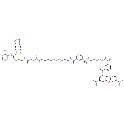
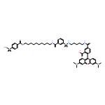


![TERT-BUTYL (2S)-2-AMINO-3-[4-(TRIFLUOROMETHYL)PHENYL]PROPANOATE](/data/chemimg/421300/1241681-43-5.png)
![TERT-BUTYL (2S)-2-AMINO-3-[4-(TRIFLUOROMETHYL)PHENYL]PROPANOATE](/data/chemimg/421300/1241681-43-5_b.png)
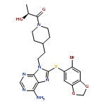
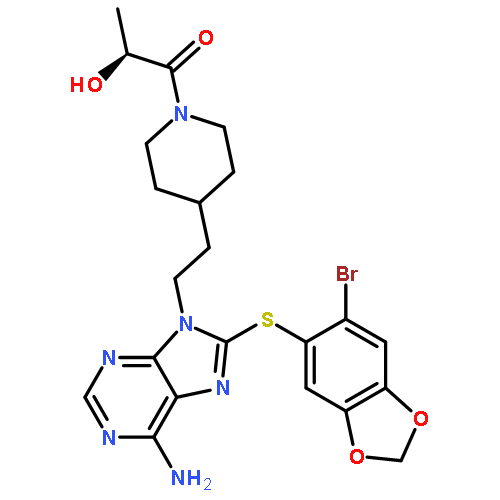

![Acetic acid, 2-[2-(2-azidoethoxy)ethoxy]-](/data/chemimg/127600/882518-90-3.png)
![Acetic acid, 2-[2-(2-azidoethoxy)ethoxy]-](/data/chemimg/127600/882518-90-3_b.png)
![L-ALANINE, 3-[1,1'-BIPHENYL]-4-YL-L-ALANYL-3-[1,1'-BIPHENYL]-4-YL-](http://img.cochemist.com/ccimg/875600/875559-54-9.png)
![L-ALANINE, 3-[1,1'-BIPHENYL]-4-YL-L-ALANYL-3-[1,1'-BIPHENYL]-4-YL-](http://img.cochemist.com/ccimg/875600/875559-54-9_b.png)
![8-(6-iodobenzo[d][1,3]dioxol-5-ylthio)-9-(3-(isopropylamino)propyl)-9H-puri n-6-amine hydrochloride](http://img.cochemist.com/ccimg/873500/873436-91-0.png)
![8-(6-iodobenzo[d][1,3]dioxol-5-ylthio)-9-(3-(isopropylamino)propyl)-9H-puri n-6-amine hydrochloride](http://img.cochemist.com/ccimg/873500/873436-91-0_b.png)
![1H-Purin-6-amine, 8-[(6-iodo-1,3-benzodioxol-5-yl)thio]-](http://img.cochemist.com/ccimg/873500/873436-89-6.png)
![1H-Purin-6-amine, 8-[(6-iodo-1,3-benzodioxol-5-yl)thio]-](http://img.cochemist.com/ccimg/873500/873436-89-6_b.png)


![Propanoic acid, 2-[(3,5-dichlorophenyl)hydrazono]-, ethyl ester, (Z)-](http://img.cochemist.com/ccimg/137900/137836-39-6.png)
![Propanoic acid, 2-[(3,5-dichlorophenyl)hydrazono]-, ethyl ester, (Z)-](http://img.cochemist.com/ccimg/137900/137836-39-6_b.png)


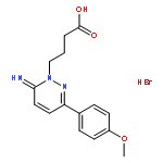
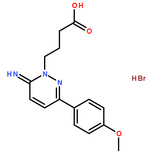
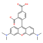
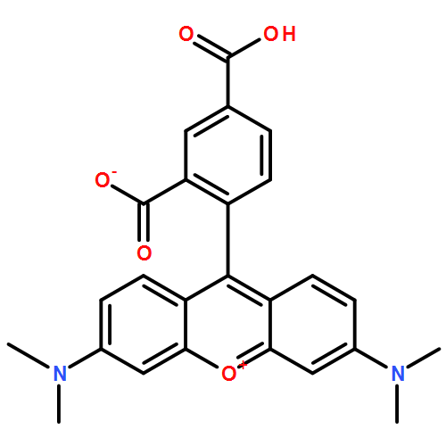


![3-Methyl-6-(3-(trifluoromethyl)phenyl)-[1,2,4]triazolo[4,3-b]pyridazine](http://img.cochemist.com/ccimg/66600/66548-69-4.png)
![3-Methyl-6-(3-(trifluoromethyl)phenyl)-[1,2,4]triazolo[4,3-b]pyridazine](http://img.cochemist.com/ccimg/66600/66548-69-4_b.png)


![3',6'-Dihydroxy-3H-spiro[isobenzofuran-1,9'-xanthen]-3-one](http://img.cochemist.com/ccimg/2400/2321-07-5.png)
![3',6'-Dihydroxy-3H-spiro[isobenzofuran-1,9'-xanthen]-3-one](http://img.cochemist.com/ccimg/2400/2321-07-5_b.png)


