Co-reporter:Mei Sun, Xiang Tian, and Zhibo Yang
Analytical Chemistry September 5, 2017 Volume 89(Issue 17) pp:9069-9069
Publication Date(Web):July 28, 2017
DOI:10.1021/acs.analchem.7b01746
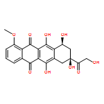
Extracellular compounds in tumors play critical roles in intercellular communication, tumor proliferation, and cancer cell metastasis. However, the lack of appropriate techniques leads to limited studies of extracellular metabolite. Here, we introduced a microscale collection device, the Micro-funnel, fabricated from biocompatible fused silica capillary. With a small probe size (∼25 μm), the Micro-funnel can be implanted into live multicellular tumor spheroids to accumulate the extracellular metabolites produced by cancer cells. Metabolites collected in the Micro-funnel device were then extracted by a microscale sampling and ionization device, the Single-probe, for real-time mass spectrometry (MS) analysis. We successfully detected the abundance change of anticancer drug irinotecan and its metabolites inside spheroids treated under a series of conditions. Moreover, we found that irinotecan treatment dramatically altered the composition of extracellular compounds. Specifically, we observed the increased abundances of a large number of lipids, which are potentially related to the drug resistance of cancer cells. This study provides a novel way to detect the extracellular compounds inside live spheroids, and the successful development of our technique can benefit the research in multiple areas, including the microenvironment inside live tissues, cell–cell communication, biomarker discovery, and drug development.
Co-reporter:Ning Pan, Wei Rao, Shawna J. Standke, and Zhibo Yang
Analytical Chemistry 2016 Volume 88(Issue 13) pp:6812
Publication Date(Web):May 30, 2016
DOI:10.1021/acs.analchem.6b01284
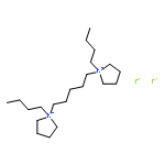
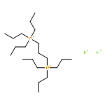
A unique mass spectrometry (MS) method has been developed to determine the negatively charged species in live single cells using the positive ionization mode. The method utilizes dicationic ion-pairing compounds through the miniaturized multifunctional device, the single-probe, for reactive MS analysis of live single cells under ambient conditions. In this study, two dicationic reagents, 1,5-pentanediyl-bis(1-butylpyrrolidinium) difluoride (C5(bpyr)2F2) and 1,3-propanediyl-bis(tripropylphosphonium) difluoride (C3(triprp)2F2), were added in the solvent and introduced into single cells to extract cellular contents for real-time MS analysis. The negatively charged (1– charged) cell metabolites, which form stable ion-pairs (1+ charged) with dicationic compounds (2+ charged), were detected in positive ionization mode with a greatly improved sensitivity. We have tentatively assigned 192 and 70 negatively charged common metabolites as adducts with (C5(bpyr)2F2) and (C3(triprp)2F2), respectively, in three separate SCMS experiments in the positive ion mode. The total number of tentatively assigned metabolites is 285 for C5(bpyr)2F2 and 143 for C3(triprp)2F2. In addition, the selectivity of dicationic compounds in the complex formation allows for the discrimination of overlapped ion peaks with low abundances. Tandem (MS/MS) analyses at the single cell level were conducted for selected adduct ions for molecular identification. The utilization of the dicationic compounds in the single-probe MS technique provides an effective approach to the detection of a broad range of metabolites at the single cell level.
Co-reporter:Wei Rao;Ning Pan;Xiang Tian
Journal of The American Society for Mass Spectrometry 2016 Volume 27( Issue 1) pp:124-134
Publication Date(Web):2016 January
DOI:10.1007/s13361-015-1287-7
We have used the Single-probe, a miniaturized sampling device utilizing in-situ surface microextraction for ambient mass spectrometry (MS) analysis, for the high resolution MS imaging (MSI) of negatively charged species in the positive ionization mode. Two dicationic compounds, 1,5-pentanediyl-bis(1-butylpyrrolidinium) difluoride [C5(bpyr)2F2] and 1,3-propanediyl-bis(tripropylphosphonium) difluoride [C3(triprp)2F2], were added into the sampling solvent to form 1+ charged adducts with the negatively charged species extracted from tissues. We were able to detect 526 and 322 negatively charged species this way using [C5(bpyr)2F2] and [C3(triprp)2F2], respectively, including oleic acid, arachidonic acid, and several species of phosphatidic acid, phosphoethanolamine, phosphatidylserine, phosphatidylglycerol, phosphatidylinositol, and others. In conjunction with the identification of the non-adduct cations, we have tentatively identified a total number of 1200 and 828 metabolites from mouse brain sections using [C5(bpyr)2F2] and [C3(triprp)2F2], respectively, through high mass accuracy measurements (mass error <5 ppm); MS/MS analyses were also performed to verify the identity of selected species. In addition to the high mass accuracy measurement, we were able to generate high spatial resolution (~17 μm) MS images of mouse brain sections. Our study demonstrated that utilization of dicationic compounds in the surface microextraction with the Single-probe device can perform high mass and spatial resolution ambient MSI measurements of broader types of compounds in tissues. Other reagents can be potentially used with the Single-probe device for a variety of reactive MSI studies to enable the analysis of species that are previously intractable.
Co-reporter:Zhibo Yang, Ning Pan
International Journal of Mass Spectrometry 2015 Volume 378() pp:364-368
Publication Date(Web):15 February 2015
DOI:10.1016/j.ijms.2014.10.015
•DFT computations were carried out to study ion–neutral reactions.•Reactions involve benzene and naphthalene.•Water and amino acid precursors can be synthesized from reactions.•Aromatic hydrocarbons participate in reactions as catalysts.•These approaches can be used as models to study reactions on dust grain surfaces.Aromatic hydrocarbons (AHs) and their derivatives have been suggested as the building blocks of interstellar dust grains and are responsible for the evolution of astrobiological molecules via surface reactions in space. Gas-phase studies of molecules and ions known to exist in space are crucial to understand relevant ion–molecule reactions and the generation of new species. Reactions catalyzed by large species such as AHs remain relatively unexplored. Our computational studies focus on the energetics and reaction mechanisms of the formation of representative molecules (i.e., hydrogen peroxide, acetamide, and amino acetonitrile) that are critical for the origin of water and amino acids in the universe. Calculations have been carried out using Gaussian 09 to obtain the structures, energetics, and reaction mechanisms to investigate the formation of hydrogen peroxide (H2O2), acetamide (CH3C(O)NH2), and amino acetonitrile (NH2CH2CN). Our results suggest that there are energetically accessible reaction pathways leading to the formation of these molecules through species which have been discovered in the interstellar medium (ISM). Ionized benzene and polycyclic aromatic hydrocarbons (PAHs) can act as catalysts to facilitate the formation of astromolecules. The theoretical studies can enhance our understanding of ion–molecule reactions that are relevant to the formation of important astromolecules in the gas phase, and provide a new way to investigate the formation of polyatomic molecules on surfaces of dust grains such as large PAHs.
Co-reporter:Wei Rao;Ning Pan
Journal of The American Society for Mass Spectrometry 2015 Volume 26( Issue 6) pp:986-993
Publication Date(Web):2015 June
DOI:10.1007/s13361-015-1091-4
Ambient mass spectrometry imaging (MSI) is an emerging field with great potential for the detailed spatial analysis of biological samples with minimal pretreatment. We have developed a miniaturized sampling and ionization device, the Single-probe, which uses in-situ surface micro-extraction to achieve high detection sensitivity and spatial resolution during MSI experiments. The Single-probe was coupled to a Thermo LTQ Orbitrap XL mass spectrometer and was able to create high spatial and high mass resolution MS images at 8 ± 2 and 8.5 μm on flat polycarbonate microscope slides and mouse kidney sections, respectively, which are among the highest resolutions available for ambient MSI techniques. Our proof-of-principle experiments indicate that the Single-probe MSI technique has the potential to obtain ambient MS images with very high spatial resolutions with minimal sample preparation, which opens the possibility for subcellular ambient tissue MSI to be performed in the future.
Co-reporter:Ning Pan, Wei Rao, Naga Rama Kothapalli, Renmeng Liu, Anthony W. G. Burgett, and Zhibo Yang
Analytical Chemistry 2014 Volume 86(Issue 19) pp:9376
Publication Date(Web):September 15, 2014
DOI:10.1021/ac5029038
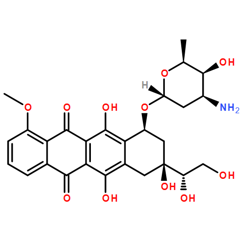
We have developed a new mass spectrometry (MS) technology, the Single-probe MS, capable of real-time, in situ metabolomic analysis of individual living cells. The Single-probe is a miniaturized multifunctional sampling and ionization device that is directly coupled to the mass spectrometer. With a sampling tip smaller than individual eukaryotic cells (<10 μm), the Single-probe can be inserted into single cells to sample the intracellular compounds for real-time MS analysis. We have used the Single-probe to detect several cellular metabolites and the anticancer small molecules paclitaxel, doxorubicin, and OSW-1 in individual cervical cancer cells (HeLa). Single cell mass spectrometry (SCMS) is an emerging scientific technology that could reshape the analytical science of many research disciplines, and the Single-probe MS technology is a novel method for SCMS that, through its accessible fabrication protocols, can be broadly applied to different research areas.
.png)

![Cholest-5-en-22-one,16-[[2-O-acetyl-3-O-[2-O-(4-methoxybenzoyl)-b-D-xylopyranosyl]-a-L-arabinopyranosyl]oxy]-3,17-dihydroxy-, (3b,16b)-](http://img.cochemist.com/ccimg/145100/145075-81-6.png)
![Cholest-5-en-22-one,16-[[2-O-acetyl-3-O-[2-O-(4-methoxybenzoyl)-b-D-xylopyranosyl]-a-L-arabinopyranosyl]oxy]-3,17-dihydroxy-, (3b,16b)-](http://img.cochemist.com/ccimg/145100/145075-81-6_b.png)