Co-reporter:Jacqueline L. Naffin-Olivos, Andrew Daab, Andre White, Nathan E. Goldfarb, Amy C. Milne, Dali Liu, Jacqueline Baikovitz, Ben M. Dunn, Jyothi Rengarajan, Gregory A. Petsko, and Dagmar Ringe
Biochemistry May 2, 2017 Volume 56(Issue 17) pp:2304-2304
Publication Date(Web):March 27, 2017
DOI:10.1021/acs.biochem.6b01066
The Mycobacterium tuberculosis (Mtb) serine protease Hip1 (hydrolase important for pathogenesis; Rv2224c) promotes tuberculosis (TB) pathogenesis by impairing host immune responses through proteolysis of a protein substrate, Mtb GroEL2. The cell surface localization of Hip1 and its immunomodulatory functions make Hip1 a good drug target for new adjunctive immune therapies for TB. Here, we report the crystal structure of Hip1 to a resolution of 2.6 Å and the kinetic studies of the enzyme against model substrates and the protein GroEL2. The structure shows a two-domain protein, one of which contains the catalytic residues that are the signature of a serine protease. Surprisingly, a threonine is located within the active site close enough to hydrogen bond with the catalytic residues Asp463 and His490. Mutation of this residue, Thr466, to alanine established its importance for function. Our studies provide insights into the structure of a member of a novel family of proteases. Knowledge of the Hip1 structure will aid in designing inhibitors that could block Hip1 activity.
Co-reporter:Aditi R. Deshpande, Thomas C. Pochapsky, and Dagmar Ringe
Chemical Reviews August 9, 2017 Volume 117(Issue 15) pp:10474-10474
Publication Date(Web):July 21, 2017
DOI:10.1021/acs.chemrev.7b00117
Acireductone dioxygenase (ARD) from the methionine salvage pathway (MSP) is a unique enzyme that exhibits dual chemistry determined solely by the identity of the divalent transition-metal ion (Fe2+ or Ni2+) in the active site. The Fe2+-containing isozyme catalyzes the on-pathway reaction using substrates 1,2-dihydroxy-3-keto-5-methylthiopent-1-ene (acireductone) and dioxygen to generate formate and the ketoacid precursor of methionine, 2-keto-4-methylthiobutyrate, whereas the Ni2+-containing isozyme catalyzes an off-pathway shunt with the same substrates, generating methylthiopropionate, carbon monoxide, and formate. The dual chemistry of ARD was originally discovered in the bacterium Klebsiella oxytoca, but it has recently been shown that mammalian ARD enzymes (mouse and human) are also capable of catalyzing metal-dependent dual chemistry in vitro. This is particularly interesting, since carbon monoxide, one of the products of off-pathway reaction, has been identified as an antiapoptotic molecule in mammals. In addition, several biochemical and genetic studies have indicated an inhibitory role of human ARD in cancer. This comprehensive review describes the biochemical and structural characterization of the ARD family, the proposed experimental and theoretical approaches to establishing mechanisms for the dual chemistry, insights into the mechanism based on comparison with structurally and functionally similar enzymes, and the applications of this research to the field of artificial metalloenzymes and synthetic biology.
Co-reporter:Aditi R. Deshpande, Karina Wagenpfeil, Thomas C. Pochapsky, Gregory A. Petsko, and Dagmar Ringe
Biochemistry 2016 Volume 55(Issue 9) pp:1398-1407
Publication Date(Web):February 9, 2016
DOI:10.1021/acs.biochem.5b01319
The two acireductone dioxygenase (ARD) isozymes from the methionine salvage pathway of Klebsiella oxytoca are the only known pair of naturally occurring metalloenzymes with distinct chemical and physical properties determined solely by the identity of the divalent transition metal ion (Fe2+ or Ni2+) in the active site. We now show that this dual chemistry can also occur in mammals. ARD from Mus musculus (MmARD) was studied to relate the metal ion identity and three-dimensional structure to enzyme function. The iron-containing isozyme catalyzes the cleavage of 1,2-dihydroxy-3-keto-5-(thiomethyl)pent-1-ene (acireductone) by O2 to formate and the ketoacid precursor of methionine, which is the penultimate step in methionine salvage. The nickel-bound form of ARD catalyzes an off-pathway reaction resulting in formate, carbon monoxide (CO), and 3-(thiomethyl) propionate. Recombinant MmARD was expressed and purified to obtain a homogeneous enzyme with a single transition metal ion bound. The Fe2+-bound protein, which shows about 10-fold higher activity than that of others, catalyzes on-pathway chemistry, whereas the Ni2+, Co2+, or Mn2+ forms exhibit off-pathway chemistry, as has been seen with ARD from Klebsiella. Thermal stability of the isozymes is strongly affected by the metal ion identity, with Ni2+-bound MmARD being the most stable, followed by Co2+ and Fe2+, and Mn2+-bound ARD being the least stable. Ni2+- and Co2+-bound MmARD were crystallized, and the structures of the two proteins found to be similar. Enzyme–ligand complexes provide insight into substrate binding, metal coordination, and the catalytic mechanism.
Co-reporter:Cheryl A. Kreinbring;Stephen P. Remillard;Paul Hubbard;Heather R. Brodkin;Finian J. Leeper;Dan Hawksley;Elaine Y. Lai;Chandler Fulton;Gregory A. Petsko;
Proceedings of the National Academy of Sciences 2014 111(1) pp:137-142
Publication Date(Web):December 18, 2013
DOI:10.1073/pnas.1315882110
Thiaminases, enzymes that cleave vitamin B1, are sporadically distributed among prokaryotes and eukaryotes. Thiaminase I enzymes
catalyze the elimination of the thiazole ring moiety from thiamin through substitution of the methylene group with a nitrogenous
base or sulfhydryl compound. In eukaryotic organisms, these enzymes are reported to have much higher molecular weights than
their bacterial counterparts. A thiaminase I of the single-celled amoeboflagellate Naegleria gruberi is the only eukaryotic thiaminase I to have been cloned, sequenced, and expressed. Here, we present the crystal structure
of N. gruberi thiaminase I to a resolution of 2.8 Å, solved by isomorphous replacement and pseudo–two-wavelength multiwavelength anomalous
diffraction and refined to an R factor of 0.231 (Rfree, 0.265). This structure was used to solve the structure of the enzyme in complex with 3-deazathiamin, a noncleavable thiamin
analog and enzyme inhibitor (2.7 Å; R, 0.233; Rfree, 0.267). These structures define the mode of thiamin binding to this class of thiaminases and indicate the involvement of
Asp272 as the catalytic base. This enzyme is able to use thiamin as a substrate and is active with amines such as aniline
and veratrylamine as well as sulfhydryl compounds such as l-cysteine and β-mercaptoethanol as cosubstrates. Despite significant differences in polypeptide sequence and length, we have
shown that the N. gruberi thiaminase I is homologous in structure and activity to a previously characterized bacterial thiaminase I.
Co-reporter:Ce Feng Liu, Dali Liu, Jessica Momb, Pei W. Thomas, Ashley Lajoie, Gregory A. Petsko, Walter Fast, and Dagmar Ringe
Biochemistry 2013 Volume 52(Issue 9) pp:
Publication Date(Web):February 6, 2013
DOI:10.1021/bi400050j
Autoinducer inactivator A (AiiA) is a metal-dependent N-acyl homoserine lactone hydrolase that displays broad substrate specificity but shows a preference for substrates with long N-acyl substitutions. Previously, crystal structures of AiiA in complex with the ring-opened product N-hexanoyl-l-homoserine revealed binding interactions near the metal center but did not identify a binding pocket for the N-acyl chains of longer substrates. Here we report the crystal structure of an AiiA mutant, F107W, determined in the presence and absence of N-decanoyl-l-homoserine. F107 is located in a hydrophobic cavity adjacent to the previously identified ligand binding pocket, and the F107W mutation results in the formation of an unexpected interaction with the ring-opened product. Notably, the structure reveals a previously unidentified hydrophobic binding pocket for the substrate’s N-acyl chain. Two aromatic residues, F64 and F68, form a hydrophobic clamp, centered around the seventh carbon in the product-bound structure’s decanoyl chain, making an interaction that would also be available for longer substrates, but not for shorter substrates. Steady-state kinetics using substrates of various lengths with AiiA bearing mutations at the hydrophobic clamp, including insertion of a redox-sensitive cysteine pair, confirms the importance of this hydrophobic feature for substrate preference. Identifying the specificity determinants of AiiA will aid the development of more selective quorum-quenching enzymes as tools and as potential therapeutics.
Co-reporter:Roman Garcia;Wei Wang;Cheryl A. Kreinbring;Gregory A. Petsko;Thomas C. Terwilliger;Boris R. Belitsky;Alicia Bach;Rui Wu;Raji Edayathumangalam;Jingling Liao;Yuguang Wang;Todd A. Stone;Quyen Q. Hoang;Dali Liu
PNAS 2013 Volume 110 (Issue 44 ) pp:17820-17825
Publication Date(Web):2013-10-29
DOI:10.1073/pnas.1315887110
Bacillus subtilis GabR is a transcription factor that regulates gamma-aminobutyric acid (GABA) metabolism. GabR is a member of the understudied
MocR/GabR subfamily of the GntR family of transcription regulators. A typical MocR/GabR-type regulator is a chimeric protein
containing a short N-terminal helix-turn-helix DNA-binding domain and a long C-terminal pyridoxal 5′-phosphate (PLP)-binding
putative aminotransferase domain. In the presence of PLP and GABA, GabR activates the gabTD operon, which allows the bacterium to use GABA as nitrogen and carbon sources. GabR binds to its own promoter and represses
gabR transcription in the absence of GABA. Here, we report two crystal structures of full-length GabR from B. subtilis: a 2.7-Å structure of GabR with PLP bound and the 2.55-Å apo structure of GabR without PLP. The quaternary structure of GabR
is a head-to-tail domain-swap homodimer. Each monomer comprises two domains: an N-terminal winged-helix DNA-binding domain
and a C-terminal PLP-binding type I aminotransferase-like domain. The winged-helix domain contains putative DNA-binding residues
conserved in other GntR-type regulators. Together with sedimentation velocity and fluorescence polarization assays, the crystal
structure of GabR provides insights into DNA binding by GabR at the gabR and gabT promoters. The absence of GabR-mediated aminotransferase activity in the presence of GABA and PLP, and the presence of an
active site configuration that is incompatible with stabilization of the GABA external aldimine suggest that a GabR aminotransferase-like
activity involving GABA and PLP is not essential to its primary function as a transcription regulator.
Co-reporter:Heather R. Brodkin, Walter R. P. Novak, Amy C. Milne, J. Alejandro D’Aquino, N. M. Karabacak, Ilana G. Goldberg, Jeffrey N. Agar, Mark S. Payne, Gregory A. Petsko, Mary Jo Ondrechen, and Dagmar Ringe
Biochemistry 2011 Volume 50(Issue 22) pp:
Publication Date(Web):April 7, 2011
DOI:10.1021/bi101761e
Active sites may be regarded as layers of residues, whereby the residues that interact directly with substrate also interact with residues in a second shell and these in turn interact with residues in a third shell. These residues in the second and third layers may have distinct roles in maintaining the essential chemical properties of the first-shell catalytic residues, particularly their spatial arrangement relative to the substrate binding pocket, and their electrostatic and dynamic properties. The extent to which these remote residues participate in catalysis and precisely how they affect first-shell residues remains unexplored. To improve our understanding of the roles of second- and third-shell residues in catalysis, we used THEMATICS to identify residues in the second and third shells of the Co-type nitrile hydratase from Pseudomonas putida (ppNHase) that may be important for catalysis. Five of these predicted residues, and three additional, conserved residues that were not predicted, have been conservatively mutated, and their effects have been studied both kinetically and structurally. The eight residues have no direct contact with the active site metal ion or bound substrate. These results demonstrate that three of the predicted second-shell residues (α-Asp164, β-Glu56, and β-His147) and one predicted third-shell residue (β-His71) have significant effects on the catalytic efficiency of the enzyme. One of the predicted residues (α-Glu168) and the three residues not predicted (α-Arg170, α-Tyr171, and β-Tyr215) do not have any significant effects on the catalytic efficiency of the enzyme.
Co-reporter:Wei Wang;Iva Perovic;Johnathan Chittuluru;Alice Kaganovich;Linh T. T. Nguyen;Jingling Liao;Jared R. Auclair;Derrick Johnson;Anuradha Landeru;Alana K. Simorellis;Shulin Ju;Mark R. Cookson;Francisco J. Asturias;Jeffrey N. Agar;Brian N. Webb;ChulHee Kang;Gregory A. Petsko;Thomas C. Pochapsky;Quyen Q. Hoang
PNAS 2011 108 (43 ) pp:17797-17802
Publication Date(Web):2011-10-25
DOI:10.1073/pnas.1113260108
A heterologously expressed form of the human Parkinson disease-associated protein α-synuclein with a 10-residue N-terminal
extension is shown to form a stable tetramer in the absence of lipid bilayers or micelles. Sequential NMR assignments, intramonomer
nuclear Overhauser effects, and circular dichroism spectra are consistent with transient formation of α-helices in the first
100 N-terminal residues of the 140-residue α-synuclein sequence. Total phosphorus analysis indicates that phospholipids are
not associated with the tetramer as isolated, and chemical cross-linking experiments confirm that the tetramer is the highest-order
oligomer present at NMR sample concentrations. Image reconstruction from electron micrographs indicates that a symmetric oligomer
is present, with three- or fourfold symmetry. Thermal unfolding experiments indicate that a hydrophobic core is present in
the tetramer. A dynamic model for the tetramer structure is proposed, based on expected close association of the amphipathic
central helices observed in the previously described micelle-associated “hairpin” structure of α-synuclein.
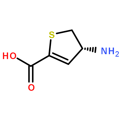
Co-reporter:Bryan W. Lepore, Dali Liu, Ying Peng, Mengmeng Fu, Chad Yasuda, James M. Manning, Richard B. Silverman and Dagmar Ringe
Biochemistry 2010 Volume 49(Issue 14) pp:
Publication Date(Web):March 1, 2010
DOI:10.1021/bi902052x
Mechanism-based inhibitors such as cycloserine and gabaculine can inactivate aminotransferases via reactions of the compounds with the pyridoxal phosphate cofactor forming an irreversible adduct. The reaction is chirally specific in that any one enzyme usually only recognizes one enantiomer of the inactivator. For instance, l-aspartate aminotransferase (l-AspAT) is inactivated by 4-amino-4,5-dihydro-2-thiophenecarboxylic acid (ADTA), however, only by the S-isomer. We have now shown that d-amino acid aminotransferase (d-a-AT) is irreversibly inactivated by the R-isomer of the same compound. The X-ray crystal structure (PDB code: 3LQS) of the inactivated enzyme shows that in the product the enzyme no longer makes a Schiff base linkage to the pyridoxal 5′-phosphate (PLP) cofactor, and instead the compound has formed a derivative of the cofactor. The adduct is similar to that formed between d-cycloserine and d-a-AT or alanine racemase (Ala-Rac) in that the thiophene ring of R-ADTA is intact and seems to be aromatic. The plane of the ring is rotated by nearly 90° with respect to the plane of the pyridine ring of the cofactor, in comparison with the enzyme inactivated by cycloserine. Based on the structure of the product, the mechanism of inactivation most probably involves a transamination followed by aromatization to form an aromatic thiophene ring.
Co-reporter:Dali Liu, Edwin Pozharski, Mengmeng Fu, Richard B. Silverman, and Dagmar Ringe
Biochemistry 2010 Volume 49(Issue 49) pp:
Publication Date(Web):October 29, 2010
DOI:10.1021/bi101325z
As a potential drug to treat neurological diseases, the mechanism-based inhibitor (S)-4-amino-4,5-dihydro-2-furancarboxylic acid (S-ADFA) has been found to inhibit the γ-aminobutyric acid aminotransferase (GABA-AT) reaction. To circumvent the difficulties in structural studies of a S-ADFA−enzyme complex using GABA-AT, l-aspartate aminotransferase (l-AspAT) from Escherichia coli was used as a model PLP-dependent enzyme. Crystal structures of the E. coli aspartate aminotransferase with S-ADFA bound to the active site were obtained via cocrystallization at pH 7.5 and 8. The complex structures suggest that S-ADFA inhibits the transamination reaction by forming adducts with the catalytic lysine 246 via a covalent bond while producing 1 equiv of pyridoxamine 5′-phosphate (PMP). Based on the structures, formation of the K246-S-ADFA adducts requires a specific initial binding configuration of S-ADFA in the l-AspAT active site, as well as deprotonation of the ε-amino group of lysine 246 after the formation of the quinonoid and/or ketimine intermediate in the overall inactivation reaction.
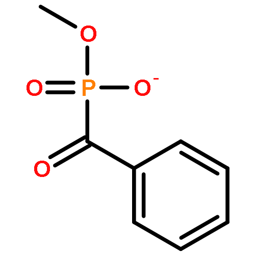
Co-reporter:Gabriel S. Brandt, Malea M. Kneen, Sumit Chakraborty, Ahmet T. Baykal, Natalia Nemeria, Alejandra Yep, David I. Ruby, Gregory A. Petsko, George L. Kenyon, Michael J. McLeish, Frank Jordan and Dagmar Ringe
Biochemistry 2009 Volume 48(Issue 15) pp:
Publication Date(Web):March 25, 2009
DOI:10.1021/bi801950k
Benzoylformate decarboxylase (BFDC) is a thiamin diphosphate- (ThDP-) dependent enzyme acting on aromatic substrates. In addition to its metabolic role in the mandelate pathway, BFDC shows broad substrate specificity coupled with tight stereo control in the carbon−carbon bond-forming reverse reaction, making it a useful biocatalyst for the production of chiral α-hydroxy ketones. The reaction of methyl benzoylphosphonate (MBP), an analogue of the natural substrate benzoylformate, with BFDC results in the formation of a stable analogue (C2α-phosphonomandelyl-ThDP) of the covalent ThDP-substrate adduct C2α-mandelyl-ThDP. Formation of the stable adduct is confirmed both by formation of a circular dichroism band characteristic of the 1′,4′-iminopyrimidine tautomeric form of ThDP (commonly observed when ThDP forms tetrahedral complexes with its substrates) and by high-resolution mass spectrometry of the reaction mixture. In addition, the structure of BFDC with the MBP inhibitor was solved by X-ray crystallography to a spatial resolution of 1.37 Å (PDB ID 3FSJ). The electron density clearly shows formation of a tetrahedral adduct between the C2 atom of ThDP and the carbonyl carbon atom of the MBP. This adduct resembles the intermediate from the penultimate step of the carboligation reaction between benzaldehyde and acetaldehyde. The combination of real-time kinetic information via stopped-flow circular dichroism with steady-state data from equilibrium circular dichroism measurements and X-ray crystallography reveals details of the first step of the reaction catalyzed by BFDC. The MBP-ThDP adduct on BFDC is compared to the recently solved structure of the same adduct on benzaldehyde lyase, another ThDP-dependent enzyme capable of catalyzing aldehyde condensation with high stereospecificity.
Co-reporter:Melissa R. Landon;Raquel L. Lieberman
Journal of Computer-Aided Molecular Design 2009 Volume 23( Issue 8) pp:491-500
Publication Date(Web):2009 August
DOI:10.1007/s10822-009-9283-2
The identification of hot spots, i.e., binding regions that contribute substantially to the free energy of ligand binding, is a critical step for structure-based drug design. Here we present the application of two fragment-based methods to the detection of hot spots for DJ-1 and glucocerebrosidase (GCase), targets for the development of therapeutics for Parkinson’s and Gaucher’s diseases, respectively. While the structures of these two proteins are known, binding information is lacking. In this study we employ the experimental multiple solvent crystal structures (MSCS) method and computational fragment mapping (FTMap) to identify regions suitable for the development of pharmacological chaperones for DJ-1 and GCase. Comparison of data derived via MSCS and FTMap also shows that FTMap, a computational method for the identification of fragment binding hot spots, is an accurate and robust alternative to the performance of expensive and difficult crystallographic experiments.
Co-reporter:Dali Liu, Jessica Momb, Pei W. Thomas, Aaron Moulin, Gregory A. Petsko, Walter Fast and Dagmar Ringe
Biochemistry 2008 Volume 47(Issue 29) pp:
Publication Date(Web):July 15, 2008
DOI:10.1021/bi800368y
Enzymes capable of hydrolyzing N-acyl-l-homoserine lactones (AHLs) used in some bacterial quorum-sensing pathways are of considerable interest for their ability to block undesirable phenotypes. Most known AHL hydrolases that catalyze ring opening (AHL lactonases) are members of the metallo-β-lactamase enzyme superfamily and rely on a dinuclear zinc site for catalysis and stability. Here we report the three-dimensional structures of three product complexes formed with the AHL lactonase from Bacillus thuringiensis. Structures of the lactonase bound with two different concentrations of the ring-opened product of N-hexanoyl-l-homoserine lactone are determined at 0.95 and 1.4 Å resolution and exhibit different product configurations. A structure of the ring-opened product of the non-natural N-hexanoyl-l-homocysteine thiolactone at 1.3 Å resolution is also determined. On the basis of these product-bound structures, a substrate-binding model is presented that differs from previous proposals. Additionally, the proximity of the product to active-site residues and observed changes in protein conformation and metal coordination provide insight into the catalytic mechanism of this quorum-quenching metalloenzyme.
Co-reporter:Jessica Momb, Canhui Wang, Dali Liu, Pei W. Thomas, Gregory A. Petsko, Hua Guo, Dagmar Ringe and Walter Fast
Biochemistry 2008 Volume 47(Issue 29) pp:
Publication Date(Web):July 15, 2008
DOI:10.1021/bi8003704
The N-acyl-l-homoserine lactone hydrolases (AHL lactonases) have attracted considerable attention because of their ability to quench AHL-mediated quorum-sensing pathways in Gram-negative bacteria and because of their relation to other enzymes in the metallo-β-lactamase superfamily. To elucidate the detailed catalytic mechanism of AHL lactonase, mutations are made on residues that presumably contribute to substrate binding and catalysis. Steady-state kinetic studies are carried out on both the wild-type and mutant enzymes using a spectrum of substrates. Two mutations, Y194F and D108N, present significant effects on the overall catalysis. On the basis of a high-resolution structural model of the enzyme−product complex, a hybrid quantum mechanical/molecular mechanical method is used to model the substrate binding orientation and to probe the effect of the Y194F mutation. Combining all experimental and computational results, we propose a detailed mechanism for the ring-opening hydrolysis of AHL substrates as catalyzed by the AHL lactonase from Bacillus thuringiensis. Several features of the mechanism that are also found in related enzymes are discussed and may help to define an evolutionary thread that connects the hydrolytic enzymes of this mechanistically diverse superfamily.
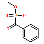
Co-reporter:Gabriel S. Brandt, Natalia Nemeria, Sumit Chakraborty, Michael J. McLeish, Alejandra Yep, George L. Kenyon, Gregory A. Petsko, Frank Jordan and Dagmar Ringe
Biochemistry 2008 Volume 47(Issue 29) pp:
Publication Date(Web):June 21, 2008
DOI:10.1021/bi8004413
Benzaldehyde lyase (BAL) catalyzes the reversible cleavage of (R)-benzoin to benzaldehyde utilizing thiamin diphosphate and Mg2+ as cofactors. The enzyme is important for the chemoenzymatic synthesis of a wide range of compounds via its carboligation reaction mechanism. In addition to its principal functions, BAL can slowly decarboxylate aromatic amino acids such as benzoylformic acid. It is also intriguing mechanistically due to the paucity of acid−base residues at the active center that can participate in proton transfer steps thought to be necessary for these types of reactions. Here methyl benzoylphosphonate, an excellent electrostatic analogue of benzoylformic acid, is used to probe the mechanism of benzaldehyde lyase. The structure of benzaldehyde lyase in its covalent complex with methyl benzoylphosphonate was determined to 2.49 Å (Protein Data Bank entry 3D7K) and represents the first structure of this enzyme with a compound bound in the active site. No large structural reorganization was detected compared to the complex of the enzyme with thiamin diphosphate. The configuration of the predecarboxylation thiamin-bound intermediate was clarified by the structure. Both spectroscopic and X-ray structural studies are consistent with inhibition resulting from the binding of MBP to the thiamin diphosphate in the active centers. We also delineated the role of His29 (the sole potential acid−base catalyst in the active site other than the highly conserved Glu50) and Trp163 in cofactor activation and catalysis by benzaldehyde lyase.
Co-reporter:Niloufar J. Ataie, Quyen Q. Hoang, Megan P. D. Zahniser, Yupeng Tu, Amy Milne, Gregory A. Petsko and Dagmar Ringe
Biochemistry 2008 Volume 47(Issue 29) pp:
Publication Date(Web):June 25, 2008
DOI:10.1021/bi702188e
The chemical properties of zinc make it an ideal metal to study the role of coordination strain in enzymatic rate enhancement. The zinc ion and the protein residues that are bound directly to the zinc ion represent a functional charge/dipole complex, and polarization of this complex, which translates to coordination distortion, may tune electrophilicity, and hence, reactivity. Conserved protein residues outside of the charge/dipole complex, such as second-shell residues, may play a role in supporting the electronic strain produced as a consequence of functional polarization. To test the correlation between charge/dipole polarity and ligand binding affinity, structure−function studies were carried out on the dizinc aminopeptidase from Vibrio proteolyticus. Alanine substitutions of S228 and M180 resulted in catalytically diminished enzymes whose crystal structures show very little change in the positions of the metal ions and the protein residues. However, more detailed inspections of the crystal structures show small positional changes that account for differences in the zinc ion coordination geometry. Measurements of the binding affinity of leucine phosphonic acid, a transition state analogue, and leucine, a product, show a correlation between coordination geometry and ligand binding affinity. These results suggest that the coordination number and polarity may tune the electrophilicity of zinc. This may have provided the evolving enzyme with the ability to discriminate between reaction coordinate species.
Co-reporter:Dali Liu;Everett M. Stone;Gregory A. Petsko;Pei W. Thomas;Walter Fast;Bryan W. Lepore
PNAS 2005 Volume 102 (Issue 33 ) pp:11882-11887
Publication Date(Web):2005-08-16
DOI:10.1073/pnas.0505255102
The three-dimensional structure of the N-acyl-l-homoserine lactone hydrolase (AHL lactonase) from Bacillus thuringiensis has been determined, by using single-wavelength anomalous dispersion (SAD) phasing, to 1.6-Å resolution. AHLs are produced
by many Gram-negative bacteria as signaling molecules used in quorum-sensing pathways that indirectly sense cell density and
regulate communal behavior. Because of their importance in pathogenicity, quorum-sensing pathways have been suggested as potential
targets for the development of novel therapeutics. Quorum-sensing can be disrupted by enzymes evolved to degrade these lactones,
such as AHL lactonases. These enzymes are members of the metallo-β-lactamase superfamily and contain two zinc ions in their
active sites. The zinc ions are coordinated to a number of ligands, including a single oxygen of a bridging carboxylate and
a bridging water/hydroxide ion, thought to be the nucleophile that hydrolyzes the AHLs to ring-opened products, which can
no longer act as quorum signals.
Co-reporter:J. Alejandro D'Aquino;Jaclyn Tetenbaum-Novatt;Andre White;Fred Berkovitch
PNAS 2005 102 (51 ) pp:18408-18413
Publication Date(Web):2005-12-20
DOI:10.1073/pnas.0500908102
The diphtheria toxin repressor (DtxR) is a metal ion-activated transcriptional regulator that has been linked to the virulence
of Corynebacterium diphtheriae. Structure determination has shown that there are two metal ion binding sites per repressor monomer, and site-directed mutagenesis
has demonstrated that binding site 2 (primary) is essential for recognition of the target DNA repressor, leaving the role
of binding site 1 (ancillary) unclear. Calorimetric techniques have demonstrated that although binding site 1 (ancillary)
has high affinity for metal ion with a binding constant of 2 × 10⁻⁷, binding site 2 (primary) is a low-affinity binding site with a binding constant of 6.3 × 10⁻⁴. These two binding sites act in an independent fashion, and their contribution can be easily dissected by traditional mutational
analysis. Our results clearly demonstrate that binding site 1 (ancillary) is the first one to be occupied during metal ion
activation, playing a critical role in stabilization of the repressor. In addition, structural data obtained for the mutants
Ni-DtxR(H79A,C102D), reported here, and the previously reported DtxR(H79A) have allowed us to propose a mechanism of metal
activation for DtxR.
Co-reporter:Bryan W. Lepore;Frank J. Ruzicka;Perry A. Frey
PNAS 2005 102 (39 ) pp:13819-13824
Publication Date(Web):2005-09-27
DOI:10.1073/pnas.0505726102
The x-ray crystal structure of the pyridoxal-5′-phosphate (PLP), S-adenosyl-l-methionine (SAM), and [4Fe–4S]-dependent lysine-2,3-aminomutase (LAM) of Clostridium subterminale has been solved to 2.1-Å resolution by single-wavelength anomalous dispersion methods on a l-selenomethionine-substituted complex of LAM with [4Fe–4S]2+, PLP, SAM, and l-α-lysine, a very close analog of the active Michaelis complex. The unit cell contains a dimer of hydrogen-bonded, domain-swapped
dimers, the subunits of which adopt a fold that contains all three cofactors in a central channel defined by six β/α structural
units. Zinc coordination links the domain-swapped dimers. In each subunit, the solvent face of the channel is occluded by
an N-terminal helical domain, with the opposite end of the channel packed against the domain-swapped subunit. Hydrogen-bonded
ionic contacts hold the external aldimine of PLP and l-α-lysine in position for abstraction of the 3-pro-R hydrogen of lysine by C5′ of SAM. The structure of the SAM/[4Fe–4S] complex confirms and extends conclusions from spectroscopic
studies of LAM and shows selenium in Se-adenosyl-l-selenomethionine poised to ligate the unique iron in the [4Fe–4S] cluster upon electron transfer and radical formation. The
chain fold in the central domain is in part analogous to other radical–SAM enzymes.
Co-reporter:J. Alejandro D’Aquino, Andrew R. Denninger, Aaron G. Moulin, Katharine E. D’Aquino, Dagmar Ringe
Journal of Molecular Biology (3 July 2009) Volume 390(Issue 1) pp:112-123
Publication Date(Web):3 July 2009
DOI:10.1016/j.jmb.2009.05.003
The metal-ion-activated diphtheria toxin repressor (DtxR) is responsible for the regulation of virulence and other genes in Corynebacterium diphtheriae. A single point mutation in DtxR, DtxR(E175K), causes this mutant repressor to have a hyperactive phenotype. Mice infected with Mycobacterium tuberculosis transformed with plasmids carrying this mutant gene show reduced signs of the tuberculosis infection. Corynebacterial DtxR is able to complement mycobacterial IdeR and vice versa. To date, an explanation for the hyperactivity of DtxR(E175K) has remained elusive. In an attempt to address this issue, we have solved the first crystal structure of DtxR(E175K) and characterized this mutant using circular dichroism, isothermal titration calorimetry, and other biochemical techniques. The results show that although DtxR(E175K) and the wild type have similar secondary structures, DtxR(E175K) gains additional thermostability upon activation with metal ions, which may lead to this mutant requiring a lower concentration of metal ions to reach the same levels of thermostability as the wild-type protein. The E175K mutation causes binding site 1 to retain metal ion bound at all times, which can only be removed by incubation with an ion chelator. The crystal structure of DtxR(E175K) shows an empty binding site 2 without evidence of oxidation of Cys102. The association constant for this low-affinity binding site of DtxR(E175K) obtained from calorimetric titration with Ni(II) is Ka = 7.6 ± 0.5 × 10⁴, which is very similar to the reported value for the wild-type repressor, Ka = 6.3 × 10⁴. Both the wild type and DtxR(E175K) require the same amount of metal ion to produce a shift in the electrophoretic mobility shift assay, but unlike the wild type, DtxR(E175K) binding to its cognate DNA [tox promoter–operator (toxPO)] does not require metal-ion supplementation in the running buffer. In the timescale of these experiments, the Mn(II)-DtxR(E175K)–toxPO complex is insensitive to changes in the environmental cation concentrations. In addition to Mn(II), Ni(II), Co(II), Cd(II), and Zn(II) are able to sustain the hyperactive phenotype. These results demonstrate a prominent role of binding site 1 in the activation of DtxR and support the hypothesis that DtxR(E175K) attenuates the expression of virulence due to the decreased ability of the Me(II)-DtxR(E175K)–toxPO complex to dissociate at low concentrations of metal ions.
.jpg)


![N-[(3s)-2-oxooxolan-3-yl]decanamide](http://img.cochemist.com/ccimg/177400/177315-87-6.png)
![N-[(3s)-2-oxooxolan-3-yl]decanamide](http://img.cochemist.com/ccimg/177400/177315-87-6_b.png)


![Pentanamide, N-[(3S)-tetrahydro-2-oxo-3-furanyl]-](http://img.cochemist.com/ccimg/148500/148497-11-4.png)
![Pentanamide, N-[(3S)-tetrahydro-2-oxo-3-furanyl]-](http://img.cochemist.com/ccimg/148500/148497-11-4_b.png)


![[4-(aminomethyl)-5-hydroxy-6-methylpyridin-3-yl]methyl Dihydrogen Phosphate](http://img.cochemist.com/ccimg/600/529-96-4.png)
![[4-(aminomethyl)-5-hydroxy-6-methylpyridin-3-yl]methyl Dihydrogen Phosphate](http://img.cochemist.com/ccimg/600/529-96-4_b.png)


