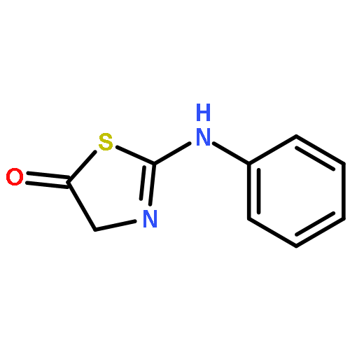
•Phage-encoded anti-CRISPRs (Acrs) inhibit CRISPR-Cas9 through unique mechanisms•AcrIIC1 controls genome editing by diverse Cas9 orthologs•AcrIIC1 binding disables Cas9’s nucleases without impacting DNA binding•AcrIIC3 promotes dimerization of Cas9 and prevents DNA bindingCRISPR-Cas9 proteins function within bacterial immune systems to target and destroy invasive DNA and have been harnessed as a robust technology for genome editing. Small bacteriophage-encoded anti-CRISPR proteins (Acrs) can inactivate Cas9, providing an efficient off switch for Cas9-based applications. Here, we show that two Acrs, AcrIIC1 and AcrIIC3, inhibit Cas9 by distinct strategies. AcrIIC1 is a broad-spectrum Cas9 inhibitor that prevents DNA cutting by multiple divergent Cas9 orthologs through direct binding to the conserved HNH catalytic domain of Cas9. A crystal structure of an AcrIIC1-Cas9 HNH domain complex shows how AcrIIC1 traps Cas9 in a DNA-bound but catalytically inactive state. By contrast, AcrIIC3 blocks activity of a single Cas9 ortholog and induces Cas9 dimerization while preventing binding to the target DNA. These two orthogonal mechanisms allow for separate control of Cas9 target binding and cleavage and suggest applications to allow DNA binding while preventing DNA cutting by Cas9.Download high-res image (141KB)Download full-size image
•Cas13a family contains two distinct subfamilies•Cas13a subfamilies possess distinct nuclease substrate preferences•Cas13a subfamilies also use orthogonal crRNAs•crRNA processing is not essential for ssRNA targeting by Cas13aCRISPR adaptive immunity pathways protect prokaryotic cells against foreign nucleic acids using CRISPR RNA (crRNA)-guided nucleases. In type VI-A CRISPR-Cas systems, the signature protein Cas13a (formerly C2c2) contains two separate ribonuclease activities that catalyze crRNA maturation and ssRNA degradation. The Cas13a protein family occurs across different bacterial phyla and varies widely in both protein sequence and corresponding crRNA sequence conservation. Although grouped phylogenetically together, we show that the Cas13a enzyme family comprises two distinct functional groups that recognize orthogonal sets of crRNAs and possess different ssRNA cleavage specificities. These functional distinctions could not be bioinformatically predicted, suggesting more subtle co-evolution of Cas13a enzymes. Additionally, we find that Cas13a pre-crRNA processing is not essential for ssRNA cleavage, although it enhances ssRNA targeting for crRNAs encoded internally within the CRISPR array. We define two Cas13a protein subfamilies that can operate in parallel for RNA detection both in bacteria and for diagnostic applications.Download high-res image (137KB)Download full-size image
Co-reporter:James K. Nuñez, Lucas B. Harrington, and Jennifer A. Doudna
ACS Chemical Biology 2016 Volume 11(Issue 3) pp:681
Publication Date(Web):February 9, 2016
DOI:10.1021/acschembio.5b01019
The application of the CRISPR–Cas9 system for genome engineering has revolutionized the ability to interrogate genomes of mammalian cells. Programming the Cas9 endonuclease to induce DNA breaks at specified sites is achieved by simply modifying the sequence of its cognate guide RNA. Although Cas9-mediated genome editing has been shown to be highly specific, cleavage events at off-target sites have also been reported. Minimizing, and eventually abolishing, unwanted off-target cleavage remains a major goal of the CRISPR–Cas9 technology before its implementation for therapeutic use. Recent efforts have turned to chemical biology and biophysical approaches to engineer inducible genome editing systems for controlling Cas9 activity at the transcriptional and protein levels. Here, we review recent advancements to modulate Cas9-mediated genome editing by engineering split-Cas9 constructs, inteins, small molecules, protein-based dimerizing domains, and light-inducible systems.
Co-reporter:David W. Taylor;Jack E. Kornfeld;Fuguo Jiang;Janice S. Chen;Aubri J. Thompson;Eva Nogales;Kaihong Zhou
Science 2016 Volume 351(Issue 6275) pp:
Publication Date(Web):
DOI:10.1126/science.aad8282
CRISPR Cas9 molecular scissors
The CRISPR-associated (Cas) protein Cas9 is a molecular scissor for cutting DNA. The first step in the cutting reaction is the RNA-guided unwinding of the DNA double helix. Jiang et al. determined the structures of Cas9 bound to DNA unwound by the targeting RNA (see the Perspective by Chen and Bailey). Cas9 bends the DNA to allow guide RNA infiltration into the double helix. The two separated DNA strands, one bound to RNA, are subsequently positioned in the dual active sites of the protein for cutting.
Co-reporter:Emine Kaya;Kevin W. Doxzen;Ross C. Wilson;Philip J. Kranzusch;Kilian R. Knoll;Steven C. Strutt
PNAS 2016 Volume 113 (Issue 15 ) pp:4057-4062
Publication Date(Web):2016-04-12
DOI:10.1073/pnas.1524385113
Eukaryotic Argonaute proteins induce gene silencing by small RNA-guided recognition and cleavage of mRNA targets. Although
structural similarities between human and prokaryotic Argonautes are consistent with shared mechanistic properties, sequence
and structure-based alignments suggested that Argonautes encoded within CRISPR-cas [clustered regularly interspaced short palindromic repeats (CRISPR)-associated] bacterial immunity operons have divergent
activities. We show here that the CRISPR-associated Marinitoga piezophila Argonaute (MpAgo) protein cleaves single-stranded target sequences using 5′-hydroxylated guide RNAs rather than the 5′-phosphorylated
guides used by all known Argonautes. The 2.0-Å resolution crystal structure of an MpAgo–RNA complex reveals a guide strand
binding site comprising residues that block 5′ phosphate interactions. Using structure-based sequence alignment, we were able
to identify other putative MpAgo-like proteins, all of which are encoded within CRISPR-cas loci. Taken together, our data suggest the evolution of an Argonaute subclass with noncanonical specificity for a 5′-hydroxylated
guide.
The CRISPR-Cas system in prokaryotes precisely identifies infecting parasitic DNAs and viruses and destroys them. The CRISPR-Cas system has been adapted for facile genome editing, heralding a new age in molecular biology. Jiang et al. show that the Cas9 nuclease adopts a distinct confirmation when it binds to the targeting guide RNA. The guide RNA then assumes a preordered shape. This RNA “seed region” is thus poised to initiate recognition of the DNA target sequence.
Co-reporter:Spencer C. Knight;Liangqi Xie;Wulan Deng;Benjamin Guglielmi;Lea B. Witkowsky;Lana Bosanac;Maxime Dahan;Zhe Liu;Mohamed El Beheiry;Robert Tjian;Elisa T. Zhang;Jean-Baptiste Masson
Science 2015 Volume 350(Issue 6262) pp:823-826
Publication Date(Web):13 Nov 2015
DOI:10.1126/science.aac6572
Genome editing with a Cas9 scalpel
The Cas9 nuclease forms the heart of the CRISPR-Cas genome editing system. Cas9 binds small guide RNAs that direct it to its target sites, where the nuclease either cleaves or binds to genomic DNA. Knight et al. used single-molecule imaging to track Cas9 in living cells. Cas9 searches the genome for its target sites using rapid threedimensional diffusion. It spends very little time binding to off-target sites, which explains the high accuracy of the CRISPRCas9 editing machine.
Co-reporter:Addison V. Wright;Samuel H. Sternberg;David W. Taylor;Brett T. Staahl;Jorge A. Bardales;Jack E. Kornfeld
PNAS 2015 112 (10 ) pp:2984-2989
Publication Date(Web):2015-03-10
DOI:10.1073/pnas.1501698112
Cas9, an RNA-guided DNA endonuclease found in clustered regularly interspaced short palindromic repeats (CRISPR) bacterial
immune systems, is a versatile tool for genome editing, transcriptional regulation, and cellular imaging applications. Structures
of Streptococcus pyogenes Cas9 alone or bound to single-guide RNA (sgRNA) and target DNA revealed a bilobed protein architecture that undergoes major
conformational changes upon guide RNA and DNA binding. To investigate the molecular determinants and relevance of the interlobe
rearrangement for target recognition and cleavage, we designed a split-Cas9 enzyme in which the nuclease lobe and α-helical
lobe are expressed as separate polypeptides. Although the lobes do not interact on their own, the sgRNA recruits them into
a ternary complex that recapitulates the activity of full-length Cas9 and catalyzes site-specific DNA cleavage. The use of
a modified sgRNA abrogates split-Cas9 activity by preventing dimerization, allowing for the development of an inducible dimerization
system. We propose that split-Cas9 can act as a highly regulatable platform for genome-engineering applications.
Co-reporter:David Baltimore;Paul Berg;Michael Botchan;Dana Carroll;R. Alta Charo;George Church;Jacob E. Corn;George Q. Daley;Marsha Fenner;Henry T. Greely;G. Steven Martin;Martin Jinek;Edward Penhoet;Jennifer Puck;Samuel H. Sternberg;Jonathan S. Weissman;Keith R. Yamamoto
Science 2015 Volume 348(Issue 6230) pp:36-38
Publication Date(Web):03 Apr 2015
DOI:10.1126/science.aab1028
A framework for open discourse on the use of CRISPR-Cas9 technology to manipulate the human genome is urgently needed
Co-reporter:David W. Taylor;Yifan Zhu;Raymond H. J. Staals;Jack E. Kornfeld;Akeo Shinkai;John van der Oost;Eva Nogales
Science 2015 Volume 348(Issue 6234) pp:581-585
Publication Date(Web):01 May 2015
DOI:10.1126/science.aaa4535
Changing shape to destroy RNA
Clustered regularly interspaced short palindromic repeats (CRISPRs) together with CRISPR-associated (Cas) proteins form an adaptive immune system that helps bacteria and archaea defend themselves against invading viruses and plasmids. CRISPR RNAs (crRNAs) target CRISPR-Cas protein complexes to the invaders, bringing about their destruction. Taylor et al. used cryo–electron microscopy to determine the structure of a 12-subunit CRISPR-Cas protein complex with crRNA from Thermus thermophilus, in the presence and absence of single-stranded target RNA. Binding to the target RNA causes a change in shape of the CRISPR-Cas complex that results in target recognition and destruction.
Co-reporter:Steven Lin;Rachel E. Gate;Dimitre R. Simeonov;Meena Subramaniam;Alexander Marson;Kathrin Schumann;Chun J. Ye;Eric Boyer;Genevieve E. Haliburton;Jeffrey A. Bluestone
PNAS 2015 Volume 112 (Issue 33 ) pp:10437-10442
Publication Date(Web):2015-08-18
DOI:10.1073/pnas.1512503112
T-cell genome engineering holds great promise for cell-based therapies for cancer, HIV, primary immune deficiencies, and autoimmune
diseases, but genetic manipulation of human T cells has been challenging. Improved tools are needed to efficiently “knock
out” genes and “knock in” targeted genome modifications to modulate T-cell function and correct disease-associated mutations.
CRISPR/Cas9 technology is facilitating genome engineering in many cell types, but in human T cells its efficiency has been
limited and it has not yet proven useful for targeted nucleotide replacements. Here we report efficient genome engineering
in human CD4+ T cells using Cas9:single-guide RNA ribonucleoproteins (Cas9 RNPs). Cas9 RNPs allowed ablation of CXCR4, a coreceptor for
HIV entry. Cas9 RNP electroporation caused up to ∼40% of cells to lose high-level cell-surface expression of CXCR4, and edited
cells could be enriched by sorting based on low CXCR4 expression. Importantly, Cas9 RNPs paired with homology-directed repair
template oligonucleotides generated a high frequency of targeted genome modifications in primary T cells. Targeted nucleotide
replacement was achieved in CXCR4 and PD-1 (PDCD1), a regulator of T-cell exhaustion that is a validated target for tumor immunotherapy. Deep sequencing of a target site confirmed
that Cas9 RNPs generated knock-in genome modifications with up to ∼20% efficiency, which accounted for up to approximately
one-third of total editing events. These results establish Cas9 RNP technology for diverse experimental and therapeutic genome
engineering applications in primary human T cells.
Co-reporter:Prashant Bhat;Megan L. Hochstrasser;Eva Nogales;Chantal K. Guegler;Samuel H. Sternberg;David W. Taylor
PNAS 2014 Volume 111 (Issue 18 ) pp:6618-6623
Publication Date(Web):2014-05-06
DOI:10.1073/pnas.1405079111
In bacteria, the clustered regularly interspaced short palindromic repeats (CRISPR)–associated (Cas) DNA-targeting complex
Cascade (CRISPR-associated complex for antiviral defense) uses CRISPR RNA (crRNA) guides to bind complementary DNA targets
at sites adjacent to a trinucleotide signature sequence called the protospacer adjacent motif (PAM). The Cascade complex then
recruits Cas3, a nuclease-helicase that catalyzes unwinding and cleavage of foreign double-stranded DNA (dsDNA) bearing a
sequence matching that of the crRNA. Cascade comprises the CasA–E proteins and one crRNA, forming a structure that binds and
unwinds dsDNA to form an R loop in which the target strand of the DNA base pairs with the 32-nt RNA guide sequence. Single-particle
electron microscopy reconstructions of dsDNA-bound Cascade with and without Cas3 reveal that Cascade positions the PAM-proximal
end of the DNA duplex at the CasA subunit and near the site of Cas3 association. The finding that the DNA target and Cas3
colocalize with CasA implicates this subunit in a key target-validation step during DNA interference. We show biochemically
that base pairing of the PAM region is unnecessary for target binding but critical for Cas3-mediated degradation. In addition,
the L1 loop of CasA, previously implicated in PAM recognition, is essential for Cas3 activation following target binding by
Cascade. Together, these data show that the CasA subunit of Cascade functions as an essential partner of Cas3 by recognizing
DNA target sites and positioning Cas3 adjacent to the PAM to ensure cleavage.
Co-reporter:Martin Jinek;Fuguo Jiang;David W. Taylor;Samuel H. Sternberg;Emine Kaya;Enbo Ma;Carolin Anders;Michael Hauer;Kaihong Zhou;Matias Kaplan;Steven Lin;Emmanuelle Charpentier;Anthony T. Iavarone;Eva Nogales
Science 2014 Volume 343(Issue 6176) pp:
Publication Date(Web):14 Mar 2014
DOI:10.1126/science.1247997
Structured Abstract
Introduction
Bacteria and archaea defend themselves against invasive DNA using adaptive immune systems comprising CRISPR (clustered regularly interspaced short palindromic repeats) loci and CRISPR-associated (Cas) genes. In association with Cas proteins, small CRISPR RNAs (crRNAs) guide the detection and cleavage of complementary DNA sequences. Type II CRISPR systems employ the RNA-guided endonuclease Cas9 to recognize and cleave double-stranded DNA (dsDNA) targets using conserved RuvC and HNH nuclease domains. Cas9-mediated cleavage is strictly dependent on the presence of a protospacer adjacent motif (PAM) in the target DNA. Recently, the biochemical properties of Cas9–guide RNA complexes have been harnessed for various genetic engineering applications and RNA-guided transcriptional control. Despite these ongoing successes, the structural basis for guide RNA recognition and DNA targeting by Cas9 is still unknown.
Structures of Cas9 endonucleases reveal RNA-mediated conformational activation. (A) Crystal structures of S. pyogenes (SpyCas9) and A. naeslundii (AnaCas9) Cas9 proteins. (B) Left: Negative-stain EM reconstructions of apo-SpyCas9 (top) and SpyCas9-RNA-target DNA complex (bottom) show that nucleic acid binding causes a reorientation of the nuclease (blue) and α-helical (gray) lobes in SpyCas9. Right: Cartoon representations of the structures. tracrRNA, trans-activating crRNA.
Rationale
To compare the architectures and domain organization of diverse Cas9 proteins,the atomic structures of Cas9 from Streptococcus pyogenes (SpyCas) and Actinomyces naeslundii (AnaCas9) were determined by x-ray crystallography. Crosslinking of target DNA containing 5-bromodeoxyuridines was conducted to identify PAM-interacting regions in SpyCas9. To test functional interactions with nucleic acid ligands, structure-based mutant SpyCas9 proteins were assayed for endonuclease activity with radiolabeled oligonucleotide dsDNA targets, and target DNA binding was monitored by electrophoretic mobility shift assays. To compare conformations of Cas9 in different states of nucleic acid binding, three-dimensional reconstructions of apo-SpyCas9, SpyCas9:RNA, and SpyCas9:RNA:DNA were obtained by negative-stain single-particle electron microscopy. Guide RNA and target DNA positions were determined with streptavidin labeling. Exonuclease protection assays were carried out to determine the extent of Cas9–target DNA interactions.
Results
The 2.6 Å–resolution structure of apo-SpyCas9 reveals a bilobed architecture comprising a nuclease domain lobe and an α-helical lobe. Both lobes contain conserved clefts that may function in nucleic acid binding. Photocrosslinking experiments show that the PAM in target DNA is engaged by two tryptophan-containing flexible loops, and mutations of both loops impair target DNA binding and cleavage. The 2.2 Å–resolution crystal structure of AnaCas9 reveals the conserved structural core shared by all Cas9 enzyme subtypes, and both SpyCas9 and AnaCas9 adopt autoinhibited conformations in their apo forms. The electron microscopic (EM) reconstructions of SpyCas9:RNA and SpyCas9:RNA:DNA complexes reveal that guide RNA binding results in a conformational rearrangement and formation of a central channel for target DNA binding. Site-specific labeling of guide RNA and target DNA define the orientations of nucleic acids in the target-bound complex.
Conclusion
The SpyCas9 and AnaCas9 structures define the molecular architecture of the Cas9 enzyme family in which a conserved structural core encompasses the two nuclease domains responsible for DNA cleavage, while structurally divergent regions, including the PAM recognition loops, are likely responsible for distinct guide RNA and PAM specificities. Cas9 enzymes adopt a catalytically inactive conformation in the apo state, necessitating structural activation for DNA recognition and cleavage. Our EM analysis shows that by triggering a conformational rearrangement in Cas9, the guide RNA acts as a critical determinant of target DNA binding.
Technologies for making and manipulating DNA have enabled advances in biology ever since the discovery of the DNA double helix. But introducing site-specific modifications in the genomes of cells and organisms remained elusive. Early approaches relied on the principle of site-specific recognition of DNA sequences by oligonucleotides, small molecules, or self-splicing introns. More recently, the site-directed zinc finger nucleases (ZFNs) and TAL effector nucleases (TALENs) using the principles of DNA-protein recognition were developed. However, difficulties of protein design, synthesis, and validation remained a barrier to widespread adoption of these engineered nucleases for routine use.
The Cas9 enzyme (blue) generates breaks in double-stranded DNA by using its two catalytic centers (blades) to cleave each strand of a DNA target site (gold) next to a PAM sequence (red) and matching the 20-nucleotide sequence (orange) of the single guide RNA (sgRNA). The sgRNA includes a dual-RNA sequence derived from CRISPR RNA (light green) and a separate transcript (tracrRNA, dark green) that binds and stabilizes the Cas9 protein. Cas9-sgRNA–mediated DNA cleavage produces a blunt double-stranded break that triggers repair enzymes to disrupt or replace DNA sequences at or near the cleavage site. Catalytically inactive forms of Cas9 can also be used for programmable regulation of transcription and visualization of genomic loci.
Advances
The field of biology is now experiencing a transformative phase with the advent of facile genome engineering in animals and plants using RNA-programmable CRISPR-Cas9. The CRISPR-Cas9 technology originates from type II CRISPR-Cas systems, which provide bacteria with adaptive immunity to viruses and plasmids. The CRISPR-associated protein Cas9 is an endonuclease that uses a guide sequence within an RNA duplex, tracrRNA:crRNA, to form base pairs with DNA target sequences, enabling Cas9 to introduce a site-specific double-strand break in the DNA. The dual tracrRNA:crRNA was engineered as a single guide RNA (sgRNA) that retains two critical features: a sequence at the 5′ side that determines the DNA target site by Watson-Crick base-pairing and a duplex RNA structure at the 3′ side that binds to Cas9. This finding created a simple two-component system in which changes in the guide sequence of the sgRNA program Cas9 to target any DNA sequence of interest. The simplicity of CRISPR-Cas9 programming, together with a unique DNA cleaving mechanism, the capacity for multiplexed target recognition, and the existence of many natural type II CRISPR-Cas system variants, has enabled remarkable developments using this cost-effective and easy-to-use technology to precisely and efficiently target, edit, modify, regulate, and mark genomic loci of a wide array of cells and organisms.
Outlook
CRISPR-Cas9 has triggered a revolution in which laboratories around the world are using the technology for innovative applications in biology. This Review illustrates the power of the technology to systematically analyze gene functions in mammalian cells, study genomic rearrangements and the progression of cancers or other diseases, and potentially correct genetic mutations responsible for inherited disorders. CRISPR-Cas9 is having a major impact on functional genomics conducted in experimental systems. Its application in genome-wide studies will enable large-scale screening for drug targets and other phenotypes and will facilitate the generation of engineered animal models that will benefit pharmacological studies and the understanding of human diseases. CRISPR-Cas9 applications in plants and fungi also promise to change the pace and course of agricultural research. Future research directions to improve the technology will include engineering or identifying smaller Cas9 variants with distinct specificity that may be more amenable to delivery in human cells. Understanding the homology-directed repair mechanisms that follow Cas9-mediated DNA cleavage will enhance insertion of new or corrected sequences into genomes. The development of specific methods for efficient and safe delivery of Cas9 and its guide RNAs to cells and tissues will also be critical for applications of the technology in human gene therapy.
Co-reporter:Ho Young Lee;Kaihong Zhou;Stephen Laderman;Rachel E. Haurwitz;Brian Smart;Alex Apffel;Craig D. Wenger;Laurakay Bruhn
PNAS 2013 Volume 110 (Issue 14 ) pp:5416-5421
Publication Date(Web):2013-04-02
DOI:10.1073/pnas.1302807110
RNA-binding proteins control the fate and function of the transcriptome in all cells. Here we present technology for isolating
RNA–protein partners efficiently and accurately using an engineered clustered regularly interspaced short palindromic repeats
(CRISPR) endoribonuclease. An inactive version of the Csy4 nuclease binds irreversibly to transcripts engineered with a 16-nt
hairpin sequence at their 5′ ends. Once immobilized by Csy4 on a solid support, contaminating proteins and other molecules
can be removed by extensive washing. Upon addition of imidazole, Csy4 is activated to cleave the RNA, removing the hairpin
tag and releasing the native transcript along with its specifically bound protein partners. This conditional Csy4 enzyme enables
recovery of specific RNA-binding partners with minimal false-positive contamination. We use this method, coupled with quantitative
MS, to identify cell type-specific human pre-microRNA-binding proteins. We also show that this technology is suitable for
analyzing diverse size transcripts, and that it is suitable for adaptation to a high-throughput discovery format.
Co-reporter:Chaomin Sun;Aleksandar Todorovic;Jordi Querol-Audí;Yun Bai;Nancy Villa;Monica Snyder;John Ashchyan;Christopher S. Lewis;Abbey Hartland;Scott Gradia;Christopher S. Fraser;Eva Nogales;Jamie H. D. Cate
PNAS 2011 108 (51 ) pp:
Publication Date(Web):2011-12-20
DOI:10.1073/pnas.1116821108
Protein fate in higher eukaryotes is controlled by three complexes that share conserved architectural elements: the proteasome,
COP9 signalosome, and eukaryotic translation initiation factor 3 (eIF3). Here we reconstitute the 13-subunit human eIF3 in
Escherichia coli, revealing its structural core to be the eight subunits with conserved orthologues in the proteasome lid complex and COP9
signalosome. This structural core in eIF3 binds to the small (40S) ribosomal subunit, to translation initiation factors involved
in mRNA cap-dependent initiation, and to the hepatitis C viral (HCV) internal ribosome entry site (IRES) RNA. Addition of
the remaining eIF3 subunits enables reconstituted eIF3 to assemble intact initiation complexes with the HCV IRES. Negative-stain
EM reconstructions of reconstituted eIF3 further reveal how the approximately 400 kDa molecular mass structural core organizes
the highly flexible 800 kDa molecular mass eIF3 complex, and mediates translation initiation.
Co-reporter:Blake Wiedenheft;Esther van Duijn;Jelle B. Bultema;Sakharam P. Waghmare;Kaihong Zhou;Arjan Barendregt;Wiebke Westphal;Albert J. R. Heck;Egbert J. Boekema;Mark J. Dickman
PNAS 2011 108 (25 ) pp:
Publication Date(Web):2011-06-21
DOI:10.1073/pnas.1102716108
Prokaryotes have evolved multiple versions of an RNA-guided adaptive immune system that targets foreign nucleic acids. In
each case, transcripts derived from clustered regularly interspaced short palindromic repeats (CRISPRs) are thought to selectively
target invading phage and plasmids in a sequence-specific process involving a variable cassette of CRISPR-associated (cas) genes. The CRISPR locus in Pseudomonas aeruginosa (PA14) includes four cas genes that are unique to and conserved in microorganisms harboring the Csy-type (CRISPR system yersinia) immune system. Here
we show that the Csy proteins (Csy1–4) assemble into a 350 kDa ribonucleoprotein complex that facilitates target recognition
by enhancing sequence-specific hybridization between the CRISPR RNA and complementary target sequences. Target recognition
is enthalpically driven and localized to a “seed sequence” at the 5′ end of the CRISPR RNA spacer. Structural analysis of
the complex by small-angle X-ray scattering and single particle electron microscopy reveals a crescent-shaped particle that
bears striking resemblance to the architecture of a large CRISPR-associated complex from Escherichia coli, termed Cascade. Although similarity between these two complexes is not evident at the sequence level, their unequal subunit
stoichiometry and quaternary architecture reveal conserved structural features that may be common among diverse CRISPR-mediated
defense systems.
Co-reporter:Sharon Aviran;Cole Trapnell;Julius B. Lucks;Stefanie A. Mortimer;Shujun Luo;Gary P. Schroth;Adam P. Arkin;Lior Pachter;
Proceedings of the National Academy of Sciences 2011 108(27) pp:11069-11074
Publication Date(Web):June 3, 2011
DOI:10.1073/pnas.1106541108
Sequence census methods reduce molecular measurements such as transcript abundance and protein-nucleic acid interactions to
counting problems via DNA sequencing. We focus on a novel assay utilizing this approach, called selective 2′-hydroxyl acylation analyzed by primer extension sequencing (SHAPE-Seq), that can be used to characterize RNA secondary and tertiary structure. We describe a fully
automated data analysis pipeline for SHAPE-Seq analysis that includes read processing, mapping, and structural inference based
on a model of the experiment. Our methods rely on the solution of a series of convex optimization problems for which we develop
efficient and effective numerical algorithms. Our results can be easily extended to other chemical probes of RNA structure,
and also generalized to modeling polymerase drop-off in other sequence census-based experiments.
Co-reporter:Julius B. Lucks;Stefanie A. Mortimer;Cole Trapnell;Shujun Luo;Sharon Aviran;Gary P. Schroth;Lior Pachter;Adam P. Arkin;
Proceedings of the National Academy of Sciences 2011 108(27) pp:11063-11068
Publication Date(Web):June 3, 2011
DOI:10.1073/pnas.1106501108
New regulatory roles continue to emerge for both natural and engineered noncoding RNAs, many of which have specific secondary
and tertiary structures essential to their function. Thus there is a growing need to develop technologies that enable rapid
characterization of structural features within complex RNA populations. We have developed a high-throughput technique, SHAPE-Seq,
that can simultaneously measure quantitative, single nucleotide-resolution secondary and tertiary structural information for
hundreds of RNA molecules of arbitrary sequence. SHAPE-Seq combines selective 2′-hydroxyl acylation analyzed by primer extension (SHAPE) chemistry with multiplexed paired-end deep sequencing of primer extension products. This generates millions
of sequencing reads, which are then analyzed using a fully automated data analysis pipeline, based on a rigorous maximum likelihood
model of the SHAPE-Seq experiment. We demonstrate the ability of SHAPE-Seq to accurately infer secondary and tertiary structural
information, detect subtle conformational changes due to single nucleotide point mutations, and simultaneously measure the
structures of a complex pool of different RNA molecules. SHAPE-Seq thus represents a powerful step toward making the study
of RNA secondary and tertiary structures high throughput and accessible to a wide array of scientific pursuits, from fundamental
biological investigations to engineering RNA for synthetic biological systems.
Transmission of influenza viruses into the human population requires surmounting barriers to cross-species infection. Changes
in the influenza polymerase overcome one such barrier. Viruses isolated from birds generally contain polymerases with the
avian-signature glutamic acid at amino acid 627 in the PB2 subunit. These polymerases display restricted activity in human
cells. An adaptive change in this residue from glutamic acid to the human-signature lysine confers high levels of polymerase
activity in human cells. This mutation permits escape from a species-specific restriction factor that targets polymerases
from avian viruses. A 2009 swine-origin H1N1 influenza A virus recently established a pandemic infection in humans, even though
the virus encodes a PB2 with the restrictive glutamic acid at amino acid 627. We show here that the 2009 H1N1 virus has acquired
second-site suppressor mutations in its PB2 polymerase subunit that convey enhanced polymerase activity in human cells. Introduction
of this polymorphism into the PB2 subunit of a primary avian isolate also increased polymerase activity and viral replication
in human and porcine cells. An alternate adaptive strategy has also been identified, whereby introduction of a human PA subunit
into an avian polymerase overcomes restriction in human cells. These data reveal a strategy used by the 2009 H1N1 influenza
A virus and identify other pathways by which avian and swine-origin viruses may evolve to enhance replication, and potentially
pathogenesis, in humans.
Co-reporter:Ian J. MacRae;Enbo Ma;Min Zhou;Carol V. Robinson
PNAS 2008 Volume 105 (Issue 2 ) pp:512-517
Publication Date(Web):2008-01-15
DOI:10.1073/pnas.0710869105
Targeted gene silencing by RNAi requires the RNA-induced silencing complex (RISC), whose core component is the protein Argonaute
(Ago) bound to a microRNA (miRNA) or an siRNA. In humans, Ago2 is loaded with miRNAs by the action of a specialized assembly
called the RISC-loading complex (RLC), comprising the proteins Ago2, Dicer, and TRBP. Here we show that the human RLC assembles
spontaneously in vitro from purified components. No cofactors or chaperones are required for the complex to form. The reconstituted RLC, containing
one copy of each protein, has the dicing, slicing, guide-strand selection, and Ago2-loading activities observed for the endogenous
RLC. Furthermore, once Ago2 is loaded with an miRNA, it tends to dissociate from the rest of the complex. These results lay
the groundwork for future structural and functional dissection of RISC loading in humans.
Co-reporter:Christopher S. Fraser
and
Jennifer A. Doudna
Nature Reviews Microbiology 2007 5(1) pp:29
Publication Date(Web):2006-11-27
DOI:10.1038/nrmicro1558
Hepatitis C virus uses an internal ribosome entry site (IRES) to control viral protein synthesis by directly recruiting ribosomes to the translation-start site in the viral mRNA. Structural insights coupled with biochemical studies have revealed that the IRES substitutes for the activities of translation-initiation factors by binding and inducing conformational changes in the 40S ribosomal subunit. Direct interactions of the IRES with initiation factor eIF3 are also crucial for efficient translation initiation, providing clues to the role of eIF3 in protein synthesis.
Co-reporter:Tamira K. Butler;Wendy V. Gilbert;Kaihong Zhou
Science 2007 Volume 317(Issue 5842) pp:1224-1227
Publication Date(Web):31 Aug 2007
DOI:10.1126/science.1144467
Abstract
Cellular internal ribosome entry sites (IRESs) are untranslated segments of mRNA transcripts thought to initiate protein synthesis in response to environmental stresses that prevent canonical 5′ cap–dependent translation. Although numerous cellular mRNAs are proposed to have IRESs, none has a demonstrated physiological function or molecular mechanism. Here we show that seven yeast genes required for invasive growth, a developmental pathway induced by nutrient limitation, contain potent IRESs that require the initiation factor eIF4G for cap-independent translation. In contrast to the RNA structure-based activity of viral IRESs, we show that an unstructured A-rich element mediates internal initiation via recruitment of the poly(A) binding protein (Pab1) to the 5′ untranslated region (UTR) of invasive growth messages. A 5′UTR mutation that impairs IRES activity compromises invasive growth, which indicates that cap-independent translation is required for physiological adaptation to stress.
Co-reporter:Ian J. MacRae;Kaihong Zhou;Fei Li;Adrian Repic;Angela N. Brooks;W. Zacheus Cande;Paul D. Adams
Science 2006 Vol 311(5758) pp:195-198
Publication Date(Web):13 Jan 2006
DOI:10.1126/science.1121638
Abstract
The specialized ribonuclease Dicer initiates RNA interference by cleaving double-stranded RNA (dsRNA) substrates into small fragments about 25 nucleotides in length. In the crystal structure of an intact Dicer enzyme, the PAZ domain, a module that binds the end of dsRNA, is separated from the two catalytic ribonuclease III (RNase III) domains by a flat, positively charged surface. The 65 angstrom distance between the PAZ and RNase III domains matches the length spanned by 25 base pairs of RNA. Thus, Dicer itself is a molecular ruler that recognizes dsRNA and cleaves a specified distance from the helical end.
Co-reporter:Bunpote Siridechadilok;Christopher S. Fraser;Richard J. Hall;Eva Nogales
Science 2005 Vol 310(5753) pp:1513-1515
Publication Date(Web):02 Dec 2005
DOI:10.1126/science.1118977
Abstract
Protein synthesis in mammalian cells requires initiation factor eIF3, a ∼750-kilodalton complex that controls assembly of 40S ribosomal subunits on messenger RNAs (mRNAs) bearing either a 5′-cap or an internal ribosome entry site (IRES). Cryo–electron microscopy reconstructions show that eIF3, a five-lobed particle, interacts with the hepatitis C virus (HCV) IRES RNA and the 5′-cap binding complex eIF4F via the same domain. Detailed modeling of eIF3 and eIF4F onto the 40S ribosomal subunit reveals that eIF3 uses eIF4F or the HCV IRES in structurally similar ways to position the mRNA strand near the exit site of 40S, promoting initiation complex assembly.
Nature Structural and Molecular Biology 2005 12(5) pp:395-402
Publication Date(Web):03 May 2005
DOI:10.1038/nsmb932
Evolution has resoundingly favored protein enzymes over RNA-based catalysts, yet ribozymes occupy important niches in modern cell biology that include the starring role in catalysis of protein synthesis on the ribosome. Recent results from structural and biochemical studies show that natural ribozymes use an impressive range of catalytic mechanisms, beyond metalloenzyme chemistry and analogous to more chemically diverse protein enzymes. These findings make it increasingly possible to compare details of RNA- and protein-based catalysis.
Co-reporter:Hong Ji;Christopher S. Fraser;Yonghao Yu;Julie Leary;
Proceedings of the National Academy of Sciences 2004 101(49) pp:16990-16995
Publication Date(Web):November 24, 2004
DOI:10.1073/pnas.0407402101
Protein synthesis in all cells begins with recruitment of the small ribosomal subunit to the initiation codon in a messenger
RNA. In some eukaryotic viruses, RNA upstream of the coding region forms an internal ribosome entry site (IRES) that directly
binds to the 40S ribosomal subunit and enables translation initiation in the absence of many canonical translation initiation
factors. The hepatitis C virus (HCV) IRES RNA requires just two initiation factors, eukaryotic initiation factor (eIF) 2 and
eIF3, to form preinitiation 48S ribosomal complexes that subsequently assemble into translation-competent ribosomes. Using
an RNA-based affinity purification approach, we show here that HCV IRES RNA facilitates eIF2 function through its interactions
with eIF3 and the 40S ribosomal subunit. Although the wild-type IRES assembles normally into 48S and 80S ribosomal complexes
in human cell extract, mutant IRES RNAs become trapped at the 48S assembly stage. Trapped 48S complexes formed by IRES mutants
with reduced eIF3 binding affinity nonetheless contain eIF3, consistent with inherent eIF3–40S subunit affinity. Intriguingly,
however, one of these IRES mutants prevents stable association of both eIF3 and eIF2, preventing initiator tRNA deposition
and explaining the block in 80S assembly. In contrast, an IRES mutant unable to induce a conformational change in the 40S
subunit, as observed previously by single-particle cryoelectron microscopy, blocks 80S formation at a later stage in assembly.
These data suggest that the IRES RNA coordinates interactions of eIF3 and eIF2 on the ribosome required to position the initiator
tRNA on the mRNA in the ribosomal peptidyl-tRNA site (P site).
The hepatitis delta virus (HDV) ribozyme catalyzes viral RNA self-cleavage through general acid-base chemistry in which an active-site cytidine and at least one metal ion are involved. Monovalent metal ions support slow catalysis and were proposed to substitute for structural, but not catalytic, divalent metal ions in the RNA. To investigate the role of monovalent cations in ribozyme structure and function, we determined the crystal structure of the precursor HDV ribozyme in the presence of thallium ions (Tl+). Two Tl+ ions can occupy a previously observed divalent metal ion hexahydrate-binding site located near the scissile phosphate, but are easily competed away by cobalt hexammine, a magnesium hexahydrate mimic and potent reaction inhibitor. Intriguingly, a third Tl+ ion forms direct inner-sphere contacts with the ribose 2′-OH nucleophile and the pro-Sp scissile phosphate oxygen. We discuss possible structural and catalytic implications of monovalent cation binding for the HDV ribozyme mechanism.
Co-reporter:Dipali G. Sashital, Blake Wiedenheft, Jennifer A. Doudna
Molecular Cell (8 June 2012) Volume 46(Issue 5) pp:606-615
Publication Date(Web):8 June 2012
DOI:10.1016/j.molcel.2012.03.020
In bacterial and archaeal CRISPR immune pathways, DNA sequences from invading bacteriophage or plasmids are integrated into CRISPR loci within the host genome, conferring immunity against subsequent infections. The ribonucleoprotein complex Cascade utilizes RNAs generated from these loci to target complementary “nonself” DNA sequences for destruction, while avoiding binding to “self” sequences within the CRISPR locus. Here we show that CasA, the largest protein subunit of Cascade, is required for nonself target recognition and binding. Combining a 2.3 Å crystal structure of CasA with cryo-EM structures of Cascade, we have identified a loop that is required for viral defense. This loop contacts a conserved three base pair motif that is required for nonself target selection. Our data suggest a model in which the CasA loop scans DNA for this short motif prior to target destabilization and binding, maximizing the efficiency of DNA surveillance by Cascade.Graphical AbstractDownload high-res image (363KB)Download full-size imageHighlights► CasA is required for dsDNA target binding by Cascade ► CasA crystal structure reveals a loop (L1) that detects foreign DNA ► CasA L1 is required in vivo for viral resistance ► CasA L1 is implicated in dsDNA destabilization and nonself target selection
Co-reporter:Cameron L. Noland, Enbo Ma, Jennifer A. Doudna
Molecular Cell (8 July 2011) Volume 43(Issue 1) pp:110-121
Publication Date(Web):8 July 2011
DOI:10.1016/j.molcel.2011.05.028
The human ribonuclease Dicer and its double-stranded RNA (dsRNA)-binding protein (dsRBP) partners TRBP and PACT play important roles in the biogenesis of regulatory RNAs. Following dicing, one dsRNA product strand is preferentially assembled into an RNA-induced silencing complex (RISC). The mechanism of strand selection in humans and the possible role of Dicer in this process remain unclear. Here we demonstrate that dsRNAs undergo significant repositioning within Dicer complexes following dicing. This repositioning enables directional binding of RNA duplexes, thereby biasing their orientation for guide strand selection according to the thermodynamic properties of the helix. Our findings indicate that Dicer is itself capable of sensing siRNA thermodynamic asymmetry regardless of the dsRBP to which it is bound. These results support a model in which Dicer employs two distinct RNA-binding sites—one for dsRNA processing and the other for sensing of siRNA thermodynamic asymmetry—during RISC loading in humans.Highlights► Dicer/TRBP repositions products along Dicer's helicase domain ► Dicer/TRBP releases nascent product RNAs prior to repositioning ► dsRBPs position siRNAs for sensing of thermodynamic asymmetry by Dicer
Co-reporter:Enbo Ma, Lucas B. Harrington, Mitchell R. O’Connell, Kaihong Zhou, Jennifer A. Doudna
Molecular Cell (5 November 2015) Volume 60(Issue 3) pp:398-407
Publication Date(Web):5 November 2015
DOI:10.1016/j.molcel.2015.10.030
•Diverse Cas9 enzymes cleave single-stranded DNA substrates•Single-stranded DNA cleavage is programmable and PAM-independent•DNA unwinding activity and guide RNA specificity varies widely among Cas9 enzymesDouble-stranded DNA (dsDNA) cleavage by Cas9 is a hallmark of type II CRISPR-Cas immune systems. Cas9-guide RNA complexes recognize 20-base-pair sequences in DNA and generate a site-specific double-strand break, a robust activity harnessed for genome editing. DNA recognition by all studied Cas9 enzymes requires a protospacer adjacent motif (PAM) next to the target site. We show that Cas9 enzymes from evolutionarily divergent bacteria can recognize and cleave single-stranded DNA (ssDNA) by an RNA-guided, PAM-independent recognition mechanism. Comparative analysis shows that in contrast to the type II-A S. pyogenes Cas9 that is widely used for genome engineering, the smaller type II-C Cas9 proteins have limited dsDNA binding and unwinding activity and promiscuous guide RNA specificity. These results indicate that inefficiency of type II-C Cas9 enzymes for genome editing results from a limited ability to cleave dsDNA and suggest that ssDNA cleavage was an ancestral function of the Cas9 enzyme family.Download high-res image (97KB)Download full-size image
Co-reporter:Ross C. Wilson, Akshay Tambe, Mary Anne Kidwell, Cameron L. Noland, ... Jennifer A. Doudna
Molecular Cell (5 February 2015) Volume 57(Issue 3) pp:397-407
Publication Date(Web):5 February 2015
DOI:10.1016/j.molcel.2014.11.030
•Key details of Dicer-TRBP interface are revealed by crystal structure•Interface mutations to Dicer disrupt binding to TRBP and related protein PACT•Fidelity of miRNA biogenesis is disrupted when Dicer cannot bind partner proteins•TRBP influences Dicer cleavage site and alters subsequent strand selectionRNA-mediated gene silencing in human cells requires the accurate generation of ∼22 nt microRNAs (miRNAs) from double-stranded RNA substrates by the endonuclease Dicer. Although the phylogenetically conserved RNA-binding proteins TRBP and PACT are known to contribute to this process, their mode of Dicer binding and their genome-wide effects on miRNA processing have not been determined. We solved the crystal structure of the human Dicer-TRBP interface, revealing the structural basis of the interaction. Interface residues conserved between TRBP and PACT show that the proteins bind to Dicer in a similar manner and by mutual exclusion. Based on the structure, a catalytically active Dicer that cannot bind TRBP or PACT was designed and introduced into Dicer-deficient mammalian cells, revealing selective defects in guide strand selection. These results demonstrate the role of Dicer-associated RNA binding proteins in maintenance of gene silencing fidelity.Download high-res image (504KB)Download full-size image
Co-reporter:Martin Jinek, Scott M. Coyle, Jennifer A. Doudna
Molecular Cell (4 March 2011) Volume 41(Issue 5) pp:600-608
Publication Date(Web):4 March 2011
DOI:10.1016/j.molcel.2011.02.004
Messenger RNA decay plays a central role in the regulation and surveillance of eukaryotic gene expression. The conserved multidomain exoribonuclease Xrn1 targets cytoplasmic RNA substrates marked by a 5′ monophosphate for processive 5′-to-3′ degradation by an unknown mechanism. Here, we report the crystal structure of an Xrn1-substrate complex. The single-stranded substrate is held in place by stacking of the 5′-terminal trinucleotide between aromatic side chains while a highly basic pocket specifically recognizes the 5′ phosphate. Mutations of residues involved in binding the 5′-terminal nucleotide impair Xrn1 processivity. The substrate recognition mechanism allows Xrn1 to couple processive hydrolysis to duplex melting in RNA substrates with sufficiently long single-stranded 5′ overhangs. The Xrn1-substrate complex structure thus rationalizes the exclusive specificity of Xrn1 for 5′-monophosphorylated substrates, ensuring fidelity of mRNA turnover, and posits a model for translocation-coupled unwinding of structured RNA substrates.Graphical AbstractDownload high-res image (263KB)Download full-size imageHighlights► Crystal structure of Xrn1 in complex with a nucleic acid substrate ► A conserved basic pocket specifically recognizes a 5′ phosphate group ► 5′-terminal nucleotide binding promotes substrate translocation and processivity ► Xrn1 couples processive hydrolysis and duplex melting via steric occlusion
Molecular Cell (3 December 2015) Volume 60(Issue 5) pp:712-714
Publication Date(Web):3 December 2015
DOI:10.1016/j.molcel.2015.11.014
In this issue, Ahl et al. (2015) and Doucet et al. (2015) illuminate structural and functional features of substrates that promote integration of RNA molecules into the human genome by LINE retrotransposons, contributing to the ∼50% of the human genome that has been colonized by mobile genetic elements.
Co-reporter:Srinivas Chakravarthy, Samuel H. Sternberg, Colleen A. Kellenberger, Jennifer A. Doudna
Journal of Molecular Biology (3 December 2010) Volume 404(Issue 3) pp:392-402
Publication Date(Web):3 December 2010
DOI:10.1016/j.jmb.2010.09.030
The specialized ribonuclease Dicer plays a central role in eukaryotic gene expression by producing small regulatory RNAs—microRNAs (miRNAs) and short interfering RNAs (siRNAs)—from larger double-stranded RNA (dsRNA) substrates. Although Dicer will cleave both imperfectly base-paired hairpin structures (pre-miRNAs) and perfect duplexes (pre-siRNAs) in vitro, it has not been clear whether these are mechanistically equivalent substrates and how dsRNA binding proteins such as trans-activation response (TAR) RNA binding protein (TRBP) influence substrate selection and RNA processing efficiency. We show here that human Dicer is much faster at processing a pre-miRNA substrate compared to a pre-siRNA substrate under both single and multiple turnover conditions. Maximal cleavage rates (Vmax) calculated by Michaelis–Menten analysis differed by more than 100-fold under multiple turnover conditions. TRBP was found to enhance dicing of both substrates to similar extents, and this stimulation required the two N-terminal dsRNA binding domains of TRBP. These results demonstrate that multiple factors influence dicing kinetics. While TRBP stimulates dicing by enhancing the stability of Dicer–substrate complexes, Dicer itself generates product RNAs at rates determined at least in part by the structural properties of the substrate.► Human Dicer shows significant variability in dicing efficiency in a substrate sequence- and/or structure-dependent manner. ► We report a clear preference for pre-miRNAs over pre-siRNAs. ► The Dicer-associated dsRNA binding protein TRBP stimulates a ∼ 5-fold increase in endonucleolytic activity with both kinds of substrates. ► This stimulatory effect of TRBP is most likely dependent on the high RNA binding affinity attributable to dsRBD1 and dsRBD2.
Co-reporter:Enbo Ma, Kaihong Zhou, Mary Anne Kidwell, Jennifer A. Doudna
Journal of Molecular Biology (28 September 2012) Volume 422(Issue 4) pp:466-476
Publication Date(Web):28 September 2012
DOI:10.1016/j.jmb.2012.06.009
The conserved ribonuclease Dicer generates microRNAs and short‐interfering RNAs that guide gene silencing in eukaryotes. The specific contributions of human Dicer's structural domains to RNA product length and substrate preference are incompletely understood, due in part to the difficulties of Dicer purification. Here, we show that active forms of human Dicer can be assembled from recombinant polypeptides expressed in bacteria. Using this system, we find that three distinct modes of RNA recognition give rise to Dicer's fidelity and product length specificity. The first involves anchoring one end of a double‐stranded RNA helix within the PAZ domain, which can assemble in trans with Dicer's catalytic domains to reconstitute an accurate but non-substrate-selective dicing activity. The second entails nonspecific RNA binding by the double-stranded RNA binding domain, an interaction that is essential for substrate recruitment in the absence of the PAZ domain. The third mode of recognition involves hairpin RNA loop recognition by the helicase domain, which ensures efficient processing of specific substrates. These results reveal distinct interactions of each Dicer domain with different RNA structural features and provide a facile system for investigating the molecular mechanisms of human microRNA biogenesis.Download high-res image (130KB)Download full-size imageHighlights► The multi-domain enzyme Dicer cleaves RNAi precursors into smaller, mature RNAs. ► Dicer was separated into two pieces for recombinant bacterial expression. ► The PAZ domain assembles with the catalytic domains to form an accurate enzyme. ► The helicase domain imparts selectivity by interacting with hairpin RNA loops.
Co-reporter:Enbo Ma, Ian J. MacRae, Jack F. Kirsch, Jennifer A. Doudna
Journal of Molecular Biology (27 June 2008) Volume 380(Issue 1) pp:237-243
Publication Date(Web):27 June 2008
DOI:10.1016/j.jmb.2008.05.005
Dicer, a member of the ribonuclease III family of enzymes, processes double-stranded RNA substrates into ∼ 21- to 27-nt products that trigger sequence-directed gene silencing by RNA interference. Although the mechanism of RNA recognition and length-specific cleavage by Dicer has been established, the way in which dicing activity is regulated is unclear. Here, we show that the N-terminal domain of human Dicer, which is homologous to DExD/H-box helicases, substantially attenuates the rate of substrate cleavage. Deletion or mutation of this domain activates human Dicer in both single- and multiple-turnover assays. The catalytic efficiency (kcat/Km) of the deletion construct is increased by 65-fold over that exhibited by the intact enzyme. Kinetic analysis shows that this activation is almost entirely due to an enhancement in kcat. Modest stimulation of catalysis by the full-length Dicer enzyme was observed in the presence of the TAR-RNA binding protein, which physically interacts with the DExD/H-box domain. These results suggest that the DExD/H-box domain likely disrupts the functionality of the Dicer active site until a structural rearrangement occurs, perhaps upon assembly with its molecular partners.
Co-reporter:Christopher S. Fraser, Katherine E. Berry, John W.B. Hershey, Jennifer A. Doudna
Molecular Cell (22 June 2007) Volume 26(Issue 6) pp:811-819
Publication Date(Web):22 June 2007
DOI:10.1016/j.molcel.2007.05.019
Protein synthesis in all cells begins with the ordered binding of the small ribosomal subunit to messenger RNA (mRNA) and transfer RNA (tRNA). In eukaryotes, translation initiation factor 3 (eIF3) is thought to play an essential role in this process by influencing mRNA and tRNA binding through indirect interactions on the backside of the 40S subunit. Here we show by directed hydroxyl radical probing that the human eIF3 subunit eIF3j binds to the aminoacyl (A) site and mRNA entry channel of the 40S subunit, placing eIF3j directly in the ribosomal decoding center. eIF3j also interacts with eIF1A and reduces 40S subunit affinity for mRNA. A high affinity for mRNA is restored upon recruitment of initiator tRNA, even though eIF3j remains in the mRNA-binding cleft in the presence of tRNA. These results suggest that eIF3j functions in part by regulating access of the mRNA-binding cleft in response to initiation factor binding.
Co-reporter:Samuel H. Sternberg, Jennifer A. Doudna
Molecular Cell (21 May 2015) Volume 58(Issue 4) pp:568-574
Publication Date(Web):21 May 2015
DOI:10.1016/j.molcel.2015.02.032
Few discoveries transform a discipline overnight, but biologists today can manipulate cells in ways never possible before, thanks to a peculiar form of prokaryotic adaptive immunity mediated by clustered regularly interspaced short palindromic repeats (CRISPR). From elegant studies that deciphered how these immune systems function in bacteria, researchers quickly uncovered the technological potential of Cas9, an RNA-guided DNA cleaving enzyme, for genome engineering. Here we highlight the recent explosion in visionary applications of CRISPR-Cas9 that promises to usher in a new era of biological understanding and control.
Co-reporter:Philip J. Kranzusch, Stephen C. Wilson, Amy S.Y. Lee, James M. Berger, ... Russell E. Vance
Molecular Cell (17 September 2015) Volume 59(Issue 6) pp:891-903
Publication Date(Web):17 September 2015
DOI:10.1016/j.molcel.2015.07.022
•Binding of CDNs is an evolutionarily ancient STING function, predating interferons•cGAS-STING function is conserved in anemone, >500 million years diverged from humans•Anemone cGAS produces a canonical 3′,3′ linked CDN similar to those in bacteria•Vertebrate 2′,3′ cGAMP signaling exploits a deeply conserved STING conformationIn humans, the cGAS-STING immunity pathway signals in response to cytosolic DNA via 2′,3′ cGAMP, a cyclic dinucleotide (CDN) second messenger containing mixed 2′–5′ and 3′–5′ phosphodiester bonds. Prokaryotes also produce CDNs, but these are exclusively 3′ linked, and thus the evolutionary origins of human 2′,3′ cGAMP signaling are unknown. Here we illuminate the ancient origins of human cGAMP signaling by discovery of a functional cGAS-STING pathway in Nematostella vectensis, an anemone species >500 million years diverged from humans. Anemone cGAS appears to produce a 3′,3′ CDN that anemone STING recognizes through nucleobase-specific contacts not observed in human STING. Nevertheless, anemone STING binds mixed-linkage 2′,3′ cGAMP indistinguishably from human STING, trapping a unique structural conformation not induced by 3′,3′ CDNs. These results reveal that human mixed-linkage cGAMP achieves universal signaling by exploiting a deeply conserved STING conformational intermediate, providing critical insight for therapeutic targeting of the STING pathway.Download high-res image (270KB)Download full-size image
Co-reporter:Katherine E. Berry, Shruti Waghray, Stefanie A. Mortimer, Yun Bai, Jennifer A. Doudna
Structure (12 October 2011) Volume 19(Issue 10) pp:1456-1466
Publication Date(Web):12 October 2011
DOI:10.1016/j.str.2011.08.002
Translation of hepatitis C viral proteins requires an internal ribosome entry site (IRES) located in the 5′ untranslated region of the viral mRNA. The core domain of the hepatitis C virus (HCV) IRES contains a four-way helical junction that is integrated within a predicted pseudoknot. This domain is required for positioning the mRNA start codon correctly on the 40S ribosomal subunit during translation initiation. Here, we present the crystal structure of this RNA, revealing a complex double-pseudoknot fold that establishes the alignment of two helical elements on either side of the four-helix junction. The conformation of this core domain constrains the open reading frame's orientation for positioning on the 40S ribosomal subunit. This structure, representing the last major domain of HCV-like IRESs to be determined at near-atomic resolution, provides the basis for a comprehensive cryoelectron microscopy-guided model of the intact HCV IRES and its interaction with 40S ribosomal subunits.Graphical AbstractDownload high-res image (398KB)Download full-size imageHighlights► Crystal structure of the most complex HCV IRES domain reveals its functional core ► A tertiary base pair creates a double pseudoknot encompassing a four-helix junction ► Final domain to be solved facilitates comprehensive IRES model bound to 40S subunit ► Orientation of two helical elements directs start-codon positioning by the IRES
Co-reporter:Blake Wiedenheft, Kaihong Zhou, Martin Jinek, Scott M. Coyle, ... Jennifer A. Doudna
Structure (10 June 2009) Volume 17(Issue 6) pp:904-912
Publication Date(Web):10 June 2009
DOI:10.1016/j.str.2009.03.019
Acquired immunity in prokaryotes is achieved by integrating short fragments of foreign nucleic acids into clustered regularly interspaced short palindromic repeats (CRISPRs). This nucleic acid-based immune system is mediated by a variable cassette of up to 45 protein families that represent distinct immune system subtypes. CRISPR-associated gene 1 (cas1) encodes the only universally conserved protein component of CRISPR immune systems, yet its function is unknown. Here we show that the Cas1 protein is a metal-dependent DNA-specific endonuclease that produces double-stranded DNA fragments of ∼80 base pairs in length. The 2.2 Å crystal structure of the Cas1 protein reveals a distinct fold and a conserved divalent metal ion-binding site. Mutation of metal ion-binding residues, chelation of metal ions, or metal-ion substitution inhibits Cas1-catalyzed DNA degradation. These results provide a foundation for understanding how Cas1 contributes to CRISPR function, perhaps as part of the machinery for processing foreign nucleic acids.
Co-reporter:Megan L. Hochstrasser, David W. Taylor, Jack E. Kornfeld, Eva Nogales, Jennifer A. Doudna
Molecular Cell (1 September 2016) Volume 63(Issue 5) pp:840-851
Publication Date(Web):1 September 2016
DOI:10.1016/j.molcel.2016.07.027
•Desulfovibrio vulgaris Cas5c cleaves pre-crRNAs and stays bound to the 5′ handle•The Cas7 backbone assembles cooperatively, capped by the crRNA stem-loop•Cascade/I-C resembles type I and III complexes; large and small subunits are fused•Cryo-EM reveals R-loop stabilization by Cas8c and pronounced target DNA bendingBacteria employ surveillance complexes guided by CRISPR (clustered, regularly interspaced, short palindromic repeats) RNAs (crRNAs) to target foreign nucleic acids for destruction. Although most type I and type III CRISPR systems require four or more distinct proteins to form multi-subunit surveillance complexes, the type I-C systems use just three proteins to achieve crRNA maturation and double-stranded DNA target recognition. We show that each protein plays multiple functional and structural roles: Cas5c cleaves pre-crRNAs and recruits Cas7 to position the RNA guide for DNA binding and unwinding by Cas8c. Cryoelectron microscopy reconstructions of free and DNA-bound forms of the Cascade/I-C surveillance complex reveal conformational changes that enable R-loop formation with distinct positioning of each DNA strand. This streamlined type I-C system explains how CRISPR pathways can evolve compact structures that retain full functionality as RNA-guided DNA capture platforms.Download high-res image (157KB)Download full-size image
.jpg)