Co-reporter:Juyi Li;Yingjie Yu;Kim Myungwoong;Kao Li;John Mikhail;Linxi Zhang;Chung-Chueh Chang;Dilip Gersappe;Marcia Simon;Christopher Ober;Miriam Rafailovich
Journal of Materials Chemistry B 2017 vol. 5(Issue 31) pp:6307-6316
Publication Date(Web):2017/08/09
DOI:10.1039/C7TB01209H
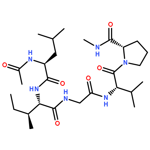
We have successfully synthesized an ABA tri-block co-polymer of poly(methacrylic acid)-block-poly(2-hydroxyethyl methacrylate)-block-poly(methacrylic acid), having Mw = 100k and 272k where we were able to insert RDG or RGD peptide sequences using thiol-acrylate Michael addition. A soft silicone stamp was then used to imprint a 0.4-micron wide grating of the copolymer with a period of 10 microns. The samples were then examined with atomic force microscopy after application of an external electric field and the pattern was observed to stretch by a factor of five. Cells plated onto these substrates showed clear preference for the striped patterns formed under the influence of the external field, and no preferential attachment to the patterns formed in the absence of the field. Cell migration experiments, using the agarose droplet method, performed on spun cast copolymer films showed minimal migration and adhesion on the substrates without peptides or those with only with the RDG peptide, while good adhesion and significant outward migration was observed for cells plated on the copolymers with the RGD sequence. Taken together these results confirmed our hypothesis that a smart biomimetic polymer substrate could be constructed where functional domains could be revealed selectively allowing us to mimic the natural design of engineered tissue constructs.
Co-reporter:Shan He;Yichen Guo;Tehila Stone;Noah Davis;Dongwhan Kim
Journal of Wood Science 2017 Volume 63( Issue 2) pp:154-160
Publication Date(Web):10 January 2017
DOI:10.1007/s10086-016-1603-2
A wood-plastic combination (WPC) was created via in situ polymerization of the l-lactide monomer (3S)-cis-3,6-dimethyl-1,4-dioxane-2,5-dione. Commercial poplar boards (Liriodendron tulipifera) were impregnated with the flame retardant chemical resorcinol bis(diphenyl phosphate)(RDP). These samples were then soaked in a solution of the monomer and deionized water with sulfuric acid 5% wt/wood as a catalyst for polymerization. The wood and solution were placed in a vacuum oven for impregnation and polymerization of the monomers. The wood RDP combination was not flame retardant and had an Izod impact strength that was slightly smaller than neat wood sample. Addition of lactide monomer tripled the Izod impact strength relative to wood, and scanning electron microscope (SEM) images indicated that a polymerized coating had formed which reinforced the porous wood structure. Addition of all three components produced a synergy. The Izod impact strength of the material was nearly 14 times greater and the WPC was flame retardant surpassing the stringent UL-94-V0 requirement.
Co-reporter:Liudi Zhang, Brendan Casey, Dennis K. Galanakis, Clement Marmorat, ... Miriam H. Rafailovich
Acta Biomaterialia 2017 Volume 54(Volume 54) pp:
Publication Date(Web):1 May 2017
DOI:10.1016/j.actbio.2017.03.002
Thrombosis is a clear risk when any foreign material is in contact with the bloodstream. Here we propose an immunohistological stain-based model for non-enzymatic clot formation that enables a facile screen for the thrombogenicity of blood-contacting materials. We exposed polymers with different surface chemistries to protease-free human fibrinogen. We observed that on hydrophilic surfaces, fibrinogen is adsorbed via αC regions, while the γ400-411 platelet-binding dodecapeptide on the D region becomes exposed, and fibrinogen fibers do not form. In contrast, fibrinogen is adsorbed on hydrophobic surfaces via the relatively hydrophobic D and E regions, exposing the αC regions while rendering the γ400-411 inaccessible. Fibrinogen adsorbed on hydrophobic surfaces is thus able to recruit other fibrinogen molecules through αC regions and polymerize into large fibrinogen fibers, similar to those formed in vivo in the presence of thrombin. Moreover, the γ400-411 is available only on the large fibers not elsewhere throughout the hydrophobic surface after fibrinogen fiber formation. When these surfaces were exposed to gel-sieved platelets or platelet rich plasma, a uniform monolayer of platelets, which appeared to be activated, was observed on the hydrophilic surfaces. In contrast, large agglomerates of platelets were clustered on fibers on the hydrophobic surfaces, resembling small nucleating thrombi. Endothelial cells were also able to adhere to the monomeric coating of fibrinogen on hydrophobic surfaces. These observations reveal that the extent and type of fibrinogen adsorption, as well as the propensity of adsorbed fibrinogen to bind platelets, may be modulated by careful selection of surface chemistry.Statements of SignificanceThrombosis is a well-known side effect of the introduction of foreign materials into the bloodstream, as might exist in medical devices including but not limited to stents, valves, and intravascular catheters. Despite many reported studies, the body’s response to foreign materials in contact with the blood remains poorly understood. Current preventive methods consist of drug eluting coatings on the devices or the systemic administration of standard anticoagulants. Here we present a potential mechanism by which surface chemistry can affects fibrinogen conformation and thus affects platelet adhesion and consequently thrombus formation. Our findings suggest a possible coating which enables endothelial cell adhesion while preventing platelet adhesion.Download high-res image (69KB)Download full-size image
Co-reporter:Hongfei Li;Zhenhua Yang;Cheng Pan;Naisheng Jiang;Sushil K. Satija;Di Xu;Dilip Gersappe;Chang-Yong Nam
Nanoscale (2009-Present) 2017 vol. 9(Issue 32) pp:11511-11522
Publication Date(Web):2017/08/17
DOI:10.1039/C7NR03789A
We report that the addition of a non-photoactive tertiary polymer phase in the binary bulk heterojunction (BHJ) polymer solar cell leads to a self-assembled columnar nanostructure, enhancing the charge mobilities and photovoltaic efficiency with surprisingly increased optimal active blend thicknesses over 300 nm, 3–4 times larger than that of the binary counterpart. Using the prototypical poly(3-hexylthiophene) (P3HT):fullerene blend as a model BHJ system, we discover that the inert poly(methyl methacrylate) (PMMA) added in the binary BHJ blend self-assembles into vertical columns, which not only template the phase segregation of electron acceptor fullerenes but also induce the out-of-plane rotation of the edge-on-orientated crystalline P3HT phase. Using complementary interrogation methods including neutron reflectivity, X-ray scattering, atomic force microscopy, transmission electron microscopy, and molecular dynamics simulations, we show that the enhanced charge transport originates from the more randomized molecular stacking of the P3HT phase and the spontaneous segregation of fullerenes at the P3HT/PMMA interface, driven by the high surface tension between the two polymeric components. The results demonstrate a potential method for increasing the thicknesses of high-performance polymer BHJ solar cells with improved photovoltaic efficiency, alleviating the burden of stringently controlling the ultrathin blend thickness during the roll-to-roll-type large-area manufacturing environment.
Co-reporter:Juyi Li;Yingjie Yu;Myungwoong Kim;Kao Li;John Mikhail;Linxi Zhang;Chung-Chueh Chang;Dilip Gersappe;Marcia Simon;Christopher Ober;Miriam Rafailovich
Journal of Materials Chemistry B 2017 vol. 5(Issue 33) pp:6973-6973
Publication Date(Web):2017/08/23
DOI:10.1039/C7TB90117H
Correction for ‘Manipulation of cell adhesion and dynamics using RGD functionalized polymers’ by Juyi Li et al., J. Mater. Chem. B, 2017, DOI: 10.1039/c7tb01209h.
Co-reporter:Yingjie Yu, Qi Zhang, Jonathan Buscaglia, Chung-Chueh Chang, Ying Liu, Zhenhua Yang, Yichen Guo, Yantian Wang, Kalle Levon and Miriam Rafailovich
Analyst 2016 vol. 141(Issue 14) pp:4424-4431
Publication Date(Web):10 May 2016
DOI:10.1039/C6AN00375C
In this study, a sensitive, yet robust, biosensing system with real-time electrochemical readout was developed. The biosensor system was applied to the detection of carcinoembryonic antigen (CEA), which is a common marker for many cancers such as pancreatic, breast, and colon cancer. Real time detection of CEA during a medical procedure can be used to make critical decisions regarding further surgical intervention. CEA was templated on gold surface (RMS roughness ∼3–4 nm) coated with a hydrophilic self-assembled monolayer (SAM) on the working electrode of an open circuit potentiometric network. The subsequent removal of template CEA makes the biosensor capable of CEA detection based on its specific structure and conformation. The molecular imprinting (MI) biosensor was further calibrated using the potentiometric responses in solutions with known CEA concentrations and a detection limit of 0.5 ng ml−1 was achieved. Potentiometric sensing was then applied to pancreatic cyst fluid samples obtained from 18 patients when the cyst fluid was also evaluated using ELISA in a certified pathology laboratory. Excellent agreement was obtained between the quantitation of CEA obtained by both the ELISA and MI biosensor detection for CEA. A 3-D MI model, using the natural rms roughness of PVD gold layers, is presented to explain the high degree of sensitivity and linearity observed in those experiments.
Co-reporter:Sisi Qin, Richard A.F. Clark, Miriam H. Rafailovich
Acta Biomaterialia 2015 Volume 25() pp:230-239
Publication Date(Web):1 October 2015
DOI:10.1016/j.actbio.2015.06.030
Abstract
Wound healing proceeds via fibroblast migration along three dimensional fibrillar substrates with multiple angles between fibers. We have developed a technique for preparation of three dimensional fibrillar scaffolds with where the fiber diameters and the angles between adjacent fiber layers could be precisely controlled. Using the agarose droplet method we were able to make accurate determinations of the dependence of the migration speed, focal adhesion distribution, and nuclear deformation on the fiber diameter, fiber spacing, and angle between adjacent fiber layers. We found that on oriented single fiber layers, whose diameters exceeded 1 μm, large focal adhesion complexes formed in a linear arrangement along the fiber axis and cell motion was highly correlated. On multi layered scaffolds most of the focal adhesion sites reformed at the junction points and the migration speed was determined by the angle between adjacent fiber layers, which followed a parabolic function with a minimum at 30°. On these surfaces we observed a 25% increase in the number of focal adhesion points and a similar decrease in the degree of nuclear deformation, both phenomena associated with decreased mobility. These results underscore the importance of substrate morphology on the en-mass migration dynamics.
Statement of Significance
En-mass fibroblast migration is an essential component of the wound healing process which can determine rate and scar formation. Yet, most publications on this topic have focused on single cell functions. Here we describe a new apparatus where we designed three dimensional fibrillar scaffolds with well controlled angles between junction points and highly oriented fiber geometries. We show that the motion of fibroblasts undergoing en-mass migration on these scaffolds can be controlled by the substrate topography. Significant differences in cell morphology and focal adhesions was found to exist between cells migrating on flat versus fibrillar scaffolds where the migration speed was found to be a function of the angle between fibers, the fiber diameter, and the distance between fibers.
Co-reporter:Sisi Qin, Richard A.F. Clark, Miriam H. Rafailovich
Acta Biomaterialia (1 October 2015) Volume 25() pp:230-239
Publication Date(Web):1 October 2015
DOI:10.1016/j.actbio.2015.06.030
Wound healing proceeds via fibroblast migration along three dimensional fibrillar substrates with multiple angles between fibers. We have developed a technique for preparation of three dimensional fibrillar scaffolds with where the fiber diameters and the angles between adjacent fiber layers could be precisely controlled. Using the agarose droplet method we were able to make accurate determinations of the dependence of the migration speed, focal adhesion distribution, and nuclear deformation on the fiber diameter, fiber spacing, and angle between adjacent fiber layers. We found that on oriented single fiber layers, whose diameters exceeded 1 μm, large focal adhesion complexes formed in a linear arrangement along the fiber axis and cell motion was highly correlated. On multi layered scaffolds most of the focal adhesion sites reformed at the junction points and the migration speed was determined by the angle between adjacent fiber layers, which followed a parabolic function with a minimum at 30°. On these surfaces we observed a 25% increase in the number of focal adhesion points and a similar decrease in the degree of nuclear deformation, both phenomena associated with decreased mobility. These results underscore the importance of substrate morphology on the en-mass migration dynamics.Statement of SignificanceEn-mass fibroblast migration is an essential component of the wound healing process which can determine rate and scar formation. Yet, most publications on this topic have focused on single cell functions. Here we describe a new apparatus where we designed three dimensional fibrillar scaffolds with well controlled angles between junction points and highly oriented fiber geometries. We show that the motion of fibroblasts undergoing en-mass migration on these scaffolds can be controlled by the substrate topography. Significant differences in cell morphology and focal adhesions was found to exist between cells migrating on flat versus fibrillar scaffolds where the migration speed was found to be a function of the angle between fibers, the fiber diameter, and the distance between fibers.Download high-res image (76KB)Download full-size image