Co-reporter:Huihui Li, Jinhui Hai, Jiang Zhou, Gu Yuan
Journal of Photochemistry and Photobiology B: Biology 2016 Volume 162() pp:625-632
Publication Date(Web):September 2016
DOI:10.1016/j.jphotobiol.2016.07.035
•The i-motif formation in human c-myb oncogene was investigated by CD and ESI-MS methods.•Increased thermal stability of the c-myb i-motif upon acid pH and more C:C+ dimers.•The molecular crowding could induce the formation of a stable i-motif in physiological pH.•The addition of H+ and K+ controls the conformational switch of double helix-i-motif DNA.C-myb proto-oncogene is a potential therapeutic target for some human solid tumors and leukemias. A long cytosine-rich sequence, which locates the downstream of the transcription initiation site, is demonstrated to fold into an intramolecular i-motif DNA using electrospray ionization mass spectrometry (ESI-MS) and circular dichroism (CD) spectroscopy. Effects of factors, including the pH value, the number of C:C+ dimers, the concentration of buffer, the molecular crowding condition, and the coexistence of the complementary DNA, on the formation and the structural stability of the i-motif DNA are systematically studied. We have demonstrated that the i-motif folding in the c-myb promoter could be accelerated upon synergistic physiological stimuli including intracellular molecular crowding and low pH values, as well as the large number of the i-motif C:C+ dimers. Meanwhile, various inputs, such as acids/bases and metal ions, have exhibited their abilities in controlling the conformational switch of the c-myb GC-rich DNA. Acidic pH values and the presence of K+ ions can induce the dissociation of the double helix. Our present strategy can greatly extend the potential usages of i-motif DNA molecules with specific sequences as conformational switch-controlled devices. Moreover, this work demonstrates the superiority of CD spectroscopy associated with ESI-MS as a rapid, more cost-effective and sensitive structural change responsive method in the research of DNA conformational switching.
Co-reporter:Yuan-Yuan JIANG, Kun WANG, Chong-Zheng XU, Xiao-Di YANG, Hui-Hui LI
Chinese Journal of Analytical Chemistry 2013 Volume 41(Issue 4) pp:481-487
Publication Date(Web):April 2013
DOI:10.1016/S1872-2040(13)60641-6
Co-reporter:Li Li, Jia Lu, Wenshu Zhou, Huihui Li, Xiaodi Yang
Journal of Photochemistry and Photobiology B: Biology 2013 Volume 123() pp:32-40
Publication Date(Web):5 June 2013
DOI:10.1016/j.jphotobiol.2013.03.009
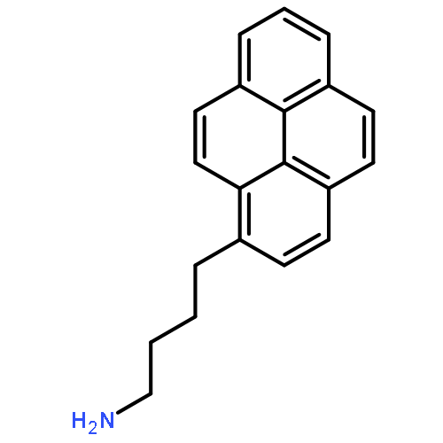
•The interaction of PAHs and DNA was investigated using spectroscopic and PAGE methods.•Amino-PAHs bind with DNA mainly via intercalative modes.•The binding ability of PAHs to p53 DNA was stronger than that for C-myc DNA.•The binding of 1-PBA induced the transformation from the duplex DNA into the G-quadruplex DNA.Polycyclic aromatic hydrocarbons derivatives (PAHs) have been confirmed to be carcinogenic, teratogenic and mutagenic, and have the potential to cause human malignant diseases. In this work, interactions of two selected amino-PAHs (aminopyrene derivatives) and human tumor-related DNA were evaluated using spectroscopic and polyacrylamide gel electrophoresis (PAGE) methods. Spectroscopic results demonstrated that there were remarkable interactions between PAHs and the targeted DNA with the order of the binding ability as 1-AP > 1-PBA. The binding constants of 1-AP with the targeted DNA were at the level of about 106 L/mol, while that of 1-PBA only to about 103 L/mol. 1-AP with a short side-chain acted mainly as an intercalator, and its interactions with DNA were strengthened with electrostatic forces. As for 1-PBA with a flexible long side-chain, the intercalation mode was dominated with an auxiliary role of Van der Wals forces and hydrogen bonds. Besides, the binding abilities of amino-PAHs to p53 DNA seemed stronger than that for C-myc DNA. PAGE results showed that the binding of amino-PAHs could further change the conformation of DNA sequences from the duplex to the antiparallel G-quadruplex.
Co-reporter:Huihui Li, Xiaoyang Bu, Jia Lu, Chongzheng Xu, Xianlong Wang, Xiaodi Yang
Spectrochimica Acta Part A: Molecular and Biomolecular Spectroscopy 2013 Volume 107() pp:227-234
Publication Date(Web):15 April 2013
DOI:10.1016/j.saa.2013.01.069
The interaction of ciprofloxacin (CIP) with human telomeric DNA was studied in vitro using multi-spectroscopy and molecular modeling methods. The hypochromic effect with a red shift in ultraviolet (UV) absorption indicated the occurrence of the interaction between CIP and DNA. The fluorescence quenching of CIP was observed with the addition of DNA and was proved to be the static quenching. The binding constant was found to be 9.62 × 10⁴ L mol−1. Electrospray ionization mass spectrometry (ESI-MS) result further confirmed the formation of 1:1 non-covalent complex between DNA and CIP. Combined with the UV melting results, circular dichroism (CD) results confirmed the existence of groove binding mode, as well as conformational changes of DNA. Molecular docking studies illustrated the visual display of the CIP binding to the GC region in the minor groove of DNA. Specific hydrogen bonds and van der Waals forces were demonstrated as main acting forces between CIP and guanine bases of DNA.Graphical abstractHighlights► Multi-spectroscopy and molecular docking methods were combined. ► CIP is likely to insert into the minor groove of DNA. ► ESI-MS was used to deduce the stoichiometric ratio of DNA–CIP complexes. ► The interaction of CIP could destabilize DNA observed by CD spectra. ► Hydrogen bonds and van der Waals were main acting forces between CIP and DNA.

![Anthra[2,1,9-def:6,5,10-d'e'f']diisoquinoline-1,3,8,10(2H,9H)-tetrone, 2,9-bis[3-(dimethylamino)propyl]-](/data/chemimg/134800/117901-97-0.png)
![Anthra[2,1,9-def:6,5,10-d'e'f']diisoquinoline-1,3,8,10(2H,9H)-tetrone, 2,9-bis[3-(dimethylamino)propyl]-](/data/chemimg/134800/117901-97-0_b.png)