Co-reporter:Jun Ye;Wujun Dong;Yanfang Yang;Huazhen Hao;Hengfeng Liao
Pharmaceutical Research 2017 Volume 34( Issue 6) pp:1244-1254
Publication Date(Web):21 March 2017
DOI:10.1007/s11095-017-2141-3
To overcome the drawbacks of high dose regimen and improve the outcomes of chemotherapy at a low dose, an immunotherapeutic nanoemulsion based combination of chemotherapeutic agent (paclitaxel) with immunomodulatory agent (vitamin E) was developed and evaluated for their antitumor effect against breast cancer.A total of five nanoemulsions loaded with various content of vitamin E were prepared and characterized. The immunoregulatory effects of vitamin E along with the overall antitumor efficacy of vitamin E-rich nanoemulsion with a low dose of paclitaxel were investigated through in vitro and in vivo experiments.Vitamin E-rich nanoemulsion exhibited relatively narrow size distribution, high entrapment efficiency and controlled in vitro release profile. In RAW264.7 cells, vitamin E-rich nanoemulsion significantly enhanced the secretion of Th1 cytokines and down-regulated the secretion of Th2 cytokine. In a co-culture system, vitamin E-rich nanoemulsion induced a high apoptosis rate in MDA-MB-231 cells as compared with vitamin E-low nanoemulsion. Furthermore, vitamin E-rich nanoemulsion exhibited superior in vivo antitumor efficacy in comparison with Taxol and vitamin E-low nanoemulsion at a paclitaxel dose of 4 mg/kg.Vitamin E-rich nanoemulsion has great potential for the treatment of breast cancers with a low dose of paclitaxel via driving Th1 immune response.
Co-reporter:Yanfang Yang;Xuejun Xia;Wujun Dong;Hongliang Wang;Lin Li;Panpan Ma;Wei Sheng;Xueqing Xu
Macromolecular Bioscience 2016 Volume 16( Issue 5) pp:759-773
Publication Date(Web):
DOI:10.1002/mabi.201500389
Co-reporter:Xuejun Xia, Xiaowei Song, Jiaming Xu, Jiuming He, Jie Peng, Xiang Zhang, Dujia Jin, Zeper Abliz, Yuling Liu
Journal of Pharmaceutical and Biomedical Analysis 2016 Volume 117() pp:532-543
Publication Date(Web):5 January 2016
DOI:10.1016/j.jpba.2015.05.023
•We developed a bioanalytical method of paclitaxel-cholesterol conjugate (CHO-PTX).•Murine plasma and tumors distribution of CHO-PTX was measured by LC–APCI-MS/MS.•We observed the high distribution of CHO-PTX in tumors relative to plasma levels.•Cholesteryl-drug conjugate is a promising approach for tumor targeting therapy.Conjugation of a cholesterol moiety to active compounds for cancer treatment or diagnosis is an attractive approach for increasing lipophilicity and improving loading into lipid carriers. We developed a highly sensitive and specific liquid chromatography atmospheric-pressure chemical ionization tandem mass spectrometry (LC–APCI-MS/MS) analytical method to investigate the in vivo plasma and tumor distribution characteristic of a cholesterol-paclitaxel conjugate (CHO-PTX) in nude mice with MDA-MB-231 human breast cancer xenografts. The samples were analyzed in positive ion, multiple reaction monitoring mode. The plasma and tumor tissue samples were processed by liquid–liquid extraction with methyl tert-butyl ether (MTBE). Docetaxel was used as the internal standard (IS) for sample processing and analysis. MS/MS detection was carried out by monitoring the transitions of m/z 1266.7 → 369.4 and 330.3 for CHO-PTX, and m/z 808.7 → 226.4 and 509.1 for IS. The calibration curves were linear over 100–25,000 ng/mL in mouse plasma and tumor homogenate samples. The limit of quantitation of CHO-PTX was 100 ng/mL in both matrices. The intra-day and inter-day precisions were less than 15%, and the accuracy was between −8.0% and 8.6% for both matrices. The developed method was successfully applied to measure CHO-PTX levels in plasma and tumor tissues in nude mice. The mean tumor concentrations in mice tumor tissues after intravenous administration of CHO-PTX emulsion at a dose equivalent to 20 mg/kg paclitaxel were 2022 ± 630 ng/mL ng/mL, 2516 ± 982 ng/mL, 3056 ± 1438 ng/mL, and 2367 ± 1029 ng/mL at 0.25, 3, 24, and 120 h, respectively. The accumulation of CHO-PTX in the tumor suggests that cholesteryl drug conjugates are a promising approach for medical treatment of various human cancers.Plasma and tumor CHO-PTX levels and the radio of CHO-PTX concentration in tumor/plasma at 0.25, 3, 24, and 120 h after a single intravenous administration of CHO-PTX lipid emulsions at a dose equivalent to 20 mg/kg paclitaxel to mice with MDA-MB-231 human breast tumor xenografts. Each point represents the mean ± SD (n = 5).
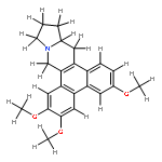
Co-reporter:Feifei Yu, Haining Lv, Wujun Dong, Jun Ye, Huazhen Hao, Shuanggang Ma, Shishan Yu, Yuling Liu
Journal of Pharmaceutical and Biomedical Analysis 2015 Volume 107() pp:223-228
Publication Date(Web):25 March 2015
DOI:10.1016/j.jpba.2014.12.042
•A method was developed for simultaneous determination of CAT and its metabolite S-4.•We develop an assay with short run time and low LLOQ.•The method showed excellent chromatographic resolution.•The validated method was successfully applied to a pharmacokinetic study in rats.CAT ((+)-(13aS)-deoxytylophorinine) is a novel anticancer drug belonging to phenanthroindolizidine alkaloids. A sensitive and reliable liquid chromatography–tandem mass spectrometry (LC–MS/MS) method for simultaneous quantification of CAT and its pharmacologically active 3-O-desmethyl metabolite (S-4) was developed and validated in rat plasma using rotundine as the internal standard (IS). CAT, S-4 and IS were extracted by acetonitrile protein precipitation and separated on an Eclipse XDB-C18 column (1.8 μm, 4.6 mm × 50 mm) with acetonitrile–water (27:73, v/v) mobile phase containing 0.1% formic acid at a 0.4 mL/min flow rate. Positive ion electrospray ionization in multiple reaction monitoring mode was employed to measure CAT, S-4 and IS by monitoring the transitions m/z 364.2 → 70.1 for CAT, 350.1 → 70.1 for S-4 and 356.2 → 192.2 for IS. Good linear correlation (r2 > 0.991) was achieved for CAT and S-4 over the range of 0.214–128.16 and 0.044–11.00 ng/mL, respectively. The lower limit of quantification was 0.214 ng/mL for CAT and 0.044 ng/mL for S-4, using 50 μL rat plasma samples. The intra- and inter-day precisions were not exceed 15% and the accuracy ranged between 94.80% and 108.22%. The average extraction recoveries of both analytes were greater than 94.62%. The method was successfully applied to the pharmacokinetic study of CAT and S-4 in rats after oral administration.
Co-reporter:Jie Peng, Wu-jun Dong, Ling Li, Jia-ming Xu, Du-jia Jin, Xue-jun Xia, Yu-ling Liu
Journal of Food and Drug Analysis (December 2015) Volume 23(Issue 4) pp:828-835
Publication Date(Web):1 December 2015
DOI:10.1016/j.jfda.2015.04.004
The effect of different high pressure homogenization energy input parameters on mean diameter droplet size (MDS) and droplets with > 5 μm of lipid injectable emulsions were evaluated. All emulsions were prepared at different water bath temperatures or at different rotation speeds and rotor-stator system times, and using different homogenization pressures and numbers of high-pressure system recirculations. The MDS and polydispersity index (PI) value of the emulsions were determined using the dynamic light scattering (DLS) method, and large-diameter tail assessments were performed using the light-obscuration/single particle optical sensing (LO/SPOS) method. Using 1000 bar homogenization pressure and seven recirculations, the energy input parameters related to the rotor-stator system will not have an effect on the final particle size results. When rotor-stator system energy input parameters are fixed, homogenization pressure and recirculation will affect mean particle size and large diameter droplet. Particle size will decrease with increasing homogenization pressure from 400 bar to 1300 bar when homogenization recirculation is fixed; when the homogenization pressure is fixed at 1000 bar, the particle size of both MDS and percent of fat droplets exceeding 5 μm (PFAT5) will decrease with increasing homogenization recirculations, MDS dropped to 173 nm after five cycles and maintained this level, volume-weighted PFAT5 will drop to 0.038% after three cycles, so the “plateau” of MDS will come up later than that of PFAT5, and the optimal particle size is produced when both of them remained at plateau. Excess homogenization recirculation such as nine times under the 1000 bar may lead to PFAT5 increase to 0.060% rather than a decrease; therefore, the high-pressure homogenization procedure is the key factor affecting the particle size distribution of emulsions. Varying storage conditions (4–25°C) also influenced particle size, especially the PFAT5.
Co-reporter:Yanfang Yang, Yang Yang, Xiangyang Xie, Xueqing Xu, Xuejun Xia, Hongliang Wang, Lin Li, Wujun Dong, Panpan Ma, Yuling Liu
International Journal of Pharmaceutics (15 June 2016) Volume 506(Issues 1–2) pp:158-173
Publication Date(Web):15 June 2016
DOI:10.1016/j.ijpharm.2016.04.035
Small interfering RNA (siRNA) offers a new and potential therapeutic strategy for tackling many diseases at the molecular level. Recently, cell-penetrating peptides (CPPs) conjugated with siRNA via disulfide-bonds (designated as siRNA-CPPs) were reported to form glutathione-sensitive carriers. However, non-cell specificity, CPPs degradation and the unwanted reduction of siRNA-CPPs before reaching the targeted tissue in vivo hampered the development of siRNA-CPPs. Herein, utilizing the dual stimulus of hyperthermia and the intracellular redox environment, we devised a thermosensitive liposome (TSL) containing an Asparagine-Glycine-Arginine (NGR) peptide and reducible siRNA-CPPs for tumor-specific siRNA transfection (siRNA-CPPs/NGR-TSL), in which siRNA-CPPs were “caged” in NGR-TSL to overcome their limitations in vivo. The functional nanocarrier possessed a small particle size of approximately 90 nm, a high drug encapsulation efficiency of approximately 86% and good serum stability. Both free siRNA-CPPs and siRNA-CPPs/NGR-TSL (preheated) silenced c-myc in human fibrosarcoma (HT-1080) cells in vitro. However, in an HT-1080 xenograft murine model, siRNA-CPPs/NGR-TSL with hyperthermia displayed superior in vivo antitumor efficacy (about 3-fold) and gene silencing efficiency (about 2-fold) compared with free siRNA-CPPs under hyperthermia. This study demonstrates that the constructed vesicle in combination with hyperthermia could greatly improve the in vivo stability of siRNA-CPPs and synergistically enhance its cancer therapy efficiency.Schematic illustration of siRNA-CPPs/NGR-TSL for specific siRNA delivery to tumor cells under dual stimulus of hyperthermia and intracellular redox environment. NGR-modified liposomes are retained in the tumor due to the active targeting effect by the NGR ligand. In the body circulation, as siRNA-CPPs were encapsulated in NGR-TSL, they can not penetrate into the normal cell membrane and be degraded by enzyme in the blood. However, upon heat-stimulus at the tumor site, siRNA-CPPs were released, the interaction of CPPs with the cell membrane is restored and mediated siRNA rapidly enter into the cells membrane and escape from the endosomal entrapment into the cytosol. At last, free siRNA was released from the CPPs via disulfide-bond broken under the stimulus of GSH in the cytosol and silenced c-myc gene over expressed in HT-1080 cells.Download high-res image (137KB)Download full-size image

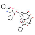
![Benzenepropanoic acid, b-(benzoylamino)-a-hydroxy-,(2aR,4S,4aS,6R,9S,11S,12S,12aR,12bS)-6-(acetyloxy)-12-(benzoyloxy)-2a,3,4,4a,5,6,9,10,11,12,12a,12b-dodecahydro-4,11,12b-trihydroxy-4a,8,13,13-tetramethyl-5-oxo-7,11-methano-1H-cyclodeca[3,4]benz[1,2-b]oxet-9-ylester, (aR,bS)-](http://img.cochemist.com/ccimg/160600/160511-46-6.png)
![Benzenepropanoic acid, b-(benzoylamino)-a-hydroxy-,(2aR,4S,4aS,6R,9S,11S,12S,12aR,12bS)-6-(acetyloxy)-12-(benzoyloxy)-2a,3,4,4a,5,6,9,10,11,12,12a,12b-dodecahydro-4,11,12b-trihydroxy-4a,8,13,13-tetramethyl-5-oxo-7,11-methano-1H-cyclodeca[3,4]benz[1,2-b]oxet-9-ylester, (aR,bS)-](http://img.cochemist.com/ccimg/160600/160511-46-6_b.png)
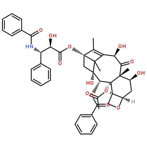
![Benzenepropanoic acid, b-(benzoylamino)-a,4-dihydroxy-,(2aR,4S,4aS,6R,9S,11S,12S,12aR,12bS)-6,12b-bis(acetyloxy)-12-(benzoyloxy)-2a,3,4,4a,5,6,9,10,11,12,12a,12b-dodecahydro-4,11-dihydroxy-4a,8,13,13-tetramethyl-5-oxo-7,11-methano-1H-cyclodeca[3,4]benz[1,2-b]oxet-9-ylester, (aR,bS)-](http://img.cochemist.com/ccimg/132200/132160-32-8.png)
![Benzenepropanoic acid, b-(benzoylamino)-a,4-dihydroxy-,(2aR,4S,4aS,6R,9S,11S,12S,12aR,12bS)-6,12b-bis(acetyloxy)-12-(benzoyloxy)-2a,3,4,4a,5,6,9,10,11,12,12a,12b-dodecahydro-4,11-dihydroxy-4a,8,13,13-tetramethyl-5-oxo-7,11-methano-1H-cyclodeca[3,4]benz[1,2-b]oxet-9-ylester, (aR,bS)-](http://img.cochemist.com/ccimg/132200/132160-32-8_b.png)


![3,5,9-Trioxa-4-phosphapentacosan-1-aminium,4-hydroxy-N,N,N-trimethyl-10-oxo-7-[(1-oxohexadecyl)oxy]-, inner salt, 4-oxide](http://img.cochemist.com/ccimg/2700/2644-64-6.png)
![3,5,9-Trioxa-4-phosphapentacosan-1-aminium,4-hydroxy-N,N,N-trimethyl-10-oxo-7-[(1-oxohexadecyl)oxy]-, inner salt, 4-oxide](http://img.cochemist.com/ccimg/2700/2644-64-6_b.png)