Co-reporter:Uthpala Seneviratne;Alexi Nott;Vadiraja B. Bhat;Kodihalli C. Ravindra;John S. Wishnok;Li-Huei Tsai
PNAS 2016 Volume 113 (Issue 15 ) pp:4152-4157
Publication Date(Web):2016-04-12
DOI:10.1073/pnas.1521318113
Protein S-nitrosation (SNO-protein), the nitric oxide-mediated posttranslational modification of cysteine thiols, is an important regulatory mechanism
of protein function in both physiological and pathological pathways. A key first step toward elucidating the mechanism by
which S-nitrosation modulates a protein’s function is identification of the targeted cysteine residues. Here, we present a strategy
for the simultaneous identification of SNO-cysteine sites and their cognate proteins to profile the brain of the CK-p25–inducible mouse model of Alzheimer’s disease-like
neurodegeneration. The approach—SNOTRAP (SNO trapping by triaryl phosphine)—is a direct tagging strategy that uses phosphine-based chemical probes, allowing enrichment
of SNO-peptides and their identification by liquid chromatography tandem mass spectrometry. SNOTRAP identified 313 endogenous SNO-sites in 251 proteins in the mouse brain, of which 135 SNO-proteins were detected only during neurodegeneration. S-nitrosation in the brain shows regional differences and becomes elevated during early stages of neurodegeneration in the
CK-p25 mouse. The SNO-proteome during early neurodegeneration identified increased S-nitrosation of proteins important for synapse function, metabolism, and Alzheimer’s disease pathology. In the latter case,
proteins related to amyloid precursor protein processing and secretion are S-nitrosated, correlating with increased amyloid formation. Sequence analysis of SNO-cysteine sites identified potential linear motifs that are altered under pathological conditions. Collectively, SNOTRAP is a direct tagging tool for global elucidation of the SNO-proteome, providing functional insights of endogenous SNO proteins in the brain and its dysregulation during neurodegeneration.
Co-reporter:Kodihalli C. Ravindra, Wanxing Eugene Ho, Chang Cheng, Luiz C. Godoy, John S. Wishnok, Choon Nam Ong, W. S. Fred Wong, Gerald N. Wogan, and Steven R. Tannenbaum
Chemical Research in Toxicology 2015 Volume 28(Issue 10) pp:1903
Publication Date(Web):September 4, 2015
DOI:10.1021/acs.chemrestox.5b00105
The antimalarial drug artesunate is a semisynthetic derivative of artemisinin, the principal active component of a medicinal plant Artemisia annua. It is hypothesized to attenuate allergic asthma via inhibition of multiple signaling pathways. We used a comprehensive approach to elucidate the mechanism of action of artesunate by designing a novel biotinylated dihydroartemisinin (BDHA) to identify cellular protein targets of this anti-inflammatory drug. By adopting an untargeted proteomics approach, we demonstrated that artesunate may exert its protective anti-inflammatory effects via direct interaction with multiple proteins, most importantly with a number of mitochondrial enzymes related to glucose and energy metabolism, along with mRNA and gene expression, ribosomal regulation, stress responses, and structural proteins. In addition, the modulatory effects of artesunate on various cellular transcription factors were investigated using a transcription factor array, which revealed that artesunate can simultaneously modulate multiple nuclear transcription factors related to several major pro- and anti-inflammatory signaling cascades in human bronchial epithelial cells. Artesunate significantly enhanced nuclear levels of nuclear factor erythroid-2-related factor 2 (Nrf2), a key promoter of antioxidant mechanisms, which is inhibited by the Kelch-like ECH-associated protein 1 (Keap1). Our results demonstrate that, like other electrophilic Nrf2 regulators, artesunate activates this system via direct molecular interaction/modification of Keap1, freeing Nrf2 for transcriptional activity. Altogether, the molecular interactions and modulation of nuclear transcription factors provide invaluable insights into the broad pharmacological actions of artesunate in inflammatory lung diseases and related inflammatory disorders.
Co-reporter:Uthpala Seneviratne ; Luiz C. Godoy ; John S. Wishnok ; Gerald N. Wogan
Journal of the American Chemical Society 2013 Volume 135(Issue 20) pp:7693-7704
Publication Date(Web):April 24, 2013
DOI:10.1021/ja401565w
Nitrosothiols (RSNOs) have been proposed as important intermediates in nitric oxide (NO•) metabolism, storage, and transport as well as mediators in numerous NO-signaling pathways. RSNO levels are finely regulated, and dysregulation is associated with the etiology of several pathologies. Current methods for RSNO quantification depend on indirect assays that limit their overall specificity and reliability. Recent developments of phosphine-based chemical probes constitute a promising approach for the direct detection of RSNOs. We report here results from a detailed mechanistic and kinetic study for trapping RSNOs by three distinct phosphine probes, including structural identification of novel intermediates and stability studies under physiological conditions. We further show that a triarylphosphine-thiophenyl ester can be used in the absolute quantification of endogenous GSNO in several cancer cell lines, while retaining the elements of the SNO functional group, using an LC–MS-based assay. Finally, we demonstrate that a common product ion (m/z = 309.0), derived from phosphine–RSNO adducts, can be used for the detection of other low-molecular weight nitrosothiols (LMW-RSNOs) in biological samples. Collectively, these findings establish a platform for the phosphine ligation-based, specific and direct detection of RSNOs in biological samples, a powerful tool for expanding the knowledge of the biology and chemistry of NO•-mediated phenomena.
Co-reporter:Liang Cui, Wenjie Ye, Erin G. Prestwich, John S. Wishnok, Koli Taghizadeh, Peter C. Dedon, and Steven R. Tannenbaum
Chemical Research in Toxicology 2013 Volume 26(Issue 2) pp:195
Publication Date(Web):November 9, 2012
DOI:10.1021/tx300294d
Oxidative damage to DNA has many origins, including irradiation, inflammation, and oxidative stress, but the chemistries are not the same. The most oxidizable base in DNA is 2-deoxyguanosine (dG), and the primary oxidation products are 8-oxodG and 2-amino-imidazolone. The latter rapidly converts to 2,2-diamino-oxazolone (Ox), and 8-oxodG is further oxidized to spiroiminodihydantoin (Sp) and guanidinohydantoin (Gh). In this study, we have examined the dose–response relationship for the formation of the above four products arising in calf thymus DNA exposed to gamma irradiation, photoactivated rose bengal, and two sources of peroxynitrite. In order to carry out these experiments, we developed a chromatographic system and synthesized isotopomeric internal standards to enable accurate and precise analysis based upon selected reaction monitoring mass spectrometry. 8-OxodG was the most abundant products in all cases, but its accumulation was highly dependent on the nature of the oxidizing agent and the subsequent conversion to Sp and Gh. Among the other oxidation products, Ox was the most abundant, and Sp was formed in significantly greater yield than Gh.
Co-reporter:Kun Lu, Charles G. Knutson, John S. Wishnok, James G. Fox, and Steven R. Tannenbaum
Journal of Proteome Research 2012 Volume 11(Issue 10) pp:4916-4926
Publication Date(Web):2017-2-22
DOI:10.1021/pr300429x
Inflammatory bowel disease (IBD) is a chronic relapsing inflammatory disorder of the bowel. The etiology remains unknown, but IBD is immune-driven and multiple factors including genetic, environmental, and microbiological components play a role. Recombinase-activating gene-2-deficient (Rag2–/–) mice infected with Helicobacter hepaticus (H. hepaticus) have been developed as an animal model to imitate naturally occurring inflammatory events and associated key features of chronic inflammatory responses in humans. In this study, we have combined mass spectrometry-based metabolomics and peptidomics to analyze serum samples of Rag2–/– mice infected with H. hepaticus. Metabolomics profiling revealed that H. hepaticus infection dramatically changed numerous metabolite pathways, including tryptophan metabolism, glycerophospholipids, methionine-homocysteine cycle, citrate cycle, fatty acid metabolism and purine metabolism, with the majority of metabolites being down-regulated. In particular, there were notable effects of gut microflora on the blood metabolites in infected animals. In addition, the peptidomics approach identified a number of peptides, originating from proteins, including fibrinogen, complement C4, and alpha-2-macroglobulin, with diverse biological functions with potentially important implications for the progress of IBD. In summary, the strategy of integrating a relevant animal model and sensitive mass spectrometry-based profiling may offer a new perspective to explore biomarkers and provide mechanistic insights into IBD.
Co-reporter:Sureshkumar Muthupalani;Aswin Mangerich;Erin Prestwich;Charles G. Knutson;Wenjie Ye;Jose L. McFaline;John S. Wishnok;Melissa Mobley;James G. Fox;Gerald N. Wogan;Peter C. Dedon;Liang Cui;Zhongming Ge;Koli Taghizadeh;Nicola M. Parry
PNAS 2012 Volume 109 (Issue 27 ) pp:
Publication Date(Web):2012-07-03
DOI:10.1073/pnas.1207829109
Helicobacter hepaticus-infected Rag2-/- mice emulate many aspects of human inflammatory bowel disease, including the development of colitis and colon cancer. To
elucidate mechanisms of inflammation-induced carcinogenesis, we undertook a comprehensive analysis of histopathology, molecular
damage, and gene expression changes during disease progression in these mice. Infected mice developed severe colitis and hepatitis
by 10 wk post-infection, progressing into colon carcinoma by 20 wk post-infection, with pronounced pathology in the cecum
and proximal colon marked by infiltration of neutrophils and macrophages. Transcriptional profiling revealed decreased expression
of DNA repair and oxidative stress response genes in colon, but not in liver. Mass spectrometric analysis revealed higher
levels of DNA and RNA damage products in liver compared to colon and infection-induced increases in 5-chlorocytosine in DNA
and RNA and hypoxanthine in DNA. Paradoxically, infection was associated with decreased levels of DNA etheno adducts. Levels
of nucleic acid damage from the same chemical class were strongly correlated in both liver and colon. The results support
a model of inflammation-mediated carcinogenesis involving infiltration of phagocytes and generation of reactive species that
cause local molecular damage leading to cell dysfunction, mutation, and cell death. There are strong correlations among histopathology,
phagocyte infiltration, and damage chemistry that suggest a major role for neutrophils in inflammation-associated cancer progression.
Further, paradoxical changes in nucleic acid damage were observed in tissue- and chemistry-specific patterns. The results
also reveal features of cell stress response that point to microbial pathophysiology and mechanisms of cell senescence as
important mechanistic links to cancer.
Co-reporter:Peter G. Slade, Michelle V. Williams, Viral Brahmbhatt, Ajit Dash, John S. Wishnok and Steven R. Tannenbaum
Chemical Research in Toxicology 2010 Volume 23(Issue 3) pp:557
Publication Date(Web):February 4, 2010
DOI:10.1021/tx9002808
The hydroperoxide of linoleic acid (13-HPODE) degrades to 9,12-dioxo-10(E)-dodecenoic acid (DODE), which readily modifies proteins. This study identified the major proteins in MCF7 cells modified by DODE. To reduce false positives, three methods were used to identify DODE-modified proteins. First, cells were treated with a synthetically biotinylated 13-HPODE (13-HPODE-biotin). Modified proteins were enriched by neutravidin affinity and identified by two-dimensional liquid chromatography−tandem mass spectrometry (2D LC-MS/MS). Second, cells were treated with native 13-HPODE. Protein carbonyls were biotinylated with an aldehyde reactive probe, and modified proteins were enriched by neutravidin affinity and identified by 2D LC-MS/MS. Third, using a newly developed DODE antibody, DODE-modified proteins were located by 2D sodium dodecyl sulfate−polyacrylamide gel electrophoresis and Western blot and identified by in-gel digestion and LC-MS/MS. Analysis of the proteins characterized by all three methods revealed a significant overlap and identified 32 primary proteins modified by DODE in MCF7 cells. These results demonstrated the feasibility for the cellular formation of DODE protein−carbonyl adducts that may be future indicators of oxidative stress.
Co-reporter:John S. Wishnok;Hongbin Yu
Journal of Labelled Compounds and Radiopharmaceuticals 2003 Volume 46(Issue 14) pp:1269-1277
Publication Date(Web):5 NOV 2003
DOI:10.1002/jlcr.789
3,7,8-15N3-N1-(β-D-erythro-pentofuranosyl)-5-guanidinohydantoin was synthesized from the oxidation of 1,7,NH2-15N3-8-oxo-7,8-dihydro-2′-deoxyguanosine with 2 equivalents of Ir(IV) in pH 4.5 potassium phosphate buffer. The synthesis of 1,7,NH2-15N3-8-oxo-7,8-dihydro-2′-deoxyguanosine started with bromination of 1,7,NH2-15N3-2′-deoxyguanosine. The resulting 1,7,NH2-15N3-8-bromo-7,8-dihydro-2′-deoxyguanosine reacted with sodium benzyloxide to afford 1,7,NH2-15N3-8-benzyloxy-7,8-dihydro-2′-deoxyguanosine. Subsequent catalytic transfer hydrogenation of 1,7,NH2-15N3-8-benzyloxy-7,8-dihydro-2′-deoxyguanosine with cyclohexene and 10% Pd/C yielded 1,7,NH2-15N3-8-oxo-7,8-dihydro-2′-deoxyguanosine. Purification of 3,7,8-15N3-N1-(β-D-erythro-pentofuranosyl)-5-guanidinohydantoin was first carried out on a C18 column and the product was further purified on a graphite column. ESI-MS was used to confirm the identity and to determine the isotopic purity of all the labeled compounds. The isotopic purity of 3,7,8-15N3-N1-(β-D-erythro-pentofuranosyl)-5-guanidinohydantoin was 99.4 atom% based on LC-MS measurements. Copyright © 2003 John Wiley & Sons, Ltd.
.jpg)


![sodium 3-({[(5aS,6R,8aS,9R,10R,12R)-3,6,9-trimethyldecahydro-3,12-epoxy[1,2]dioxepino[4,3-i]isochromen-10-yl]carbonyl}oxy)propanoate](http://img.cochemist.com/ccimg/83600/83507-69-1.png)
![sodium 3-({[(5aS,6R,8aS,9R,10R,12R)-3,6,9-trimethyldecahydro-3,12-epoxy[1,2]dioxepino[4,3-i]isochromen-10-yl]carbonyl}oxy)propanoate](http://img.cochemist.com/ccimg/83600/83507-69-1_b.png)


![1-Propanaminium,3-carboxy-N,N,N-trimethyl-2-[(1-oxohexadecyl)oxy]-, inner salt, (2R)-](http://img.cochemist.com/ccimg/2400/2364-67-2.png)
![1-Propanaminium,3-carboxy-N,N,N-trimethyl-2-[(1-oxohexadecyl)oxy]-, inner salt, (2R)-](http://img.cochemist.com/ccimg/2400/2364-67-2_b.png)


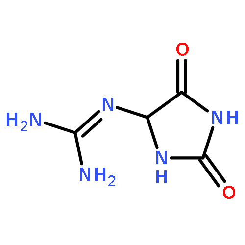
![2-AMINO-1,3,6,8-TETRAZASPIRO[4.4]NON-1-ENE-4,7,9-TRIONE](http://img.cochemist.com/ccimg/481000/480996-13-2.png)
![2-AMINO-1,3,6,8-TETRAZASPIRO[4.4]NON-1-ENE-4,7,9-TRIONE](http://img.cochemist.com/ccimg/481000/480996-13-2_b.png)
![1,3,6,8-Tetraazaspiro[4.4]non-6-ene-2,4,9-trione, 7-amino-1-(2-deoxy-β-D-erythro-pentofuranosyl)-](/data/chemimg/124700/426265-32-9.png)
![1,3,6,8-Tetraazaspiro[4.4]non-6-ene-2,4,9-trione, 7-amino-1-(2-deoxy-β-D-erythro-pentofuranosyl)-](/data/chemimg/124700/426265-32-9_b.png)


![Guanidine, [3-(2-deoxy-β-D-erythro-pentofuranosyl)-2,5-dioxo-4-imidazolidinyl]-](/data/chemimg/119300/265986-31-0.png)
![Guanidine, [3-(2-deoxy-β-D-erythro-pentofuranosyl)-2,5-dioxo-4-imidazolidinyl]-](/data/chemimg/119300/265986-31-0_b.png)


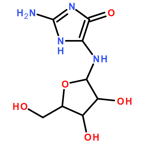
![4H-Imidazol-4-one, 2-amino-5-[(2-deoxy-β-D-erythro-pentofuranosyl)amino]-](/data/chemimg/124700/142559-87-3.png)
![4H-Imidazol-4-one, 2-amino-5-[(2-deoxy-β-D-erythro-pentofuranosyl)amino]-](/data/chemimg/124700/142559-87-3_b.png)
![1H-Thieno[3,4-d]imidazole-4-pentanoicacid, hexahydro-2-oxo-, 2-[2-(aminooxy)acetyl]hydrazide, (3aS,4S,6aR)-](http://img.cochemist.com/ccimg/139600/139585-03-8.png)
![1H-Thieno[3,4-d]imidazole-4-pentanoicacid, hexahydro-2-oxo-, 2-[2-(aminooxy)acetyl]hydrazide, (3aS,4S,6aR)-](http://img.cochemist.com/ccimg/139600/139585-03-8_b.png)


![(11S,11aS)-9-Hydroxy-11-(1H-indol-3-yl)-1,2,3,10,11,11a-hexahydro -5H-pyrrolo[2,1-c][1,4]benzodiazepin-5-one](http://img.cochemist.com/ccimg/80300/80279-24-9.png)
![(11S,11aS)-9-Hydroxy-11-(1H-indol-3-yl)-1,2,3,10,11,11a-hexahydro -5H-pyrrolo[2,1-c][1,4]benzodiazepin-5-one](http://img.cochemist.com/ccimg/80300/80279-24-9_b.png)
![3H-Imidazo[2,1-i]purine,3-(2-deoxy-b-D-erythro-pentofuranosyl)-](http://img.cochemist.com/ccimg/68500/68498-25-9.png)
![3H-Imidazo[2,1-i]purine,3-(2-deoxy-b-D-erythro-pentofuranosyl)-](http://img.cochemist.com/ccimg/68500/68498-25-9_b.png)
![Imidazo[1,2-c]pyrimidin-5(6H)-one](http://img.cochemist.com/ccimg/55700/55662-66-3.png)
![Imidazo[1,2-c]pyrimidin-5(6H)-one](http://img.cochemist.com/ccimg/55700/55662-66-3_b.png)


![3H-Imidazo[2,1-i]purine](http://img.cochemist.com/ccimg/13900/13875-63-3.png)
![3H-Imidazo[2,1-i]purine](http://img.cochemist.com/ccimg/13900/13875-63-3_b.png)


![4-amino-5-chloro-1-[(2r,4s,5r)-4-hydroxy-5-(hydroxymethyl)oxolan-2-yl]pyrimidin-2-one](http://img.cochemist.com/ccimg/32400/32387-56-7.png)
![4-amino-5-chloro-1-[(2r,4s,5r)-4-hydroxy-5-(hydroxymethyl)oxolan-2-yl]pyrimidin-2-one](http://img.cochemist.com/ccimg/32400/32387-56-7_b.png)


