Co-reporter:Jie Ma, Fang Wang, Jingyu Yang, Yingxu Dong, Guangyue Su, Kuo Zhang, Xing Pan, Ping Ma, Tingshuo Zhou, Chunfu Wu
Journal of Ethnopharmacology 2017 Volume 208(Volume 208) pp:
Publication Date(Web):17 August 2017
DOI:10.1016/j.jep.2017.07.005
Ethnopharmacological relevanceXiaochaihutang (XCHT), as a classical herbal formula for the treatment of “Shaoyang syndrome” has been demonstrated to exert an antidepressant effect in multiple animal models of depression as shown in our previous studies. However, the effects of XCHT on social isolation (SI)-reared mice have not been investigated. This study aims to explore the effects of XCHT on depressive/anxiety-like behaviors of SI-reared mice, and its implicated mechanisms, including alterations in the monoaminergic system, neurogenesis and neurotrophin expression.Materials and methodsMale C57 BL/6 J mice (aged 4 weeks after weaning) were reared isolatedly for 8 weeks and XCHT (0.8, 2.3, 7.0 g/kg) were given by gavage once a day. Forced swimming test (FST), tail suspension test (TST), open field test (OFT), elevated-plus maze test (EPM) and intruder-induced aggression test were used to explore the effects of XCHT on depressive/anxiety-like behaviors of SI-reared mice after administration of XCHT for 6 weeks. HPLC-MS/MS was performed to quantify the levels of neurotransmitters in the hippocampus by in vivo microdialysis, while western immunoblotting was used to evaluate the action of XCHT on the synthesis, transport and degradation of monoamine neurotransmitters. Immunofluorescence was used to study the effects of XCHT on neurogenesis and neurotrophin expression, including Ki-67, DCX, BrdU and BDNF.ResultsOur results showed that administration of XCHT (0.8, 2.3 and 7.0 g/kg) for 6 weeks significantly attenuated the increase in immobility time in TST and FST, improved the anxiety-like behaviors in OFT and EPM, and improved the aggressive behaviors of SI-reared mice. XCHT significantly elevated monoamine neurotransmitters levels and inhibited 5-HT turnover (5-HIAA/5-HT) in hippocampal microdialysates of SI-reared mice. In addition, we found XCHT enhanced monoamine neurotransmitter synthesis enzymes (TPH2 and TH) expressions, inhibited serotonin transporter (SERT) expression and decreased monoamine neurotransmitter degradation enzyme (MAOA) expression in the hippocampus of SI-reared mice for the first time. Moreover, XCHT significantly augmented hippocampal neurogenesis and BDNF expression in hippocampus of SI-reared mice.ConclusionsOur results showed for the first time that XCHT improved depressive/anxiety-like behaviors of SI-reared mice by regulating the monoaminergic system, neurogenesis and neurotrophin expression. The findings indicate that XCHT may have a therapeutic application for early-life stress model of depression and in turn provide further evidence supporting XCHT a novel potential antidepressant from a distinct perspective.Download high-res image (195KB)Download full-size image
Co-reporter:Ping Liu, Xiaohang Che, Lisha Yu, Xiaofeng Yang, Nina An, Wu Song, Chunfu Wu, Jingyu Yang
Pharmacology Biochemistry and Behavior 2017 Volume 163(Volume 163) pp:
Publication Date(Web):1 December 2017
DOI:10.1016/j.pbb.2017.10.004
•Uridine blocked the rewarding effects of morphine.•Levels of glutamate and GABA in the mPFC may be related to stress-induced relapse.•Uridine depressed the glutamate overflow in the stress-induced relapse.•Uridine increased the GABA levels in the stress-induced relapse.Several lines of evidence suggest that uridine, as a neuromodulator, plays an important role in drug addiction. We previously found that uridine circumvents morphine-induced behavioral sensitization by decreasing the extracellular dopamine levels in the dorsal striatum. In the present study, the effects of uridine on morphine-induced conditioned place preference (CPP) and the possible roles of glutamate and GABA in the stress-induced reinstatement of CPP were investigated. First, the effects of uridine (1, 10 and 100 mg/kg, i.p.) on the four defined phases - acquisition, expression, extinction and reinstatement (drug priming and restraint stress) - of morphine-induced CPP were studied. The results showed that pretreatment with uridine significantly blocked the acquisition and expression phases of CPP. Additionally, uridine also facilitated CPP extinction and inhibited stress-induced CPP reinstatement, although it failed to affect drug-induced CPP reinstatement. Since glutamatergic and GABAergic systems are both involved in CPP reinstatement, the extracellular levels of glutamate and GABA in the mPFC during the stress-induced CPP reinstatement were determined using in vivo microdialysis. The results showed that uridine attenuated the stress-induced glutamate increase in the mPFC without influencing the basal glutamate levels, and increased the levels of extracellular GABA in the mPFC both under normal physiological conditions and after the stress stimulus. Thus, our results indicate that uridine depresses the stress-induced reinstatement of CPP, simultaneously regulating glutamatergic and GABAergic neurotransmission in the mPFC. The present work provides further understanding of the role of uridine in morphine-induced neurobehavioral changes.
Co-reporter:Dan-Qi Li, Le Zhou, Di Wang, Jie Wu, Ling-Zhi Li, Xiao-Xiao Huang, Qing-Bo Liu, Ying-Ying Wu, Song Lin, Jing-Yu Yang, Shao-Jiang Song, Chun-Fu Wu
Bioorganic & Medicinal Chemistry Letters 2016 Volume 26(Issue 6) pp:1594-1598
Publication Date(Web):15 March 2016
DOI:10.1016/j.bmcl.2016.02.004
The discovery of new natural compounds with pharmacological properties is an increasingly important field, and a continuous phytochemical investigation of the roots of Bupleurum chinense D.C. has led to the isolation of 17 triterpenoids, including three new oleanane triterpenes (1–3) together with 14 known ones. Their structures were determined on the basis of 1D and 2D NMR spectra as well as HR-ESI-MS data. The cytotoxicities of all compounds against five selected human cancer cell lines were assayed. Only compounds 9 and 14 exhibited moderate activities. Recently, a number of investigations have focused on the neuroprotective properties of triterpenoids in B. chinense. In order to expand our knowledge about this herb, the neuroprotective effects of compounds 1–17 against hydrogen peroxide (H2O2)-induced neuronal cell damage in human neuroblastoma SH-SY5Y cells were evaluated. Compounds 1–3, 6–7 showed significant neuroprotective effects against H2O2-induced SH-SY5Y cell death. Preliminary structure–activity relationships (SARs) between neuroprotective effects and the isolates were also discussed.
Co-reporter:Fangyang Wang;Yajing Liu;Lihui Wang;Jingyu Yang;Yanfang Zhao;Nannan Wang;Qi Cao;Ping Gong;Chunfu Wu
Journal of Cellular and Molecular Medicine 2015 Volume 19( Issue 8) pp:1916-1928
Publication Date(Web):
DOI:10.1111/jcmm.12566
Abstract
Caspase-3 is a critical effector caspase in apoptosis cascade, and is often over-expressed in many cancer tissues. The first synthesized procaspase-3 activator, PAC-1, induces cancer cell apoptosis and exhibits antitumour activity in murine xenograft models. To identify more potent procaspase-3 activators, a series of compounds were designed, synthesized and evaluated for their ability of inducing cancer cell death in culture. Among these compounds, WF-208 stood out by its high cytotoxicity against procaspase-3 overexpressed HL-60 cells. Compared with PAC-1, WF-208 showed higher cytotoxicity in cancer cells and lower toxicity in normal cells. The further investigation described herein showed that WF-208 activated procaspase-3, degraded IAPs (The Inhibitors of apoptosis proteins) and leaded to caspase-3-dependent cell death in tumour cells, which possibly because of the zinc-chelating properties. WF-208 also showed greater antitumour activity than PAC-1 in murine xenograft model. In conclusion, we have discovered WF-208 as a promising procaspase-3 activating compound, with higher activity and higher cell selectivity than PAC-1.
Co-reporter:Lihui Wang, Guoliang Chen, Xiuhong Lu, Shuang Wang, Shanghe Han, Yi Li, Guanfang Ping, Xiaorui Jiang, Huahuan Li, Jingyu Yang, Chunfu Wu
European Journal of Medicinal Chemistry 2015 Volume 89() pp:88-97
Publication Date(Web):7 January 2015
DOI:10.1016/j.ejmech.2014.10.036
•Synthesis and in vitro anti-HIF-1 activity of a novel series of chalcone derivatives.•Studying of structure activity relationship.•The tested compound inhibited tumor invasion and angiogenesis in vitro and in vivo.•The tested compound was well tolerated and presented a good therapeutic window.A novel series of chalcone derivatives were synthesized and their biological activities against HIF-1 were evaluated. Among these compounds, 5d exhibited clearly inhibitory effects on HIF-1 by downregulating the expression of HIF-1α under hypoxic conditions. Meanwhile, it also significantly suppressed VEGF-induced migration and invasion of Hep3B and HUVEC cells in nontoxic concentrations. Additionally, tube formation assay demonstrated its anti-angiogenesis activity. Moreover, the in vivo study indicated that compound 5d could retard tumor growth of Hep3B xenograft models and reduced CD31 and MMP-2 expression in tumor tissues. Finally, in acute intravenous toxicity, 5d was well tolerated and was found to be non-toxic up to 200 mg/kg in Swiss mice. These findings support the further investigation on the anti-invasive and anti-angiogenic potential of this class of compounds as HIF-1 inhibitor.
Co-reporter:Dan-Qi Li, Jie Wu, Li-Yin Liu, Ying-Ying Wu, Ling-Zhi Li, Xiao-Xiao Huang, Qing-Bo Liu, Jing-Yu Yang, Shao-Jiang Song, Chun-Fu Wu
Bioorganic & Medicinal Chemistry Letters 2015 Volume 25(Issue 18) pp:3887-3892
Publication Date(Web):15 September 2015
DOI:10.1016/j.bmcl.2015.07.053
As a part of our ongoing studies on cytotoxic triterpenoid saponins from herbal medicines, phytochemical investigation of the roots of Bupleurum chinense DC. afforded four new saikosaponins (1–4), along with 16 known ones (5–20). Their structures were established by direct interpretation of their spectral data, mainly HR-ESI-MS, 1D NMR and 2D NMR, and by comparison with literature data. Among them, compound 20 was isolated from the natural product for the first time. The cytotoxicities of all compounds against five selected human cancer cell lines (A549, HepG2, Hep3B, Bcap-37 and MCF-7) were assayed. In general, a number of the isolated compounds exhibited potent cytotoxic activities against the five selected human cancer cell lines. In particular, compounds 3, 8–9, 11–13, 16 and 20 showed more potent cytotoxic activities against the HepG2 and A549 cell lines than the positive control 5-fluorouracil. Based on the primary screening results, the preliminary structure–activity relationship (SAR) studies were also discussed. The SAR results suggest that the 13,28-epoxy bridge, the orientation of the hydroxyl group and the type of the sugar units are important requirements for cytotoxicity and selectivity.
Co-reporter:Guo-Liang Chen, Li-Hui Wang, Jian Wang, Kang Chen, Man Zhao, Zhao-Zhu Sun, Shuang Wang, Hong-Li Zheng, Jing-Yu Yang, Chun-Fu Wu
Bioorganic & Medicinal Chemistry Letters 2013 Volume 23(Issue 13) pp:3891-3895
Publication Date(Web):1 July 2013
DOI:10.1016/j.bmcl.2013.04.067
Retinoid X receptor (RXR) and Histone deacetylase (HDAC) are considered important targets for anti-cancer therapy due to their crucial roles in genetic or epigenetic regulations of cancer development and progression. Here, we have designed and synthesized a novel compound which targets both RXR and HADC. This dual-targeting agent is derived from bexarotene and suberoylanilide hydroxamic acid (SAHA), prototypical RXR agonist and HDAC inhibitor, respectively. Molecular docking studies demonstrate that this agent has a relatively strong affinity to RXR and HADC. Importantly, it presents the potentials of activation of RXR and inhibition of HDAC in both cell-free and whole-cell assays, and displays anti-proliferative effect on representative cancer cell lines and drug-resistant cancer cell lines.
Co-reporter:Jingyu Yang, Yan Li, Fang Wang and Chunfu Wu
Journal of Agricultural and Food Chemistry 2010 Volume 58(Issue 10) pp:6525-6531
Publication Date(Web):April 23, 2010
DOI:10.1021/jf903070a
In this study, the hepatoprotective effects of apple polyphenols (AP, Appjfnol) against CCl4-induced acute liver damage in Kunming mice as well as the possible mechanisms were investigated. Mice were treated with AP (200, 400, and 800 mg/kg, ig) for seven consecutive days prior to the administration of CCl4 (0.1%, intraperitoneally). The serum levels of alanine aminotransferase (ALT) and aspartate aminotransferase (AST), the malondialdehyde (MDA), superoxide dismutase (SOD), and reduced glutathione (GSH) concentrations in the hepatic homogenate, and histopathological changes in mouse liver sections were determined. Levels of ferrous sulfate-l-cysteine (FeSO4-l-Cys)-induced lipid peroxidation and 1,1-diphenyl-2-picrylhydrazyl (DPPH) were also determined in vitro. AP significantly prevented the increase in serum ALT and AST levels in acute liver injury induced by CCl4 and produced a marked amelioration in the histopathological hepatic lesions coupled to weight loss. The extent of MDA formation was reduced; the SOD activity was enhanced, and the GSH concentration was increased in the hepatic homogenate in AP-treated groups compared with the CCl4-intoxicated group. AP also exhibited antioxidant effects on FeSO4-l-Cys-induced lipid peroxidation in rat liver homogenate and DPPH free radical scavenging activity in vitro. These results indicate that AP has a significant protective effect against acute hepatotoxicity induced by CCl4 in mice, which may be due to its free radical scavenging effect, inhibition of lipid peroxidation, and its ability to increase antioxidant activity.
Co-reporter:Lei Guo;Jing Yu Yang ;Chun Fu Wu
Basic & Clinical Pharmacology & Toxicology 2008 Volume 103( Issue 3) pp:222-227
Publication Date(Web):
DOI:10.1111/j.1742-7843.2008.00258.x
Abstract: It is suggested that reactive oxygen species produced during ethanol intake may induce oxidative DNA damage. The present study, by the use of single cell gel electrophoresis (comet assay), investigated the potential genotoxicity of acute and long-term ethanol administration in mouse peripheral leucocytes. Urinary 8-hydroxy-2′-deoxyguanosine and total antioxidant capacity and reactive oxygen species in whole blood were also detected. Acute or long-term administration of ethanol, at the dose of 2.5 or 5.0 g/kg intraperitoneally, for 30 days, could induce significant DNA damage in peripheral leucocytes, which could be repaired at least 3 days after ethanol withdrawal in long-term ethanol treatment. A significant increase of urinary 8-hydroxy-2′-deoxyguanosine and generations of reactive oxygen species in whole blood after ethanol administration were observed, which demonstrated the ethanol-induced oxidative DNA damage. Interestingly, it was found that the total antioxidant capacity was significantly increased in whole blood after long-term ethanol administration compared to the control group, which suggested enhancement in the activities of the antioxidative defence system against oxidative tissue damage caused by excessive ethanol consumption. Thus, the present studies provide a clear evidence that ethanol induced DNA damage in peripheral leucocytes, which might result from ethanol-induced oxidative stress in the body.
Co-reporter:Jia Qi;Jing-Yu Yang;Ming Song;Yan Li
Naunyn-Schmiedeberg's Archives of Pharmacology 2008 Volume 376( Issue 6) pp:441-448
Publication Date(Web):2008 February
DOI:10.1007/s00210-007-0245-8
Accumulated data have shown the neuroactive properties of oxytocin (OT), a neurohypophyseal neuropeptide, and its capability of reducing the abuse potential of drugs. The present study investigated the effect of OT on methamphetamine (MAP)-induced hyperactivity in mice and its possible mechanism of action. Locomotor activity was measured after administered with MAP using an infrared sensor. High-performance liquid chromatography with electrochemical detection (HPLC-ECD) was used to detect the content of monoamines and their metabolites in the striatum and accumbens and prefrontal cortex in mice after the behavioral test. OT (0.1, 0.5, and 2.5 μg/mouse, i.c.v.) had no effect on locomotor activity in naïve mice, but inhibited, in a dose-dependent manner, the hyperactivity induced by acute administration of MAP. Atosiban (Ato) (2.0 μg/mouse, i.c.v.), the selective inhibitor of OT receptor, attenuated the inhibitory effect of OT on MAP. A marked reduction of the ratios of 3, 4-dihydroxyphenylacetic acid (DOPAC) and homovanillic acid (HVA) to dopamine (DA) was observed in the striatum and accumbens of mice after acute administration of MAP. OT (2.5 μg, i.c.v.) significantly inhibited the reduction of DOPAC/DA and HVA/DA ratios. However, Ato decreased the ratio of DOPAC/DA significantly in mice compared with OT (2.5 μg) in combination with MAP. There was no significant change in serotonin (5-HT) metabolism in mice after a single administration of MAP. These results suggested that OT inhibited the MAP-induced hyperactivity by altering the DA turnover in the mesolimbic region of mice.
Co-reporter:Qiu Hua Zhang;Chun Fu Wu;Lian Duan;Jing Yu Yang
Archives of Toxicology 2008 Volume 82( Issue 2) pp:117-123
Publication Date(Web):2008 February
DOI:10.1007/s00204-007-0224-3
Despite the significant anti-tumor activities, cyclophosphamide (CP) also shows cytotoxicity to normal cells. In order to explore the protective effects of drugs against CP-induced adverse effects, 20(S)-ginsenoside Rg3 was tested for its possibly protective activities on CP-induced DNA damage and cell apoptosis in mouse bone marrow cells or peripheral lymphocyte cells. In the current study, the alkaline single cell gel electrophoresis (comet assay), flow cytometry assay with annexin V-FITC/PI and AO/EB staining assay were employed to measure DNA strand breakage and cell apoptosis, respectively. The activities of SOD and GPx and the contents of MDA were also tested by the various colormetric methods. The results showed that CP at a dose of 100 mg/kg, i.p. significantly caused DNA damages in both mouse bone marrow cells and peripheral lymphocyte cells, and markedly inhibited the activities of GPx and SOD and increased MDA contents in mouse blood. Moreover, CP at a dose of 200 mg/kg, i.p. triggered apoptosis in mouse bone marrow cells. On the other hand, 20(S)-ginsenoside Rg3 orally administered at a dose of 20 mg/kg to the animals once a day for 2 days significantly inhibited CP-induced DNA damages in mouse bone marrow cells and peripheral lymphocyte cells, decrease the apoptotic numbers of bone marrow cells, antagonized the reduction of the activities of SOD and GPx, and the increase in MDA contents. In conclusion, 20(S)-ginsenoside Rg3 showed the significant protective effects on CP-induced cell DNA damage and apoptosis. These effects might be partially attributed to its protective actions against CP-induced oxidative stress.
Co-reporter:Lei Zhu, Jing-yu Yang, Xue Xue, Ying-xu Dong, Yang Liu, Feng-rong Miao, Yong-feng Wang, Hong Xue, Chun-fu Wu
Mechanisms of Ageing and Development (September 2015) Volume 150() pp:34-45
Publication Date(Web):1 September 2015
DOI:10.1016/j.mad.2015.07.002
•Yonkenafil improves cognitive deficits in AD mice.•Yonkenafil reduces the β-amyloid burden.•Yonkenafil inhibits the activation of microglia and astrocytes.•Yonkenafil protects hippocampal neurogenesis in AD.In Alzheimer’s disease (AD), activated microglia invade and surround β-amyloid plaques, possibly contributing to the aggregation of amyloid β (Aβ), which affect the survival of neurons and lead to memory loss. Phosphodiesterase-5 (PDE-5) inhibitors have recently been shown a potential therapeutic effect on AD. In this study, the effects of yonkenafil (yonk), a novel PDE-5 inhibitor, on cognitive behaviors as well as the pathological features in transgenic AD mice were investigated. Seven-month-old APP/PS1 transgenic mice were treated with yonk (2, 6, or 18 mg/kg, intraperitoneal injection (i.p.)) or sildenafil (sild) (6 mg/kg, i.p.) daily for 3 months and then behavioral tests were performed. The results demonstrated that yonk improved nesting-building ability, ameliorated working memory deficits in the Y-maze tasks, and significantly improved learning and memory function in the Morris water maze (MWM) tasks. In addition, yonk reduced the area of Aβ plaques, and inhibited over-activation of microglia and astrocytes. Furthermore, yonk increased neurogenesis in the dentate granule brain region of APP/PS1 mice, indicated by increased BrdU+/NeuN+ and BrdU+/DCX+ cells compared to vehicle-treated transgenic mice. These results suggest that yonk could rescue cognitive deficits by ameliorated amyloid burden through regulating APP processing, inhibited the over-activation of microglia and astrocytes as well as restored neurogenesis.
Co-reporter:Xuemei Chen, Nannan Wang, Yueyang Liu, Yinglu Liu, Tianyu Zhang, Lei Zhu, Yongfeng Wang, Chunfu Wu, Jingyu Yang
Experimental Neurology (November 2014) Volume 261() pp:267-277
Publication Date(Web):1 November 2014
DOI:10.1016/j.expneurol.2014.07.007
•Yonkenafil protects against ischemic brain injury.•Yonkenafil enlarges the range of penumbra, and reduces ischemic cell apoptosis.•Yonkenafil reduces the loss of neurons via a cGMP-dependent Nogo-R axis.•Yonkenafil improves synaptic dysfunction via BDNF/TrkB and NGF/TrkA.Yonkenafil is a novel phosphodiesterase type 5 (PDE5) inhibitor. Here we evaluated the effect of yonkenafil on ischemic injury and its possible mechanism of action. Male Sprague–Dawley rats underwent middle cerebral artery occlusion, followed by intraperitoneal or intravenous treatment with yonkenafil starting 2 h later. Behavioral tests were carried out on day 1 or day 7 after reperfusion. Nissl staining, Fluoro-Jade B staining and electron microscopy studies were carried out 24 h post-stroke, together with an analysis of infarct volume and severity of edema. Levels of cGMP-dependent Nogo-66 receptor (Nogo-R) pathway components, hsp70, apaf-1, caspase-3, caspase-9, synaptophysin, PSD-95/neuronal nitric oxide synthases (nNOS), brain-derived neurotrophic factor (BDNF)/tropomyosin-related kinase B (TrkB) and nerve growth factor (NGF)/tropomyosin-related kinase A (TrkA) were also measured after 24 h. Yonkenafil markedly inhibited infarction and edema, even when administration was delayed until 4 h after stroke onset. This protection was associated with an improvement in neurological function and was sustained for 7 d. Yonkenafil enlarged the range of penumbra, reduced ischemic cell apoptosis and the loss of neurons, and modulated the expression of proteins in the Nogo-R pathway. Moreover, yonkenafil protected the structure of synapses and increased the expression of synaptophysin, BDNF/TrkB and NGF/TrkA. In conclusion, yonkenafil protects neuronal networks from injury after stroke.
Co-reporter:Yan Hua Mou, Jing Yu Yang, Nan Cui, Ji Ming Wang, Yue Hou, Shuang Song, Chun Fu Wu
International Immunopharmacology (May 2012) Volume 13(Issue 1) pp:120-125
Publication Date(Web):1 May 2012
DOI:10.1016/j.intimp.2012.03.017
The involvement of microglial activation in metal neurotoxicity is becoming increasingly recognized. Some metal ions, such as zinc (II) and manganese (II), have been recently reported as microglial activators to induce the release of inflammatory mediators including cytokines, chemokines and nitric oxide (NO) which are involved in the pathogenesis of neurological diseases. Cobalt is essential for human life. However, excessive cobalt is cytotoxic and neurotoxic. In the present study, we determined cobalt-induced production of NO and cytokines/chemokines in N9 cells, a murine microglial cell line. High levels of cobalt significantly up-regulated iNOS mRNA and protein expression, which resulted in the release of NO. Cobalt induced the production of tumor necrosis factor α (TNF-α) and interleukin-6 (IL-6) in a concentration- and time-dependent manner in both N9 cells and primary mouse microglia and increased lipopolysaccharides (LPS)-induced cytokine production. Further study showed that cobalt induced cytokine production by a mechanism involving both nuclear factor kappa B (NF-κB) and p38 mitogen-activated protein kinase (MAPK) signaling pathways. The involvement of reactive oxygen species (ROS) in microglial activation was also confirmed. These findings suggested that cobalt neurotoxicity should be attributed not only directly to neuronal damage but also indirectly to microglial activation which might potentiate neuronal injury via elevation of proinflammatory mediator levels.Highlights► Cobalt induced NO release via iNOS mRNA and protein expression upregulation. ► Cobalt significantly increased TNF-α and IL-6 levels in medium. ► TNF-α and IL-6 levels were amplified when combined with LPS. ► Cobalt induced cytokine production by a mechanism involving both NF-κB and p38 MAPK pathways. ► Reactive oxygen species were involved in cobalt-induced microglial activation.
Co-reporter:Jing-Yu Yang, Xue Xue, Hua Tian, Xiao-Xiao Wang, Ying-Xu Dong, Fang Wang, Ya-Nan Zhao, Xue-Chun Yao, Wei Cui, Chun-Fu Wu
Pharmacology & Therapeutics (December 2014) Volume 144(Issue 3) pp:321-337
Publication Date(Web):1 December 2014
DOI:10.1016/j.pharmthera.2014.07.002
Alcohol abuse can result in significant alterations to the structure of the brain and ultimately to behavioral dysfunctions. Epidemiological studies have shown that alcoholism is closely associated with impaired memory and judgment. However, the degree of deficit (brain injury) depends on factors such as the age of onset, duration of heavy drinking, continuous versus periodic (binge) drinking and the typical amount consumed per session. In recent years, neuroinflammation has been proposed as one of the alcoholism-induced neuropathological mechanisms, since increased levels of microglial markers are observed in the brains of both post-mortem human alcoholics and various alcohol-treated animals, from newborn or adolescent rodents to adult rodents. Many studies have investigated how microglia modulate alcohol-induced behavioral changes such as cognitive deficits, abnormal locomotor activity, motor impairment and mood disturbance. Importantly, we try to characterize and compare the distinct features in different ethanol (EtOH)-induced neurodegenerative disease (NDD) models. Moreover, mounting evidence indicates that in response to certain environmental toxins, microglia can become over-activated under oxidative stress, releasing pro-inflammatory mediators that cause central nervous system (CNS) disease. The molecular mechanisms involve free radical formation and the release of pro-inflammatory cytokines that are detrimental to neighboring neurons and interfere with the molecules regulating cell–cell interactions. The identification and understanding of the cellular and molecular mechanisms of microglial activation are described, as well as multiple downstream targets, in different alcohol-treated animal models. This review might contribute to the development of treatments and/or therapeutic agents that can reduce or eliminate the deleterious effects of alcohol-induced NDD.
Co-reporter:Wen-Yan Han, Ping Du, Shi-Yuan Fu, Fang Wang, Ming Song, Chun-Fu Wu, Jing-Yu Yang
Pharmacology Biochemistry and Behavior (April 2014) Volume 119() pp:80-87
Publication Date(Web):1 April 2014
DOI:10.1016/j.pbb.2013.11.014
•OT inhibited stress-induced reinstatement via OT receptor in mPFC and DHC.•OT blocked VGLUT2, GLT1, and NR2B–CaMKII–CREB pathway activity in PFC.•OT increased VGLUT2, and ERK1/2–CREB pathway deficit in hippocampus.Our previous study revealed that intracerebroventricular oxytocin (OT) markedly inhibited the restraint stress-priming conditioned place preference (CPP) reinstatement induced by methamphetamine (MAP) via the glutamatergic system. In this study, the effect of microinjection with OT into mesocorticolimbic regions, the medial prefrontal cortex (mPFC) and the dorsal hippocampus (DHC), on the restraint stress-priming CPP reinstatement were further studied. The results showed that a 15-min restraint stress significantly reinstated MAP-induced CPP, which was inhibited by the microinjection of OT (0.5 and 2.5 μg/μl/mouse) into the mPFC. Atosiban (Ato), a selective inhibitor of OT receptor, could absolutely block the effect of OT (2.5 μg/μl/mouse). The reinstatement was inhibited by microinjecting with OT (2.5 but not 0.5 μg/μl/mouse) into the DHC, which could not be reversed by Ato. Western blotting results showed that the levels of GLT1, VGLUT2, NR2B, p-ERK1/2 and p-CREB expressions in the mPFC were increased and CaMKII was decreased markedly after the stress-priming MAP-induced CPP reinstatement test. OT blocked the changing levels of GLT1, VGLUT2, NR2B, p-CREB and CaMK II, which were reversed by Ato, but failed to affect the elevated expression of p-ERK1/2. In DHC, the levels of VGLUT2, p-ERK1/2 and CREB expressions were reduced during the stress-induced reinstatement, which could be reversed by OT and further abolished by Ato. The present results suggest that mPFC and DHC play differential roles in restraint stress-priming CPP reinstatement induced by MAP and OT via OT receptor affects the reinstatement in which the glutamatergic system is involved.
Co-reporter:Xiaoxiao Wang, Chunming Wang, Jiming Wang, Siqi Zhao, Kuo Zhang, Jingmin Wang, Wei Zhang, Chunfu Wu, Jingyu Yang
Neuropharmacology (April 2014) Volume 79() pp:642-656
Publication Date(Web):1 April 2014
DOI:10.1016/j.neuropharm.2014.01.022
•PF11 down-regulated LPS-induced TNF-α, IL-1β and IL-6 expression in N9 cells.•PF11 suppressed LPS-induced IKK/IκB/NF-κB, MAPKs and Akt signaling pathways.•PF11 inhibited interactions of TLR4 with MyD88 or TAK1 in LPS-stimulated N9 cells.•PF11 alleviated the death of neuronal cells induced by C-CM from activated microglia.•PF11 inhibited microglial activation in mice injected intrahippocampally with LPS.Pseudoginsenoside-F11 (PF11), an ocotillol-type ginsenoside, has been shown to possess significant neuroprotective activity. Since microglia-mediated inflammation is critical for induction of neurodegeneration, this study was designed to investigate the effect of PF11 on activated microglia. PF11 significantly suppressed the release of ROS and proinflammatory mediators induced by LPS in a microglial cell line N9 including NO, PGE2, IL-1β, IL-6 and TNF-α. Moreover, PF11 inhibited interaction and expression of TLR4 and MyD88 in LPS-activated N9 cells, resulting in an inhibition of the TAK1/IKK/NF-κB signaling pathway. PF11 also inhibited the phosphorylation of Akt and MAPKs induced by LPS in N9 cells. Importantly, PF11 significantly alleviated the death of SH-SY5Y neuroblastoma cells and primary cortical neurons induced by the conditioned-medium from activated microglia. At last, the effect of PF11 on neuroinflammation was confirmed in vivo: PF11 mitigated the microglial activation and proinflammatory factors expression obviously in both cortex and hippocampus in mice injected intrahippocampally with LPS. These findings indicate that PF11 exerts anti-neuroinflammatory effects on LPS-activated microglial cells by inhibiting TLR4-mediated TAK1/IKK/NF-κB, MAPKs and Akt signaling pathways, suggesting its therapeutic implication for neurodegenerative disease associated with neuroinflammation.Download full-size image
Co-reporter:Jingyu Yang, Qing Wang, Ruijun Zhao, Baoshan Sun, Lihui Wang, Yue Hou, Xiaoqin Li, Chunfu Wu
International Immunopharmacology (April 2013) Volume 15(Issue 4) pp:756-763
Publication Date(Web):1 April 2013
DOI:10.1016/j.intimp.2013.03.007
•F2 is an FPR1 ligand.•F2 is a partial agonist.•F2 inhibits activation of FPR1.Formyl peptide receptor 1 (FPR1) plays an important role in the rapid progression of glioblastoma and has been considered as a molecular target for the treatment. Previously, we have shown that oligomer proanthocyanidins (F2, degree of polymerization 2–15), isolated from grape seeds, inhibited FPR1-mediated chemotaxis of U-87 glioblastoma cells. In the present study, we investigated the capacity of F2 to interact with FPR1. The cross attenuation of chemotaxis revealed that F2 shared FPR1 with formyl-methionyl-leucyl-phenylalanine (fMLF), which is a prototype agonist of FPR1. F2 was chemotactic for U-87 cells, and the chemotactic response was abolished when FPR1 gene was silenced or FPR1 was competitively occupied. We further show that F2 specifically blocked the binding of fluorescent agonist to FPR1. Interestingly, F2 exhibited the characteristic of a partial agonist for FPR1, as shown by its capacity to activate FPR1-mediated PI3K–PKC–MAPK pathways. Meanwhile, F2 also attenuated fMLF-triggered MAPK activation, suggesting that F2 could antagonize the effect of an agonist. Furthermore, F2 abolished the invasion of U-87 cells induced by fMLF. Thus, we have identified F2 as a novel, partial agonist for FPR1, which may be useful for glioblastoma therapy.
Co-reporter:Siqi Zhao, Lijia Zhang, Guoning Lian, Xiaoxiao Wang, Haotian Zhang, Xuechun Yao, Jingyu Yang, Chunfu Wu
International Immunopharmacology (April 2011) Volume 11(Issue 4) pp:468-474
Publication Date(Web):1 April 2011
DOI:10.1016/j.intimp.2010.12.017
Excessive activation of microglial cells has been implicated in various neuroinflammation. The present study showed that sildenafil, a PDE5 inhibitor, significantly suppressed NO, interleukin 1β (IL-1β) and tumor necrosis factor α (TNF-α) production induced by LPS in microglial cells through decreasing the protein and/or mRNA expressions of inducible NO synthase (iNOS), IL-1β and TNF-α in a concentration-dependent manner. Sildenafil also blocked IκBα phosphorylation and degradation, inhibited the phosphorylation of mitogen-activated protein kinases (MAPKs), extracellular signal-regulated kinases 1 and 2 (ERK1/2), p38 MAPK, and c-Jun N-terminal kinase (JNK). Moreover, the increase of the expression of gp91phox, a critical and catalytic subunit of NADPH oxidase, and the levels of intracellular reactive oxygen species (iROS) induced by LPS were markedly inhibited by sildenafil. In summary, these data suggest that sildenafil exerts its in vitro anti-inflammatory effect in LPS-activated N9 microglial cells by blocking nuclear factor-κB (NF-κB) and MAPKs activation, which may be partly due to its potent down-regulation of the NADPH-derived iROS production.Research Highlights► Sildenafil attenuates LPS-induced pro-inflammatory responses in N9 microglia. ► Sildenafil down-regulates MAPK/NF-κB activation in LPS-induced N9 microglia. ► Sildenafil inhibits the generation of iROS in LPS-activated microglia. ► Sildenafil suppresses the expression of gp91phox induced by LPS in N9 microglia.
Co-reporter:Yue Hou, Guanbo Xie, Fengrong Miao, Lingling Ding, Yanhua Mou, Lihui Wang, Guangyue Su, Guoliang Chen, Jingyu Yang, Chunfu Wu
Progress in Neuro-Psychopharmacology and Biological Psychiatry (3 October 2014) Volume 54() pp:92-102
Publication Date(Web):3 October 2014
DOI:10.1016/j.pnpbp.2014.03.015
•Pterostilbene attenuated LPS-induced learning and memory impairment.•Pterostilbene inhibited LPS-induced microglia activation in vivo and in vitro.•Pterostilbene suppressed DCX expression and increased NeuN expression.The present study aims to evaluate the effects of pterostilbene on lipopolysaccharide (LPS)-induced learning and memory impairment as well as the possible changes of microglia and neurons. Firstly, learning and memory function was investigated by behavioral tests. Pterostilbene attenuated LPS-induced learning and memory impairment tested by Y-maze and Morris water maze. Secondly, immunohistochemical method was used to study the changes of microglia and neurons. The results showed that pterostilbene produced a significant decrease in the number of Iba-1 and Doublecortin (DCX) positive cells and a significant increase in neuronal nuclear antigen (NeuN)-stained area of neurons in mouse hippocampal compared to the LPS group. Finally, an in vitro study was performed to further confirm the inhibitory effect on microglia activation and protective effect on neurons exerted by pterostilbene. The results demonstrated that pterostilbene significantly inhibited microglia activation, showing the obvious decrease of LPS-induced production of NO, TNF-α and IL-6 in N9 microglial cells. In addition, the viability of SH-SY5Y cells decreased by conditioned media of LPS-activated N9 microglial cells was remarkably recovered by pterostilbene. In summary, the present study demonstrated for the first time that pterostilbene attenuated LPS-induced learning and memory impairment, which may be associated with its inhibitory effect on microglia activation and protective effect on neuronal injury.
Co-reporter:Chun-Li Li, Bao-Feng Yang, Jing-Hai Zhang, Jun-Dong Jiao, ... Chun-Fu Wu
Regulatory Peptides (24 September 2010) Volume 164(Issues 2–3) pp:105-112
Publication Date(Web):24 September 2010
DOI:10.1016/j.regpep.2010.05.010
Previous studies have shown that the recombinant neurotoxic polypeptide BmK ANEP (ANEPIII) displayed good anti-neuroexcitation activity as demonstrated by pharmacological tests of the blockade of chemical-induced convulsive seizures. In order to search for further anticonvulsant mechanism of action of ANEPIII, the effects of ANEPIII on sodium channels were assessed using the whole-cell patch clamp recordings in primary cultures of rat hippocampal and cortical neurons. ANEPIII decreased the sodium currents in a voltage-dependent manner, which appeared as a shift of the current–voltage relation to positive potentials. The effect was reversible after washing. The concentration-responsiveness measured in hippocampal and cortical neurons revealed an IC50 value of 124.6 nM and 192.7 nM, respectively. Furthermore, ANEPIII 1000 nM significantly shifted the activation curves of sodium current in hippocampal and cortical neurons to more positive potentials and the recovery from inactivation of sodium current was significantly slower. Voltage-dependent inactivation curves of sodium channels in hippocampal and cortical neurons did not change in the presence of 1000 nM ANEPIII. Thus, our results demonstrated that ANEPIII in submicromolar concentrations was a voltage-dependent, reversible blocker of sodium current in hippocampal and cortical neurons. It is concluded that these phenomena may explain, at least in part, the anti-neuroexciting properties of this peptide.
Co-reporter:Kuo Zhang, Jingyu Yang, Fang Wang, Xing Pan, Jian Liu, Lijuan Wang, Guangyue Su, Jie Ma, Yingxu Dong, Zhili Xiong, Chunfu Wu
Journal of Ethnopharmacology (24 December 2016) Volume 194() pp:674-683
Publication Date(Web):24 December 2016
DOI:10.1016/j.jep.2016.10.028
Ethnopharmacological relevanceHyperactivity of hypothalamic-pituitary-adrenal (HPA) axis is often observed in the pathophysiology of depression. Antidepressant therapy can restore hippocampal neurogenesis to rescue the HPA axis regulation defects. Xiaochaihutang (XCHT), a famous Chinese herbal formula, has been used clinically in depressive disorders in China. Our previous studies have demonstrated XCHT improved depressive-like behaviors in chronic unpredictable mild stress rat, but the underlying mechanisms of XCHT on hippocampal neurogenesis and the HPA axis were still unclear.Materials and methodsWe used chronic corticosterone (CORT)-induced mouse model of anxiety/depression to investigate antidepressant-like effects of XCHT by several physical and behavioral testing, including body weight, coat state, open field test, elevated plus maze, tail suspension test and forced swimming test. The integrity of negative feedback function on HPA axis was assessed by the dexamethasone (DEX) suppression test. In addition, Ki-67 and doublecortin (DCX) were performed to assess hippocampal cell proliferation and neurogenesis by immunohistochemistry. Chemical profile of active constituents in brain after oral administration of XCHT was revealed by ultra performance liquid chromatography-tandem mass spectrometry (UPLC-MS/MS).ResultsOur results showed that oral administration of XCHT (2.3, 7 and 21 g/kg) for 30 days remarkably normalized chronic CORT-induced the slowness in weight gain, the deterioration in coat state, the escape behavior in open field test and elevated plus maze, and the increase of immobility time in tail suspension test and forced swimming test. Moreover, XCHT significantly reversed chronic CORT-induced the reduction of DEX-induced plasma corticosterone/c-Fos suppression and Ki-67/DCX positive cells. Finally, a total 13 potential active constituents in brain were identified by UPLC-MS/MS after oral administration of XCHT, including 10 prototype components and 3 metabolites.ConclusionsOur findings showed that XCHT could remarkably alleviate chronic CORT-induced anxiety/depression-like behaviors, which were probably attribute to promoting hippocampal neurogenesis and remodeling the integrity of the negative feedback loop on HPA axis. The constituents identified in brain might contribute to understanding the therapeutic basis of XCHT on depression.Download high-res image (203KB)Download full-size image
Co-reporter:Kuo Zhang, Fang Wang, Jing-yu Yang, Li-juan Wang, Huan-huan Pang, Guang-yue Su, Jie Ma, Shao-jiang Song, Zhi-li Xiong, Chun-fu Wu
Journal of Ethnopharmacology (21 July 2016) Volume 188() pp:
Publication Date(Web):21 July 2016
DOI:10.1016/j.jep.2016.05.007

![acetic acid; (2S)-2-amino-N-[(1R)-1-[[[(1S)-1-(2-hydroxyethylcarbamoyl)-2-phenyl-ethyl]-methyl-carbamoyl]methylcarbamoyl]ethyl]-3-(4-hydroxyphenyl)propanamide](http://img.cochemist.com/ccimg/101000/100929-53-1.png)
![acetic acid; (2S)-2-amino-N-[(1R)-1-[[[(1S)-1-(2-hydroxyethylcarbamoyl)-2-phenyl-ethyl]-methyl-carbamoyl]methylcarbamoyl]ethyl]-3-(4-hydroxyphenyl)propanamide](http://img.cochemist.com/ccimg/101000/100929-53-1_b.png)
![1H-Pyrazolo[3,4-g]quinoline,4,4a,5,6,7,8,8a,9-octahydro-5-propyl-, hydrochloride (1:1), (4aR,8aR)-](http://img.cochemist.com/ccimg/85800/85798-08-9.png)
![1H-Pyrazolo[3,4-g]quinoline,4,4a,5,6,7,8,8a,9-octahydro-5-propyl-, hydrochloride (1:1), (4aR,8aR)-](http://img.cochemist.com/ccimg/85800/85798-08-9_b.png)
![(2r)-2-amino-6-[4-[(2s)-2-amino-2-carboxyethyl]-3-[(3s)-3-amino-3-carboxypropyl]-5-hydroxypyridin-1-ium-1-yl]hexanoate](http://img.cochemist.com/ccimg/83500/83462-55-9.png)
![(2r)-2-amino-6-[4-[(2s)-2-amino-2-carboxyethyl]-3-[(3s)-3-amino-3-carboxypropyl]-5-hydroxypyridin-1-ium-1-yl]hexanoate](http://img.cochemist.com/ccimg/83500/83462-55-9_b.png)
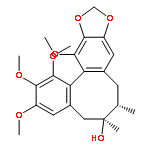
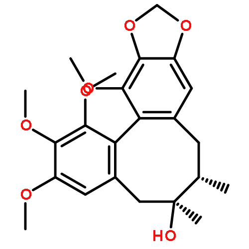
![(6S,7R)-2,3,13-Trimethoxy-6,7-dimethyl-5,6,7,8-tetrahydrobenzo[3' ,4']cycloocta[1',2':4,5]benzo[1,2-d][1,3]dioxol-1-ol](http://img.cochemist.com/ccimg/82500/82467-50-3.png)
![(6S,7R)-2,3,13-Trimethoxy-6,7-dimethyl-5,6,7,8-tetrahydrobenzo[3' ,4']cycloocta[1',2':4,5]benzo[1,2-d][1,3]dioxol-1-ol](http://img.cochemist.com/ccimg/82500/82467-50-3_b.png)

![Benzaldehyde, 4-[(4-chlorophenyl)methoxy]-2-hydroxy-](http://img.cochemist.com/ccimg/929300/929291-04-3.png)
![Benzaldehyde, 4-[(4-chlorophenyl)methoxy]-2-hydroxy-](http://img.cochemist.com/ccimg/929300/929291-04-3_b.png)


![13,14-dimethoxy-6,7-dimethyl-5,6,7,8-tetrahydro[1,3]benzodioxolo[5',6':3,4]cycloocta[1,2-f][1,3]benzodioxole (non-preferred name)](http://img.cochemist.com/ccimg/61400/61301-33-5.png)
![13,14-dimethoxy-6,7-dimethyl-5,6,7,8-tetrahydro[1,3]benzodioxolo[5',6':3,4]cycloocta[1,2-f][1,3]benzodioxole (non-preferred name)](http://img.cochemist.com/ccimg/61400/61301-33-5_b.png)


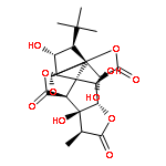
![9H-1,7a-(Epoxymethano)-1H,6aH-cyclopenta[c]furo[2,3-b]furo[3',2':3,4]cyclopenta[1,2-d]furan-5,9,12(4H)-trione,3-(1,1-dimethylethyl)hexahydro-2,4,11-trihydroxy-8-methyl-, (1S,2R,3S,3aS,4R,6aR,7aR,7bR,8S,10aS,11R,11aS)-](http://img.cochemist.com/ccimg/15300/15291-78-8.png)
![9H-1,7a-(Epoxymethano)-1H,6aH-cyclopenta[c]furo[2,3-b]furo[3',2':3,4]cyclopenta[1,2-d]furan-5,9,12(4H)-trione,3-(1,1-dimethylethyl)hexahydro-2,4,11-trihydroxy-8-methyl-, (1S,2R,3S,3aS,4R,6aR,7aR,7bR,8S,10aS,11R,11aS)-](http://img.cochemist.com/ccimg/15300/15291-78-8_b.png)


![Spiro[3H-indole-3,1'(5'H)-indolizine]-7'-aceticacid, 6'-ethenyl-1,2,2',3',6',7',8',8'a-octahydro-a-(methoxymethylene)-2-oxo-, methyl ester, (aE,1'R,6'R,7'S,8'aS)-](http://img.cochemist.com/ccimg/700/630-94-4.png)
![Spiro[3H-indole-3,1'(5'H)-indolizine]-7'-aceticacid, 6'-ethenyl-1,2,2',3',6',7',8',8'a-octahydro-a-(methoxymethylene)-2-oxo-, methyl ester, (aE,1'R,6'R,7'S,8'aS)-](http://img.cochemist.com/ccimg/700/630-94-4_b.png)
![(2s)-7-hydroxy-2-[4-[(2s,3r,4s,5s,6r)-3,4,5-trihydroxy-6-(hydroxymethyl)oxan-2-yl]oxyphenyl]-2,3-dihydrochromen-4-one](http://img.cochemist.com/ccimg/600/551-15-5.png)
![(2s)-7-hydroxy-2-[4-[(2s,3r,4s,5s,6r)-3,4,5-trihydroxy-6-(hydroxymethyl)oxan-2-yl]oxyphenyl]-2,3-dihydrochromen-4-one](http://img.cochemist.com/ccimg/600/551-15-5_b.png)

![4H-Dibenzo[de,g]quinoline-10,11-diol,5,6,6a,7-tetrahydro-6-methyl-, (6aR)-](http://img.cochemist.com/ccimg/100/58-00-4.png)
![4H-Dibenzo[de,g]quinoline-10,11-diol,5,6,6a,7-tetrahydro-6-methyl-, (6aR)-](http://img.cochemist.com/ccimg/100/58-00-4_b.png)


![3-O-[beta-D-glucuronopyranosyl-(1->2)-beta-D-glucuronopyranosyl]liquiritic acid](http://img.cochemist.com/ccimg/118500/118441-85-3.png)
![3-O-[beta-D-glucuronopyranosyl-(1->2)-beta-D-glucuronopyranosyl]liquiritic acid](http://img.cochemist.com/ccimg/118500/118441-85-3_b.png)


![(E)-3-(4-hydroxyphenyl)-1-[2-hydroxy-4-[(2S,3R,4S,5S,6R)-3,4,5-trihydroxy-6-(hydroxymethyl)tetrahydropyran-2-yl]oxy-phenyl]prop-2-en-1-one](http://img.cochemist.com/ccimg/59200/59122-93-9.png)
![(E)-3-(4-hydroxyphenyl)-1-[2-hydroxy-4-[(2S,3R,4S,5S,6R)-3,4,5-trihydroxy-6-(hydroxymethyl)tetrahydropyran-2-yl]oxy-phenyl]prop-2-en-1-one](http://img.cochemist.com/ccimg/59200/59122-93-9_b.png)

![5,7-dihydroxy-2-(4-hydroxyphenyl)-6-[(2s,3r,4r,5s,6r)-3,4,5-trihydroxy-6-(hydroxymethyl)oxan-2-yl]-8-[(2s,3r,4s,5s)-3,4,5-trihydroxyoxan-2-yl]chromen-4-one](http://img.cochemist.com/ccimg/52000/51938-32-0.png)
![5,7-dihydroxy-2-(4-hydroxyphenyl)-6-[(2s,3r,4r,5s,6r)-3,4,5-trihydroxy-6-(hydroxymethyl)oxan-2-yl]-8-[(2s,3r,4s,5s)-3,4,5-trihydroxyoxan-2-yl]chromen-4-one](http://img.cochemist.com/ccimg/52000/51938-32-0_b.png)


![2-METHYL-2-PROPANYL 4-{[(6-CHLORO-3-PYRIDAZINYL)AMINO]METHYL}-1-P<WBR />IPERIDINECARBOXYLATE](http://img.cochemist.com/ccimg/37000/36948-76-2.png)
![2-METHYL-2-PROPANYL 4-{[(6-CHLORO-3-PYRIDAZINYL)AMINO]METHYL}-1-P<WBR />IPERIDINECARBOXYLATE](http://img.cochemist.com/ccimg/37000/36948-76-2_b.png)


![(e)-1-(2,4-dihydroxyphenyl)-3-[4-[(2s,3r,4s,5s,6r)-3,4,5-trihydroxy-6-(hydroxymethyl)oxan-2-yl]oxyphenyl]prop-2-en-1-one](http://img.cochemist.com/ccimg/5100/5041-81-6.png)
![(e)-1-(2,4-dihydroxyphenyl)-3-[4-[(2s,3r,4s,5s,6r)-3,4,5-trihydroxy-6-(hydroxymethyl)oxan-2-yl]oxyphenyl]prop-2-en-1-one](http://img.cochemist.com/ccimg/5100/5041-81-6_b.png)


![1,3,5-Naphthalenetrisulfonicacid,8,8'-[carbonylbis[imino-3,1-phenylenecarbonylimino(4-methyl-3,1-phenylene)carbonylimino]]bis-](http://img.cochemist.com/ccimg/200/145-63-1.png)
![1,3,5-Naphthalenetrisulfonicacid,8,8'-[carbonylbis[imino-3,1-phenylenecarbonylimino(4-methyl-3,1-phenylene)carbonylimino]]bis-](http://img.cochemist.com/ccimg/200/145-63-1_b.png)