Co-reporter:Brendan M. Watson;Tiffany N. Vo;Paul S. Engel
Industrial & Engineering Chemistry Research October 28, 2015 Volume 54(Issue 42) pp:10206-10211
Publication Date(Web):2017-2-22
DOI:10.1021/acs.iecr.5b01388
Hydroxyapatite nanoparticles (HANPs) were suspended in injectable, biodegradable, phosphate-containing, dual-gelling macromer solutions that were used to encapsulate mesenchymal stem cells (MSCs) within stable hydrogel composites when elevated to physiologic temperature. The suspension of HANPs at 0.75%w/v within the hydrogels was found to have no significant effect on the swelling ratio or the compressive modulus. The MSCs were shown to survive the encapsulation process, and live cells were detected within the hydrogel composites for up to 28 days. The activity of osteogenic marker alkaline phosphatase increased with time in cell-laden hydrogel composites over the 28 days in osteogenic medium, suggesting that the joint combination of encapsulated MSCs within a HANP- and phosphate-containing hydrogel can enhance enzyme activity to assist in hydrogel mineralization. This is observed in the calcium biochemical assays, where the incorporation of HANPs significantly improved both cell-laden and acellular hydrogel composite mineralization over time. Hydrogel nanocomposites that form in situ while facilitating cell delivery and mineralization are promising materials for craniofacial bone tissue engineering.
Co-reporter:Yingying Du, Haoming Liu, Qin Yang, Shuai Wang, Jianglin Wang, Jun Ma, Insup Noh, Antonios G. Mikos, Shengmin Zhang
Biomaterials 2017 Volume 137(Volume 137) pp:
Publication Date(Web):1 August 2017
DOI:10.1016/j.biomaterials.2017.05.021
Osteochondral defects cannot be adequately self-repaired due to the presence of the sophisticated hierarchical structure and the lack of blood supply in cartilage. Thus, one of the major challenges remaining in this field is the structural design of a biomimetic scaffold that satisfies the specific requirements for osteochondral repair. To address this hurdle, a bio-inspired multilayer osteochondral scaffold that consisted of the poly(ε-caprolactone) (PCL) and the hydroxyapatite (HA)/PCL microspheres, was constructed via selective laser sintering (SLS) technique. The SLS-derived scaffolds exhibited an excellent biocompatibility to support cell adhesion and proliferation in vitro. The repair effect was evaluated by implanting the acellular multilayer scaffolds into osteochondral defects of a rabbit model. Our findings demonstrated that the multilayer scaffolds were able to induce articular cartilage formation by accelerating the early subchondral bone regeneration, and the newly formed tissues could well integrate with the native tissues. Consequently, the current study not only achieves osteochondral repair, but also suggests a promising strategy for the fabrication of bio-inspired multilayer scaffolds with well-designed architecture and gradient composition via SLS technique.
Co-reporter:Laura G. Bracaglia, Brandon T. Smith, Emma Watson, Navein Arumugasaamy, ... John P. Fisher
Acta Biomaterialia 2017 Volume 56(Volume 56) pp:
Publication Date(Web):1 July 2017
DOI:10.1016/j.actbio.2017.03.030
To accurately mimic the native tissue environment, tissue engineered scaffolds often need to have a highly controlled and varied display of three-dimensional (3D) architecture and geometrical cues. Additive manufacturing in tissue engineering has made possible the development of complex scaffolds that mimic the native tissue architectures. As such, architectural details that were previously unattainable or irreproducible can now be incorporated in an ordered and organized approach, further advancing the structural and chemical cues delivered to cells interacting with the scaffold. This control over the environment has given engineers the ability to unlock cellular machinery that is highly dependent upon the intricate heterogeneous environment of native tissue. Recent research into the incorporation of physical and chemical gradients within scaffolds indicates that integrating these features improves the function of a tissue engineered construct. This review covers recent advances on techniques to incorporate gradients into polymer scaffolds through additive manufacturing and evaluate the success of these techniques. As covered here, to best replicate different tissue types, one must be cognizant of the vastly different types of manufacturing techniques available to create these gradient scaffolds. We review the various types of additive manufacturing techniques that can be leveraged to fabricate scaffolds with heterogeneous properties and discuss methods to successfully characterize them.Statement of significanceAdditive manufacturing techniques have given tissue engineers the ability to precisely recapitulate the native architecture present within tissue. In addition, these techniques can be leveraged to create scaffolds with both physical and chemical gradients. This work offers insight into several techniques that can be used to generate graded scaffolds, depending on the desired gradient. Furthermore, it outlines methods to determine if the designed gradient was achieved. This review will help to condense the abundance of information that has been published on the creation and characterization of gradient scaffolds and to provide a single review discussing both methods for manufacturing gradient scaffolds and evaluating the establishment of a gradient.Download high-res image (52KB)Download full-size image
Co-reporter:Brandon T. Smith, Marco Santoro, Eline C. Grosfeld, Sarita R. Shah, ... Antonios G. Mikos.
Acta Biomaterialia 2017 Volume 50(Volume 50) pp:
Publication Date(Web):1 March 2017
DOI:10.1016/j.actbio.2016.12.024
Calcium phosphate cements (CPCs) have been extensively investigated as scaffolds in bone tissue engineering in light of their chemical composition closely resembling the mineral component of bone extracellular matrix. Yet, the degradation kinetics of many CPCs is slow compared to de novo bone formation. In order to overcome this shortcoming, the use of porogens within CPCs has been suggested as a potential strategy to increase scaffold porosity and promote surface degradation. This study explored the usage of glucose microparticles (GMPs) as porogens for the introduction of macroporosity within CPCs, and characterized the handling properties and physicochemical characteristics of CPCs containing GMPs. Samples were fabricated with four different weight fractions of GMPs (10, 20, 30, and 40%) and two different size ranges (100–150 μm and 150–300 μm), and were assayed for porosity, pore size distribution, morphology, and compressive mechanical properties. Samples were further tested for their handling properties – specifically, setting time and cohesiveness. Additionally, these same analyses were conducted on samples exposed to a physiological solution in order to estimate the dissolution kinetics of GMPs and its effect on the properties of the composite. GMPs were efficiently encapsulated and homogeneously dispersed in the resulting composite. Although setting times increased for GMP/CPC formulations compared to control CPC material, increasing the Na2HPO4 concentration in the liquid phase decreased the initial setting time to clinically acceptable values (i.e. <15 min). Incorporation of GMPs led to the formation of instant macroporosity upon cement setting, and encapsulated GMPs completely dissolved in three days, resulting in a further increase in scaffold porosity. However, the dissolution of GMPs decreased scaffold compressive strength. Overall, the introduction of GMPs into CPC resulted in macroporous scaffolds with good handling properties, as well as designer porosity and pore size distribution via selection of the appropriate size/weight fraction of GMPs. The data demonstrate that GMPs are promising porogens for the production of highly tunable porous CPC scaffolds.Statement of SignificanceCalcium phosphate cements have shown great promise for the regeneration of bone. However, macropores (>100 μm) are required for promoting bone ingrowth. Several studies have investigated methods to generate macroporosity within calcium phosphate cements but many of these methods either affect the cement setting or take weeks or months to generate the maximum porosity. This work offers a new method for generating macroporosity within calcium phosphate cements by utilizing glucose microparticles. The microparticles dissolve in less then 72 h, thereby generating scaffolds with maximum porosity in short period of time. The results will offer a new method for generating macroporosity within calcium phosphate cements.Download high-res image (89KB)Download full-size image
Co-reporter:Alexander M. Tatara, Sarita R. Shah, Nagi Demian, Tang Ho, Jonathan Shum, Jeroen J.J.P. van den Beucken, John A. Jansen, Mark E. Wong, Antonios G. Mikos
Acta Biomaterialia 2016 Volume 45() pp:72-84
Publication Date(Web):November 2016
DOI:10.1016/j.actbio.2016.09.013
Abstract
Reconstruction of large mandibular defects is clinically challenging due to the need for donor tissue of appropriate shape and volume to facilitate high fidelity repair. In order to generate large vascularized tissues of custom geometry, bioreactors were implanted against the rib periosteum of 3–4 year-old sheep for nine weeks. Bioreactors were filled with either morcellized autologous bone, synthetic ceramic particles, or a combination thereof. Tissues generated within synthetic graft-filled bioreactors were transferred into a large right-sided mandibular angle defect as either avascular grafts (n = 3) or vascularized free flaps (n = 3). After twelve additional weeks, reconstructed mandibular angles were harvested and compared to contralateral control angles. Per histologic and radiologic evaluation, a greater amount of mineralized tissue was generated in bioreactors filled with autologous graft although the quality of viable bone was not significantly different between groups. Genetic analyses of soft tissue surrounding bioreactor-generated tissues demonstrated similar early and late stage osteogenic biomarker expression (Runx2 and Osteocalcin) between the bioreactors and rib periosteum. Although no significant differences between the height of reconstructed and control mandibular angles were observed, the reconstructed mandibles had decreased bone volume. There were no differences between mandibles reconstructed with bioreactor-generated tissues transferred as flaps or grafts. Tissues used for mandibular reconstruction demonstrated integration with native bone as well as evidence of remodeling. In this study, we have demonstrated that synthetic scaffolds are sufficient to generate large volumes of mineralized tissue in an in vivo bioreactor for mandibular reconstruction.
Statement of Significance
A significant clinical challenge in craniofacial surgery is the reconstruction of large mandibular defects. In this work, we demonstrated that vascularized tissues of large volume and custom geometry can be generated from in vivo bioreactors implanted against the rib periosteum in an ovine model. The effects of different bioreactor scaffold material on tissue ingrowth were measured. To minimize donor site morbidity, tissues generated from bioreactors filled with synthetic graft were transferred as either vascularized free flaps or avascular grafts to a large mandibular defect. It was demonstrated that synthetic graft in an in vivo bioreactor is sufficient to produce free tissue bone flaps capable of integrating with native tissues when transferred to a large mandibular defect in an ovine model.
Co-reporter:P. M. Mountziaris, S. R. Shah, J. Lam, G. N. Bennett and A. G. Mikos
Biomaterials Science 2016 vol. 4(Issue 1) pp:121-129
Publication Date(Web):04 Sep 2015
DOI:10.1039/C5BM00175G
Severe injuries in the craniofacial complex, resulting from trauma or pathology, present several challenges to functional and aesthetic reconstruction. The anatomy and position of the craniofacial region make it vulnerable to injury and subsequent local infection due to external bacteria as well as those from neighbouring structures like the sinuses, nasal passages, and mouth. Porous polymethylmethacrylate (PMMA) “space maintainers” have proven useful in staged craniofacial reconstruction by promoting healing of overlying soft tissue prior to reconstruction of craniofacial bones. We describe herein a method by which the porosity of a prefabricated porous PMMA space maintainer, generated by porogen leaching, can be loaded with a thermogelling copolymer-based drug delivery system. Porogen leaching, space maintainer prewetting, and thermogel loading all significantly affected the loading of a model antibiotic, colistin. Weeks-long release of antibiotic at clinically relevant levels was achieved with several formulations. In vitro assays confirmed that the released colistin maintained its antibiotic activity against several bacterial targets. Our results suggest that this method is a valuable tool in the development of novel therapeutic approaches for the treatment of severe complex, infected craniofacial injuries.
Co-reporter:T.N. Vo, S.R. Shah, S. Lu, A.M. Tatara, E.J. Lee, T.T. Roh, Y. Tabata, A.G. Mikos
Biomaterials 2016 83() pp: 1-11
Publication Date(Web):1 March 2016
DOI:10.1016/j.biomaterials.2015.12.026
The present work investigated the osteogenic potential of injectable, dual thermally and chemically gelable composite hydrogels for mesenchymal stem cell (MSC) delivery in vitro and in vivo. Composite hydrogels comprising copolymer macromers of N-isopropylacrylamide were fabricated through the incorporation of gelatin microparticles (GMPs) as enzymatically digestible porogens and sites for cellular attachment. High and low polymer content hydrogels with and without GMP loading were shown to successfully encapsulate viable MSCs and maintain their survival over 28 days in vitro. GMP incorporation was also shown to modulate alkaline phosphatase production, but enhanced hydrogel mineralization along with higher polymer content even in the absence of cells. Moreover, the regenerative capacity of 2 mm thick hydrogels with GMPs only, MSCs only, or GMPs and MSCs was evaluated in vivo in an 8 mm rat critical size cranial defect for 4 and 12 weeks. GMP incorporation led to enhanced bony bridging and mineralization within the defect at each timepoint, and direct bone-implant contact as determined by microcomputed tomography and histological scoring, respectively. Encapsulation of both GMPs and MSCs enabled hydrogel degradation leading to significant tissue infiltration and osteoid formation. The results suggest that these injectable, dual-gelling cell-laden composite hydrogels can facilitate bone ingrowth and integration, warranting further investigation for bone tissue engineering.
Co-reporter:Sarita R. Shah, Alexander M. Tatara, Johnny Lam, Steven Lu, David W. Scott, George N. Bennett, Jeroen J. J. P. van den Beucken, John A. Jansen, Mark E. Wong, and Antonios G. Mikos
ACS Biomaterials Science & Engineering 2016 Volume 2(Issue 4) pp:558
Publication Date(Web):March 14, 2016
DOI:10.1021/acsbiomaterials.5b00545
Antibiotic-releasing porous poly(methyl methacrylate) (PMMA) space maintainers, comprising PMMA with an aqueous porogen and a poly(DL-lactic-co-glycolic acid) (PLGA) antibiotic carrier, have been developed to facilitate local delivery of antibiotics and tissue integration. In this study, clindamycin-loaded space maintainers were used to investigate the effects of antibiotic release kinetics and dose upon bacterial clearance and bone and soft tissue healing in a pathogen-contaminated rabbit mandibular defect. Three formulations were fabricated for either high dose burst release (7 days) or with PLGA microparticles for extended release (28 days) at high and low dose. Although inoculated bacteria were not recovered from any specimens, the burst release formulation showed less inflammation and fibrous capsule formation and more bone formation close to the implant than the low dose extended release formulation by histologic analysis. These results suggest that local antibiotic release kinetics and dose affect soft and hard tissue healing independent from its ability to clear bacteria.Keywords: controlled antibiotic release; odontogenic infection; osteomyelitis; PLGA; PMMA
Co-reporter:Jordan E. Trachtenberg, Jesse K. Placone, Brandon T. Smith, Charlotte M. Piard, Marco Santoro, David W. Scott, John P. Fisher, and Antonios G. Mikos
ACS Biomaterials Science & Engineering 2016 Volume 2(Issue 10) pp:1771
Publication Date(Web):March 3, 2016
DOI:10.1021/acsbiomaterials.6b00026
3D printing has emerged as an important technique for fabricating tissue engineered scaffolds. However, systematic evaluations of biomaterials for 3D printing have not been widely investigated. We evaluated poly(propylene fumarate) (PPF) as a model material for extrusion-based printing applications. A full-factorial design evaluating the effects of four factors (PPF concentration, printing pressure, printing speed, and programmed fiber spacing) on viscosity, fiber diameter, and pore size was performed layer-by-layer on 3D scaffolds. We developed a linear model of printing solution viscosity, where concentration of PPF had the greatest effect on viscosity, and the polymer exhibited shear thinning behavior. Additionally, linear models of pore size and fiber diameter revealed that fiber spacing and pressure had the greatest effect on pore size and fiber diameter, respectively, but interplay among the factors also influenced scaffold architecture. This study serves as a platform to determine if novel biomaterials are suitable for extrusion-based 3D printing applications.Keywords: extrusion; fused deposition modeling; layer analysis; poly(propylene fumarate); rapid prototyping; viscosity
Co-reporter:Steven Lu;Esther J. Lee;Johnny Lam;Yasuhiko Tabata
Annals of Biomedical Engineering 2016 Volume 44( Issue 6) pp:1894-1907
Publication Date(Web):2016 June
DOI:10.1007/s10439-016-1582-x
Due to the lack of cell-adhesive moieties in traditional synthetic hydrogels, the present work investigated the use of degradable gelatin microparticles (GMPs) as temporary adherent substrates for anchorage-dependent mesenchymal stem cells (MSCs). MSCs were seeded onto GMPs of varying crosslinking densities and sizes to investigate their role on influencing MSC differentiation and aggregation. The MSC-seeded GMPs were then encapsulated in poly(ethylene glycol)-based hydrogels and cultured in serum-free, growth factor-free osteochondral medium. Non-seeded MSCs co-encapsulated with GMPs in the hydrogels were used as a control for comparison. Over the course of 35 days, MSCs seeded on GMPs exhibited more cell–cell contacts, greater chondrogenic potential, and a down-regulation of osteogenic markers compared to the controls. Although the factors of GMP crosslinking and size had nominal influence on MSC differentiation and aggregation, GMPs demonstrate potential as an adherent-substrate for improving cell delivery from hydrogel scaffolds by facilitating cell–cell contacts and improving MSC differentiation.
Co-reporter:Eric R. Molina, Brandon T. Smith, Sarita R. Shah, Heungsoo Shin, Antonios G. Mikos
Journal of Controlled Release 2015 Volume 219() pp:107-118
Publication Date(Web):10 December 2015
DOI:10.1016/j.jconrel.2015.08.038
The immune system plays a crucial role in the success of tissue engineering strategies. Failure to consider the interactions between implantable scaffolds, usually containing cells and/or bioactive molecules, and the immune system can result in rejection of the implant and devastating clinical consequences. However, recent research into mesenchymal stem cells, which are commonly used in many tissue engineering applications, indicates that they may play a beneficial role modulating the immune system. Likewise, direct delivery of bioactive molecules involved in the inflammatory process can promote the success of tissue engineering constructs. In this article, we will review the various mechanisms in which modulation of the immune system is achieved through delivered bioactive molecules and cells and contextualize this information for future strategies in tissue engineering.
Co-reporter:Tiffany N. Vo, Adam K. Ekenseair, Patrick P. Spicer, Brendan M. Watson, Stephanie N. Tzouanas, Terrence T. Roh, Antonios G. Mikos
Journal of Controlled Release 2015 Volume 205() pp:25-34
Publication Date(Web):10 May 2015
DOI:10.1016/j.jconrel.2014.11.028
In this study, we investigated the mineralization capacity and biocompatibility of injectable, dual-gelling hydrogels in a rat cranial defect as a function of hydrogel hydrophobicity from either the copolymerization of a hydrolyzable lactone ring or the hydrogel polymer content. The hydrogel system comprised a poly(N-isopropylacrylamide)-based thermogelling macromer (TGM) and a polyamidoamine crosslinker. The thermogelling macromer was copolymerized with (TGM/DBA) or without (TGM) a dimethyl-γ-butyrolactone acrylate (DBA)-containing lactone ring that modulated the lower critical solution temperature and thus, the hydrogel hydrophobicity, over time. Three hydrogel groups were examined: (1) 15 wt.% TGM, (2) 15 wt.% TGM/DBA, and (3) 20 wt.% TGM/DBA. The hydrogels were implanted within an 8 mm critical size rat cranial defect for 4 and 12 weeks. Implants were harvested at each timepoint and analyzed for bone formation, hydrogel mineralization and tissue response using microcomputed tomography (microCT). Histology and fibrous capsule scoring showed a light inflammatory response at 4 weeks that was mitigated by 12 weeks for all groups. MicroCT scoring and bone volume quantification demonstrated a similar bone formation at 4 weeks that was significantly increased for the more hydrophobic hydrogel formulations – 15 wt.% TGM and 20 wt.% TGM/DBA – from 4 weeks to 12 weeks. A complementary in vitro acellular mineralization study revealed that the hydrogels exhibited calcium binding properties in the presence of serum-containing media, which was modulated by the hydrogel hydrophobicity. The tailored mineralization capacity of these injectable, dual-gelling hydrogels with hydrolysis-dependent hydrophobicity presents an exciting property for their use in bone tissue engineering applications.
Co-reporter:Allan M. Henslee;Diana M. Yoon;Benjamin Y. Lu;Joseph Yu;Andrew A. Arango;Liann P. Marruffo;Luke Seng;Tamir D. Anver;Hunaiza Ather;Manitha B. Nair;Sean O. Piper;Nagi Demian;Mark E. K. Wong;F. Kurtis Kasper
Journal of Biomedical Materials Research Part B: Applied Biomaterials 2015 Volume 103( Issue 3) pp:529-538
Publication Date(Web):
DOI:10.1002/jbm.b.33216
Abstract
This study investigated the use of injectable poly(propylene fumarate) (PPF) formulations for mandibular fracture stabilization applications. A full factorial design with main effects analysis was employed to evaluate the effects of the PPF:N-vinyl pyrrolidone (NVP, crosslinking agent) ratio and dimethyl toluidine (DMT, accelerator) concentration on key physicochemical properties including setting time, maximum temperature, mechanical properties, sol fraction, and swelling ratio. Additionally, the effects of formulation crosslinking time on the mechanical and swelling properties were investigated. The results showed that increasing the PPF:NVP ratio from 3:1 to 4:1 or decreasing the DMT concentration from 0.05 to 0.01 v/w % significantly decreased all mechanical properties as well as significantly increased the sol fraction and swelling ratio. Also, increasing the crosslinking time at 37°C from 1 to 7 days significantly increased all mechanical properties and decreased both the sol fraction and swelling ratio. This study further showed that the flexural stiffness of ex vivo stabilized rabbit mandibles increased from 1.7 ± 0.3 N/mm with a traditional mini-plate fixator to 14.5 ± 4.1 N/mm for the 4:1 (0.05 v/w % DMT) PPF formulation at day 1. Overall, the formulations tested in this study were found to have properties suitable for potential further consideration in mandibular fracture fixation applications. © 2014 Wiley Periodicals, Inc. J Biomed Mater Res Part B: Appl Biomater, 103B: 529–538, 2015.
Co-reporter:Sajeesh Kumar Madhurakkat Perikamana, Jinkyu Lee, Yu Bin Lee, Young Min Shin, Esther J. Lee, Antonios G. Mikos, and Heungsoo Shin
Biomacromolecules 2015 Volume 16(Issue 9) pp:
Publication Date(Web):August 17, 2015
DOI:10.1021/acs.biomac.5b00852
Current advances in biomaterial fabrication techniques have broadened their application in different realms of biomedical engineering, spanning from drug delivery to tissue engineering. The success of biomaterials depends highly on the ability to modulate cell and tissue responses, including cell adhesion, as well as induction of repair and immune processes. Thus, most recent approaches in the field have concentrated on functionalizing biomaterials with different biomolecules intended to evoke cell- and tissue-specific reactions. Marine mussels produce mussel adhesive proteins (MAPs), which help them strongly attach to different surfaces, even under wet conditions in the ocean. Inspired by mussel adhesiveness, scientists discovered that dopamine undergoes self-polymerization at alkaline conditions. This reaction provides a universal coating for metals, polymers, and ceramics, regardless of their chemical and physical properties. Furthermore, this polymerized layer is enriched with catechol groups that enable immobilization of primary amine or thiol-based biomolecules via a simple dipping process. Herein, this review explores the versatile surface modification techniques that have recently been exploited in tissue engineering and summarizes polydopamine polymerization mechanisms, coating process parameters, and effects on substrate properties. A brief discussion of polydopamine-based reactions in the context of engineering various tissue types, including bone, blood vessels, cartilage, nerves, and muscle, is also provided.
Co-reporter:Jordan E. Trachtenberg;Tiffany N. Vo
Annals of Biomedical Engineering 2015 Volume 43( Issue 3) pp:681-696
Publication Date(Web):2015 March
DOI:10.1007/s10439-014-1151-0
Pre-clinical animal models play a crucial role in the translation of biomedical technologies from the bench top to the bedside. However, there is a need for improved techniques to evaluate implanted biomaterials within the host, including consideration of the care and ethics associated with animal studies, as well as the evaluation of host tissue repair in a clinically relevant manner. This review discusses non-invasive, quantitative, and real-time techniques for evaluating host-materials interactions, quality and rate of neotissue formation, and functional outcomes of implanted biomaterials for bone and cartilage tissue engineering. Specifically, a comparison will be presented for pre-clinical animal models, histological scoring systems, and non-invasive imaging modalities. Additionally, novel technologies to track delivered cells and growth factors will be discussed, including methods to directly correlate their release with tissue growth.
Co-reporter:Brian A. Menegaz;Joseph A. Ludwig;Marco Santoro;Salah-Eddine Lamhamedi-Cherradi
PNAS 2015 Volume 112 (Issue 33 ) pp:10304-10309
Publication Date(Web):2015-08-18
DOI:10.1073/pnas.1506684112
Three-dimensional tumor models accurately describe different aspects of the tumor microenvironment and are readily available
for mechanistic studies of tumor biology and for drug screening. Nevertheless, these systems often overlook biomechanical
stimulation, another fundamental driver of tumor progression. To address this issue, we cultured Ewing sarcoma (ES) cells
on electrospun poly(ε-caprolactone) 3D scaffolds within a flow perfusion bioreactor. Flow-derived shear stress provided a
physiologically relevant mechanical stimulation that significantly promoted insulin-like growth factor-1 (IGF1) production
and elicited a superadditive release in the presence of exogenous IGF1. This finding is particularly relevant, given the central
role of the IGF1/IGF-1 receptor (IGF-1R) pathway in ES tumorigenesis and as a promising clinical target. Additionally, flow
perfusion enhanced in a rate-dependent manner the sensitivity of ES cells to IGF-1R inhibitor dalotuzumab (MK-0646) and showed
shear stress-dependent resistance to the IGF-1R blockade. This study demonstrates shear stress-dependent ES cell sensitivity
to dalotuzumab, highlighting the importance of biomechanical stimulation on ES-acquired drug resistance to IGF-1R inhibition.
Furthermore, flow perfusion increased nutrient supply throughout the scaffold, enriching ES culture over static conditions.
Our use of a tissue-engineered model, rather than human tumors or xenografts, enabled precise control of the forces experienced
by ES cells, and therefore provided at least one explanation for the remarkable antineoplastic effects observed by some ES
tumor patients from IGF-1R targeted therapies, in contrast to the lackluster effect observed in cells grown in conventional
monolayer culture.
Co-reporter:Lucas A. Kinard, Rebecca L. Dahlin, Johnny Lam, Steven Lu, Esther J. Lee, F. Kurtis Kasper, Antonios G. Mikos
Acta Biomaterialia 2014 Volume 10(Issue 11) pp:4574-4582
Publication Date(Web):November 2014
DOI:10.1016/j.actbio.2014.07.011
Abstract
There exists a strong clinical need for a more capable and robust method to achieve bone augmentation, and a system with fine-tuned delivery of demineralized bone matrix (DBM) has the potential to meet that need. As such, the objective of the present study was to investigate a synthetic biodegradable hydrogel for the delivery of DBM for bone augmentation in a rat model. Oligo(poly(ethylene glycol) fumarate) (OPF) constructs were designed and fabricated by varying the content of rat-derived DBM particles (either 1:3, 1:1 or 3:1 DBM:OPF weight ratio on a dry basis) and using two DBM particle size ranges (50–150 or 150–250 μm). The physical properties of the constructs and the bioactivity of the DBM were evaluated. Selected formulations (1:1 and 3:1 with 50–150 μm DBM) were evaluated in vivo compared to an empty control to investigate the effect of DBM dose and construct properties on bone augmentation. Overall, 3:1 constructs with higher DBM content achieved the greatest volume of bone augmentation, exceeding 1:1 constructs and empty implants by 3- and 5-fold, respectively. As such, we have established that a synthetic, biodegradable hydrogel can function as a carrier for DBM, and that the volume of bone augmentation achieved by the constructs correlates directly to the DBM dose.
Co-reporter:Clark J. Needham, Sarita R. Shah, Rebecca L. Dahlin, Lucas A. Kinard, Johnny Lam, Brendan M. Watson, Steven Lu, F. Kurtis Kasper, Antonios G. Mikos
Acta Biomaterialia 2014 Volume 10(Issue 10) pp:4103-4112
Publication Date(Web):October 2014
DOI:10.1016/j.actbio.2014.05.011
Abstract
Native osteochondral repair is often inadequate owing to the inherent properties of the tissue, and current clinical repair strategies can result in healing with a limited lifespan and donor site morbidity. This work investigates the use of polymeric gene therapy to address this problem by delivering DNA encoding for transcription factors complexed with the branched poly(ethylenimine)–hyaluronic acid (bPEI–HA) delivery vector via a porous oligo[poly(ethylene glycol) fumarate] hydrogel scaffold. To evaluate the potential of this approach, a bilayered scaffold mimicking native osteochondral tissue organization was loaded with DNA/bPEI–HA complexes. Next, bilayered implants either unloaded or loaded in a spatial fashion with bPEI–HA and DNA encoding for either Runt-related transcription factor 2 (RUNX2) or SRY (sex determining region Y)-box 5, 6, and 9 (the SOX trio), to generate bone and cartilage tissues respectively, were fabricated and implanted in a rat osteochondral defect. At 6 weeks post-implantation, micro-computed tomography analysis and histological scoring were performed on the explants to evaluate the quality and quantity of tissue repair in each group. The incorporation of DNA encoding for RUNX2 in the bone layer of these scaffolds significantly increased bone growth. Additionally, a spatially loaded combination of RUNX2 and SOX trio DNA loading significantly improved healing relative to empty hydrogels or either factor alone. Finally, the results of this study suggest that subchondral bone formation is necessary for correct cartilage healing.
Co-reporter:Erica J. Levorson, Marco Santoro, F. Kurtis Kasper, Antonios G. Mikos
Acta Biomaterialia 2014 Volume 10(Issue 5) pp:1824-1835
Publication Date(Web):May 2014
DOI:10.1016/j.actbio.2013.12.026
Abstract
In this work, the influence of direct cell–cell contact in co-cultures of mesenchymal stem cells (MSCs) and chondrocytes for the improved deposition of cartilage-like extracellular matrix (ECM) within nonwoven fibrous poly(∊-caprolactone) (PCL) scaffolds was examined. To this end, chondrocytes and MSCs were either co-cultured in direct contact by mixing on a single PCL scaffold or produced via indirect co-culture, whereby the two cell types were seeded on separate scaffolds which were then cultured together in the same system either statically or under media perfusion in a bioreactor. In static cultures, the chondrocyte scaffold of an indirectly co-cultured group generated significantly greater amounts of glycosaminoglycan and collagen than the direct co-culture group initially seeded with the same number of chondrocytes. Furthermore, improved ECM production was linked to greater cellular proliferation and distribution throughout the scaffold in static culture. In perfusion cultures, flow had a significant effect on the proliferation of the chondrocytes. The ECM contents within the chondrocyte-containing scaffolds of the indirect co-culture groups either approximated or surpassed the amounts generated within the direct co-culture group. Additionally, within bioreactor culture there were indications that chondrocytes had an influence on the chondrogenesis of MSCs as evidenced by increases in cartilaginous ECM synthetic capacity. This work demonstrates that it is possible to generate PCL/ECM hybrid scaffolds for cartilage regeneration by utilizing the factors secreted by two different cell types, chondrocytes and MSCs, even in the absence of juxtacrine signaling.
Co-reporter:Stephanie N. Tzouanas;Adam K. Ekenseair;F. Kurtis Kasper
Journal of Biomedical Materials Research Part A 2014 Volume 102( Issue 5) pp:1222-1230
Publication Date(Web):
DOI:10.1002/jbm.a.35093
Abstract
In this work, we investigated the viability and osteogenic differentiation of mesenchymal stem cells encapsulated with gelatin microparticles (GMPs) in an injectable, chemically and thermally gelling hydrogel system combining poly(N-isopropylacrylamide)-based thermogelling macromers containing pendant epoxy rings with polyamidoamine-based hydrophilic and degradable diamine crosslinking macromers. Specifically, we studied how the parameters of GMP size and loading ratio affected the viability and differentiation of cells encapsulated within the hydrogel. We also examined the effects of cell and GMP co-encapsulation on hydrogel mineralization. Cells demonstrated long-term viability within the hydrogels, which was shown to depend on GMP size and loading ratio. In particular, increased interaction of cells and GMPs through greater available GMP surface area, use of an epoxy-based chemical gelation mechanism, and the tunable high water content of the thermogelled hydrogels led to favorable long-term cell viability. Compared with cellular hydrogels without GMPs, hydrogels co-encapsulating cells and GMPs demonstrated greater production of alkaline phosphatase by cells at all time-points and a transient early enhancement of hydrogel mineralization for larger GMPs at higher loading ratios. Such injectable, in situ forming hydrogels capable of delivering and maintaining populations of encapsulated mesenchymal stem cells and promoting mineralization in vitro offer promise as novel therapies for applications in tissue engineering and regenerative medicine. © 2014 Wiley Periodicals, Inc. J Biomed Mater Res Part A: 102A: 1222–1230, 2014.
Co-reporter:Lucas A. Kinard;Rebecca L. Dahlin;Allan M. Henslee;Patrick P. Spicer;Chia-Ye Chu;Yasuhiko Tabata;Jeroen J.J.P. van den Beucken;John A. Jansen;Simon Young;Mark E. Wong;F. Kurtis Kasper
Journal of Biomedical Materials Research Part A 2014 Volume 102( Issue 7) pp:2079-2088
Publication Date(Web):
DOI:10.1002/jbm.a.34878
Abstract
The objective of the present study was to develop a preclinical animal model for evaluating bone augmentation and to examine the level of bone augmentation induced by hydrogel composites. Design criteria outlined for the development of the animal model included rigid immobilization of bilateral implants apposed to the parietal bone of the rat, while avoiding the calvarial sutures. The animal model was evaluated through the implantation of hydrogel composites of oligo(poly(ethylene glycol) fumarate) (OPF) and gelatin microparticles releasing bone morphogenetic protein-2 (BMP-2). The BMP-2 release profile was varied and compared to the implantation of a material control without BMP-2. Each hydrogel composite was implanted within a polypropylene cassette, which was immobilized to the calvarial bone using screws, and empty cassettes were implanted as a control. The design criteria for the animal model were realized; however, the level of bone augmentation did not vary between any of the groups after 4 weeks. Osteoclastic bone resorption occurred to a higher extent in groups releasing BMP-2, but the cause could not be elucidated. In conclusion, a promising bone augmentation model was established in the rat; however, refinement of the hydrogel composites was suggested to optimize the constructs for bone augmentation applications. © 2013 Wiley Periodicals, Inc. J Biomed Mater Res Part A: 102A: 2079–2088, 2014.
Co-reporter:Jordan E. Trachtenberg;Paschalia M. Mountziaris;Jordan S. Miller;Matthew Wettergreen;Fred K. Kasper
Journal of Biomedical Materials Research Part A 2014 Volume 102( Issue 12) pp:4326-4335
Publication Date(Web):
DOI:10.1002/jbm.a.35108
Abstract
The fabrication of scaffolds for tissue engineering requires elements of customization depending on the application and is often limited due to the flexibility of the processing technique. This investigation seeks to address this obstacle by utilizing an open-source three-dimensional printing (3DP) system that allows vast customizability and facilitates reproduction of experiments. The effects of processing parameters on printed poly(ε-caprolactone) scaffolds with uniform and gradient pore architectures have been characterized with respect to fiber and pore morphology and mechanical properties. The results demonstrate the ability to tailor the fiber diameter, pore size, and porosity through modification of pressure, printing speed, and programmed fiber spacing. A model was also used to predict the compressive mechanical properties of uniform and gradient scaffolds, and it was found that modulus and yield strength declined with increasing porosity. The use of open-source 3DP technologies for printing tissue-engineering scaffolds provides a flexible system that can be readily modified at a low cost and is supported by community documentation. In this manner, the 3DP system is more accessible to the scientific community, which further facilitates the translation of these technologies toward successful tissue-engineering strategies. © 2014 Wiley Periodicals, Inc. J Biomed Mater Res Part A: 102A: 4326–4335, 2014.
Co-reporter:Brendan M. Watson, F. Kurtis Kasper, Paul S. Engel, and Antonios G. Mikos
Biomacromolecules 2014 Volume 15(Issue 5) pp:
Publication Date(Web):April 23, 2014
DOI:10.1021/bm500175e
Novel, injectable, biodegradable macromer solutions that form hydrogels when elevated to physiologic temperature via a dual chemical and thermo-gelation were fabricated and characterized. A thermogelling, poly(N-isopropylacrylamide)-based macromer with pendant phosphate groups was synthesized and subsequently functionalized with chemically cross-linkable methacrylate groups via degradable phosphate ester bonds, yielding a dual-gelling macromer. These dual-gelling macromers were tuned to have transition temperatures between room temperature and physiologic temperature, allowing them to undergo instantaneous thermogelation as well as chemical gelation when elevated to physiologic temperature. Additionally, the chemical cross-linking of the hydrogels was shown to mitigate hydrogel syneresis, which commonly occurs when thermogelling materials are raised above their transition temperature. Finally, degradation of the phosphate ester bonds of the cross-linked hydrogels yielded macromers that were soluble at physiologic temperature. Further characterization of the hydrogels demonstrated minimal cytotoxicity of hydrogel leachables as well as in vitro calcification, making these novel, injectable macromers promising materials for use in bone tissue engineering.
Co-reporter:Tiffany N. Vo, Adam K. Ekenseair, F. Kurtis Kasper, and Antonios G. Mikos
Biomacromolecules 2014 Volume 15(Issue 1) pp:
Publication Date(Web):December 10, 2013
DOI:10.1021/bm401413c
Injectable, dual-gelling hydrogels were successfully developed through the combination of physical thermogellation at 37 °C and favorable amine:epoxy chemical cross-linking. Poly(N-isopropylacrylamide)-based thermogelling macromers with a hydrolyzable lactone ring and epoxy pendant groups and a biodegradable diamine-functionalized polyamidoamine cross-linker were synthesized, characterized, and combined to produce nonsyneresing and bioresorbable hydrogels. Differential scanning calorimetry and oscillatory rheometry demonstrated the rapid and dual-gelling nature of the hydrogel formation. The postgelation dimensional stability, swelling, and mechanical behavior of the hydrogel system were shown to be easily tuned in the synthesis and formulation stages. The leachable products were found to be cytocompatible under all conditions, while the degradation products demonstrated a dose- and time-dependent response due to solution osmolality. Preliminary encapsulation studies showed mesenchymal stem cell viability could be maintained for 7 days. The results suggest that injectable and thermally and chemically cross-linkable hydrogels are promising alternatives to prefabricated biomaterials for tissue engineering applications, particularly for cell delivery.
Co-reporter:Rebecca L. Dahlin, Lucas A. Kinard, Johnny Lam, Clark J. Needham, Steven Lu, F. Kurtis Kasper, Antonios G. Mikos
Biomaterials 2014 35(26) pp: 7460-7469
Publication Date(Web):
DOI:10.1016/j.biomaterials.2014.05.055
Co-reporter:Steven Lu, Johnny Lam, Jordan E. Trachtenberg, Esther J. Lee, Hajar Seyednejad, Jeroen J.J.P. van den Beucken, Yasuhiko Tabata, Mark E. Wong, John A. Jansen, Antonios G. Mikos, F. Kurtis Kasper
Biomaterials 2014 35(31) pp: 8829-8839
Publication Date(Web):
DOI:10.1016/j.biomaterials.2014.07.006
Co-reporter:Rebecca L. Dahlin, Mengwei Ni, Ville V. Meretoja, F. Kurtis Kasper, Antonios G. Mikos
Biomaterials 2014 35(1) pp: 123-132
Publication Date(Web):
DOI:10.1016/j.biomaterials.2013.09.086
Co-reporter:Rebecca L. Dahlin;Julia G. Gershovich
Annals of Biomedical Engineering 2014 Volume 42( Issue 7) pp:1381-1390
Publication Date(Web):2014 July
DOI:10.1007/s10439-013-0862-y
In this study, we investigated the effect of flow perfusion culture on the mineralization of co-cultures of human umbilical vein endothelial cells (HUVECs) and human mesenchymal stem cells (hMSCs). Osteogenically precultured hMSCs were seeded onto electrospun scaffolds in monoculture or a 1:1 ratio with HUVECs, cultured for 7 or 14 days in osteogenic medium under static or flow perfusion conditions, and the resulting constructs were analyzed for cellularity, alkaline phosphatase (ALP) activity and calcium content. In flow perfusion, constructs with monocultures of hMSCs demonstrated higher cellularity and calcium content, but lower ALP activity compared to corresponding static controls. ALP activity was enhanced in co-cultures under flow perfusion conditions, compared to hMSCs alone; however unlike the static controls, the calcium content of the co-cultures in flow perfusion was not different from the corresponding hMSC monocultures. The data suggest that co-cultures of hMSCs and HUVECs did not contribute to enhanced mineralization compared to hMSCs alone under the flow perfusion conditions investigated in this study. However, flow perfusion culture resulted in an enhanced spatial distribution of cells and matrix compared to static cultures, which were limited to a thin surface layer.
Co-reporter:Clark J. Needham;Sarita R. Shah;Paschalia M. Mountziaris
Pharmaceutical Research 2014 Volume 31( Issue 1) pp:77-85
Publication Date(Web):2014 January
DOI:10.1007/s11095-013-1133-1
This work investigates the effects of hyaluronic acid (HA) conjugated onto branched poly(ethylenimine) (bPEI) and varying loading concentrations of these polymers complexed with DNA on their release from poly(DL-lactic-co-glycolic acid) (PLGA) microparticles and the transfection of target cells.To examine the effect of alteration of the gene delivery polymer on the system, we observed the morphology, size, loading efficiency, polymer and DNA release, and the transfection efficiency for the microparticles formed with three internal phase loading concentrations during microparticle formation.Addition of HA to this vector allowed for increased loading concentration within these systems and significantly altered release kinetics without changing the morphology of the particles. The incorporation of HA onto the bPEI backbone significantly increased the transfection efficiency of the complexes released from the corresponding microparticle formulation.The results show that the modification of bPEI with HA and the concentration of loaded polymer/DNA complexes can significantly alter the entrapment and release profiles from PLGA microparticles. This is significant in that it offers insight into the effects of modification of gene delivery vectors on a controlled release system designed to achieve a sustained therapeutic response.
Co-reporter:Esther J. Lee;F. Kurtis Kasper
Annals of Biomedical Engineering 2014 Volume 42( Issue 2) pp:323-337
Publication Date(Web):2014 February
DOI:10.1007/s10439-013-0859-6
Biomaterials serve as an integral component of tissue engineering. They are designed to provide architectural framework reminiscent of native extracellular matrix in order to encourage cell growth and eventual tissue regeneration. Bone and cartilage represent two distinct tissues with varying compositional and mechanical properties. Despite these differences, both meet at the osteochondral interface. This article presents an overview of current biomaterials employed in bone and cartilage applications, discusses some design considerations, and alludes to future prospects within this field of research.
Co-reporter:Sarita R. Shah;Allan M. Henslee;Patrick P. Spicer;Shun Yokota
Pharmaceutical Research 2014 Volume 31( Issue 12) pp:3379-3389
Publication Date(Web):2014 December
DOI:10.1007/s11095-014-1427-y
This study investigated the effects of the physicochemical properties of antibiotics on the morphology, loading efficiency, size, release kinetics, and antibiotic efficacy of loaded poly(DL-lactic-co-glycolic acid) (PLGA) microparticles (MPs) at different loading percentages.Cefazolin, ciprofloxacin, clindamycin, colistin, doxycycline, and vancomycin were loaded at 10 and 20 wt% into PLGA MPs using a water-in-oil-in water double emulsion fabrication protocol. Microparticle morphology, size, loading efficiency, release kinetics, and antibiotic efficacy were assessed.The results from this study demonstrate that the chemical nature of loaded antibiotics, especially charge and molecular weight, influence the incorporation into and release of antibiotics from PLGA MPs. Drugs with molecular weights less than 600 Da displayed biphasic release while those with molecular weights greater than 1,000 Da displayed triphasic release kinetics. Large molecular weight drugs also had a longer delay before release than smaller molecular weight drugs. The negatively charged antibiotic cefazolin had lower loading efficiency than positively charged antibiotics. Microparticle size appeared to be mainly controlled by fabrication parameters, and partition and solubility coefficients did not appear to have an obvious effect on loading efficiency or release. Released antibiotics maintained their efficacy against susceptible strains over the duration of release. Duration of release varied between 17 and 49 days based on the type of antibiotic loaded.The data from this study indicate that the chemical nature of antibiotics affects properties of antibiotic-loaded PLGA MPs and allows for general prediction of loading and release kinetics.
Co-reporter:Jordan E. Trachtenberg;Paschalia M. Mountziaris;F. Kurtis Kasper
Israel Journal of Chemistry 2013 Volume 53( Issue 9-10) pp:646-654
Publication Date(Web):
DOI:10.1002/ijch.201300051
Abstract
Drug delivery through tissue-engineered scaffolds provides a composite approach to address the regenerative limitations of simple material implantation, providing expanded avenues for therapeutic tissue-repair strategies in the clinic. Both nano- and microfibrous scaffolds generated by a variety of techniques have been investigated for their potential in drug-delivery applications. While nanofibers mimic the structure and organization of natural extracellular matrix, microfibers provide more sustained release of drugs, larger pores to facilitate cell infiltration, and improved mechanical support. Various methods exist to embed drugs within the fiber matrix to modulate the release kinetics specific to the tissue-engineering application. The current article reviews the established and emerging fabrication methods for drug-loaded fiber-based scaffolds and addresses how further combination into composite scaffolds can enhance drug delivery and tissue regeneration.
Co-reporter:Limin Wang;Diana M. Yoon;Patrick P. Spicer;Allan M. Henslee;David W. Scott;Mark E. Wong;F. Kurtis Kasper
Journal of Biomedical Materials Research Part B: Applied Biomaterials 2013 Volume 101B( Issue 5) pp:813-825
Publication Date(Web):
DOI:10.1002/jbm.b.32885
Abstract
Porous polymethylmethacrylate (PMMA) has been used as an alloplastic bone substitute in the craniofacial complex, showing integration with the surrounding soft and hard tissue. This study investigated the physicochemical properties of curing and cured mixtures of a PMMA-based bone cement and a carboxymethylcellulose (CMC) gel porogen. Four formulations yielding porous PMMA of varied porosity were examined; specifically, two groups containing 30% (w/w) CMC gel in the mixture using a 7% (w/v) or 9% (w/v) stock CMC gel (30-7 and 30-9, respectively) and two groups containing 40% (w/w) CMC gel (40-7 and 40-9). An additional group comprising solid PMMA without CMC was investigated. The incorporation of the CMC gel into the PMMA bone cement during polymerization decreased the setting time from 608 ± 12 s for the solid PMMA to 427 ± 10 s for the 40-9 group, and decreased the maximum temperature from 81 ± 4°C for the solid PMMA to 38 ± 2°C for the 40-9 group. The porous PMMA groups exhibited reduced compressive strength and bending modulus and strength relative to the solid PMMA. All the porous PMMA formulations released more unconverted methylmethacrylate (MMA) monomer and N,N-dimethyl-p-toluidine (DMT) from cured specimens and less MMA and DMT from curing specimens than the solid PMMA. The data suggest that the physicochemical properties of the porous PMMA formulations are appropriate for their application in craniofacial space maintenance. © 2013 Wiley Periodicals, Inc. J Biomed Mater Res Part B: Appl Biomater, 2013.
Co-reporter:Ville V. Meretoja, Rebecca L. Dahlin, Sarah Wright, F. Kurtis Kasper, Antonios G. Mikos
Biomaterials 2013 34(17) pp: 4266-4273
Publication Date(Web):
DOI:10.1016/j.biomaterials.2013.02.064
Co-reporter:Eliza Li Shan Fong;Emily Burdett;Alexander J. Lazar;Vandhana Ramamoorthy;Elizabeth G. Demicco;Brian A. Menegaz;Mary C. Farach-Carson;F. Kurtis Kasper;Hesham M. Amin;Deeksha Vishwamitra;Salah-Eddine Lamhamedi-Cherradi;Joseph A. Ludwig
PNAS 2013 Volume 110 (Issue 16 ) pp:6500-6505
Publication Date(Web):2013-04-16
DOI:10.1073/pnas.1221403110
The pronounced biological influence of the tumor microenvironment on cancer progression and metastasis has gained increased
recognition over the past decade, yet most preclinical antineoplastic drug testing is still reliant on conventional 2D cell
culture systems. Although monolayer cultures recapitulate some of the phenotypic traits observed clinically, they are limited
in their ability to model the full range of microenvironmental cues, such as ones elicited by 3D cell–cell and cell–extracellular
matrix interactions. To address these shortcomings, we established an ex vivo 3D Ewing sarcoma model that closely mimics the
morphology, growth kinetics, and protein expression profile of human tumors. We observed that Ewing sarcoma cells cultured
in porous 3D electrospun poly(ε-caprolactone) scaffolds not only were more resistant to traditional cytotoxic drugs than were
cells in 2D monolayer culture but also exhibited remarkable differences in the expression pattern of the insulin-like growth
factor-1 receptor/mammalian target of rapamycin pathway. This 3D model of the bone microenvironment may have broad applicability
for mechanistic studies of bone sarcomas and exhibits the potential to augment preclinical evaluation of antineoplastic drug
candidates for these malignancies.
Co-reporter:Lucas A. Kinard;Chia-Ye Chu;Yasuhiko Tabata;F. Kurtis Kasper
Pharmaceutical Research 2013 Volume 30( Issue 9) pp:2332-2343
Publication Date(Web):2013 September
DOI:10.1007/s11095-013-1077-5
Hydrogel composites of oligo(poly(ethylene glycol) fumarate) (OPF) and gelatin microparticles (GMs) were investigated as carriers of bone morphogenetic protein-2 (BMP-2) for bone tissue engineering applications.Hydrogel composites with different physical characteristics were prepared by changing the amount and type (acidic vs. basic) of gelatin incorporated in the OPF bulk phase. Composites with differing physical properties (degradation, swelling, and mechanical properties) and differing BMP-2 loading phase were investigated to determine the effect of these factors on BMP-2 release profiles over 28 days.Overall, higher gelatin amount increased the degradation and swelling of composites, and acidic GMs further increased the degradation and swelling and reduced the compressive modulus of the composites. The most significant factor affecting the release of BMP-2 from composites was the loading phase of the growth factor: GM loading reduced the burst release, increased BMP-2 release during the later phases of the experiment, and increased the cumulative release in faster degrading samples.The results indicate that the physical properties and the BMP-2 release kinetics of hydrogel composites can be controlled by adjusting multiple parameters at the time of the hydrogel composite fabrication.
Co-reporter:Sarita R. Shah;F. Kurtis Kasper
Science Bulletin 2013 Volume 58( Issue 35) pp:4342-4348
Publication Date(Web):2013/12/01
DOI:10.1007/s11434-013-5780-8
The emergence of orthopedic tissue engineering devices as viable clinical solutions demands that the field address significant clinical complications associated with the implantation of foreign materials in the body, especially infection. While tissue engineering has focused on the development of methods to regenerate and repair, until recently there has been a relative dearth of literature regarding the intersection of tissue engineering and infection. In particular, local delivery of antimicrobials has long been of clinical interest, but only recently has that been translated into the realm of tissue engineering. In this perspective, we briefly review major modes of local delivery for infection prevention and treatment and discuss possible strategies for preventing implant-associated infections.
Co-reporter:Eliza L. S. Fong;Brendan M. Watson;F. Kurtis Kasper
Advanced Materials 2012 Volume 24( Issue 36) pp:4995-5013
Publication Date(Web):
DOI:10.1002/adma.201201762
Abstract
Our laboratory at Rice University has forged numerous collaborations with clinicians and basic scientists over the years to advance the development of novel biomaterials and the modification of existing materials to meet clinical needs. This review highlights collaborative advances in biomaterials research from our laboratory in the areas of scaffold development, drug delivery, and gene therapy, especially as related to applications in bone and cartilage tissue engineering.
Co-reporter:Paschalia M. Mountziaris, Stephanie N. Tzouanas, David C. Sing, Phillip R. Kramer, F. Kurtis Kasper, Antonios G. Mikos
Acta Biomaterialia 2012 Volume 8(Issue 10) pp:3552-3560
Publication Date(Web):October 2012
DOI:10.1016/j.actbio.2012.06.031
We investigated the in vivo therapeutic efficacy of an intra-articular controlled release system consisting of biodegradable poly(dl-lactic-co-glycolic acid) (PLGA) microparticles (MPs) encapsulating anti-inflammatory small interfering RNA (siRNA), together with branched poly(ethylenimine) (PEI) as a transfecting agent, in a rat model of painful temporomandibular joint (TMJ) inflammation. The in vivo effects of PLGA MP dose and siRNA–PEI polyplex delivery were examined via non-invasive meal pattern analysis and by quantifying the protein level of the siRNA target as well as of several downstream inflammatory cytokines. Controlled release of siRNA–PEI from PLGA MPs significantly reduced inflammation-induced changes in meal patterns compared to untreated rats with inflamed TMJs. These changes correlated to decreases in tissue-level protein expression of the siRNA target to 20–50% of the amount present in the corresponding control groups. Similar reductions were also observed in the expression of downstream inflammatory cytokines, e.g. interleukin-6, whose tissue levels in the siRNA–PEI PLGA MP groups were 50% of the values for the corresponding controls. This intra-articular sustained release system has significant implications for the treatment of severe TMJ pain, and also has the potential to be readily adapted and applied to mitigate painful, chronic inflammation in a variety of conditions.
Co-reporter:Haibin Wang;Zhiqiang Liu;Dexue Li;Xuan Guo;F. Kurtis Kasper;Cuimi Duan;Jin Zhou;Changyong Wang
Journal of Cellular and Molecular Medicine 2012 Volume 16( Issue 6) pp:1310-1320
Publication Date(Web):
DOI:10.1111/j.1582-4934.2011.01409.x
Abstract
In this study, an injectable, biodegradable hydrogel composite of oligo[poly(ethylene glycol) fumarate] (OPF) was investigated as a carrier of mouse embryonic stem cells (mESCs) for the treatment of myocardial infarction (MI). The OPF hydrogels were used to encapsulate mESCs. The cell differentiation in vitro over 14 days was determined via immunohistochemical examination. Then, mESCs encapsulated in OPF hydrogels were injected into the LV wall of a rat MI model. Detailed histological analysis and echocardiography were used to determine the structural and functional consequences after 4 weeks of transplantation. With ascorbic acid induction, mESCs could differentiate into cardiomyocytes and other cell types in all three lineages in the OPF hydrogel. After transplantation, both the 24-hr cell retention and 4-week graft size were significantly greater in the OPF + ESC group than that of the PBS + ESC group (P < 0.01). Four weeks after transplantation, OPF hydrogel alone significantly reduced the infarct size and collagen deposition and improved the cardiac function. The heart function and revascularization improved significantly, while the infarct size and fibrotic area decreased significantly in the OPF + ESC group compared with that of the PBS + ESC, OPF and PBS groups (P < 0.01). All treatments had significantly reduced MMP2 and MMP9 protein levels compared to the PBS control group, and the OPF + ESC group decreased most by Western blotting. Transplanted mESCs expressed cardiovascular markers. This study suggests the potential of a method for heart regeneration involving OPF hydrogels for stem cell encapsulation and transplantation.
Co-reporter:Paschalia M. Mountziaris;Stephanie N. Tzouanas
Journal of Biomedical Materials Research Part A 2012 Volume 100A( Issue 5) pp:1097-1106
Publication Date(Web):
DOI:10.1002/jbm.a.34058
Abstract
As an initial step in the development of a bone tissue engineering strategy to rationally control inflammation, we investigated the interplay of bone-like extracellular matrix (ECM) and varying doses of the inflammatory cytokine tumor necrosis factor alpha (TNF-α) on osteogenically differentiating mesenchymal stem cells (MSCs) cultured in vitro on 3D poly(ε-caprolactone) (PCL) microfiber scaffolds containing pregenerated bone-like ECM. To generate the ECM, PCL scaffolds were seeded with MSCs and cultured in medium containing the typically required osteogenic supplement dexamethasone. However, since dexamethasone antagonizes TNF-α, the interplay of ECM and TNF-α was investigated by culturing naïve MSCs on the decellularized scaffolds in the absence of dexamethasone. MSCs cultured on ECM-coated scaffolds continued to deposit mineralized matrix, a late stage marker of osteogenic differentiation. Mineralized matrix deposition was not adversely affected by exposure to TNF-α for 4–8 days, but was significantly reduced after continuous exposure to TNF-α over 16 days, which simulates the in vivo response, where brief TNF-α signaling stimulates bone regeneration, while prolonged exposure has damaging effects. This underscores the exciting potential of PCL/ECM constructs as a more clinically realistic in vitro culture model to facilitate the design of new bone tissue engineering strategies that rationally control inflammation to promote regeneration. © 2012 Wiley Periodicals, Inc. J Biomed Mater Res Part A, 2012.
Co-reporter:Clark J. Needham, Austin K. Williams, Sue Anne Chew, F. Kurtis Kasper, and Antonios G. Mikos
Biomacromolecules 2012 Volume 13(Issue 5) pp:
Publication Date(Web):March 29, 2012
DOI:10.1021/bm300145q
In this work, the effects of primary amines, ligand targeting, and overall charge on the effectiveness of branched poly(ethylenimine)-hyaluronic acid conjugate (bPEI-HA) zwitterionic gene delivery vectors are investigated. To elucidate the relative importance of each of these parameters, we explored the zeta potential, cytotoxicity, and transfection efficiency for a variety of formulations of bPEI-HA. It was found that the length of the hyaluronic acid (HA) oligosaccharide had the most significant effect on cytotoxicity and transfection efficiency with human mesenchymal stem cells. Test groups of bPEI incorporating HA with a length of 10 saccharides had significantly higher transfection efficiency (14.6 ± 2.0%) and lower cytotoxicity than other formulations tested, with the cytotoxicity of the group containing the greatest mass of 10 saccharide showing similar results as the positive controls at the highest polymer concentration (100 μg/mL). Additionally, molar incorporation of HA, as opposed to the saccharide length and HA mass incorporation, had the greatest effect on zeta potential but a minor effect on both cytotoxicity and transfection efficiency. This work demonstrates the relative importance of each of these tunable design criteria when creating a zwitterionic polymeric gene delivery vector and provides useful specific information regarding the design of bPEI-HA gene delivery vectors.
Co-reporter:Adam K. Ekenseair, Kristel W. M. Boere, Stephanie N. Tzouanas, Tiffany N. Vo, F. Kurtis Kasper, and Antonios G. Mikos
Biomacromolecules 2012 Volume 13(Issue 9) pp:
Publication Date(Web):August 10, 2012
DOI:10.1021/bm300797m
The impact of synthesis and solution formulation parameters on the swelling and mechanical properties of a novel class of thermally and chemically gelling hydrogels combining poly(N-isopropylacrylamide)-based thermogelling macromers containing pendant epoxy rings with polyamidoamine-based hydrophilic and degradable diamine cross-linking macromers was evaluated. Through variation of network hydrophilicity and capacity for chain rearrangement, the often problematic tendency of thermogelling hydrogels to undergo significant syneresis was addressed. The demonstrated ability to tune postformation dimensional stability easily at both the synthesis and formulation stages represents a significant novel contribution toward efforts to utilize poly(N-isopropylacrylamide)-based polymers as injectable biomaterials. Furthermore, the cytocompatibility of the hydrogel system under relevant conditions was established while demonstrating time- and dose-dependent cytotoxicity at high solution osmolality. Such injectable in situ forming degradable hydrogels with tunable water content are promising candidates for many tissue-engineering applications, particularly for cell delivery to promote rapid tissue regeneration in non-load-bearing defects.
Co-reporter:Ville V. Meretoja, Rebecca L. Dahlin, F. Kurtis Kasper, Antonios G. Mikos
Biomaterials 2012 33(27) pp: 6362-6369
Publication Date(Web):
DOI:10.1016/j.biomaterials.2012.05.042
Co-reporter:Adam K. Ekenseair, Kristel W. M. Boere, Stephanie N. Tzouanas, Tiffany N. Vo, F. Kurtis Kasper, and Antonios G. Mikos
Biomacromolecules 2012 Volume 13(Issue 6) pp:
Publication Date(Web):May 3, 2012
DOI:10.1021/bm300429e
Novel, injectable hydrogels were developed that solidify through a physical and chemical dual-gelation mechanism upon preparation and elevation of temperature to 37 °C. A thermogelling, poly(N-isopropylacrylamide)-based macromer with pendant epoxy rings and a hydrolytically degradable polyamidoamine-based diamine cross-linker were synthesized, characterized, and combined to produce in situ forming hydrogel constructs. Network formation through the epoxy-amine reaction was shown to be rapid and facile, and the progressive incorporation of the hydrophilic polyamidoamine cross-linker into the hydrogel was shown to mitigate the often problematic tendency of thermogelling materials to undergo significant postformation gel syneresis. The results suggest that this novel class of injectable hydrogels may be attractive substrates for tissue engineering applications due to the synthetic versatility of the component materials and beneficial hydrogel gelation kinetics and stability.
Co-reporter:Antonios G. Mikos;Anthony Atala;F. Kurtis Kasper
Science Translational Medicine 2012 Volume 4(Issue 160) pp:160rv12
Publication Date(Web):14 Nov 2012
DOI:10.1126/scitranslmed.3004890
Advances in tissue engineering technologies will enable regeneration of complex tissues and organs.
Co-reporter:Leda Klouda, Kevin R. Perkins, Brendan M. Watson, Michael C. Hacker, Stephanie J. Bryant, Robert M. Raphael, F. Kurtis Kasper, Antonios G. Mikos
Acta Biomaterialia 2011 Volume 7(Issue 4) pp:1460-1467
Publication Date(Web):April 2011
DOI:10.1016/j.actbio.2010.12.027
Abstract
Hydrogels that solidify in response to a dual, physical and chemical, mechanism upon temperature increase were fabricated and characterized. The hydrogels were based on N-isopropylacrylamide, which renders them thermoresponsive, and contained covalently cross-linkable moieties in the macromers. The effects of the macromer end group, acrylate or methacrylate, and the fabrication conditions on the degradative and swelling properties of the hydrogels were investigated. The hydrogels exhibited higher swelling below their lower critical solution temperature (LCST). When immersed in cell culture medium at physiological temperature, which was above their LCST, hydrogels showed constant swelling and no degradation over 8 weeks, with the methacrylated hydrogels showing greater swelling than their acrylated analogs. In addition, hydrogels immersed in cell culture medium under the same conditions showed lower swelling compared with phosphate-buffered saline. The interplay between chemical cross-linking and thermally induced phase separation affected the swelling characteristics of the hydrogels in different media. Mesenchymal stem cells encapsulated in the hydrogels in vitro were viable over 3 weeks and markers of osteogenic differentiation were detected when the cells were cultured with osteogenic supplements. Hydrogel mineralization in the absence of cells was observed in cell culture medium with the addition of fetal bovine serum and β-glycerol phosphate. The results suggest that these hydrogels may be suitable as carriers for cell delivery in tissue engineering.
Co-reporter:James D. Kretlow
Journal of Biomedical Materials Research Part A 2011 Volume 98A( Issue 3) pp:323-331
Publication Date(Web):
DOI:10.1002/jbm.a.33154
Abstract
For biomaterials scientists focusing on tissue engineering applications, the gold standard material is healthy, autologous tissue. Ideal material properties and construct design parameters are thus both obvious and often times unachievable; additional considerations such as construct delivery and the underlying pathology necessitating new tissue yield additional design challenges with solutions that are not evident in nature. For the past nearly two decades, our laboratory and collaborators have aimed to develop both new biomaterials and a better understanding of the complex interplay between material and host tissue to facilitate bone and cartilage regeneration. Various approaches have ranged from mimicking native tissue material properties and architecture to developing systems for bioactive molecule delivery as soluble factors or bound directly to the biomaterial substrate. Such technologies have allowed others and us to design synthetic biomaterials incorporating increasing levels of complexity found in native tissues with promising advances made toward translational success. Recent work focuses on translation of these technologies in specific clinical situations through the use of adjunctive biomaterials designed to address existing pathologies or guide host-material integration. © 2011 Wiley Periodicals, Inc. J Biomed Mater Res Part A:, 2011.
Co-reporter:Paschalia M. Mountziaris;David C. Sing;Sue Anne Chew
Pharmaceutical Research 2011 Volume 28( Issue 6) pp:1370-1384
Publication Date(Web):2011 June
DOI:10.1007/s11095-010-0354-9
As the next step in the development of an intra-articular controlled release system to treat painful temporomandibular joint (TMJ) inflammation, we developed several biodegradable poly(DL-lactic-co-glycolic acid) (PLGA)-based microparticle (MP) formulations encapsulating a model anti-inflammatory small interfering RNA (siRNA) together with branched poly(ethylenimine) (PEI) as a transfecting agent. The effect of siRNA loading and N:P ratio on the release kinetics of siRNA-PEI polyplexes was determined, and the size and N:P ratio of the polyplexes released over time was characterized.Polyplex-loaded PLGA MPs were prepared using an established double emulsion technique. Increasing the pH of the release samples enabled siRNA-PEI dissociation and subsequent measurement of the release of each component over 28 days. Polyplex diameter was measured for all release samples and compared to freshly prepared siRNA-PEI under simulated physiologic conditions.Systematic variation of siRNA loading and N:P ratio resulted in distinct siRNA and PEI release profiles. Polyplex diameter remained constant despite large variations in the relative amounts of siRNA and PEI. Excess PEI was sequestered through complexation with 500–1,000 nm diameter PLGA MP-derived particles, including small MPs and PLGA degradation products.These PLGA MP formulations show exciting potential as the first intra-articular TMJ controlled release system.
Co-reporter:Patrick P. Spicer, Antonios G. Mikos
Journal of Controlled Release 2010 Volume 148(Issue 1) pp:49-55
Publication Date(Web):20 November 2010
DOI:10.1016/j.jconrel.2010.06.025
Fibrin glue has been used surgically for decades for hemostasis as well as a sealant. It has also been researched as both a gel for cell delivery and a vehicle for drug delivery. The drug delivery applications for fibrin glue span tissue engineering to chemotherapy and involve several mechanisms for drug matrix interactions and control of release kinetics. Additionally, drugs or factors can be loaded in the gel via impregnation and tethering to the gel through covalent linkages or affinity-based systems. This review highlights recent research of fibrin glue as a drug delivery vehicle.
Co-reporter:Anita Saraf, L. Scott Baggett, Robert M. Raphael, F. Kurtis Kasper, Antonios G. Mikos
Journal of Controlled Release 2010 Volume 143(Issue 1) pp:95-103
Publication Date(Web):2 April 2010
DOI:10.1016/j.jconrel.2009.12.009
In an effort to add to the versatility of three-dimensional scaffolds for tissue engineering applications, recent experimental designs are incorporating biological molecules such as plasmids and proteins within the scaffold structure. Such scaffolds act as reservoirs for the biological molecules of interest while regulating their release over various durations of time. Here, we describe the use of coaxial electrospinning as a means for the fabrication of fiber mesh scaffolds and the encapsulation and subsequent release of a non-viral gene delivery vector over a period of up to 60 days. Various fiber mesh scaffolds containing plasmid DNA (pDNA) within the core and the non-viral gene delivery vector poly(ethylenimine)-hyaluronic acid (PEI-HA) within the sheath of coaxial fibers were fabricated based on a fractional factorial design that investigated the effects of four processing parameters at two levels. Poly(ε-caprolactone) sheath polymer concentration, poly(ethylene glycol) core polymer molecular weight and concentration, and the concentration of pDNA were investigated for their effects on average fiber diameter, release kinetics of PEI-HA, and transfection efficiency. It was determined that increasing the values of each of the investigated parameters caused an increase in the average diameter of the fibers. The release kinetics of PEI-HA from the fibers were affected by the loading concentration of pDNA (with PEI-HA concentration adjusted accordingly to maintain a constant nitrogen to phosphorous (N:P) ratio within the complexes). Two-dimensional cell culture experiments with model fibroblast-like cells demonstrated that complexes of pDNA with PEI-HA released from fiber mesh scaffolds could successfully transfect cells and induce expression of enhanced green fluorescent protein (EGFP). Peak EGFP expression varied with the investigated processing parameters, and the average transfection observed was a function of poly(ethylene glycol) (core) molecular weight and concentration. Furthermore, fibroblast-like cells seeded directly onto coaxial fiber mesh scaffolds containing PEI-HA and pDNA showed EGFP expression over 60 days, which was significantly greater than the EGFP expression observed with scaffolds containing pDNA alone. Hence, variable transfection activity can be achieved over extended periods of time upon release of pDNA and non-viral gene delivery vectors from electrospun coaxial fiber mesh scaffolds, with release and subsequent transfection controlled by tunable coaxial fiber mesh fabrication parameters.
Co-reporter:X. Guo, J. Liao, H. Park, A. Saraf, R.M. Raphael, Y. Tabata, F.K. Kasper, A.G. Mikos
Acta Biomaterialia 2010 Volume 6(Issue 8) pp:2920-2931
Publication Date(Web):August 2010
DOI:10.1016/j.actbio.2010.02.046
Abstract
In this work, injectable, biodegradable hydrogel composites of crosslinked oligo(poly(ethylene glycol) fumarate) and gelatin microparticles (MPs) were used to fabricate a bilayered osteochondral construct. Rabbit marrow mesenchymal stem cells (MSCs) were encapsulated with transforming growth factor-β3 (TGF-β3)-loaded MPs in the chondrogenic layer and cocultured with cells of different periods of osteogenic preculture (0, 3, 6 and 12 days) in the osteogenic layer to investigate the effects of TGF-β3 delivery and coculture on the proliferation and differentiation of cells in both layers. The results showed that, in the chondrogenic layer, TGF-β3 significantly stimulated chondrogenic differentiation of MSCs. In addition, cells of various osteogenic preculture periods in the osteogenic layer, along with TGF-β3, enhanced gene expression for MSC chondrogenic markers to different extents. In the osteogenic layer, cells maintained their alkaline phosphatase activity during the coculture; however, mineralization was delayed by the presence of TGF-β3. Overall, this study demonstrated the fabrication of bilayered hydrogel composites which mimic the structure and function of osteochondral tissue, along with the application of these composites as cell and growth factor carriers, while illustrating that encapsulated cells of different degrees of osteogenic differentiation can significantly influence the chondrogenic differentiation of cocultured progenitor cells in both the presence and absence of chondrogenic growth factors.
Co-reporter:Jiehong Liao, Xuan Guo, Dan Nelson, F. Kurtis Kasper, Antonios G. Mikos
Acta Biomaterialia 2010 Volume 6(Issue 7) pp:2386-2393
Publication Date(Web):July 2010
DOI:10.1016/j.actbio.2010.01.011
Abstract
In this study, composite scaffolds consisting of both synthetic and natural components with controllable properties were generated by incorporating mineralized extracellular matrix (ECM) and electrospun poly(ε-caprolactone) (PCL) microfiber scaffolds. Mesenchymal stem cells (MSCs) were cultured on PCL scaffolds under flow perfusion conditions with culture medium supplemented with dexamethasone to investigate the effect of culture duration on mineralized extracellular matrix deposition. MSCs differentiated down the osteogenic lineage and produced extracellular matrix with different compositions of mineral, collagen, and glycosaminoglycan with distinct morphologies at various stages of osteogenesis. To determine whether the presence and maturity of mineralized extracellular matrix influences osteogenic differentiation in vitro, PCL/ECM constructs were decellularized to yield PCL/ECM composite scaffolds that were subsequently seeded with MSCs and cultured in the absence of dexamethasone. The presence of mineralized matrix reduced cellular proliferation while stimulating alkaline phosphatase activity with increasing amounts of calcium deposition over time. PCL/ECM composite scaffolds containing the most mature mineralized matrix resulted in the most rapid increase and highest levels of alkaline phosphatase activity and calcium deposition compared to all other scaffold groups. Therefore, we demonstrate that mineralized extracellular matrix generated under controlled flow perfusion conditions can impart osteogenic properties to an osteoconductive polymer scaffold, and that the maturity of this matrix influences osteogenic differentiation in vitro, even in the absence of dexamethasone.
Co-reporter:James D. Kretlow, Michael C. Hacker, Leda Klouda, Brandy B. Ma and Antonios G. Mikos
Biomacromolecules 2010 Volume 11(Issue 3) pp:
Publication Date(Web):February 1, 2010
DOI:10.1021/bm9014182
Stimulus responsive materials hold great promise in biological applications as they can react to changes in physiological stimuli to produce a desired effect. Stimulus responsive macromers designed to respond to temperature changes at or around 37 °C and the presence of divalent cations were synthesized from N-isopropylacrylamide, pentaerythritol diacrylate monostearate, 2-hydroxyethyl acrylate, and vinylphosphonic acid by free radical polymerization. Monomers were incorporated into the macromers in ratios approximating the molar feed ratios, and macromers underwent thermogelation around normal body temperature (36.2−40.5 °C) as determined by rheology and differential scanning calorimetry. Macromers containing vinylphosphonic acid interacted with calcium ions in solution, displaying decreased sol−gel transition temperatures (27.6−34.4 °C in 100 mM CaCl2), with decreases of greater magnitude observed for macromers with higher relative vinylphosphonic acid content. Critical micellar concentrations also decreased in a dose-dependent manner with increased vinylphosphonic acid incorporation in solutions with CaCl2 but not in solutions with NaCl. These dually responsive macromers allow examination of the effect of increasing vinylphosphonic acid content in a macromer, which holds promise in biological applications such as drug and cell delivery or tissue engineering due to the macromer responsiveness at physiological temperatures and concentrations of calcium.
Co-reporter:Sue Anne Chew, Michael C. Hacker, Anita Saraf, Robert M. Raphael, F. Kurtis Kasper and Antonios G. Mikos
Biomacromolecules 2010 Volume 11(Issue 3) pp:
Publication Date(Web):February 19, 2010
DOI:10.1021/bm901147k
In this work, biodegradable branched polycationic polymers were synthesized by Michael addition polymerization from different amine monomers and the triacrylate monomer trimethylolpropane triacrylate. The polymers varied in the number of amines that dissociate in different pH ranges, which are considered to be beneficial to different parts of the gene delivery process. P-DED, a polymer synthesized from trimethylolpropane triacrylate and dimethylethylenediamine, had the highest number of protonated amines that are available for plasmid DNA (pDNA) complexation at pH 7.4 of all polymers synthesized. P-DED formed a positive polyplex (13.9 ± 0.5 mV) at a polymer/pDNA weight ratio of 10:1 in contrast with the other polymers synthesized, which formed positive polyplexes only at higher weight ratios. Polyplexes formed with the synthesized polymers at the highest polymer/pDNA weight ratio tested (300:1) resulted in higher transfection with enhanced green fluorescent protein reporter gene (5.3 ± 1.0 to 30.6 ± 6.6%) compared with naked pDNA (0.8 ± 0.4%), as quantified by flow cytometry. Polyplexes formed with P-DED (weight ratio of 300:1) also showed higher transfection (30.6 ± 6.6%) as compared with polyplexes formed with branched polyethylenimine (weight ratio of 2:1, 25.5 ± 2.7%). The results from this study demonstrated that polymers with amines that dissociate above pH 7.4, which are available as positively charged groups for pDNA complexation at pH 7.4, can be synthesized to produce stable polyplexes with increased ζ potential and decreased hydrodynamic size that efficiently transfect cells. This work indicated that polymers containing varying amine functionalities with different buffering capabilities can be synthesized by using different amine monomers and used as effective gene delivery vectors.
Co-reporter:Paschalia M. Mountziaris, Stephanie N. Tzouanas, Antonios G. Mikos
Biomaterials 2010 31(7) pp: 1666-1675
Publication Date(Web):
DOI:10.1016/j.biomaterials.2009.11.058
Co-reporter:Meng Shi, James D. Kretlow, Anson Nguyen, Simon Young, L. Scott Baggett, Mark E. Wong, F. Kurtis Kasper, Antonios G. Mikos
Biomaterials 2010 31(14) pp: 4146-4156
Publication Date(Web):
DOI:10.1016/j.biomaterials.2010.01.112
Co-reporter:Jiehong Liao, Xuan Guo, K. Jane Grande-Allen, F. Kurtis Kasper, Antonios G. Mikos
Biomaterials 2010 31(34) pp: 8911-8920
Publication Date(Web):
DOI:10.1016/j.biomaterials.2010.07.110
Co-reporter:James D. Kretlow;Simon Young;Leda Klouda;Mark Wong
Advanced Materials 2009 Volume 21( Issue 32-33) pp:3368-3393
Publication Date(Web):
DOI:10.1002/adma.200802009
Abstract
Engineering complex tissues requires a precisely formulated combination of cells, spatiotemporally released bioactive factors, and a specialized scaffold support system. Injectable materials, particularly those delivered in aqueous solution, are considered ideal delivery vehicles for cells and bioactive factors and can also be delivered through minimally invasive methods and fill complex 3D shapes. In this review, we examine injectable materials that form scaffolds or networks capable of both replacing tissue function early after delivery and supporting tissue regeneration over a time period of weeks to months. The use of these materials for tissue engineering within the craniofacial complex is challenging but ideal as many highly specialized and functional tissues reside within a small volume in the craniofacial structures and the need for minimally invasive interventions is desirable due to aesthetic considerations. Current biomaterials and strategies used to treat craniofacial defects are examined, followed by a review of craniofacial tissue engineering, and finally an examination of current technologies used for injectable scaffold development and drug and cell delivery using these materials.
Co-reporter:M. C. Hacker;A. Haesslein;H. Ueda;W. J. Foster;C. A. Garcia;D. M. Ammon;R. N. Borazjani;J. F. Kunzler;J. C. Salamone;A. G. Mikos
Journal of Biomedical Materials Research Part A 2009 Volume 88A( Issue 4) pp:976-989
Publication Date(Web):
DOI:10.1002/jbm.a.31942
Abstract
The function of a photocrosslinked poly(propylene fumarate) (PPF)/poly(N-vinyl pyrrolidone) (PVP) matrix for the sustained release of three ophthalmic model drugs, acetazolamide (AZ), dichlorphenamide (DP), and timolol maleate (TM), was investigated. The drugs differ in molecular weight and degree of dissociation in aqueous environments; both are parameters that significantly influence drug diffusivity. AZ, DP, and TM-loaded cylindrical rods (10 mm length, 0.6 mm diameter) were fabricated by photoinduced cross-copolymerization of PPF and N-vinyl pyrrolidone (NVP) in molds. The released amounts of AZ, DP, TM, and NVP were determined by high-performance liquid chromatography (HPLC). The effects of drug properties and loading on the release kinetics were investigated. The in vitro release of AZ, DP, and TM was well sustained from the polymer matrices over a period of ∼210, 270, and 250 days, respectively. The release kinetics correlated with the HPLC retention profiles of the different drugs. Following a small initial burst release (<10%), a dual modality release controlled by diffusion and bulk erosion was found for all drugs. Drug release rates of up to 4 μg/day were reached. Matrix drug loading generally affected the extent of the burst release, release kinetics, as well as the matrix water content and matrix degradation that were determined gravimetrically. Microcomputed tomography was used to image structural and dimensional changes of the devices. A preliminary rabbit implantation study revealed promising ocular biocompatibility of drug-free PPF/PVP matrices. All results indicate the potential of photocrosslinked PPF-based matrices as polymeric carriers for long-term ophthalmic drug delivery. © 2008 Wiley Periodicals, Inc. J Biomed Mater Res, 2009
Co-reporter:Hansoo Park;Johnna S. Temenoff;Yasuhiko Tabata;Arnold I. Caplan;Robert M. Raphael;John A. Jansen
Journal of Biomedical Materials Research Part A 2009 Volume 88A( Issue 4) pp:889-897
Publication Date(Web):
DOI:10.1002/jbm.a.31948
Abstract
An injectable hydrogel composite consisting of oligo(poly(ethylene glycol)fumarate) (OPF) and gelatin microparticles has been developed as a novel carrier system for cells and growth factors. Rabbit marrow mesenchymal stem cells (MSCs) and gelatin microparticles (MPs) loaded with insulin-like growth factor-1 (IGF-1), transforming growth factor-β1 (TGF-β1), or a combination of both growth factors were mixed with OPF, a poly(ethylene glycol)-diacrylate crosslinker and the radical initiators ammonium persulfate and N,N,N′,N′-tetramethylethylenediamine, and then crosslinked at 37°C for 8 min to form hydrogel composites. Hydrogel composites encapsulating rabbit marrow MSCs and blank MPs served as controls. At day 14, confocal fluorescent images of OPF hydrogels showed a strong aggregation of rabbit marrow MSCs when encapsulated with IGF-1-loaded MPs with or without TGF-β1-loaded MPs. Quantitative RT-PCR results showed that rabbit marrow MSCs encapsulated with MPs loaded with TGF-β1 or both TGF-β1 and IGF-1 had a significant increase in the expression of chondrocyte-specific genes such as collagen type II and aggrecan at day 14 as compared with the control group. Specifically, samples with both TGF-β1-loaded MPs and IGF-1-loaded MPs exhibited a 121 ± 20-fold increase of type II collagen gene expression and a 71 ± 24-fold increase of aggrecan gene expression after 14 days of in vitro culture as compared with controls at day 0. These results suggest that hydrogel composites based on OPF and gelatin microparticles have great potential as carriers for MSCs and multiple growth factors for cartilage tissue engineering applications. © 2008 Wiley Periodicals, Inc. J Biomed Mater Res, 2009
Co-reporter:Amit S. Mistry;Stacy H. Cheng;Tiffany Yeh;Elizabeth Christenson;John A. Jansen
Journal of Biomedical Materials Research Part A 2009 Volume 89A( Issue 1) pp:68-79
Publication Date(Web):
DOI:10.1002/jbm.a.32010
Abstract
In this work, the fabrication and in vitro degradation of porous fumarate-based/alumoxane nanocomposites were evaluated for their potential as bone tissue engineering scaffolds. The biodegradable polymer poly (propylene fumarate)/propylene fumarate-diacrylate (PPF/PF-DA), a macrocomposite composed of PPF/PF-DA and boehmite microparticles, and a nanocomposite composed of PPF/PF-DA and surface-modified alumoxane nanoparticles were used to fabricate porous scaffolds by photo-crosslinking and salt-leaching. Scaffolds then underwent 12 weeks of in vitro degradation in phosphate buffered saline at 37°C. The presence of boehmite microparticles and alumoxane nanoparticles in the polymer inhibited scaffold shrinkage during crosslinking. Furthermore, the incorporation of alumoxane nanoparticles into the polymer limited salt-leaching, perhaps due to tighter crosslinking within the nanocomposite. Analysis of crosslinking revealed that the acrylate and overall double bond conversions in the nanocomposite were higher than in the PPF/PF-DA polymer alone, though these differences were not significant. During 12 weeks of in vitro degradation, the nanocomposite lost 5.3% ± 2.4% of its mass but maintained its compressive mechanical properties and porous architecture. The addition of alumoxane nanoparticles into the fumarate-based polymer did not significantly affect the degradation of the nanocomposite compared with the other materials in terms of mass loss, compressive properties, and porous structure. These results demonstrate the feasibility of fabricating degradable nanocomposite scaffolds for bone tissue engineering by photo-crosslinking and salt-leaching mixtures of fumarate-based polymers, alumoxane nanoparticles, and salt microparticles. © 2008 Wiley Periodicals, Inc. J Biomed Mater Res, 2009
Co-reporter:Quynh P. Pham;F. Kurtis Kasper;Amit S. Mistry;Upma Sharma;Alan W. Yasko;John A. Jansen
Journal of Biomedical Materials Research Part A 2009 Volume 88A( Issue 2) pp:295-303
Publication Date(Web):
DOI:10.1002/jbm.a.31875
Abstract
In this study, the osteoinductive potential of an in vitro generated extracellular matrix (ECM) deposited by marrow stromal cells seeded onto titanium fiber mesh scaffolds and cultured in a flow perfusion bioreactor was investigated. Culture periods of 8, 12, and 16 days were selected to allow for different amounts of ECM deposition by the cells as well as ECM with varying degrees of maturity (Ti/ECM/d8, Ti/ECM/d12, and Ti/ECM/d16, respectively). These ECM-containing constructs were implanted intramuscularly in a rat animal model. After 56 days, histologic analysis of retrieved constructs revealed no bone formation in any of the implants. Surrounding many of the implants was a fibrous capsule, which was often interspersed with fat cells. Within the pore spaces, the predominant tissue response was the presence of blood vessels and young fibroblasts or fat cells. The number of blood vessels on a per area basis calculated from a histomorphometric analysis increased as a function of the amount of ECM within the implanted constructs, with a significant difference between Ti/ECM/d16 and plain Ti constructs. These results indicate that although an in vitro generated ECM alone may not induce bone formation at an ectopic site, its use may enhance the vascularization of implanted constructs. © 2008 Wiley Periodicals, Inc. J Biomed Mater Res 2009
Co-reporter:Leda Klouda
Pharmaceutical Research 2009 Volume 26( Issue 3) pp:599-600
Publication Date(Web):2009 March
DOI:10.1007/s11095-008-9814-x
Co-reporter:Sue Anne Chew, Michael C. Hacker, Anita Saraf, Robert M. Raphael, F. Kurtis Kasper and Antonios G. Mikos
Biomacromolecules 2009 Volume 10(Issue 9) pp:
Publication Date(Web):August 14, 2009
DOI:10.1021/bm9003783
Biodegradable branched polycationic polymers with varying hydrophilic spacer lengths were synthesized from different triacrylate monomers and the amine monomer 1-(2-aminoethyl)piperazine by Michael addition polymerization. The hydrophilic spacers were varied by the number of ethyleneoxy groups in the triacrylate monomer (E/M) that ranged from 0 to 14. The polymer degradation depended on the spacer length and pH; the amount of ester degraded as determined by 1H NMR after 14 days was 43.4 ± 2.1% (pH 5.0) and 89.7 ± 1.3% (pH 7.4) for the polymer with 0 E/M compared to 55.7 ± 2.6% (pH 5.0) and 98.5 ± 1.6% (pH 7.4) for the polymer with 14 E/M. Cell viability of rat fibroblasts after exposure to polymer solutions of concentrations up to 1000 μg/mL remained high (above 66.9 ± 12.1% compared to below 7.6 ± 1.1% for polyethylenimine at a concentration of 50 μg/mL or higher) and increased with the spacer length. The polyplexes made with all the synthesized polymers showed higher transfection efficiency (4.5 ± 1.7% to 9.4 ± 2.0%, dependent on the polymer/pDNA weight ratio) with an enhanced green fluorescent protein reporter gene compared to naked pDNA (0.8 ± 0.4%) as quantified by flow cytometry. This study demonstrates that hydrophilic spacers can be incorporated into polycationic polymers to reduce their cytotoxicity and enhance their degradability for nonviral gene delivery.
Co-reporter:Hansoo Park, Xuan Guo, Johnna S. Temenoff, Yasuhiko Tabata, Arnold I. Caplan, F. Kurtis Kasper and Antonios G. Mikos
Biomacromolecules 2009 Volume 10(Issue 3) pp:
Publication Date(Web):January 27, 2009
DOI:10.1021/bm801197m
An injectable, biodegradable hydrogel composite of oligo(poly(ethylene glycol) fumarate) (OPF) and gelatin microparticles (MPs) has been investigated as a cell and growth factor carrier for cartilage tissue engineering applications. In this study, hydrogel composites with different swelling ratios were prepared by cross-linking OPF macromers with poly(ethylene glycol) (PEG) repeating units of varying molecular weights from 1000∼35000. Rabbit marrow mesenchymal stem cells (MSCs) and MPs loaded with transforming growth factor-β1 (TGF-β1) were encapsulated in the hydrogel composites to examine the effect of the swelling ratio of the hydrogel composites on the chondrogenic differentiation of encapsulated rabbit marrow MSCs both in the presence and in the absence of TGF-β1. The swelling ratio of the hydrogel composites increased as the PEG molecular weight in the OPF macromers increased. Chondrocyte-specific genes were expressed at higher levels in groups containing TGF-β1-loaded MPs and varied with the swelling ratio of the hydrogel composites. OPF hydrogel composites with PEG repeating units of molecular weight 35000 and 10000 with TGF-β1-loaded MPs exhibited a 159 ± 95- and a 89 ± 31-fold increase in type II collagen gene expression at day 28, respectively, while OPF hydrogel composites with PEG repeating units of molecular weight 3000 and 1000 with TGF-β1-loaded MPs showed a 27 ± 10- and a 17 ± 7-fold increase in type II collagen gene expression, respectively, as compared to the composites with blank MPs at day 0. The results indicate that chondrogenic differentiation of encapsulated rabbit marrow MSCs within OPF hydrogel composites could be affected by their swelling ratio, thus suggesting the potential of OPF composite hydrogels as part of a novel strategy for controlling the differentiation of stem cells.
Co-reporter:Zarana S. Patel, Masaya Yamamoto, Hiroki Ueda, Yasuhiko Tabata, Antonios G. Mikos
Acta Biomaterialia 2008 Volume 4(Issue 5) pp:1126-1138
Publication Date(Web):September 2008
DOI:10.1016/j.actbio.2008.04.002
Abstract
This work evaluated gelatin microparticles and biodegradable composite scaffolds for the controlled release of bone morphogenetic protein-2 (BMP-2) in vitro and in vivo. Gelatin crosslinking (10 and 40 mM glutaraldehyde), BMP-2 dose (6 and 60 ng BMP-2 per mg dry microparticles), buffer type (phosphate buffered saline (PBS) and collagenase-containing PBS), and gelatin type (acidic and basic) were investigated for their effects on BMP-2 release. Release profiles were also observed using poly(lactic-co-glycolic acid) (PLGA) microparticles with varying molecular weights (8300 and 57,500). In vitro and in vivo studies were conducted using radiolabeled BMP-2; the chloramine-T method was preferred over Bolton–Hunter reagent for radioiodination with this system. BMP-2 release from PLGA microparticles resulted in a moderate burst release followed by minimal cumulative release, while BMP-2 release from gelatin microparticles exhibited minimal burst release followed by linear release kinetics in vitro. Growth factor dose had a small effect on its normalized release kinetics probably because of an equilibrium between gelatin-bound and unbound BMP-2. Differences in release from acidic and basic gelatin microparticles may result from the different pretreatment conditions used for gelatin synthesis. The in vitro release kinetics for both gelatin microparticles alone and within composite scaffolds were dependent largely on the extent of gelatin crosslinking; varying buffer type served to confirm that controlled release relies on enzymatic degradation of the gelatin for controlled release. Finally, in vivo studies with composite scaffolds exhibited minimal burst and linear release up to 28 days. In summary, dose effects on BMP-2 release were found to be minimal while varying gelatin type and release medium can alter release kinetics. These results demonstrate that a systematic control of BMP-2 delivery from gelatin microparticles can be achieved by altering the extent of basic gelatin crosslinking.
Co-reporter:A. Haesslein, M.C. Hacker, A.G. Mikos
Acta Biomaterialia 2008 Volume 4(Issue 1) pp:1-10
Publication Date(Web):January 2008
DOI:10.1016/j.actbio.2007.08.011
Abstract
The objective of this study was to investigate the effect of poly(propylene fumarate) (PPF) molecular weight on the release kinetics of two ophthalmic model drugs, acetazolamide (AZ) and timolol maleate (TM), from matrices prepared by photo-induced copolymerization of PPF with N-vinyl pyrrolidone (NVP). PPF macromers of different number average molecular weight (Mn) and polydispersity index (PI) were used in the experiments. Photo-crosslinked matrices were loaded with 5 wt.% AZ or TM. The amounts of released drug and NVP were determined using high-performance liquid chromatography (HPLC). The release kinetics of both drugs was influenced by the molecular weight of the constituent PPF macromer. An increased Mn led to an increased burst release and an accelerated drug release. Dependent on the PPF Mn, the initial AZ loading was released within periods ranging from 35 days (Mn = 3670, PI = 1.9) to 220 days (Mn = 2050, PI = 1.5). TM-loaded matrices revealed similar release kinetics dependent on the PPF Mn. The amount of released NVP from photo-crosslinked matrices during the course of a release experiment was independent of the PPF Mn for both drugs. Matrix swelling and erosion were determined gravimetrically. The network structures of non-loaded matrices were further characterized by determining their crosslinking densities and the relative double bond conversions of fumaric acid (FAA) and NVP. Independent of PPF Mn, PPF and NVP similarly participated in the formation of the PPF/polyNVP copolymer network. The observed differences in drug release might therefore be explained by differences in the microstructural organization of the copolymer networks. Overall, the results demonstrate that drug release kinetics from photo-crosslinked PPF/polyNVP matrices is strongly dependent on the Mn of the PPF macromer.
Co-reporter:Xinfeng Shi;Balaji Sitharaman;Quynh P. Pham;Patrick P. Spicer;Jared L. Hudson;Lon J. Wilson;James M. Tour;Robert M. Raphael
Journal of Biomedical Materials Research Part A 2008 Volume 86A( Issue 3) pp:813-823
Publication Date(Web):
DOI:10.1002/jbm.a.31671
Abstract
Injectable nanocomposites made of biodegradable poly(propylene fumarate) and the crosslinking agent propylene fumarate–diacrylate as well as each of three forms of single-walled carbon nanotubes (SWNTs) were evaluated for their in vitro cytotoxicity. Unreacted components, crosslinked networks, and degradation products of the nanocomposites were investigated for their effects on cell viability using a fibroblast cell line in vitro. The results did not reveal any in vitro cytotoxicity for purified SWNTs, SWNTs functionalized with 4-tert-butylphenylene, and ultra-short SWNTs at 1– 100 μg/mL concentrations. Moreover, nearly 100% cell viability was observed on all crosslinked nanocomposites and cell attachment on their surfaces was comparable with that on tissue culture polystyrene. The degradation products of the nanocomposites displayed a dose-dependent adverse effect on cells, which was partially due to increased osmolarity by the conditions of accelerated degradation and could be overcome at diluted concentrations. These results demonstrate that all three tested nanocomposites have favorable cytocompatibility for potential use as scaffolds for bone tissue engineering applications. © 2007 Wiley Periodicals, Inc. J Biomed Mater Res 2008
Co-reporter:Simon Young;Alex G. Bashoura;Timothy Borden;L. Scott Baggett;John A. Jansen;Mark Wong
Journal of Biomedical Materials Research Part A 2008 Volume 86A( Issue 1) pp:182-194
Publication Date(Web):
DOI:10.1002/jbm.a.31639
Abstract
The aim of this study was to develop an easily accessible and reproducible, nonhealing alveolar bone defect in the rabbit mandible. Twenty-four adult male New Zealand white rabbits underwent unilateral mandibular defect surgery. Two types of defect in the premolar/molar region were compared: (1) a 10-mm “full thickness” cylindrical defect removing both cortical plates and the intervening trabecular bone and tooth roots; (2) a 10-mm “partial thickness” cylindrical defect removing only the lateral bony cortex, trabecular bone, and tooth roots. Both types of defect were examined at 0, 8, and 16 weeks using histology and/or microcomputed tomography to determine the quality and quantity of bone formation. The partial thickness defect displayed significant bone fill at 8 weeks (86.9% ± 10.8%), and complete regeneration of bony contours and bridging by 16 weeks. In contrast, the full thickness defect was never able to bridge itself and displayed no significant difference in bone regeneration between the 8-week (61.5% ± 3.7%) and 16-week (55.1% ± 18.5%) time points. These results indicate that a nonhealing defect can be created with a 10-mm bicortical cylindrical ostectomy placed in the premolar/molar region of the rabbit mandible, demonstrating the potential of this animal model as a test bed for mandibular biomaterials and tissue-engineering constructs. © 2007 Wiley Periodicals, Inc. J Biomed Mater Res, 2008
Co-reporter:Anita Saraf, Michael C. Hacker, Balaji Sitharaman, K. Jane Grande-Allen, Michael A. Barry and Antonios G. Mikos
Biomacromolecules 2008 Volume 9(Issue 3) pp:
Publication Date(Web):February 5, 2008
DOI:10.1021/bm701146f
We have synthesized a novel gene delivery vector by covalently combining branched polyethylenimine (bPEI) and hyaluronic acid (HA) with the aim of improving transfection of bPEI into human mesenchymal stem cells (hMSCs) while maintaining cell viability. Because of the opposite charges on bPEI and HA, the bPEI-HA vector forms a zwitterionic polymer capable of inter- and intramolecular interactions. We have characterized the hydrodynamic radius of bPEI-HA and bPEI-HA/DNA complexes at ambient and physiological temperatures, as well as at a range of salt concentrations using light scattering, and investigated the effect of the size of transfecting complexes on gene delivery. We found that by increasing the salt concentration from 150 to 1000 mM of NaCl, the mean hydrodynamic radius (Rh) of bPEI-HA increases from 2.0 ± 1.1 to 366.0 ± 149.0 nm. However, increasing the salt concentration decreases the mean Rh of bPEI-HA/DNA complexes from 595.0 ± 44.6 to 106.0 ± 19.2 nm at 25 °C and from 767.0 ± 137.2 to 74.0 ± 23.0 nm at 37 °C. hMSCs transfected with smaller complexes showed a significant increase in transfection from 3.8 ± 1.5% to 19.1 ± 4.4%. Similarly, bPEI-HA performed significantly better than bPEI in terms of cell viability (86.0 ± 6.7% with bPEI-HA versus 7.0 ± 2.8% with bPEI, 24 h post exposure at the highest concentration of 500 mg/mL) and maximum transfection efficiencies (12.0 ± 4.2% with bPEI/DNA complexes and 33.6 ± 13.9% with bPEI-HA/DNA complexes). Thus, modifying bPEI by covalent conjugation with HA improves its performance as a gene delivery vector in hMSCs. This presents a promising approach to altering hMSCs for tissue engineering and other applications.
Co-reporter:Michael C. Hacker, Leda Klouda, Brandy B. Ma, James D. Kretlow and Antonios G. Mikos
Biomacromolecules 2008 Volume 9(Issue 6) pp:
Publication Date(Web):May 16, 2008
DOI:10.1021/bm8000414
In this study, we synthesized and characterized a series of macromers based on poly(N-isopropylacrylamide) that undergo thermally induced physical gelation and, following chemical modification, can be chemically cross-linked. Macromers with number average molecular weights typically ranging from 2000–3500 Da were synthesized via free radical polymerization from, in addition to N-isopropylacrylamide, pentaerythritol diacrylate monostearate, a bifunctional monomer containing a long hydrophobic chain, acrylamide, a hydrophilic monomer, and hydroxyethyl acrylate, a hydrophilic monomer used to provide hydroxyl groups for further chemical modification. Results indicated that the hydrophobic−hydrophilic balance achieved by varying the relative concentrations of comonomers used during synthesis was an important parameter in controlling the transition temperature of the macromers in solution and stability of the resultant gels. Storage moduli of the macromers increased over 4 orders of magnitude once gelation occurred above the transition temperature. Furthermore, chemical cross-linking of these macromers resulted in gels with increased stability compared to uncross-linked controls. These results demonstrate the feasibility of synthesizing poly(N-isopropylacrylamide)-based macromers that undergo tandem gelation and establish key criteria relating to the transition temperature and stability of these materials. The data suggest that these materials may be attractive substrates for tissue engineering and cellular delivery applications as the combination of mechanistically independent gelation techniques used in tandem may offer superior materials with regard to gelation kinetics and stability.
Co-reporter:H. Ueda;M.C. Hacker;A. Haesslein;S. Jo;D.M. Ammon;R.N. Borazjani;J.F. Kunzler;J.C. Salamone;A.G. Mikos
Journal of Biomedical Materials Research Part A 2007 Volume 83A(Issue 3) pp:656-666
Publication Date(Web):18 MAY 2007
DOI:10.1002/jbm.a.31226
This study sought to develop an injectable formulation for long-term ocular delivery of fluocinolone acetonide (FA) by dissolving the anti-inflammatory drug and the biodegradable polymer poly(propylene fumarate) (PPF) in the biocompatible, water-miscible, organic solvent N-methyl-2-pyrrolidone (NMP). Upon injection of the solution into an aqueous environment, a FA-loaded PPF matrix is precipitated in situ through the diffusion/extraction of NMP into surrounding aqueous fluids. Fabrication of the matrices and in vitro release studies were performed in phosphate buffered saline at 37°C. Drug loadings up to 5% were achieved. High performance liquid chromatography was employed to determine the released amount of FA. The effects of drug loading, PPF content of the injectable formulation, and additional photo-crosslinking of the matrix surface were investigated. Overall, FA release was sustained in vitro over up to 400 days. After an initial burst release of 22 to 68% of initial FA loading, controlled drug release driven by diffusion and bulk erosion was observed. Drug release rates in a therapeutic range were demonstrated. Release kinetics were found to be dependent on drug loading, formulation PPF content, and extent of surface crosslinking. The results suggest that injectable, in situ formed PPF matrices are promising candidates for the formulation of long-term, controlled delivery devices for intraocular drug delivery. © 2007 Wiley Periodicals, Inc. J Biomed Mater Res 2007
Co-reporter:Elizabeth M. Christenson, Wafa Soofi, Jennifer L. Holm, Neil R. Cameron and Antonios G. Mikos
Biomacromolecules 2007 Volume 8(Issue 12) pp:
Publication Date(Web):November 3, 2007
DOI:10.1021/bm7007235
PolyHIPEs show great promise as tissue engineering scaffolds due to the tremendous control of pore size and interconnectivity afforded by this technique. Highly porous, fully biodegradable scaffolds were prepared by polymerization of the continuous phase of high internal phase emulsions (HIPEs) containing the macromer poly(propylene fumarate) (PPF) and the cross-linker propylene fumarate diacrylate (PFDA). Toluene was used as a diluent to reduce the viscosity of the organic phase to enable HIPE formation. A range of polyHIPE scaffolds of different pore sizes and morphologies were generated by varying the diluent concentration (40–60 wt %), cross-linker concentration (25–75 wt %), and macromer molecular weight (Mn = 800–1000 g/mol). Although some formulations resulted in macroporous monoliths (pore diameter >500 µm), the majority of the polyHIPEs studied were rigid, microporous monoliths with average pore diameters in the range 10–300 µm. Gravimetric analysis confirmed the porosity of the microporous monoliths as 80–89% with most scaffolds above 84%. These studies demonstrate that emulsion templating can be used to generate rigid, biodegradable scaffolds with highly interconnected pores suitable for tissue engineering scaffolds.
Co-reporter:A. Haesslein, H. Ueda, M.C. Hacker, S. Jo, D.M. Ammon, R.N. Borazjani, J.F. Kunzler, J.C. Salamone, A.G. Mikos
Journal of Controlled Release 2006 Volume 114(Issue 2) pp:251-260
Publication Date(Web):28 August 2006
DOI:10.1016/j.jconrel.2006.05.024
Intraocular drug delivery systems made from biodegradable polymers hold great potential to effectively treat chronic diseases of the posterior segment of the eye. This study is based on the hypothesis that crosslinked poly(propylene fumarate) (PPF)-based matrices are suitable long-term delivery devices for the sustained release of the anti-inflammatory drug fluocinolone acetonide (FA) due to their hydrophobicity and network density. FA-loaded rods of 10 mm length and 0.6 mm diameter were fabricated by photo-crosslinking PPF with N-vinyl pyrrolidone (NVP). The released amounts of FA and NVP were determined by HPLC analysis. The effects of drug loading and the ratio of PPF to NVP on the release kinetics were investigated using a 23-1 factorial design. Overall, FA release was sustained in vitro over almost 400 days by all tested formulations. Low burst release was followed by a dual modality release controlled by diffusion and bulk erosion with release rates up to 1.7 μg/day. The extent of the burst effect and the release kinetics were controlled by the drug loading and the matrix composition. Matrix water content and degradation were determined gravimetrically. Micro-computed tomography was used to image structural and dimensional changes of the devices. The results show that photo-crosslinked PPF-based matrices are promising long-term delivery devices for intraocular drug delivery.
Co-reporter:F. Kurtis Kasper;Simon Young;Kazuhiro Tanahashi;Michael A. Barry;Yasuhiko Tabata;John A. Jansen
Journal of Biomedical Materials Research Part A 2006 Volume 78A(Issue 2) pp:335-342
Publication Date(Web):25 APR 2006
DOI:10.1002/jbm.a.30698
This research examines the bone formation response to release of plasmid DNA encoding human Bone Morphogenetic Protein-2 from hydrogel composites consisting of cationized gelatin microspheres (CGMS) embedded within a crosslinked oligo(poly(ethylene glycol) fumarate) (OPF) hydrogel network in a critical-sized rat cranial defect model after 30 days. Four composite groups were investigated: (1) composites with 10 μg DNA loaded into the CGMS phase, (2) composites with 10 μg DNA loaded into the OPF phase, (3) composites with 100 μg DNA loaded into the OPF phase, and (4) composites without DNA (material control). Light microscopy revealed no enhancement in bone formation for groups releasing plasmid DNA, relative to the material control group. Limited formation of new bone was observed from the defect margins and within the defect for some samples. The hydrogels swelled appreciably and fragmentation of the implants was noted to varying degrees among samples within groups, with a presence of inflammatory cells related to the degree of fragmentation. The lack of enhancement in bone formation indicates that the release of plasmid DNA from the composites was not sufficient to elicit a bone regeneration response. © 2006 Wiley Periodicals, Inc. J Biomed Mater Res, 2006
Co-reporter:F. Kurtis Kasper;Erin Jerkins;Kazuhiro Tanahashi;Michael A. Barry;Yasuhiko Tabata
Journal of Biomedical Materials Research Part A 2006 Volume 78A(Issue 4) pp:823-835
Publication Date(Web):1 JUN 2006
DOI:10.1002/jbm.a.30736
This research investigates the release of plasmid DNA from novel hydrogel composites of oligo(poly(ethylene glycol) fumarate) (OPF) and cationized gelatin microspheres (CGMS), as well as the swelling and degradation of these materials in vitro. The release of total DNA and of double-stranded DNA was measured fluorescently, and the swelling properties and polymer mass loss of the hydrogels were assessed. Further, the structural integrity of the released DNA was determined through electrophoresis. It was found that plasmid DNA can be released in a sustained fashion over the course of up to 49–140 days in vitro from hydrogels of OPF synthesized from poly(ethylene glycol) of nominal molecular weights of 10 kDa and 3 kDa, respectively, with the release kinetics depending upon the material composition and the method of DNA loading. Released DNA was predominately double-stranded DNA (dsDNA) in structure and of the open-circular conformation. The results suggest that DNA release from hydrogel composites of OPF and CGMS is dominated by the degradation of the OPF component of the gels. Electrophoresis results indicate that the released DNA retains suitable conformation for potential bioactivity over the course of at least 63 days of release. Thus, these studies demonstrate the potential of composites of OPF and CGMS in controlled gene delivery applications. © 2006 Wiley Periodicals, Inc. J Biomed Mater Res, 2006
Co-reporter:Néha Datta;John A. Jansen;Upma Sharma;Quynh P. Pham;Vassilios I. Sikavitsas
PNAS 2006 Volume 103 (Issue 8 ) pp:2488-2493
Publication Date(Web):2006-02-21
DOI:10.1073/pnas.0505661103
This study instituted a unique approach to bone tissue engineering by combining effects of mechanical stimulation in the form
of fluid shear stresses and the presence of bone-like extracellular matrix (ECM) on osteodifferentiation. Rat marrow stromal
cells (MSCs) harvested from bone marrow were cultured on titanium (Ti) fiber mesh discs for 12 days in a flow perfusion system
to generate constructs containing bone-like ECM. To observe osteodifferentiation and bone-like matrix deposition, these decellularized
constructs and plain Ti fiber meshes were seeded with MSCs (Ti/ECM and Ti, respectively) and cultured in the presence of fluid
shear stresses either with or without the osteogenic culture supplement dexamethasone. The calcium content, alkaline phosphatase
activity, and osteopontin secretion were monitored as indicators of MSC differentiation. Ti/ECM constructs demonstrated a
75-fold increase in calcium content compared with their Ti counterparts after 16 days of culture. After 16 days, the presence
of dexamethasone enhanced the effects of fluid shear stress and the bone-like ECM by increasing mineralization 50-fold for
Ti/ECM constructs; even in the absence of dexamethasone, the Ti/ECM constructs exhibited approximately a 40-fold increase
in mineralization compared with their Ti counterparts. Additionally, denatured Ti/ECM* constructs demonstrated a 60-fold decrease
in calcium content compared with Ti/ECM constructs after 4 days of culture. These results indicate that the inherent osteoinductive
potential of bone-like ECM along with fluid shear stresses synergistically enhance the osteodifferentiation of MSCs with profound
implications on bone-tissue-engineering applications.
Co-reporter:Simon Young, Mark Wong, Yasuhiko Tabata, Antonios G. Mikos
Journal of Controlled Release 2005 Volume 109(1–3) pp:256-274
Publication Date(Web):5 December 2005
DOI:10.1016/j.jconrel.2005.09.023
Gelatin is a commonly used natural polymer which is derived from collagen. The isoelectric point of gelatin can be modified during the fabrication process to yield either a negatively charged acidic gelatin, or a positively charged basic gelatin at physiological pH. This theoretically allows electrostatic interactions to take place between a charged biomolecule and gelatin of the opposite charge, forming polyion complexes. Various forms of gelatin carrier matrices can be fabricated for controlled-release studies, and characterization studies have been performed which show that gelatin carriers are able to sorb charged biomolecules such as proteins and plasmid DNA through polyion complexation. The crosslinking density of gelatin hydrogels has been shown to affect their degradation rate in vivo, and the rate of biomolecule release from gelatin carriers has been shown to have a similar profile, suggesting that complexed gelatin/biomolecule fragments are released by enzymatic degradation of the carrier in vivo. This review will emphasize how biomolecules released from gelatin controlled-release systems are able to retain their biological activity, allowing for their use in tissue engineering, therapeutic angiogenesis, gene therapy, and drug delivery applications.
Co-reporter:Theresa A. Holland, Yasuhiko Tabata, Antonios G. Mikos
Journal of Controlled Release 2005 Volume 101(1–3) pp:111-125
Publication Date(Web):3 January 2005
DOI:10.1016/j.jconrel.2004.07.004
This work describes the development of a non-invasive means of simultaneously delivering insulin-like growth factor-1 (IGF-1) and transforming growth factor-β1 (TGF-β1) to injured cartilage tissue in a controlled manner. This novel delivery technology employs the water-soluble polymer, oligo(poly(ethylene glycol) fumarate) (OPF), in the fabrication of biodegradable hydrogels which encapsulate gelatin microparticles. Release studies first examined the effect of gelatin isoelectric point (IEP) and crosslinking extent on IGF-1 release from these microparticles. In the presence of collagenase, highly crosslinked, acidic gelatin (IEP=5.0) provided sustained release of IGF-1, 95.2±2.9% cumulative release at day 28, while less crosslinked microparticles and microparticles of alternate IEP exhibited similar release values after only 6 days. Encapsulation of these highly crosslinked microparticles in a network of OPF provided a means to further control release, reducing final cumulative release to 70.2±4.7% in collagenase-containing PBS. Final release values from OPF–gelatin microparticle composites could be altered by incorporating less crosslinked, non-loaded microparticles within these constructs. Finally, this technology was extended to the dual delivery of IGF-1 and TGF-β1 by loading these growth factors into either the OPF hydrogel phase or gelatin microparticle phase of composites. Release profiles were successfully manipulated by altering the phase of growth factor loading and microparticle crosslinking extent. For instance, by loading TGF-β1 into the gelatin microparticle phase, a burst release of 10.8±0.7% was achieved, while loading this growth factor into the OPF hydrogel phase resulted in a burst release of 25.2±1.5%. With either system, simultaneous, slow release of IGF-1 over a 4-week period was accomplished by selectively loading this protein into highly crosslinked, encapsulated microparticles. These results demonstrate the utility of these systems in future studies to assess the interplay and time course of multiple growth factors in cartilage repair.
Co-reporter:F. Kurtis Kasper, Stephanie K. Seidlits, Andrew Tang, Roger S. Crowther, Darrell H. Carney, Michael A. Barry, Antonios G. Mikos
Journal of Controlled Release 2005 Volume 104(Issue 3) pp:521-539
Publication Date(Web):2 June 2005
DOI:10.1016/j.jconrel.2005.03.008
This research investigates the release of plasmid DNA in vitro from novel, injectable hydrogels based on the polymer oligo(poly(ethylene glycol) fumarate) (OPF). These biodegradable hydrogels can be crosslinked under physiological conditions to physically entrap plasmid DNA. The DNA release kinetics were characterized fluorescently with the PicoGreen and OliGreen Reagents as well as through the use of radiolabeled plasmid. Further, the ability of the released DNA to be expressed was assessed through bacterial transformations. It was found that plasmid DNA can be released in a sustained, linear fashion over the course of 45–62 days, with the release kinetics depending upon the molecular weight of the poly(ethylene glycol) from which the OPF was synthesized. Two formulations of OPF were synthesized from poly(ethylene glycol) of a nominal molecular weight of either 3.35K (termed OPF 3K) or 10K (termed OPF 10K). By the time the gels had completely degraded, 97.8 ± 0.3% of the initially loaded DNA was recovered from OPF 3K hydrogels, with 80.8 ± 1.9% of the initial DNA retaining its double-stranded form. Likewise, for OPF 10K gels, 92.1 ± 4.3% of the initially loaded DNA was recovered upon complete degradation of the gels, with 81.6 ± 3.8% of the initial DNA retaining double-stranded form. Experiments suggest that the release of plasmid DNA from OPF hydrogels is dominated by the degradation of the gels. Bacterial transformation results indicated that the DNA retained bioactivity over the course of 42 days of release. Thus, these studies demonstrate the potential of OPF hydrogels in controlled gene delivery applications.
Co-reporter:John A. Jansen;Heidi L. Holtorf
Journal of Biomedical Materials Research Part A 2005 Volume 72A(Issue 3) pp:326-334
Publication Date(Web):18 JAN 2005
DOI:10.1002/jbm.a.30251
Flow perfusion culture of scaffold/cell constructs has been shown to enhance the osteoblastic differentiation of rat bone marrow stromal cells (MSCs) over static culture in the presence of osteogenic supplements including dexamethasone. Although dexamethasone is known to be a powerful induction agent of osteoblast differentiation in MSCs, we hypothesized that the mechanical shear force caused by fluid flow in a flow perfusion bioreactor would be sufficient to induce osteoblast differentiation in the absence of dexamethasone. In this study, we examined the ability of MSCs seeded on titanium fiber mesh scaffolds to differentiate into osteoblasts in a flow perfusion bioreactor in both the presence and absence of dexamethasone. Scaffold/cell constructs were cultured for 8 or 16 days and osteoblastic differentiation was determined by analyzing the constructs for cellularity, alkaline phosphatase activity, and calcium content as well as media samples for osteopontin. For scaffold/cell constructs cultured under flow perfusion, there was greater scaffold cellularity, alkaline phosphatase activity, osteopontin secretion, and calcium deposition compared with static controls, even in the absence of dexamethasone. When dexamethasone was present in the cell culture medium under flow perfusion conditions, there was further enhancement of osteogenic differentiation as evidenced by lower scaffold cellularity, greater osteopontin secretion, and greater calcium deposition. These results suggest that flow perfusion culture alone induces osteogenic differentiation of rat MSCs and that there is a synergistic effect of enhanced osteogenic differentiation when both dexamethasone and flow perfusion culture are used. © 2005 Wiley Periodicals, Inc. J Biomed Mater Res 72A: 326–334, 2005
Co-reporter:Heidi L. Holtorf;John A. Jansen;Néha Datta
Journal of Biomedical Materials Research Part A 2005 Volume 74A(Issue 2) pp:171-180
Publication Date(Web):17 JUN 2005
DOI:10.1002/jbm.a.30330
In this study, we cultured marrow stromal cells on titanium fiber meshes in a flow perfusion bioreactor and examined the effect of altering scaffold mesh size on cell behavior in an effort to develop a bone tissue construct composed of a scaffold, osteogenic cells, and extracellular matrix. Scaffolds of differing mesh size, that is, distance between fibers, were created by altering the diameter of the mesh fibers (20 or 40 μm) while maintaining a constant porosity. These scaffolds had a porosity of 80% and mesh sizes of 65 μm (20-μm fibers) or 119 μm (40-μm fibers). Cell/scaffold constructs were grown in static culture or under flow for up to 16 days and assayed for osteoblastic differentiation. Cellularity was higher at early time points and Ca2+ deposition was higher at later time points for flow constructs over static controls. The 20-μm mesh had reduced cellularity in static culture. Under flow conditions, mass transport limitations are mitigated allowing uniform cell growth throughout the scaffold, and there was no difference in cellularity between mesh types. There was greater alkaline phosphatase (ALP) activity, osteopontin levels, and calcium under flow at 8 days for the 40-μm mesh compared to the 20-μm mesh. However, by day 16, the trend was reversed, suggesting the time course of differentiation was dependent on scaffold mesh size under flow conditions. However, this dependence was not linear with respect to time; larger mesh size was conducive to early osteoblast differentiation while smaller mesh size was conducive to later differentiation and matrix deposition. © 2005 Wiley Periodicals, Inc. J Biomed Mater Res, 2005
Co-reporter:P. Quinten Ruhé;Elizabeth L. Hedberg;Nestor Torio Padron;Paul H. M. Spauwen;John A. Jansen
Journal of Biomedical Materials Research Part A 2005 Volume 74A(Issue 4) pp:533-544
Publication Date(Web):22 JUL 2005
DOI:10.1002/jbm.a.30341
Injectable calcium phosphate (Ca-P) cement materials exhibit favorable osteocompatible behavior but are resorbed slowly because of a lack of a bone ingrowth–enabling macroporosity. In this study, poly(DL-lactic-co-glycolic acid) (PLGA) microparticles (average size 66 ± 25 μm) were incorporated into Ca-P cement to obtain a macroporous Ca-P cement scaffold after PLGA hydrolysis in vivo. Preset PLGA/Ca-P cement composite discs of various weight ratios (0/100, 15/85, 30/70, and 50/50) were implanted subcutaneously and in cranial defects in rats for 12 weeks. Histological analysis revealed that all macropores in the PLGA-containing composites (average pore size 73 ± 27 μm) were filled with fibrous tissue and blood vessels (subcutaneous implants) and/or bone (cranial implants). Histologically, bone formation appeared most abundant and most consistent in the 30/70 PLGA/Ca-P cement composites. Histomorphometrical evaluation revealed a significant increase in defect fill in the 15/85 and 30/70 PLGA/Ca-P cement composites. Finally, subcutaneous and cranial 50/50 PLGA/Ca-P cement composites had degraded to a large extent, without adequate replacement by bone in the cranial implants. Therefore, we conclude that PLGA/Ca-P cement composites enable tissue ingrowth and show excellent osteocompatibility in weight ratios of 15/85 and 30/70 PLGA/Ca-P cement. In this model, 30/70 PLGA/Ca-P cement composites showed the most favorable biological response. © 2005 Wiley Periodicals, Inc. J Biomed Mater Res, 2005
Co-reporter:Hansoo Park, Johnna S. Temenoff, Theresa A. Holland, Yasuhiko Tabata, Antonios G. Mikos
Biomaterials 2005 Volume 26(Issue 34) pp:7095-7103
Publication Date(Web):December 2005
DOI:10.1016/j.biomaterials.2005.05.083
In this work, novel hydrogel composites, based on the biodegradable polymer, oligo(poly(ethylene glycol) fumarate) (OPF) and gelatin microparticles (MPs) were utilized as injectable cell and growth factor carriers for cartilage tissue engineering applications. Specifically, bovine chondrocytes were embedded in composite hydrogels co-encapsulating gelatin MPs loaded with transforming growth factor-β1 (TGF-β1). Hydrogels with embedded cells co-encapsulating unloaded MPs and those with no MPs served as controls in order to assess the effects of MPs and TGF-β1 on chondrocyte function. Samples were cultured up to 28 days in vitro. By 14 days, cell attachment to embedded gelatin MPs within the constructs was observed via light microscopy. Bioassay results showed that, over the 21 day period, there was a statistically significant increase in cellular proliferation for samples containing gelatin MPs, but no increase was exhibited in samples without MPs over the culture period. The release of TGF-β1 further increased cell construct cellularity. Over the same time period, glycosaminoglycan content per cell remained constant for all formulations, suggesting that the dramatic increase in cell number for samples with TGF-β1-loaded MPs was accompanied by maintenance of the cell phenotype. Overall, these data indicate the potential of OPF hydrogel composites containing embedded chondrocytes and TGF-β1-loaded gelatin MPs as a novel strategy for cartilage tissue engineering.
Co-reporter:Néha Datta, Heidi L. Holtorf, Vassilios I. Sikavitsas, John A. Jansen, Antonios G. Mikos
Biomaterials 2005 Volume 26(Issue 9) pp:971-977
Publication Date(Web):March 2005
DOI:10.1016/j.biomaterials.2004.04.001
Alternative materials for bone grafts are gaining greater importance in dentistry and orthopaedics, as the limitations of conventional methods become more apparent. We are investigating the generation of osteoinductive matrix in vitro by culturing cell/scaffold constructs for tissue engineering applications. The main strategy involves the use of a scaffold composed of titanium (Ti) fibers seeded with progenitor cells. In this study, we investigated the effect of extracellular matrix (ECM) laid down by osteoblastic cells on the differentiation of marrow stromal cells (MSCs) towards osteoblasts. Primary rat MSCs were harvested from bone marrow, cultured in dexamethasone containing medium and seeded directly onto the scaffolds. Constructs were grown in static culture for 12 days and then decellularized by rapid freeze–thaw cycling. Decellularized scaffolds were re-seeded with pre-cultured MSCs at a density of 2.5×10⁵ cells/construct and osteogenicity was determined according to DNA, alkaline phosphatase, calcium and osteopontin analysis. DNA content was higher for cells grown on decellularized scaffolds with a maximum content of about 1.3×10⁶ cells/construct. Calcium was deposited at a greater rate by cells grown on decellularized scaffolds than the constructs with only one seeding on day-16. The Ti/MSC constructs showed negligible calcium content by day-16, compared with 213.2 (±13.6) μg/construct for the Ti/ECM/MSC constructs cultured without any osteogenic supplements after 16 days. These results indicate that bone-like ECM synthesized in vitro can enhance the osteoblastic differentiation of MSCs.
Co-reporter:Heungsoo Shin, Johnna S. Temenoff, Gregory C. Bowden, Kyriacos Zygourakis, Mary C. Farach-Carson, Michael J. Yaszemski, Antonios G. Mikos
Biomaterials 2005 Volume 26(Issue 17) pp:3645-3654
Publication Date(Web):June 2005
DOI:10.1016/j.biomaterials.2004.09.050
In this study, we investigated the effect of signaling peptides incorporated into oligo(poly(ethylene glycol) fumarate) (OPF) hydrogels on in vitro differentiation and mineralization of marrow stromal cells (MSCs) cultured in media without soluble osteogenic supplements (dexamethasone and β-glycerol phosphate). When MSCs were cultured for 16 days on OPF hydrogels modified with Arg–Gly–Asp (RGD) containing peptides, the normalized cell number was dependent on the peptide concentration between days 0 and 5 and reached comparable values at day 10 regardless of the concentration. The alkaline phosphatase (ALP) activity of MSCs on the peptide-modified OPF hydrogels was also concentration-dependent: ALP activity showed peaks on day 10 or day 13 on OPF hydrogels modified with 2.0 and 1.0 μmol peptide/g, which were significantly greater than those on the OPF hydrogels modified with 0.1 μmol peptides/g or no peptide. A characteristic marker of osteoblastic differentiation, osteopontin (OPN), was detected for all the test groups. However, OPN secretion between days 0 and 10 was significantly higher on the peptide modified hydrogels compared to that on tissue culture-treated polystyrene. Taken together, the results indicate that the presence of signaling peptide allows for a favorable microenvironment for MSCs to differentiate into osteoblasts and produce mineralized matrix, although the soluble factors may further enhance calcium deposition. These findings further support the usefulness of OPF hydrogels as scaffolds for guided bone regeneration, and represent an initial step in exploring the complex relationship between soluble and insoluble factors in osteogenic differentiation on biodegradable materials.
Co-reporter:Heidi L. Holtorf, John A. Jansen, Antonios G. Mikos
Biomaterials 2005 Volume 26(Issue 31) pp:6208-6216
Publication Date(Web):November 2005
DOI:10.1016/j.biomaterials.2005.04.006
Titanium fiber mesh scaffolds have been shown to be a suitable material for culture of primary marrow stromal cells in an effort to create tissue engineered constructs for bone tissue replacement. In native bone tissue, these cells are known to attach to extracellular matrix molecules via integrin receptors for specific peptide sequences, and these attachments can be a source of cell signaling, affecting cell behaviors such as differentiation. In this study, we examined the ability of primary rat marrow stromal cells at two different stages of osteoblastic differentiation to further differentiate into osteoblasts both in vitro and in vivo when seeded on titanium fiber mesh scaffolds either with or without RGD peptide tethered to the surface. In vitro, the tethered RGD peptide resulted in reduced initial cell proliferation. In vivo, there was no effect of tethered RGD peptide on ectopic bone formation in a rat subcutaneous implant model. Scaffold/cell constructs exposed to dexamethasone for 4 days prior to implantation (+dex constructs) resulted in significant bone formation whereas no bone formation was observed in −dex constructs. These results show that the osteoblastic differentiation of marrow stromal cells was not dependent on surface tethered RGD peptide, and that the initial differentiation stage of implanted cells plays an important role in bone formation in titanium fiber mesh bone tissue engineering constructs.
Co-reporter:Elizabeth L. Hedberg, Charles K. Shih, Luis A. Solchaga, Arnold I. Caplan, Antonios G. Mikos
Journal of Controlled Release 2004 Volume 100(Issue 2) pp:257-266
Publication Date(Web):24 November 2004
DOI:10.1016/j.jconrel.2004.08.020
In the present study, biodegradable microparticles of blends of poly(dl-lactic-co-glycolic acid) (PLGA) and poly(ethylene glycol) (PEG) were explored as a potential carrier for the controlled release of polysaccharide oligomers. To this end, hyaluronan (HY) oligomers of varying molecular weights were incorporated into PLGA/PEG microparticles. Using a two-level fractional factorial experimental design, four microparticle formulation parameters, the amount of PEG included in the microparticles, the initial HY loading of the microparticles, the molecular weight of HY, and the molecular weight of PLGA, were studied for their influence on the incorporation and in vitro release of HY over the period of 28 days. The entrapment efficiencies were found to range between 10±1% and 24±2% depending on the initial loading and the molecular weight of the HY oligomer used in the fabrication of the microparticles. The HY was released in a multiphasic fashion including an initial burst release, followed by two separate periods of linear release. The normalized cumulative mass released during the burst release ranged from 25.1±9.2% to 93.0±0.7% and was found to be significantly influenced by the initial HY loading, the HY molecular weight, and the PLGA molecular weight. The initial period of linear release lasted from day 1 to day 14 and displayed normalized cumulative rates of release from 0.1±0.0%/day to 1.4±0.2%/day. During this period, PEG content of the microparticles and HY molecular weight exerted the greatest influence on the rate of release. Finally, the second period of linear release lasted through the final time-point at day 28. Here, the normalized cumulative rate of release values ranged from 0.2±0.1%/day to 3.6±0.7%/day and were dependent on all formulation parameters studied. These results demonstrate the potential of PLGA/PEG blend microparticles for the controlled release of HY oligomers.
Co-reporter:Theresa A. Holland, Joerg K.V. Tessmar, Yasuhiko Tabata, Antonios G. Mikos
Journal of Controlled Release 2004 Volume 94(Issue 1) pp:101-114
Publication Date(Web):8 January 2004
DOI:10.1016/j.jconrel.2003.09.007
This research demonstrates that controlled material degradation and transforming growth factor-β1 (TGF-β1) release can be achieved by encapsulation of TGF-β1-loaded gelatin microparticles within the biodegradable polymer oligo(poly(ethylene glycol) fumarate) (OPF), so that these microparticles function as both a digestible porogen and a delivery vehicle. Release studies performed with non-encapsulated microparticles confirmed that at normal physiological pH, TGF-β1 complexes with acidic gelatin, resulting in slow release rates. At pH 4.0, this complexation no longer persists, and TGF-β1 release is enhanced. However, by encapsulating TGF-β1-loaded microparticles in a network of OPF, release at either pH can be diffusionally controlled. For instance, after 28 days of incubation at pH 4.0, final cumulative release from non-encapsulated microparticles crosslinked in 10 and 40 mM glutaraldehyde (GA) was 75.4±1.6% and 76.6±1.1%, respectively. However, when either microparticle formulation was encapsulated in an OPF hydrogel (noted as OPF-10 mM and OPF-40 mM, respectively), these values were reduced to 44.7±14.6% and 47.4±4.7%. More interestingly, release studies, in conditions that model the expected collagenase concentration of injured cartilage, demonstrated that by altering the microparticle crosslinking extent and loading within OPF hydrogels, TGF-β1 release, composite swelling, and polymer loss could be systematically altered. Composites encapsulating less crosslinked microparticles (OPF-10 mM) exhibited 100% release after only 18 days and were completely degraded by day 24 in collagenase-containing phosphate-buffered saline (PBS). Hydrogels encapsulating 40 mM GA microparticles did not exhibit 100% release or polymer loss until day 28. Hydrogels with no microparticle component demonstrated only 79.3±9.2% release and 89.2±3.4% polymer loss after 28 days in enzyme-containing PBS. Accordingly, these studies confirm that the rate of TGF-β1 release and material degradation can be controlled by altering key parameters of these novel, in situ crosslinkable biomaterials, so that TGF-β1 release and scaffold degradation may be tailored to optimize cartilage repair.
Co-reporter:John P. Fisher;Zahid Lalani;Carla M. Bossano;Eric M. Brey;Nagi Demian;John A. Jansen;David Dean;Carol M. Johnston;Mark E. K. Wong
Journal of Biomedical Materials Research Part A 2004 Volume 68A(Issue 3) pp:428-438
Publication Date(Web):17 DEC 2003
DOI:10.1002/jbm.a.20073
In this work we sought to understand the effect of biomaterial properties upon healing bone tissue. We hypothesized that a hydrophilic polymer gel implanted into a bone tissue defect would impede the healing process owing to the biomaterial's prevention of protein adsorption and thus cell adhesion. To test this hypothesis, healing bone was investigated within a rabbit incisor extraction socket, a subcritical size bone defect that resists significant soft tissue invasion by virtue of its conformity. After removal of the incisor teeth, one tooth socket was left as an empty control, one was filled with crosslinked polymer networks formed from the hydrophobic polymer poly(propylene fumarate) (PPF), and one was filled with a hydrogel formed from the hydrophilic oligomer oligo(poly(ethylene glycol) fumarate) (OPF). At five different times (4 days as well as 1, 2, 4, and 8 weeks), jaw bone specimens containing the tooth sockets were removed. We analyzed bone healing by histomorphometrical analysis of hematoxylin and eosin stained sections as well as immunohistochemically stained sections. The proposed hypothesis, that a hydrophilic material would hinder bone healing, was supported by the histomorphometrical results. In addition, the immunohistochemical results reflect molecular signaling indicative of the early invasion of platelets, the vascularization of wound-healing tissue, the differentiation of migrating progenitor cells, and the formation and remodeling of bone tissue. Finally, the results emphasize the need to consider biomaterial properties and their differing effects upon endogenous growth factors, and thus bone healing, during the development of tissue engineering devices. © 2003 Wiley Periodicals, Inc. J Biomed Mater Res 68A: 428–438, 2004
Co-reporter:Kyriacos Zygourakis;Mary C. Farach-Carson;Heungsoo Shin;Michael J. Yaszemski
Journal of Biomedical Materials Research Part A 2004 Volume 69A(Issue 3) pp:535-543
Publication Date(Web):9 APR 2004
DOI:10.1002/jbm.a.30027
We synthesized biomimetic hydrogels modified with an osteopontin-derived peptide (ODP) and used them as a substrate for in vitro culture of marrow stromal cells (MSCs) to investigate the effect of the biomimetic surface on differentiation of MSCs into osteoblasts. Proliferation and biological assays for 16 days proved that MSCs became differentiated into osteoblasts secreting osteogenic phenotypic markers such as alkaline phosphatase (ALP), osteopontin, and mineralized calcium. In addition, there was an additive effect of the cell-binding peptide on differentiation and mineralization of MSCs cultured in the presence of soluble osteogenic supplements in cell culture media. For example, calcium content at day 16 on peptide-modified hydrogels was significantly higher than on tissue culture polystyrene. Two general trends were observed: (1) proliferation of MSCs decreased as the amount of differentiation markers increased, and (2) higher peptide concentrations accelerated the differentiation of MSCs. On the hydrogel modified with ODP, ALP activity exhibited a maximum value of 36.7 ± 4.2 pmol/cell/h at day 10 for the concentration of 2 μmol/g while the culture time needed for maximum ALP activity occurred on day 13 for the lower concentrations. On the same hydrogel, the calcium content at day 10 was 21.4 ± 2.3 ng/cell for the peptide concentration of 2 μmol/g and 1.0 ± 0.3 ng/cell for 1.0 μmol/g. We used Gly-Arg-Gly-Asp-Ser (GRGDS) for modification of the hydrogel as a comparison to the results with ODP. However, osteoblast development was not significantly affected by the nature of the binding peptide sequences. These results suggest that MSC function can be modulated by variation of the peptide concentration in biomimetic hydrogels used for scaffold-based bone tissue engineering. © 2004 Wiley Periodicals, Inc. J Biomed Mater Res 69A: 535–543, 2004
Co-reporter:Johnna S. Temenoff;Richard G. LeBaron;Hansoo Park;Tiffany L. Sheffield;Esmaiel Jabbari;Catherine G. Ambrose
Journal of Biomedical Materials Research Part A 2004 Volume 70A(Issue 2) pp:235-244
Publication Date(Web):25 MAY 2004
DOI:10.1002/jbm.a.30064
Novel hydrogel materials based on oligo(poly(ethylene glycol) fumarate) (OPF) crosslinked with a redox radical initiation system were recently developed in our laboratory as injectable cell carriers for orthopedic tissue engineering applications. The effect of OPF hydrogel material properties on in vitro osteogenic differentiation of encapsulated rat marrow stromal cells (MSCs) with and without the presence of osteogenic supplements (dexamethasone) was investigated. Two OPF formulations that resulted in hydrogels with different swelling properties were used to encapsulate rat MSCs (seeding density ∼13 million cells/mL, samples 6 mm diameter × 0.5 mm thick before swelling) and osteogenic differentiation in these constructs over 28 days in vitro was determined via histology and biochemical assays for alkaline phosphatase, osteopontin and calcium. Evidence of MSC differentiation was apparent over the culture period for samples without dexamethasone, but there was large variability in calcium production between constructs using cells of the same source. Differentiation was also seen in samples cultured with osteogenic supplements, but calcium deposition varied depending on the source pool of MSCs. By day 28, osteopontin and calcium results suggested that, in the presence of dexamethasone, OPF hydrogels with greater swelling promoted embedded MSC differentiation over those that swelled less (43.7 ± 16.5 μg calcium/sample and 16.4 ± 2.8 μg calcium/sample, respectively). In histological sections, mineralized areas were apparent in all sample types many microns away from the cells. These experiments indicate that OPF hydrogels are promising materials for use as injectable MSC carriers and that hydrogel swelling properties can influence osteogenic differentiation of encapsulated progenitor cells. © 2004 Wiley Periodicals, Inc. J Biomed Mater Res 70A: 235–244, 2004
Co-reporter:Theresa A Holland, Yasuhiko Tabata, Antonios G Mikos
Journal of Controlled Release 2003 Volume 91(Issue 3) pp:299-313
Publication Date(Web):4 September 2003
DOI:10.1016/S0168-3659(03)00258-X
This research investigates the in vitro release of transforming growth factor-β1 (TGF-β1) from novel, injectable hydrogels based on the polymer oligo(poly(ethylene glycol) fumarate) (OPF). These hydrogels can be used to encapsulate TGF-β1-loaded-gelatin microparticles and can be crosslinked at physiological conditions within a clinically relevant time period. Experiments revealed that OPF formulation and crosslinking time may be adjusted to influence the equilibrium swelling ratio, elastic modulus, strain at fracture, and mesh size of these hydrogels. Studies with OPF–gelatin microparticle composites revealed that OPF formulation and crosslinking time, as well as microparticle loading and crosslinking extent, influence composite swelling. In vitro TGF-β1 release studies demonstrated that burst release from OPF hydrogels with a mesh size of 136 Å was approximately 53%, while burst release from hydrogels with a mesh size of 93 Å was only 34%. For hydrogels with a large mesh size (136 Å), encapsulation of loaded gelatin microparticles allowed burst release to be reduced to 29–32%, depending on microparticle loading. Likewise, final cumulative release after 28 days was reduced from 71% to 48–66% by encapsulation of loaded microparticles. However, inclusion of gelatin microparticles within OPF hydrogels of smaller mesh size (93 Å) was seen to increase TGF-β1 release rates. The equilibrium swelling ratio of the microparticle component of these composites was shown to be greater than the equilibrium swelling ratio of the OPF component. Therefore, increased release rates are the result of disruption of the polymer network during swelling. These combined results indicate that the kinetics of TGF-β1 release can be controlled by adjusting OPF formulation and microparticle loading, factors affecting the swelling behavior these composites. By systematically altering these parameters, in vitro release rates from hydrogels and composites loaded with TGF-β1 at concentrations of 200 ng/ml can be varied from 13 to 170 pg TGF-β1/day for days 1–3 and from 7 to 47 pg TGF-β1/day for days 6–21. Therefore, these studies demonstrate the potential of these novel hydrogels and composites in the sustained delivery of low dosages of TGF-β1 to articular cartilage defects.
Co-reporter:Esfiar Behravesh;Kyriacos Zygourakis
Journal of Biomedical Materials Research Part A 2003 Volume 65A(Issue 2) pp:
Publication Date(Web):20 MAR 2003
DOI:10.1002/jbm.a.10461
Marrow-derived osteoblasts were cultured on poly(propylene fumarate-co-ethylene glycol) (P(PF-co-EG)) based hydrogels modified in bulk with a covalently linked RGDS model peptide. A poly(ethylene glycol) spacer arm was utilized to covalently link the peptide to the hydrogel. Three P(PF-co-EG) block copolymers were synthesized with varying poly(ethylene glycol) block lengths relative to poly(ethylene glycol) spacer arm. A poly(ethylene glycol) block length of nominal molecular weight 2000 and spacer arm of nominal molecular weight 3400 were found to reduce nonspecific cell adhesion and show RGDS concentration dependent marrow-derived osteoblast adhesion. A concentration of 100 nmol/mL RGDS was sufficient to promote adhesion of 84 ± 17% of the initial seeded marrow-derived osteoblasts compared with 9 ± 1% for the unmodified hydrogel after 12 h. Cell spreading was quantified as a method for evaluating adhesivity of cells to the hydrogel. A megacolony migration assay was utilized to assess the migration characteristics of the marrow-derived osteoblasts on RGDS modified hydrogels. Marrow-stromal osteoblasts migration was greater on hydrogels modified with 100 nmol/mL linked RGDS when compared with hydrogels modified with 1000 nmol/mL linked RGDS, while proliferation was not affected. These P(PF-co-EG) hydrogels modified in the bulk with RGDS peptide are potential candidates as in situ forming scaffolds for bone tissue engineering applications. © 2003 Wiley Periodicals, Inc. J Biomed Mater Res 65A: 260–270, 2003
Co-reporter:Juliette van den Dolder;Gregory N. Bancroft;Vassilios I. Sikavitsas;Paul H. M. Spauwen;John A. Jansen
Journal of Biomedical Materials Research Part A 2003 Volume 64A(Issue 2) pp:
Publication Date(Web):19 DEC 2002
DOI:10.1002/jbm.a.10365
The objective of this study was to evaluate the effect of two cell culture techniques, static and flow perfusion, on the osteogenic expression of rat bone marrow cells seeded into titanium fiber mesh for a period up to 16 days. A cell suspension of rat bone marrow stromal osteoblasts (5 × 10⁵ cells/300 μL) was seeded into the mesh material. Thereafter, the constructs were cultured under static conditions or in a flow perfusion system for 4, 8, and 16 days. To evaluate cellular proliferation and differentiation, constructs were examined for DNA, calcium content, and alkaline phosphatase activity. Samples were also examined with scanning electron microscopy (SEM) and plastic-embedded histological sections. Results showed an increase in DNA from day 4 to day 8 for the flow perfusion system. At day 8, a significant enhancement in DNA content was observed for flow perfusion culture compared with static culture conditions, but similar cell numbers were found for each culture system at 16 days. Calcium measurements showed a large increase in calcium content of the meshes subjected to flow perfusion at day 16. The SEM examination revealed that the 16-day samples subjected to flow perfusion culture were completely covered with layers of cells and mineralized matrix. In addition, this matrix extended deep into the scaffolds. In contrast, meshes cultured under static conditions had only a thin sheet of matrix present on the upper surface of the meshes. Evaluation of the light microscopy sections confirmed the SEM observations. On the basis of our results, we conclude that a flow perfusion system can enhance the early proliferation, differentiation, and mineralized matrix production of bone marrow stromal osteoblasts seeded in titanium fiber mesh. © 2002 Wiley Periodicals, Inc. J Biomed Mater Res 64A: 235–241, 2003
Co-reporter:Kazuhiro Tanahashi
Journal of Biomedical Materials Research Part A 2003 Volume 67A(Issue 4) pp:1148-1154
Publication Date(Web):4 NOV 2003
DOI:10.1002/jbm.a.10147
We synthesized positively charged biodegradable hydrogels with different poly(propylene fumarate-co-ethylene glycol) [P(PF-co-EG)] block copolymers and agmatine-modified poly(ethylene glycol)-tethered fumarate [Agm-PEGF] by radical crosslinking and investigated the effect of copolymer composition and agmatine modification on their degradation. Hydrogels were incubated in phosphate-buffered saline (PBS) at 37°C with periodic PBS changes. All hydrogels experienced a slight mass loss over 4 weeks, ranging from 10–20%. Hydrogels with a molar ratio of ethylene glycol repeating units to propylene fumarate repeating units (EG/PF) of 9.3 degraded faster than hydrogels with an EG/PF ratio of 0.6. Agmatine-modified hydrogels degraded faster than unmodified hydrogels. The weight swelling ratio of the hydrogels at pH 7 increased over 4 weeks while increases in the EG/PF ratio and agmatine modification showed greater swelling. After 2 weeks, degraded hydrogels swollen at pH 3 demonstrated significantly lower weight swelling ratios than at pH 5, 7, and 9. Our results suggest that the hydrophilicity of the P(PF-co-EG) copolymer and agmatine modification have a small effect on the degradation of P(PF-co-EG) hydrogels. © 2003 Wiley Periodicals, Inc. J Biomed Mater Res 67A: 1148–1154, 2003
Co-reporter:Esfiar Behravesh and
Journal of Biomedical Materials Research Part A 2003 Volume 66A(Issue 3) pp:698-706
Publication Date(Web):6 AUG 2003
DOI:10.1002/jbm.a.10003
This study assesses the ability of biomimetic poly(propylene fumarate-co-ethylene glycol)-based hydrogels to sustain the differentiation of marrow stromal cells (MSCs) to the osteoblastic phenotype and to produce a mineralized matrix in vitro. Macroporous hydrogels based on poly(propylene fumarate-co-ethylene glycol) with and without covalently linked RGD cell-adhesive peptide were synthesized and seeded with rat MSCs suspended in media or in a type I collagen solution. Cells suspended in media were found to adhere to RGD-modified but not to unmodified hydrogels. Cells suspended in a collagen solution were entrapped after collagen gelation and proliferated independent of the peptide modification of the hydrogel. Hydrogel modification with RGD peptide was sufficient to allow for the adhesion and differentiation of MSCs to the osteoblastic phenotype in the presence of osteogenic culture supplements. MSCs seeded with a collagen gel onto RGD-modified macroporous hydrogels after 28 days of culture showed a significant increase in cell numbers, from 15,200 ± 2,000 to 208,600 ± 69,700 cells (p < 0.05). Moreover, significant calcium deposition was apparent after 28 days of culture in RGD-modified hydrogels for cells suspended in a collagen gel in comparison to cells suspended in media, 3.47 ± 0.26 compared to 0.82 ± 0.20 mg Ca2+ per scaffold (p < 0.05). Confocal microscopy revealed that MSCs suspended in a collagen gel and cultured on RGD-modified hydrogels for 28 days were adhered to the surface of the hydrogel while MSCs suspended in a collagen gel and cultured on unmodified hydrogels were located within the pores of and not in direct contact with the hydrogel surface. The results demonstrate that these biomimetic hydrogels facilitate the adhesion and support the differentiation of MSCs to the osteoblastic phenotype in the presence of osteogenic culture media. © 2003 Wiley Periodicals, Inc. J Biomed Mater Res 66A: 698–706, 2003
Co-reporter:Catherine G. Ambrose;Mark D. Timmer
Journal of Biomedical Materials Research Part A 2003 Volume 66A(Issue 4) pp:811-818
Publication Date(Web):8 AUG 2003
DOI:10.1002/jbm.a.10011
Biodegradable networks of poly(propylene fumarate) (PPF) and the crosslinking reagent poly(propylene fumarate)-diacrylate (PPF-DA) were prepared with thermal- and photo-initiator systems. Thermal-crosslinking was performed with benzoyl peroxide (BP), which is accelerated by N,N-dimethyl-p-toluidine (DMT) and enables injection and in situ polymerization. Photo-crosslinking was accomplished with bis(2,4,6-trimethylbenzoyl) phenylphosphine oxide (BAPO), which is activated by long-wavelength UV light and facilitates material processing with rapid manufacturing techniques, such as stereolithography. Networks were evaluated to assess the effects of the initiators and the PPF/PPF-DA double bond ratio on the mechanical properties. Regardless of the initiator system, the compressive properties of the PPF/PPF-DA networks increased as the double bond ratio decreased from 2 to 0.5. BAPO/UV-initiated networks were significantly stronger than those formed with BP/DMT. The compressive modulus of the photo- and thermal-crosslinked PPF/PPF-DA networks ranged from 310 ± 25 to 1270 ± 286 MPa and 75 ± 8 to 332 ± 89 MPa, respectively. The corresponding fracture strengths varied from 58 ± 7 to 129 ± 17 MPa and 31 ± 13 to 105 ± 12 MPa. The mechanical properties were not affected by the initiator concentration. Characterization of the network structures indicated that BAPO was a more efficient initiator for the crosslinking of PPF/PPF-DA, achieving a higher double bond conversion and crosslinking density than its BP counterpart. Estimated average molecular weights between crosslinks (Mc) confirmed the effects of the initiators and PPF/PPF-DA double bond ratio on the mechanical properties. This work demonstrates the capability to control the properties of PPF/PPF-DA networks as well as their versatility to be used as an injectable material or a prefabricated implant. © 2003 Wiley Periodicals, Inc. J Biomed Mater Res 66A: 811–818, 2003
Co-reporter:Manuela E. Gomes;Esfiar Behravesh;Vassilios I. Sikavitsas;Rui L. Reis
Journal of Biomedical Materials Research Part A 2003 Volume 67A(Issue 1) pp:87-95
Publication Date(Web):25 AUG 2003
DOI:10.1002/jbm.a.10075
This study aims to investigate the effect of culturing conditions (static and flow perfusion) on the proliferation and osteogenic differentiation of rat bone marrow stromal cells seeded on two novel scaffolds exhibiting distinct porous structures. Specifically, scaffolds based on SEVA-C (a blend of starch with ethylene vinyl alcohol) and SPCL (a blend of starch with polycaprolactone) were examined in static and flow perfusion culture. SEVA-C scaffolds were formed using an extrusion process, whereas SPCL scaffolds were obtained by a fiber bonding process. For this purpose, these scaffolds were seeded with marrow stromal cells harvested from femoras and tibias of Wistar rats and cultured in a flow perfusion bioreactor and in 6-well plates for 3, 7, and 15 days. The proliferation and alkaline phosphatase activity patterns were similar for both types of scaffolds and for both culture conditions. However, calcium content analysis revealed a significant enhancement of calcium deposition on both scaffold types cultured under flow perfusion. This observation was confirmed by Von Kossa-stained sections and tetracycline fluorescence. Histological analysis and confocal images of the cultured scaffolds showed a much better distribution of cells within the SPCL scaffolds than the SEVA-C scaffolds, which had limited pore interconnectivity, under flow perfusion conditions. In the scaffolds cultured under static conditions, only a surface layer of cells was observed. These results suggest that flow perfusion culture enhances the osteogenic differentiation of marrow stromal cells and improves their distribution in three-dimensional, starch-based scaffolds. They also indicate that scaffold architecture and especially pore interconnectivity affect the homogeneity of the formed tissue. © 2003 Wiley Periodicals, Inc. J Biomed Mater Res 67A: 87–95, 2003
Co-reporter:Antonios G. Mikos;Kazuhiro Tanahashi
Journal of Biomedical Materials Research Part A 2003 Volume 67A(Issue 2) pp:448-457
Publication Date(Web):29 SEP 2003
DOI:10.1002/jbm.a.10077
We synthesized positively charged biodegradable hydrogels from poly(propylene fumarate-co-ethylene glycol) block copolymer and agmatine-modified poly(ethylene glycol)-tethered fumarate by radical crosslinking, and investigated the effect of the guanidino group of agmatine on vascular smooth muscle cell adhesion and protein adsorption to the hydrogels. In the presence of serum, the number of adherent smooth muscle cells per unit surface area increased dose-dependently from 15 to 75% of the initial seeding density at 20 h as the initial agmatine-modified monomer content increased from 0 to 200 mg/g. Cell spreading also depended on the initial monomer content. In the absence of serum, the number of adherent cells per unit surface area increased slightly from 10 to 17% of the initial seeding density as the initial monomer content increased from 0 to 200 mg/g. Cell adhesion increased significantly by adding exogenous vitronectin to serum-free medium, whereas exogenous fibronectin addition did not enhance cell adhesion. The enzyme-linked immunosorbent assay of fibronectin and vitronectin adsorbed onto the hydrogels revealed that the incorporation of positive charges into the hydrogels enhanced vitronectin, but not fibronectin, adsorption significantly. These results suggest that the guanidino group of agmatine enhanced cell adhesion by promoting the adsorption of serum components, and vitronectin may be one of the components. © 2003 Wiley Periodicals, Inc. J Biomed Mater Res 67A: 448–457, 2003
Co-reporter:Gregory N. Bancroft;John A. Jansen;Vassilios I. Sikavitsas;Juliette van den Dolder
Journal of Biomedical Materials Research Part A 2003 Volume 67A(Issue 3) pp:944-951
Publication Date(Web):21 OCT 2003
DOI:10.1002/jbm.a.10126
The aim of this study was to investigate the in vivo performance in bone-regenerating capability of cell/scaffold constructs implanted into an orthotopic site. Bone marrow stromal osteoblasts were seeded on titanium fiber mesh scaffolds using a cell suspension (5 × 10⁵ cells per scaffold) and cultured for 1, 4, and 8 days under either static or flow perfusion conditions forming six different treatment groups. A total of 16 constructs from each one of the six treatment groups were then implanted into an 8-mm critical size calvarial defect created in the cranium of adult syngeneic male Fisher rats. Half of the constructs from each group were retrieved 7 days postimplantation, and the other half of the constructs were retrieved 30 days postimplantation and examined for new bone formation and tissue response. Constructs retrieved 7 days postimplantation were filled with fibrous tissue and capillaries, but no bone formation was observed in any of the six treatment groups. Constructs retrieved 30 days postimplantation showed bone formation (at least 7 out of 8 constructs in all treatment groups). Titanium fiber meshes seeded with bone marrow stromal osteoblasts and cultured for 1 day under flow perfusion conditions before implantation appeared to give the highest percentage of bone formation per implant (64 ± 17%). They also showed the highest ratio of critical size cranial defects that resulted in union of the defect 30 days postimplantation (7 out of 8) together with the constructs cultured for 1 day under static conditions before implantation. There were no significant differences between the different treatment groups; this finding is most likely due to the large variability of the results and the small number of animals per group. However, these results show that titanium fiber mesh scaffolds loaded with bone marrow stromal osteoblasts can have osteoinductive properties when implanted in an orthotopic site. They also indicate the importance of the stage of the osteoblastic differentiation and the quality of the in vitro generated extracellular matrix in the observed osteoinductive potential. © 2003 Wiley Periodicals, Inc. J Biomed Mater Res 67A: 944–951, 2003
Co-reporter:Mark D. Timmer, Cory Carter, Catherine G. Ambrose, Antonios G. Mikos
Biomaterials 2003 Volume 24(Issue 25) pp:4707-4714
Publication Date(Web):November 2003
DOI:10.1016/S0142-9612(03)00364-8
This work presents a new molding process for photo-crosslinked, degradable polymeric networks of poly(propylene fumarate) (PPF) and the crosslinking agent poly(propylene fumarate)-diacrylate (PPF-DA). Transparent room temperature vulcanizing silicone molds were fabricated for parts ranging from simple test coupons to orthopaedic implants. The PPF/PPF-DA resin blend was injected into the cavity and photo-crosslinked as light was transmitted through the mold wall. The volumetric shrinkage, mechanical properties, and the effects of gamma sterilization were reported for molded PPF/PPF-DA networks prepared with varying compositions of the two polymer components. The shrinkage decreased while the mechanical properties displayed a general increasing trend when more of the crosslinking agent was incorporated into the network. Gamma irradiation resulted in an improvement of the mechanical properties. In addition, PPF/PPF-DA replicates of a 70:30 poly(l/dl-lactide) biodegradable fixation plate and a bone allograft interbody fusion spacer were produced to evaluate the performance of PPF/PPF-DA as an orthopaedic implant and allow for a comparison to be made with materials that have been established for clinical use.
Co-reporter:Elizabeth L Hedberg, Andrew Tang, Roger S Crowther, Darrell H Carney, Antonios G Mikos
Journal of Controlled Release 2002 Volume 84(Issue 3) pp:137-150
Publication Date(Web):5 December 2002
DOI:10.1016/S0168-3659(02)00261-4
Poly(d,l-lactic-co-glycolic acid)/poly(ethylene glycol) (PLGA/PEG) blend microparticles loaded with the osteogenic peptide TP508 were added to a mixture of poly(propylene fumarate) (PPF), poly(propylene fumarate)–diacrylate (PPF–DA), and sodium chloride (NaCl) for the fabrication of PPF composite scaffolds that could allow for tissue ingrowth as well as for the controlled release of TP508 when implanted in an orthopedic defect site. In this study, PPF composites were fabricated and the in vitro release kinetics of TP508 were determined. TP508 loading within the PLGA/PEG microparticles, PEG content within the PLGA/PEG microparticles, the microparticle content of the PPF composite polymer component, and the leachable porogen initial mass percent of the PPF composites were varied according to a fractional factorial design and the effect of each variable on the release kinetics was determined for up to 28 days. Each composite formulation released TP508 with a unique release profile. The initial release (release through day 1) of the PLGA/PEG microparticles was reduced upon inclusion in the PPF composite formulations. Day 1 normalized cumulative mass release from PPF composites ranged from 0.14±0.01 to 0.41±0.01, whereas the release from PLGA/PEG microparticles ranged from 0.31±0.02 to 0.58±0.01. After 28 days, PPF composites released 53±4% to 86±2% of the entrapped peptide resulting in cumulative mass releases ranging from 0.14±0.01 μg TP508/mm3 scaffold to 2.46±0.05 μg TP508/mm3 scaffold. The results presented here demonstrate that PPF composites can be used for the controlled release of TP508 and that alterations in the composite’s composition can lead to modulation of the TP508 release kinetics. These composites can be used to explore the effects varied release kinetics and dosages on the formation of bone in vivo.
Co-reporter:Heungsoo Shin;Seongbong Jo
Journal of Biomedical Materials Research Part A 2002 Volume 61(Issue 2) pp:169-179
Publication Date(Web):17 APR 2002
DOI:10.1002/jbm.10193
Novel oligo[poly(ethylene glycol) fumarate] (OPF) hydrogels functionalized with cell adhesion peptides were prepared, and the effects of incorporated peptide density and macromolecular structure of hydrogels on attachment and morphology of marrow stromal cells (MSCs) were evaluated. Poly(ethylene glycol) (PEG; number average molecular weight of 930, 2860, and 6090) was used to synthesize OPF. A model peptide, Gly-Arg-Gly-Asp (GRGD), was incorporated into OPF hydrogels after being coupled to acrylated PEG of molecular weight 3400. The increase of incorporated peptide concentration enhanced MSC attachment to OPF hydrogels of PEG of molecular weight of 930 and 2860. However, the number of attached MSCs to OPF hydrogels of PEG (molecular weight 6090) remained constant regardless of the peptide density. The length of PEG in OPF also influenced cell attachment. When 1 μmole peptide/g hydrogel was incorporated into the OPF hydrogels, the degree of cell attachment at 12 h relative to the initial seeding density was 93.9 ± 5.9%, 64.7 ± 8.2%, and 9.3 ± 6.6% for OPF hydrogels prepared with PEG of molecular weights of 930, 2860, and 6090, respectively. However, the crosslinking density of hydrogels did not significantly affect cell attachment. The interaction was sequence specific, in that MSC attachment to GRGD-modified hydrogels was competitively inhibited when cells were incubated in the presence of 0.5 mM soluble GRGD before cell seeding. These results suggest that we can modulate MSC attachment to OPF hydrogels by altering the peptide density and the molecular structure of OPF hydrogels. © 2002 Wiley Periodicals, Inc. J Biomed Mater Res 61: 169–179, 2002
Co-reporter:Vassilios I. Sikavitsas;Gregory N. Bancroft
Journal of Biomedical Materials Research Part A 2002 Volume 62(Issue 1) pp:136-148
Publication Date(Web):3 JUL 2002
DOI:10.1002/jbm.10150
The aim of this study is to investigate the effect of the cell culture conditions of three-dimensional polymer scaffolds seeded with rat marrow stromal cells (MSCs) cultured in different bioreactors concerning the ability of these cells to proliferate, differentiate towards the osteoblastic lineage, and generate mineralized extracellular matrix. MSCs harvested from male Sprague–Dawley rats were culture expanded, seeded on three-dimensional porous 75:25 poly(D,L-lactic-co-glycolic acid) biodegradable scaffolds, and cultured for 21 days under static conditions or in two model bioreactors (a spinner flask and a rotating wall vessel) that enhance mixing of the media and provide better nutrient transport to the seeded cells. The spinner flask culture demonstrated a 60% enhanced proliferation at the end of the first week when compared to static culture. On day 14, all cell/polymer constructs exhibited their maximum alkaline phosphatase activity (AP). Cell/polymer constructs cultured in the spinner flask had 2.4 times higher AP activity than constructs cultured under static conditions on day 14. The total osteocalcin (OC) secretion in the spinner flask culture was 3.5 times higher than the static culture, with a peak OC secretion occurring on day 18. No considerable AP activity and OC secretion were detected in the rotating wall vessel culture throughout the 21-day culture period. The spinner flask culture had the highest calcium content at day 14. On day 21, the calcium deposition in the spinner flask culture was 6.6 times higher than the static cultured constructs and over 30 times higher than the rotating wall vessel culture. Histological sections showed concentration of cells and mineralization at the exterior of the foams at day 21. This phenomenon may arise from the potential existence of nutrient concentration gradients at the interior of the scaffolds. The better mixing provided in the spinner flask, external to the outer surface of the scaffolds, may explain the accelerated proliferation and differentiation of marrow stromal osteoblasts, and the localization of the enhanced mineralization on the external surface of the scaffolds. © 2002 Wiley Periodicals, Inc. J Biomed Mater Res 62: 136–148, 2002
Co-reporter:Kazuhiro Tanahashi
Journal of Biomedical Materials Research Part A 2002 Volume 62(Issue 4) pp:558-566
Publication Date(Web):5 SEP 2002
DOI:10.1002/jbm.10284
We synthesized poly(propylene fumarate-co-ethylene glycol) block copolymers [P(PF-co-EG)] that were crosslinked to form hydrogels and investigated the effect of copolymer composition on cell adhesion to the hydrogels. These copolymers were water soluble when the molar ratio of ethylene glycol repeating unit to propylene fumarate repeating unit was higher than 4.4. The water content of swollen hydrogels increased from 29 to 63% and the water contact angle decreased from 38 to 21° as the molar ratio increased from 0.6 to 4.4. No significant change in either property was observed for ratios higher than 4.4. In a cell adhesion assay under serum-free conditions, the number of adherent platelets and smooth muscle cells decreased from 21 to 2% and from 78 to 20% of the initial seeding density, respectively, as the molar ratio increased from 0.6 to 7.8. Adherent smooth muscle cells did not spread on the hydrogels of the compositions tested. Adherent platelets did not show any filopodia. These results suggest that the hydrophilicity of P(PF-co-EG) hydrogels is one of the factors affecting cell adhesion, and that copolymer modification may be required for enhancing cell adhesion for an application involving the copolymers as in situ crosslinkable cell carriers. © 2002 Wiley Periodicals, Inc. J Biomed Mater Res 62: 558–566, 2002
Co-reporter:Johnna S. Temenoff;Richard G. Lebaron;Kyriacos A. Athanasiou
Journal of Biomedical Materials Research Part A 2002 Volume 59(Issue 3) pp:429-437
Publication Date(Web):15 NOV 2001
DOI:10.1002/jbm.1259
This study was designed to determine the effect of changes in poly(ethylene glycol) (PEG) molecular weight on swelling and mechanical properties of hydrogels made from a novel polymer, oligo(poly(ethylene glycol) fumarate) (OPF), recently developed in our laboratory. Properties of hydrogels made from OPF with initial PEG molecular weights of 860, 3900, and 9300 were examined. The PEG 3900 formulation had a tensile modulus of 23.1 ± 12.4 kPa and percent elongation at fracture of 53.2 ± 13.7%; the PEG 9300 formulation had similar tensile properties (modulus: 16.5 ± 4.6 kPa, elongation: 76.0 ± 26.4%). However, the PEG 860 gels had a significantly higher modulus (89.5 ± 50.7 kPa) and a significantly smaller percent elongation at fracture (30.1 ± 6.4%), when compared with other formulations. Additionally, there were significant differences in percent swelling between each of the formulations. Molecular weight between crosslinks (Mc) and mesh size were calculated for each OPF formulation. Mc increased from 2010 ± 116 g/mol with PEG 860 to 6250 ± 280 g/mol with PEG 9300. Mesh size calculations showed a similar trend (76 ± 2 Å for PEG 860 to 160 ± 6 Å for PEG 9300). It was also found that these hydrogels could be laminated if a second layer was added before the first had completely crosslinked. Mechanical testing of these laminated gels revealed that the presence of an interfacial area did not significantly alter their tensile properties. These results suggest that the material properties of OPF-based hydrogels can be altered by changing the molecular weight of PEG used in synthesis and that multilayered OPF hydrogel constructs can be produced, with each layer having distinct mechanical properties. © 2001 Wiley Periodicals, Inc. J Biomed Mater Res 59: 429–437, 2002
Co-reporter:John P Fisher, David Dean, Antonios G Mikos
Biomaterials 2002 Volume 23(Issue 22) pp:4333-4343
Publication Date(Web):November 2002
DOI:10.1016/S0142-9612(02)00178-3
The development of tissue engineered materials for the treatment of large bone defects would provide attractive alternatives to the autografts, allografts, non-degradable polymers, ceramics, and metals that are currently used in clinical settings. To this end, poly(propylene fumarate) (PPF), a viscous polyester synthesized from diethyl fumarate (DEF), has been studied for use as an engineered bone graft. We have investigated the photocrosslinking of PPF dissolved in its precursor, DEF, using the photoinitiator bis(2,4,6-trimethylbenzoyl) phenylphosphine oxide (BAPO) and low levels of ultraviolet light exposure. A three factor, 2×2×4 factorial design was developed, studying the effects of PPF number average molecular weight, BAPO initiator content, and DEF content upon photocrosslinking characteristics and mechanical properties. Uncured DEF/PPF solution viscosity fell over three orders of magnitude as DEF content was increased from 0% to 75%. The exothermic photocrosslinking reaction released low levels of heat, with no more than 160 J/g released from any formulation tested. As a result, the maximum photocrosslinking temperature remained below 47°C for all samples. Both sol fraction and swelling degree generally increased with increasing DEF content. Compressive mechanical properties were within the range of trabecular bone, with the strongest samples possessing an elastic modulus of 195.3±17.5 MPa and a fracture strength of 68.8±9.4 MPa. Finally, the results indicate that PPF crosslinking was facilitated at low DEF precursor concentrations, but hindered at higher precursor concentrations. These novel DEF/PPF solutions may be preferred over pure PPF as the basis for an engineered bone graft because they (1) exhibit reduced viscosity and thus are easily handled, (2) form polymer networks with compressive strength at fracture suitable for consideration for trabecular bone replacement, and (3) may be readily fabricated into solids with a wide range of structures.
Co-reporter:Vassilios I. Sikavitsas;Tiffany L. Sheffield;John A. Jansen;Gregory N. Bancroft;Juliette van den Dolder;Catherine G. Ambrose
PNAS 2002 Volume 99 (Issue 20 ) pp:12600-12605
Publication Date(Web):2002-10-01
DOI:10.1073/pnas.202296599
Bone is a complex highly structured mechanically active 3D tissue composed of cellular and matrix elements. The true biological
environment of a bone cell is thus derived from a dynamic interaction between responsively active cells experiencing mechanical
forces and a continuously changing 3D matrix architecture. To investigate this phenomenon in vitro, marrow stromal osteoblasts were cultured on 3D scaffolds under flow perfusion with different rates of flow for an extended
period to permit osteoblast differentiation and significant matrix production and mineralization. With all flow conditions,
mineralized matrix production was dramatically increased over statically cultured constructs with the total calcium content
of the cultured scaffolds increasing with increasing flow rate. Flow perfusion induced de novo tissue modeling with the formation of pore-like structures in the scaffolds and enhanced the distribution of cells and matrix
throughout the scaffolds. These results represent reporting of the long-term effects of fluid flow on primary differentiating
osteoblasts and indicate that fluid flow has far-reaching effects on osteoblast differentiation and phenotypic expression
in vitro. Flow perfusion culture permits the generation and study of a 3D, actively modeled, mineralized matrix and can therefore
be a valuable tool for both bone biology and tissue engineering.
Co-reporter:Richard G. Payne, Michael J. Yaszemski, Alan W. Yasko, Antonios G. Mikos
Biomaterials 2002 Volume 23(Issue 22) pp:4359-4371
Publication Date(Web):November 2002
DOI:10.1016/S0142-9612(02)00184-9
This study investigated the temporary encapsulation of rat marrow stromal osteoblasts in surface crosslinked gelatin microparticles. Cells were encapsulated in uncrosslinked gelatin microparticles of average diameter of 630 μm containing ∼53 cells. Gelatin microparticles were crosslinked to shell thicknesses of ∼75 μm via exposure to 1 mm dithiobis(succinimidylpropionate) (DSP) solution for 15 min or 5 mm DSP solution for 5 min for the production of microparticles dispersing ∼60 min after placement into a physiologic fluid at 37°C. Formed microparticles were placed into culture wells at a cell seeding density of 5.3×10⁴ cells/cm2 and, following the degradation and/or dissolution of gelatin, the cells were cultured in the presence of osteogenic supplements for 28 days. Samples were taken at specified time points and analyzed by a DNA assay for cell number and a 3H-thymidine incorporation assay for proliferative potential. Samples were also obtained and analyzed at several time points by alkaline phosphatase, osteocalcin, and mineralization assays for early and late phenotypic expression markers of osteoblastic differentiation. The measurements from the different assays for encapsulated cells (EC) in uncrosslinked and crosslinked gelatin microparticles were normalized with the cell numbers from the DNA assay and compared with those for nonencapsulated control cells. The results demonstrated that the marrow stromal cells survived the encapsulation procedure in uncrosslinked gelatin microparticles and also retained their proliferative potential and osteoblastic phenotype over a 28 day period, although at a slightly lower level than the nonencapsulated cells. The results further showed that the marrow stromal cells survived the encapsulation in crosslinked gelatin microparticles prepared via exposure to 5 mm DSP for 5 min and also retained their proliferative potential and osteoblastic phenotype over a 28 day period, but at a slightly lower level than the EC in uncrosslinked gelatin microparticles. In contrast, exposure to 1 mm DSP for 15 min led to severely limited cell viability and phenotypic expression probably due to the increased crosslinking time. These results suggest that temporary encapsulation of cells in gelatin microparticles may protect cells from short-term environmental effects such as those associated with the crosslinking of an injectable polymeric carrier for bone tissue engineering.
Co-reporter:Richard G. Payne, Joseph S. McGonigle, Michael J. Yaszemski, Alan W. Yasko, Antonios G. Mikos
Biomaterials 2002 Volume 23(Issue 22) pp:4373-4380
Publication Date(Web):November 2002
DOI:10.1016/S0142-9612(02)00185-0
The effect of temporary encapsulation of rat marrow stromal osteoblasts in crosslinked gelatin microparticles on cell viability and proliferation was investigated in this study for microparticles placed on a crosslinking poly(propylene fumarate) (PPF) composite over a 7 day time period. Encapsulated cells were seeded on crosslinking PPF composites at times up to 10 min following initiation of the crosslinking reaction, and also on fully crosslinked PPF composites and tissue culture polystyrene controls, with a cell seeding density of 5.3×10⁴ cells/cm2. The crosslinked PPF composite exhibited an average gel point of 10.3 min and an average maximum crosslinking temperature of 47.5°C. Cell viability and proliferation were assessed by DNA and 3H-thymidine assays and the results were compared with those for nonencapsulated cells. The results showed that the addition time of cells to a crosslinking PPF composite had a large effect on cell viability and proliferation for both encapsulated and nonencapsulated cells with more surviving cells added at later time points. Most importantly, the temporary encapsulation of cells significantly enhanced cell viability at earlier time points. The data indicate that the presence of gelatin microparticles does not affect the crosslinking of a PPF composite. They further suggest that the temporary encapsulation of cells in crosslinked gelatin microparticles may preserve the viability of cells contained in an actively crosslinking PPF composite used as an injectable polymeric scaffold serving also as a carrier for osteogenic cell populations.
Co-reporter:Richard G. Payne, Joseph S. McGonigle, Michael J. Yaszemski, Alan W. Yasko, Antonios G. Mikos
Biomaterials 2002 Volume 23(Issue 22) pp:4381-4387
Publication Date(Web):November 2002
DOI:10.1016/S0142-9612(02)00186-2
This study investigated the effect of temporary encapsulation of rat marrow stromal osteoblasts in crosslinked gelatin microparticles on long-term cell proliferation and phenotypic expression for microparticles placed on crosslinking poly(propylene fumarate) (PPF) composites using N-vinyl pyrollidinone (N-VP) as a crosslinking agent over a 28 day time period. Encapsulated cells (ECs) were seeded on actively crosslinking PPF composites 6 min after initiation of the crosslinking reaction, and also on fully crosslinked PPF composites and tissue culture polystyrene controls, with a cell seeding density of 5.3×10⁴ cells/cm2. Composites prepared with three PPF:N-VP ratios were examined: 1:0.5, 1:0.1, and 1:0.05. Samples were taken at specified time points and analyzed by DNA, 3H-thymidine, alkaline phosphatase, osteocalcin, and calcium assays, and the measurements were compared with those for nonencapsulated cells (NCs). The results showed that encapsulated marrow stromal cells exhibited much higher viability, proliferation, and phenotypic expression when placed on crosslinking PPF composites than NCs. The assay results for ECs on crosslinking PPF composites were also similar to those on fully crosslinked PPF composites. The data further demonstrated that the PPF:N-VP ratio had no effect on the viability, proliferation, or phenotypic expression of the ECs. These results suggest that cells encapsulated in crosslinked gelatin microparticles may be part of an injectable, in situ crosslinkable, biodegradable polymeric composite for bone tissue engineering applications.
Co-reporter:Guizhen Liu;Suzanne G. Eskin
Journal of Cellular Biochemistry 2001 Volume 83(Issue 1) pp:129-135
Publication Date(Web):16 JUL 2001
DOI:10.1002/jcb.1208
We examined the effects of basic fibroblast growth factor (bFGF) and platelet-derived growth factor (PDGF) on the migration of vascular adventitial fibroblasts (VAFs) isolated from rat aortic adventitiae. Both bFGF and PDGF significantly stimulated VAF migration in vitro. An antibody to rat β3 integrin reduced bFGF-stimulated migration in a dose dependent manner. Moreover, VAF migration was inhibited in the presence of cyclic RGD (cRGD) peptide. However, PDGF-directed migration was blocked only by equivalent cRGD peptide but not by antibody to β3 integrin. These data suggest that αvβ3 integrin mediates VAF migration stimulated by bFGF and that chemoattractant directed migration may be through distinct integrins. J. Cell. Biochem. 83: 129–135, 2001. © 2001 Wiley-Liss, Inc.
Co-reporter:Lichun Lu, Michael J. Yaszemski, Antonios G. Mikos
Biomaterials 2001 Volume 22(Issue 24) pp:3345-3355
Publication Date(Web):15 December 2001
DOI:10.1016/S0142-9612(01)00172-7
Retinal pigment epithelium (RPE) plays a key role in the maintenance of the normal functions of the retina, especially photoreceptors. Alteration in RPE structure and function is implicated in a variety of ocular disorders. Tissue engineering strategies using synthetic biodegradable polymers as temporary substrates for RPE cell culture and subsequent transplantation may provide a promising new therapy. In this review article, the manufacture of thin biodegradable poly(dl-lactic-co-glycolic acid) (PLGA) films and their degradation behavior in vitro are discussed. RPE cell proliferation and differentiation on these PLGA films are reviewed. The fabrication of model substrates with desired chemical micropatterns in the micrometer scale is discussed and the effects of surface patterning on RPE morphology and function are assessed. Finally, the preparation of biodegradable micropatterns with adhesive PLGA and non-adhesive poly(ethylene glycol)/PLA domains to modulate RPE cell adhesion is presented.
Co-reporter:W.T. Godbey, Kenneth K. Wu, Antonios G. Mikos
Biomaterials 2001 Volume 22(Issue 5) pp:471-480
Publication Date(Web):1 March 2001
DOI:10.1016/S0142-9612(00)00203-9
Poly(ethylenimine) (PEI) was used to transfect the endothelial cell line EA.hy 926, and the secreted levels of three gene products, tissue-type plasminogen activator (tPA), plasminogen activator inhibitor type 1 (PAI-1), and von Willebrand Factor (vWF), were assessed via ELISA. We found that the levels of these gene products in cell supernatants increased by factors up to 16.3 (tPA), 8.3 (PAI-1), or 6.7 (vWF) times the levels recorded for untreated cells, and roughly correlated with the percentage of cells that expressed the reporter plasmid. Transfections carried out using promotorless constructs of the same reporter plasmid also yielded increases in tPA, PAI-1, and vWF to similar extents. Additionally, data regarding cell viability were gathered and found to inversely relate to both the effectiveness of the PEI used for transfection and the secreted levels of the three mentioned products. There appeared to be two distinct types of cell death, resulting from the use of either free PEI (which acts within 2 h) or PEI/DNA complexes (which cause death 7–9 h after transfection). Cells were also transfected by poly(l-lysine) and liposomal carriers, and increases in secreted tPA similar to those seen with PEI-mediated transfection were observed for positively transfected cells. The results of these investigations indicate that non-viral gene delivery can induce a state of endothelial cell dysfunction, and that PEI-mediated transfection can lead to two distinct types of cell death.
Co-reporter:Susan J. Peter;Lichun Lu;Daniel J. Kim;Michael J. Miller;Georgios N. Stamatas;Michael J. Yaszemski
Journal of Biomedical Materials Research Part A 2000 Volume 50(Issue 3) pp:452-462
Publication Date(Web):23 MAR 2000
DOI:10.1002/(SICI)1097-4636(20000605)50:3<452::AID-JBM20>3.0.CO;2-0
Recombinant human transforming growth factor β1 (TGF-β1) was incorporated into microparticles of blends of poly(DL-lactic-co-glycolic acid) (PLGA) and poly(ethylene glycol) (PEG) to create a delivery vehicle for the growth factor. The entrapment efficiency of TGF-β1 in the microparticles containing 5% PEG was 40.3 ± 1.2% for a TGF-β1 loading density of 6.0 ng/1 mg of microparticles. For the same loading, 17.9 ± 0.6 and 32.1 ± 2.5% of the loaded TGF-β1 was released after 1 and 8 days, respectively, followed by a plateau for the remaining 3 weeks. Rat marrow stromal cells showed a dose response to TGF-β1 released from the microparticles similar to that of added TGF-β1, indicating the activity of TGF-β1 was retained during microparticle fabrication and after TGF-β1 release. An optimal TGF-β1 dosage of 1.0 ng/mL was determined through a 3-day dose response study for maximal alkaline phosphatase (ALP) activity. The TGF-β1 released from the microparticles loaded with 6.0 ng TGF-β1/1 mg of microparticles for the optimal dosage of TGF-β1 enhanced the proliferation and osteoblastic differentiation of marrow stromal cells cultured on poly(propylene fumarate) substrates. The cells showed significantly increased total cell number, ALP activity, and osteocalcin production with values reaching 138,700 ± 3300 cells/cm2, 22.8 ± 1.5 × 10⁻⁷ μmol/min/cell, and 15.9 ± 1.5 × 10⁻⁶ ng/cell, respectively, after 21 days as compared to cells cultured under control conditions without TGF-β1. These results suggest that controlled release of TGF-β1 from the PLGA/PEG blend microparticles may find applications in modulating cellular response during bone healing at a skeletal defect site. © 2000 John Wiley & Sons, Inc. J Biomed Mater Res, 50, 452–462, 2000.
Co-reporter:Georgios N. Stamatas;Lichun Lu
Journal of Biomedical Materials Research Part A 2000 Volume 50(Issue 3) pp:440-451
Publication Date(Web):23 MAR 2000
DOI:10.1002/(SICI)1097-4636(20000605)50:3<440::AID-JBM19>3.0.CO;2-G
Recombinant human transforming growth factor β1 (TGF-β1) was incorporated into biodegradable microparticles of blends of poly(dl-lactic-co-glycolic acid) (PLGA) and poly(ethylene glycol) (PEG) at 6 ng/1 mg microparticles. Fluorescein isothiocynate labeled bovine serum albumin (FITC-BSA) was coencapsulated as a porogen at 4 μg/1 mg of microparticles. The effects of PEG content (0, 1, or 5 wt %) and buffer pH (3, 5, or 7.4) on the protein release kinetics and the degradation of PLGA were determined in vitro for up to 28 days. The entrapment yield of TGF-β1 was 83.4 ± 13.1 and 54.2 ± 12.1% for PEG contents of 0 and 5%, respectively. The FITC-BSA and TGF-β1 were both released in a multiphasic fashion including an initial burst effect. Increasing the PEG content resulted in the decreased cumulative mass of released proteins. By day 28, 3.8 ± 0.1 and 2.8 ± 0.3 μg (based on 1 mg microparticles) of loaded FITC-BSA and 3.4 ± 0.2 and 2.2 ± 0.3 ng of loaded TGF-β1 were released into pH 7.4 phosphate buffered saline (PBS) from microparticles with 0 and 5% PEG, respectively. Aggregation of FITC-BSA occurred at lower buffer pH, which led to decreased release rates of both proteins. For microparticles with 5% PEG, 2.3 ± 0.1 μg of FITC-BSA and 2.0 ± 0.2 ng of TGF-β1 were released in pH 7.4 buffer after 28 days, while only 1.7 ± 0.3 μg and 1.3 ± 0.4 ng of the corresponding proteins were released in pH 3 buffer. The degradation of PLGA was also enhanced at 5% PEG content, which was significantly accelerated at acidic pH conditions. The calculated half-lives of PLGA were 20.3 ± 0.9 and 15.9 ± 1.2 days for PEG contents of 0 and 5%, respectively, in pH 7.4 PBS and 14.8 ± 0.4 and 5.5 ± 0.1 days for 5% PEG in pH 7.4 and 3 buffers, respectively. These results suggest that PLGA/PEG blend microparticles are useful as delivery vehicles for controlled release of growth factors. © 2000 John Wiley & Sons, Inc. J Biomed Mater Res, 50, 440–451, 2000.
Co-reporter:Peter Saggau;Kenneth K. Wu;W.T. Godbey;Michael A. Barry
Journal of Biomedical Materials Research Part A 2000 Volume 51(Issue 3) pp:321-328
Publication Date(Web):28 JUN 2000
DOI:10.1002/1097-4636(20000905)51:3<321::AID-JBM5>3.0.CO;2-R
Poly(ethylenimine) (PEI) is a synthetic polycation that has been used successfully for gene delivery both in vitro and in vivo due, in theory, to a form of protection that is afforded to the carried plasmids. In this study the stability of PEI/DNA complexes was demonstrated using deoxyribonuclease (DNase) 1 and DNase 2, various levels of pH, and increasing exposure times. DNA that was complexed with PEI was not degraded when exposed to at least 25 Units of either enzyme for 24 h while uncomplexed forms of the same plasmid were digested when exposed to 0.010 Units of DNase 1 for 0.05 h or 0.003 Units of DNase 2 for 1 h. For further comparison, the stability of complexes made with poly(L-lysine) (PLL) and DNA was examined and found to be lower than that of PEI/DNA complexes; PLL-complexed DNA was digested on exposure to 1.25 Units of DNase 1 for 3 min. Cells were transfected with PEI/DNA complexes and, by using a pH indicator and optical recording techniques, it was found that the normal lysosomal pH value of 5.0 was not altered, bringing into question PEI's hypothesized lysosomal entry. Confocal microscopy showed that PEI/DNA complexes and lysosomes do not merge during transfection (although PLL/DNA complexes do). The lack of lysosomal involvement in PEI-mediated transfection is surprising because it goes against the conventional wisdom that has attempted to explain how PEI functions during transfection. PEI forms a stable complex with DNA, which moves from endocytosis to nuclear entry without significant cellular obstacles. © 2000 John Wiley & Sons, Inc. J Biomed Mater Res, 51, 321–328, 2000.
Co-reporter:Lichun Lu, Susan J Peter, Michelle D. Lyman, Hui-Lin Lai, Susan M Leite, Janet A Tamada, Shiro Uyama, Joseph P Vacanti, Robert Langer, Antonios G Mikos
Biomaterials 2000 Volume 21(Issue 18) pp:1837-1845
Publication Date(Web):September 2000
DOI:10.1016/S0142-9612(00)00047-8
This study investigated the in vitro degradation of porous poly(dl-lactic-co-glycolic acid) (PLGA) foams during a 20-week period in pH 7.4 phosphate-buffered saline (PBS) at 37°C and their in vivo degradation following implantation in rat mesentery for up to 8 weeks. Three types of PLGA 85 : 15 and three types of 50 : 50 foams were fabricated using a solvent-casting, particulate-leaching technique. The two types had initial salt weight fraction of 80 and 90%, and a salt particle size of 106–150 μm, while the third type had 90% initial weight fraction of salt in the size range 0–53 μm. The porosities of the resulting foams were 0.82, 0.89, and 0.85 for PLGA 85 : 15, and 0.73, 0.87, and 0.84 for PLGA 50 : 50 foams, respectively. The corresponding median pore diameters were 30, 50, and 17 μm for PLGA 85 : 15, and 19, 17, and 17 μm for PLGA 50 : 50. The in vitro and in vivo degradation kinetics of PLGA 85 : 15 foams were independent of pore morphology with insignificant variation in foam weight, thickness, pore distribution, compressive creep behavior, and morphology during degradation. The in vitro foam half-lives based on the weight average molecular weight were 11.1±1.8 (80%, 106–150 μm), 12.0±2.0 (90%, 106–150 μm), and 11.6±1.3 (90%, 0–53 μm) weeks, similar to the corresponding values of 9.4±2.2, 14.3±1.5, and 13.7±3.3 weeks for in vivo degradation. In contrast, all PLGA 50 : 50 foams exhibited significant change in foam weight, water absorption, and pore distribution after 6–8 weeks of incubation with PBS. The in vitro foam half-lives were 3.3±0.3 (80%, 106–150 μm), 3.0±0.3 (90%, 106–150 μm), and 3.2±0.1 (90%, 0–53 μm) weeks, and the corresponding in vivo half-lives were 1.9±0.1, 2.2±0.2, and 2.4±0.2 weeks. The significantly shorter half-lives of PLGA 50 : 50 compared to 85 : 15 foams indicated their faster degradation both in vitro and in vivo. In addition, PLGA 50 : 50 foams exhibited significantly faster degradation in vivo as compared to in vitro conditions due to an autocatalytic effect of the accumulated acidic degradation products in the medium surrounding the implants. These results suggest that the polymer composition and environmental conditions have significant effects on the degradation rate of porous PLGA foams.
Co-reporter:Johnna S Temenoff, Antonios G Mikos
Biomaterials 2000 Volume 21(Issue 5) pp:431-440
Publication Date(Web):March 2000
DOI:10.1016/S0142-9612(99)00213-6
Joint pain due to cartilage degeneration is a serious problem, affecting people of all ages. Although many techniques, often surgical, are currently employed to treat this affliction, none have had complete success. Recent advances in biology and materials science have pushed tissue engineering to the forefront of new cartilage repair techniques. This review seeks to condense information for the biomaterialist interested in developing materials for this application. Articular cartilage anatomy, types of injury, and current repair methods are explained. The need for biomaterials, current commonly used materials for tissue-engineered cartilage, and considerations in scale-up of cell–biomaterial constructs are summarized.
Co-reporter:Shulin He, Michael J. Yaszemski, Alan W Yasko, Paul S Engel, Antonios G Mikos
Biomaterials 2000 Volume 21(Issue 23) pp:2389-2394
Publication Date(Web):1 December 2000
DOI:10.1016/S0142-9612(00)00106-X
New injectable, in situ crosslinkable biodegradable polymer composites were investigated consisting of poly(propylene fumarate) (PPF), poly(ethylene glycol)-dimethacrylate (PEG-DMA), and β-tricalcium phosphate (β-TCP). We examined the effects of the PEG-DMA/PPF double-bond ratio and β-TCP content on the crosslinking characteristics of the composites including the maximum crosslinking temperature and the gel point, as well as the properties of the crosslinked composites such as the compressive strength and modulus, and the water-holding capacity. The maximum crosslinking temperature was constant averaging 39.7°C for the composite formulations tested. The gel points varied from 8.0±1.0 to 12.6±2.5 min and were not affected by the relative amounts of PEG-DMA. The compressive strength at yield of PEG-DMA/PPF composites without β-TCP increased from 5.9±1.0 to 11.2±2.2 MPa as the double-bond ratio of PEG-DMA/PPF increased from 0.38 to 1.88. An increase in compressive modulus was also observed from 30.2±3.5 to 58.4±6.2 MPa for the same range of the PEG–DMA/PPF double-bond ratio. Also, the addition of β-TCP (33 wt%) enhanced the mechanical properties of all composites. The equilibrium water content of networks without β-TCP increased from 21.7±0.2 to 30.6±0.2% for a double-bond ratio of PEG-DMA/PPF ranging from 0.38 to 1.88. However, the mechanical properties of the swollen composites under compression were smaller than the dry ones. These data demonstrate the feasibility of fabricating injectable biodegradle polymer composites with engineered mechanical properties for orthopedic tissue engineering.
Co-reporter:Johnna S Temenoff, Antonios G Mikos
Biomaterials 2000 Volume 21(Issue 23) pp:2405-2412
Publication Date(Web):1 December 2000
DOI:10.1016/S0142-9612(00)00108-3
The large number of orthopedic procedures performed each year, including many performed arthroscopically, have led to great interest in injectable biodegradable materials for regeneration of bone and cartilage. A variety of materials have been developed for these applications, including ceramics, naturally derived substances and synthetic polymers. These materials demonstrate overall biocompatibility and appropriate mechanical properties, as well as promote tissue formation, thus providing an important step towards minimally invasive orthopedic procedures. This review provides a comparison of these materials based on mechanical properties, biocompatibility and regeneration efficacy. Advantages and disadvantages of each material are explained and design criteria for injectable biodegradable systems are provided.
Co-reporter:Lichun Lu, Susan J Peter, Michelle D Lyman, Hui-Lin Lai, Susan M Leite, Janet A. Tamada, Joseph P Vacanti, Robert Langer, Antonios G Mikos
Biomaterials 2000 Volume 21(Issue 15) pp:1595-1605
Publication Date(Web):August 2000
DOI:10.1016/S0142-9612(00)00048-X
This study investigated the in vitro degradation of porous poly(l-lactic acid) (PLLA) foams during a 46-week period in pH 7.4 phosphate-buffered saline at 37°C. Four types of PLLA foams were fabricated using a solvent-casting, particulate-leaching technique. The three types had initial salt weight fraction of 70, 80, and 90%, and a salt particle size of 106–150 μm, while the fourth type had 90% initial weight fraction of salt in the size range 0–53 μm. The porosities of the resulting foams were 0.67, 0.79, 0.91, and 0.84, respectively. The corresponding median pore diameters were 33, 52, 91, and 34 μm. The macroscopic degradation of PLLA foams was independent of pore morphology with insignificant variation in foam weight, thickness, pore distribution, compressive creep behavior, and morphology during degradation. However, decrease in melting temperature and slight increase in crystallinity were observed at the end of degradation. The foam half-lives based on the weight average molecular weight were 11.6±0.7 (70%, 106–150 μm), 15.8±1.2 (80%, 106–150 μm), 21.5±1.5 (90%, 106–150 μm), and 43.0±2.7 (90%, 0–53 μm) weeks. The thicker pore walls of foams prepared with 70 or 80% salt weight fraction as compared to those with 90% salt weight fraction contributed to an autocatalytic effect resulting in faster foam degradation. Also, the increased pore surface/volume ratio of foams prepared with salt in the range 0–53 μm enhanced the release of degradation products thus diminishing the autocatalytic effect and resulting in slower foam degradation compared to those with salt in the range 106–150 μm. Formation and release of crystalline PLLA particulates occurred for foams fabricated with 90% salt weight fraction at early stages of degradation. These results suggest that the degradation rate of porous foams can be engineered by varying the pore wall thickness and pore surface/volume ratio.
Co-reporter:Susan J. Peter;Paul Kim;Alan W. Yasko;Michael J. Yaszemski
Journal of Biomedical Materials Research Part A 1999 Volume 44(Issue 3) pp:314-321
Publication Date(Web):29 JAN 1999
DOI:10.1002/(SICI)1097-4636(19990305)44:3<314::AID-JBM10>3.0.CO;2-W
We investigated the crosslinking characteristics of an injectable composite paste of poly(propylene fumarate) (PPF), N-vinyl pyrrolidinone (N-VP), benzoyl peroxide (BP), sodium chloride (NaCl), and β-tricalcium phosphate (β-TCP). We examined the effects of PPF molecular weight, N-VP/PPF ratio, BP/PPF ratio, and NaCl weight percent on the crosslinking temperature, heat release upon crosslinking, gel point, and the composite compressive strength and modulus. The maximum crosslinking temperature did not vary widely among formulations, with the absolute values falling between 38° and 48°C, which was much lower than that of 94°C for poly(methyl methacrylate) bone cement controls tested under the same conditions. The total heat released upon crosslinking was decreased by an increase in PPF molecular weight and a decrease in N-VP/PPF ratio. The gel point was affected strongly by the PPF molecular weight, with a decrease in PPF molecular weight more rapidly leading to a gel point. An increase in initiator concentration had the same effect to a lesser degree. The time frame for curing was varied from 1–121 min, allowing the composite to be tailored to specific applications. The compressive strength and compressive modulus values increased with decreasing N-VP/PPF, increasing NaCl content, and increasing BP/PPF ratio. For all formulations, the compressive strength values fell between 1 and 12 MPa, and the compressive modulus values fell between 23 and 265 MPa. These data suggest that injectable PPF/β-TCP pastes can be prepared with handling characteristics appropriate for clinical orthopedic applications and that the mechanical properties of the cured composites are suitable for trabecular bone replacement. © 1999 John Wiley & Sons, Inc. J Biomed Mater Res, 44, 314–321, 1999.
Co-reporter:W. T. Godbey;Kenneth K. Wu
Journal of Biomedical Materials Research Part A 1999 Volume 45(Issue 3) pp:268-275
Publication Date(Web):31 MAR 1999
DOI:10.1002/(SICI)1097-4636(19990605)45:3<268::AID-JBM15>3.0.CO;2-Q
Poly(ethylenimine) (PEI) samples of various molecular weights and pHs were used to transfect endothelial cells to achieve levels of gene expression for comparison. PEIs with nominal molecular weights of 600, 1200, 1800, 10,000, and 70,000 Da were examined at pHs of 5.0, 6.0, 7.0, and 8.0, and the results were recorded in terms of transfection efficiencies at 24, 48, 68, 92, and 120 h post-transfection. Trials were performed on the human endothelial cell-derived cell line EA.hy 926. We found that, for the polymers tested, transfection efficiency increased as the molecular weight of PEI increased. Representative values of PEIs at pH 6 and molecular weight 70,000 produced average transfection efficiencies of 25.6 ± 7.9% (n = 8) at the greatest average expression levels, while PEI of molecular weight 10,000 yielded efficiencies of only 11.4 ± 1.7% (n = 6). Transfection efficiencies for molecular weight 1,800 PEI were essentially zero, and PEIs of lower molecular weights produced no transfection at all. In contrast, the pH of the PEI solutions had no discernible effect on transfection. Optimal expression of the green fluorescent protein reporter occurred between 2 and 3 days post-transfection. The amount of reporter expression also was noted, as determined by the brightness of fluorescing cells under UV. The data obtained demonstrate that the molecular weight of the PEI carrier has an effect on transfection efficiency while the pH of the PEI solutions prior to DNA complexation has no such effect. © 1999 John Wiley & Sons, Inc. J Biomed Mater Res, 45, 268–275, 1999.
Co-reporter:Charles A. Garcia;Lichun Lu
Journal of Biomedical Materials Research Part A 1999 Volume 46(Issue 2) pp:236-244
Publication Date(Web):25 MAY 1999
DOI:10.1002/(SICI)1097-4636(199908)46:2<236::AID-JBM13>3.0.CO;2-F
This study was designed to investigate the in vitro degradation of thin poly(DL-lactic-co-glycolic acid) (PLGA) films for applications in retinal pigment epithelium transplantation and guided tissue regeneration. PLGA films of copolymer ratios of 75:25 and 50:50 were manufactured with thickness levels of 10 μm (thin) and 100 μm (thick). Degradation of the films occurred during sample processing, and thin films with a higher surface area to volume ratio degraded faster. Sample weight loss, molecular weight loss, dimensional, and morphological changes were analyzed over a 10-week period of degradation in 0.2 M of phosphate-buffered saline (PBS), pH 7.4, at 37°C. All PLGA films degraded by heterogeneous bulk degradation. Sample weights remained relatively constant for the first several weeks and then decreased dramatically. The molecular weights of PLGA films decreased immediately upon placement in PBS and continued to decrease throughout the time course. PLGA 50:50 films degraded faster than 75:25 films due to their higher content of hydrophilic glycolic units. The results also demonstrated that thick films degrade faster than corresponding thin films with the same composition. This was attributed to the greater extent of the autocatalytic effect, which further was confirmed by heterogeneous gel permeation chromatograms. These studies suggest that the degradation rate of thin films can be engineered by varying film thicknesses. © 1999 John Wiley & Sons, Inc. J Biomed Mater Res, 46, 236–244, 1999.
Co-reporter:S. J. Peter;M. J. Miller;A. W. Yasko;M. J. Yaszemski;A. G. Mikos
Journal of Biomedical Materials Research Part A 1998 Volume 43(Issue 4) pp:422-427
Publication Date(Web):7 JAN 1999
DOI:10.1002/(SICI)1097-4636(199824)43:4<422::AID-JBM9>3.0.CO;2-1
Traumatic injuries, cancer treatment, and congenital abnormalities are often associated with abnormal bone shape or segmental bone loss. Restoration of normal structure and function in these cases requires replacement of the missing bone that may be accomplished by surgical transfer of natural tissue from an uninjured location elsewhere in the body. However, this procedure is limited by availability, adequate blood supply, and secondary deformities at the donor site. One strategy to overcome these problems is to develop living tissue substitutes based on synthetic biodegradable polymers. Three methods of bone regeneration using biodegradable polymers are being studied in our laboratory: tissue induction, cell transplantation, and fabrication of vascularized bone flaps. Injectable polymers are used for filling skeletal defects and guiding bone tissue growth. Their main advantage is minimizing the surgical intervention or the severity of the surgery. Polymer-cell constructs also hold great promise in the field of tissue engineering. They provide a scaffold on which cells grow and organize themselves. As the cells begin to secrete their own extracellular matrix, the polymer degrades and is eventually eliminated from the body, resulting in completely natural tissue replacement. Bone flaps can be fabricated ectopically into precise shapes and sizes. With an attached vascular supply, these flaps can be transferred into areas deficient in vascularity. This article discusses polymer concepts regarding bone tissue engineering and reviews recent advances of our laboratory on guided bone regeneration using biodegradable polymer scaffolds. © 1998 John Wiley & Sons, Inc. J Biomed Mater Res (Appl Biomater) 43: 422–427, 1998
Co-reporter:Susan J. Peter;Catalina R. Liang;Daniel J. Kim;Markus S. Widmer
Journal of Cellular Biochemistry 1998 Volume 71(Issue 1) pp:55-62
Publication Date(Web):7 DEC 1998
DOI:10.1002/(SICI)1097-4644(19981001)71:1<55::AID-JCB6>3.0.CO;2-0
We investigated the effects of the time course of addition of osteogenic supplements dexamethasone, β-glycerolphosphate, and L-ascorbic acid to rat marrow stromal cells, and the exposure time on the proliferation and differentiation of the cells. It was the goal of these experiments to determine the time point for supplement addition to optimize marrow stromal cell proliferation and osteoblastic differentiation. To determine this, two studies were performed; one study was based on the age of the cells from harvest, and the other study was based on the duration of exposure to supplemented medium. Cells were seen to proliferate rapidly at early time points in the presence and absence of osteogenic supplements as determined by 3H-thymidine incorporation into the DNA of replicating cells. These results were supported by cell counts ascertained through total DNA analysis. Alkaline phosphatase (ALP) activity and osteocalcin production at 21 days were highest for both experimental designs when the cells were exposed to supplemented medium immediately upon harvest. The ALP levels at 21 days were six times greater for cells maintained in supplements throughout than for control cells cultured in the absence of supplements for both studies, reaching an absolute value of 75 × 10−7 μmole/min/cell. Osteocalcin production reached 20 × 10−6 ng/cell at 21 days in both studies for cells maintained in supplemented medium throughout the study, whereas the control cells produced an insignificant amount of osteocalcin. These results suggest that the addition of osteogenic supplements to marrow-derived cells early in the culture period did not inhibit proliferation and greatly enhanced the osteoblastic phenotype of cells in a rat model. J. Cell. Biochem. 71:55–62, 1998. © 1998 Wiley-Liss, Inc.
Co-reporter:Robert C. Thomson, Michael J. Yaszemski, John M. Powers, Antonios G. Mikos
Biomaterials 1998 Volume 19(Issue 21) pp:1935-1943
Publication Date(Web):November 1998
DOI:10.1016/S0142-9612(98)00097-0
A process has been developed to manufacture biodegradable composite foams of poly(DL-lactic-co-glycolic acid) (PLGA) and hydroxyapatite short fibers for use in bone regeneration. The processing technique allows the manufacture of three-dimensional foam scaffolds and involves the formation of a composite material consisting of a porogen material (either gelatin microspheres or salt particles) and hydroxyapatite short fibers embedded in a PLGA matrix. After the porogen is leached out, an open-cell composite foam remains which has a pore size and morphology defined by the porogen. By changing the weight fraction of the leachable component it was possible to produce composite foams with controlled porosities ranging from 0.47±0.02 to 0.85±0.01 (n=3). Up to a polymer : fiber ratio of 7 : 6, short hydroxyapatite fibers served to reinforce low-porosity PLGA foams manufactured using gelatin microspheres as a porogen. Foams with a compressive yield strength up to 2.82±0.63 MPa (n=3) and a porosity of 0.47±0.02 (n=3) were manufactured using a polymer : fiber weight ratio of 7 : 6. In contrast, high-porosity composite foams (up to 0.81±0.02, n=3) suitable for cell seeding were not reinforced by the introduction of increasing quantities of hydroxyapatite short fibers. We were therefore able to manufacture high-porosity foams which may be seeded with cells but which have minimal compressive yield strength, or low porosity foams with enhanced osteoconductivity and compressive yield strength.
Co-reporter:Markus S. Widmer, Puneet K. Gupta, Lichun Lu, Rudolf K. Meszlenyi, Gregory R.D. Evans, Keith Brandt, Tom Savel, Ali Gurlek, Charles W. Patrick Jr, Antonios G. Mikos
Biomaterials 1998 Volume 19(Issue 21) pp:1945-1955
Publication Date(Web):November 1998
DOI:10.1016/S0142-9612(98)00099-4
We have fabricated porous, biodegradable tubular conduits for guided tissue regeneration using a combined solvent casting and extrusion technique. The biodegradable polymers used in this study were poly(dl-lactic-co-glycolic acid) (PLGA) and poly(l-lactic acid) (PLLA). A polymer/salt composite was first prepared by a solvent casting process. After drying, the composite was extruded to form a tubular construct. The salt particles in the construct were then leached out leaving a conduit with an open-pore structure. PLGA was studied as a model polymer to analyze the effects of salt weight fraction, salt particle size, and processing temperature on porosity and pore size of the extruded conduits. The porosity and pore size were found to increase with increasing salt weight fraction. Increasing the salt particle size increased the pore diameter but did not affect the porosity. High extrusion temperatures decreased the pore diameter without altering the porosity. Greater decrease in molecular weight was observed for conduits manufactured at higher temperatures. The mechanical properties of both PLGA and PLLA conduits were tested after degradation in vitro for up to 8 weeks. The modulus and failure strength of PLLA conduits were approximately 10 times higher than those of PLGA conduits. Failure strain was similar for both conduits. After degradation for 8 weeks, the molecular weights of the PLGA and PLLA conduits decreased to 38% and 43% of the initial values, respectively. However, both conduits maintained their shape and did not collapse. The PLGA also remained amorphous throughout the time course, while the crystallinity of PLLA increased from 5.2% to 11.5%. The potential of seeding the conduits with cells for transplantation or with biodegradable polymer microparticles for drug delivery was also tested with dyed microspheres. These porous tubular structures hold great promise for the regeneration of tissues which require tubular scaffolds such as peripheral nerve, long bone, intestine, or blood vessel.
Co-reporter:Susan L. Ishaug-Riley, Genevieve M. Crane-Kruger, Michael J. Yaszemski, Antonios G. Mikos
Biomaterials 1998 Volume 19(Issue 15) pp:1405-1412
Publication Date(Web):August 1998
DOI:10.1016/S0142-9612(98)00021-0
Neonatal rat calvarial osteoblasts were cultured in 90% porous, 75 : 25 poly(dl-lactic-co-glycolic acid) (PLGA) foam scaffolds for up to 56 days to examine the effects of the cell seeding density, scaffold pore size, and foam thickness on the proliferation and function of the cells in this three-dimensional environment. Osteoblasts were seeded at either 11.1×10⁵ or 22.1×10⁵ cells per cm2 onto PLGA scaffolds having pore sizes in the range of 150–300 or 500–710 μm with a thickness of either 1.9 or 3.2 mm. After 1 day in culture, 75.6 and 68.6% of the seeded cells attached and proliferated on the 1.9 mm thick scaffolds of 150–300 μm pore size for the low and high seeding densities, respectively. The number of osteoblasts continued to increase throughout the study and eventually leveled off near 56 days, as indicated by a quantitative DNA assay. Osteoblast/foam constructs with a low cell seeding density achieved comparable DNA content and alkaline phosphatase (ALPase) activity after 14 days, and mineralization results after 56 days to those with a high cell seeding density. A maximum penetration depth of osseous tissue of 220±40 μm was reached after 56 days in the osteoblast/foam constructs of 150–300 μm pore size initially seeded with a high cell density. For constructs of 500–710 μm pore size, the penetration depth was 190±40 μm under the same conditions. Scaffold pore size and thickness did not significantly affect the proliferation or function of osteoblasts as demonstrated by DNA content, ALPase activity, and mineralized tissue formation. These data show that comparable bone-like tissues can be engineered in vitro over a 56 day period using different rat calvarial osteoblast seeding densities onto biodegradable polymer scaffolds with pore sizes in the range of 150–710 μm. When compared with the results of a previous study where similar polymer scaffolds were seeded and cultured with marrow stromal cells, this study demonstrates that PLGA foams are suitable substrates for osteoblast growth and differentiated function independent of cell source.
Co-reporter:
Science 1918 Vol 47(1225) pp:607-608
Publication Date(Web):21 Jun 1918
DOI:10.1126/science.47.1225.607
Co-reporter:J. Lam, S. Lu, E.J. Lee, J.E. Trachtenberg, ... F.K. Kasper
Osteoarthritis and Cartilage (September 2014) Volume 22(Issue 9) pp:1291-1300
Publication Date(Web):1 September 2014
DOI:10.1016/j.joca.2014.06.035
ObjectiveTo investigate the ability of cell-laden bilayered hydrogels encapsulating chondrogenically and osteogenically (OS) pre-differentiated mesenchymal stem cells (MSCs) to effect osteochondral defect repair in a rabbit model. By varying the period of chondrogenic pre-differentiation from 7 (CG7) to 14 days (CG14), the effect of chondrogenic differentiation stage on osteochondral tissue repair was also investigated.MethodsRabbit MSCs were subjected to either chondrogenic or osteogenic pre-differentiation, encapsulated within respective chondral/subchondral layers of a bilayered hydrogel construct, and then implanted into femoral condyle osteochondral defects. Rabbits were randomized into one of four groups (MSC/MSC, MSC/OS, CG7/OS, and CG14/OS; chondral/subchondral) and received two similar constructs bilaterally. Defects were evaluated after 12 weeks.ResultsAll groups exhibited similar overall neo-tissue filling. The delivery of OS cells when compared to undifferentiated MSCs in the subchondral construct layer resulted in improvements in neo-cartilage thickness and regularity. However, the addition of CG cells in the chondral layer, with OS cells in the subchondral layer, did not augment tissue repair as influenced by the latter when compared to the control. Instead, CG7/OS implants resulted in more irregular neo-tissue surfaces when compared to MSC/OS implants. Notably, the delivery of CG7 cells, when compared to CG14 cells, with OS cells stimulated morphologically superior cartilage repair. However, neither osteogenic nor chondrogenic pre-differentiation affected detectable changes in subchondral tissue repair.ConclusionsCartilage regeneration in osteochondral defects can be enhanced by MSCs that are chondrogenically and osteogenically pre-differentiated prior to implantation. Longer chondrogenic pre-differentiation periods, however, lead to diminished cartilage repair.
Co-reporter:Tiffany N. Vo, F. Kurtis Kasper, Antonios G. Mikos
Advanced Drug Delivery Reviews (September 2012) Volume 64(Issue 12) pp:1292-1309
Publication Date(Web):1 September 2012
DOI:10.1016/j.addr.2012.01.016
The controlled delivery of growth factors and cells within biomaterial carriers can enhance and accelerate functional bone formation. The carrier system can be designed with pre-programmed release kinetics to deliver bioactive molecules in a localized, spatiotemporal manner most similar to the natural wound healing process. The carrier can also act as an extracellular matrix-mimicking substrate for promoting osteoprogenitor cellular infiltration and proliferation for integrative tissue repair. This review discusses the role of various regenerative factors involved in bone healing and their appropriate combinations with different delivery systems for augmenting bone regeneration. The general requirements of protein, cell and gene therapy are described, with elaboration on how the selection of materials, configurations and processing affects growth factor and cell delivery and regenerative efficacy in both in vitro and in vivo applications for bone tissue engineering.Download high-res image (189KB)Download full-size image
Co-reporter:Johnny Lam, Elisa C. Clark, Eliza L.S. Fong, Esther J. Lee, Steven Lu, Yasuhiko Tabata, Antonios G. Mikos
Biomaterials (March 2016) Volume 83() pp:332-346
Publication Date(Web):March 2016
DOI:10.1016/j.biomaterials.2016.01.020
To address the lack of reliable long-term solutions for cartilage injuries, strategies in tissue engineering are beginning to leverage developmental processes to spur tissue regeneration. This study focuses on the use of poly(l-lysine) (PLL), previously shown to up-regulate mesenchymal condensation during developmental skeletogenesis in vitro, as an early chondrogenic stimulant of mesenchymal stem cells (MSCs). We characterized the effect of PLL incorporation on the swelling and degradation of oligo(poly(ethylene) glycol) fumarate) (OPF)-based hydrogels as functions of PLL molecular weight and dosage. Furthermore, we investigated the effect of PLL incorporation on the chondrogenic gene expression of hydrogel-encapsulated MSCs. The incorporation of PLL resulted in early enhancements of type II collagen and aggrecan gene expression and type II/type I collagen expression ratios when compared to blank controls. The presentation of PLL to MSCs encapsulated in OPF hydrogels also enhanced N-cadherin gene expression under certain culture conditions, suggesting that PLL may induce the expression of condensation markers in synthetic hydrogel systems. In summary, PLL can function as an inductive factor that primes the cellular microenvironment for early chondrogenic gene expression but may require additional biochemical factors for the generation of fully functional chondrocytes.
Co-reporter:Johnny Lam, Elisa C. Clark, Eliza L.S. Fong, Esther J. Lee, Steven Lu, Yasuhiko Tabata, Antonios G. Mikos
Biomaterials (March 2016) Volume 83() pp:
Publication Date(Web):1 March 2016
DOI:10.1016/j.biomaterials.2016.01.020
To address the lack of reliable long-term solutions for cartilage injuries, strategies in tissue engineering are beginning to leverage developmental processes to spur tissue regeneration. This study focuses on the use of poly(l-lysine) (PLL), previously shown to up-regulate mesenchymal condensation during developmental skeletogenesis in vitro, as an early chondrogenic stimulant of mesenchymal stem cells (MSCs). We characterized the effect of PLL incorporation on the swelling and degradation of oligo(poly(ethylene) glycol) fumarate) (OPF)-based hydrogels as functions of PLL molecular weight and dosage. Furthermore, we investigated the effect of PLL incorporation on the chondrogenic gene expression of hydrogel-encapsulated MSCs. The incorporation of PLL resulted in early enhancements of type II collagen and aggrecan gene expression and type II/type I collagen expression ratios when compared to blank controls. The presentation of PLL to MSCs encapsulated in OPF hydrogels also enhanced N-cadherin gene expression under certain culture conditions, suggesting that PLL may induce the expression of condensation markers in synthetic hydrogel systems. In summary, PLL can function as an inductive factor that primes the cellular microenvironment for early chondrogenic gene expression but may require additional biochemical factors for the generation of fully functional chondrocytes.
Co-reporter:Adam K. Ekenseair, F. Kurtis Kasper, Antonios G. Mikos
Advanced Drug Delivery Reviews (January 2013) Volume 65(Issue 1) pp:89-92
Publication Date(Web):1 January 2013
DOI:10.1016/j.addr.2012.08.017
Controlled drug delivery of bioactive molecules continues to be an essential component of engineering strategies for tissue defect repair. This article surveys the current challenges associated with trying to regenerate complex tissues utilizing drug delivery and gives perspectives on the development of translational tissue engineering therapies which promote spatiotemporal cell-signaling cascades to maximize the rate and quality of repair.Download high-res image (287KB)Download full-size image
Co-reporter:EL Fong, M Santoro, MC Farach-Carson, FK Kasper, AG Mikos
Current Opinion in Chemical Engineering (February 2014) Volume 3() pp:112-117
Publication Date(Web):1 February 2014
DOI:10.1016/j.coche.2013.12.008
•Role of fluid flow in cancer progression remains inadequately elucidated.•Recapitulation of fluid dynamics requires both microfluidic and macroscale models.•Microfluidic models enable oncology investigations at the single-cell level.•Macroscale models can reproducibly mimic the hierarchical nature of tumorigenesis.The effect of fluid flow on cancer progression is currently not well understood, highlighting the need for perfused tumor models to close this gap in knowledge. Enabling biological processes at the cellular level to be modeled with high spatiotemporal control, microfluidic tumor models have demonstrated applicability as platforms to study cell–cell interactions, effect of interstitial flow on tumor migration and the role of vascular barrier function. To account for the multi-scale nature of cancer growth and invasion, macroscale models are also necessary. The consideration of fluid dynamics within tumor models at both the microscopic and macroscopic levels may greatly improve our ability to more fully mimic the tumor microenvironment.
Co-reporter:Alexander M. Tatara, Sarita R. Shah, Carissa E. Livingston, Antonios G. Mikos
Methods (August 2015) Volume 84() pp:17-24
Publication Date(Web):1 August 2015
DOI:10.1016/j.ymeth.2015.03.025
•Factors for choosing infected animal models for tissue engineering strategies are underscored.•Established models of infection for cutaneous, fascial, and orthopedic defects are reviewed.•Areas that require further model development are discussed.Infection is one of the most common complications associated with medical interventions and implants. As tissue engineering strategies to replace missing or damaged tissue advance, the focus on prevention and treatment of concomitant infection has also begun to emerge as an important area of research. Because the in vivo environment is a complex interaction between host tissue, implanted materials, and native immune system that cannot be replicated in vitro, animal models of infection are integral in evaluating the safety and efficacy of experimental treatments for infection. In this review, considerations for selecting an animal model, established models of infection, and areas that require further model development are discussed with regard to cutaneous, fascial, and orthopedic infections.Download full-size image
Co-reporter:Johnny Lam, Steven Lu, F. Kurtis Kasper, Antonios G. Mikos
Advanced Drug Delivery Reviews (April 2015) Volume 84() pp:123-134
Publication Date(Web):1 April 2015
DOI:10.1016/j.addr.2014.06.006
The delivery of biologics is an important component in the treatment of osteoarthritis and the functional restoration of articular cartilage. Numerous factors have been implicated in the cartilage repair process, but the uncontrolled delivery of these factors may not only reduce their full reparative potential but can also cause unwanted morphological effects. It is therefore imperative to consider the type of biologic to be delivered, the method of delivery, and the temporal as well as spatial presentation of the biologic to achieve the desired effect in cartilage repair. Additionally, the delivery of a single factor may not be sufficient in guiding neo-tissue formation, motivating recent research toward the delivery of multiple factors. This review will discuss the roles of various biologics involved in cartilage repair and the different methods of delivery for appropriate healing responses. A number of spatiotemporal strategies will then be emphasized for the controlled delivery of single and multiple bioactive factors in both in vitro and in vivo cartilage tissue engineering applications.Download high-res image (245KB)Download full-size image
Co-reporter:James D. Kretlow, Leda Klouda, Antonios G. Mikos
Advanced Drug Delivery Reviews (30 May 2007) Volume 59(Issues 4–5) pp:263-273
Publication Date(Web):30 May 2007
DOI:10.1016/j.addr.2007.03.013
Injectable matrices and depots have been the subject of much research in the field of drug delivery. The classical tissue engineering paradigm includes a matrix or scaffold to facilitate tissue growth and provide structural support, cells, and the delivery of bioactive molecules. As both tissue engineering and drug delivery techniques benefit from the use of injectable materials due to the minimal invasiveness of an injection, significant crossover should be observed between injectable materials in both fields. This review aims to outline injectable materials and processing techniques used in both tissue engineering and drug delivery and to describe methods by which current injectable materials in the field of drug delivery can be adapted for use as injectable scaffolds for tissue engineering.
Co-reporter:Avi Domb, Antonios G. Mikos
Advanced Drug Delivery Reviews (30 May 2007) Volume 59(Issues 4–5) pp:185-186
Publication Date(Web):30 May 2007
DOI:10.1016/j.addr.2007.05.001
Co-reporter:Marco Santoro, Sarita R. Shah, Jennifer L. Walker, Antonios G. Mikos
Advanced Drug Delivery Reviews (15 December 2016) Volume 107() pp:206-212
Publication Date(Web):15 December 2016
DOI:10.1016/j.addr.2016.04.019
Poly(lactic acid) (PLA) is a synthetic polyester that has shown extensive utility in tissue engineering. Synthesized either by ring opening polymerization or polycondensation, PLA hydrolytically degrades into lactic acid, a metabolic byproduct, making it suitable for medical applications. Specifically, PLA nanofibers have widened the possible uses of PLA scaffolds for regenerative medicine and drug delivery applications. The use of nanofibrous scaffolds imparts a host of desirable properties, including high surface area, biomimicry of native extracellular matrix architecture, and tuning of mechanical properties, all of which are important facets of designing scaffolds for a particular organ system. Additionally, nanofibrous PLA scaffolds hold great promise as drug delivery carriers, where fabrication parameters and drug-PLA compatibility greatly affect the drug release kinetics. In this review, we present the latest advances in the use of PLA nanofibrous scaffolds for musculoskeletal, nervous, cardiovascular, and cutaneous tissue engineering and offer perspectives on their future use.Download high-res image (298KB)Download full-size image
Co-reporter:P. M. Mountziaris, S. R. Shah, J. Lam, G. N. Bennett and A. G. Mikos
Biomaterials Science (2013-Present) 2016 - vol. 4(Issue 1) pp:
Publication Date(Web):
DOI:10.1039/C5BM00175G
.jpg)
![4-amino-5-fluoro-3-[5-(4-methylpiperazin-1-yl)-1h-benzimidazol-2-yl]quinolin-2(1h)-one](http://img.cochemist.com/ccimg/405200/405169-16-6.png)
![4-amino-5-fluoro-3-[5-(4-methylpiperazin-1-yl)-1h-benzimidazol-2-yl]quinolin-2(1h)-one](http://img.cochemist.com/ccimg/405200/405169-16-6_b.png)
![POLY[1,4-PIPERAZINEDIYL(3-OXO-1,3-PROPANEDIYL)IMINOMETHYLENEIMINO(1-OXO-1,3-PROPANEDIYL)]](http://img.cochemist.com/ccimg/53200/53161-79-8.png)
![POLY[1,4-PIPERAZINEDIYL(3-OXO-1,3-PROPANEDIYL)IMINOMETHYLENEIMINO(1-OXO-1,3-PROPANEDIYL)]](http://img.cochemist.com/ccimg/53200/53161-79-8_b.png)
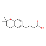


![Poly[oxy[(1S)-1-methyl-2-oxo-1,2-ethanediyl]]](http://img.cochemist.com/ccimg/26200/26161-42-2.png)
![Poly[oxy[(1S)-1-methyl-2-oxo-1,2-ethanediyl]]](http://img.cochemist.com/ccimg/26200/26161-42-2_b.png)


![Poly[oxy(1-oxo-1,6-hexanediyl)]](http://img.cochemist.com/ccimg/25300/25248-42-4.png)
![Poly[oxy(1-oxo-1,6-hexanediyl)]](http://img.cochemist.com/ccimg/25300/25248-42-4_b.png)