Co-reporter:Kusal T. G. Samarasinghe, Dhanushka N. P. Munkanatta Godage, Yani Zhou, Fidelis T. Ndombera, Eranthie Weerapana and Young-Hoon Ahn
Molecular BioSystems 2016 vol. 12(Issue 8) pp:2471-2480
Publication Date(Web):10 May 2016
DOI:10.1039/C6MB00175K
Glucose metabolism and mitochondrial function are closely interconnected with cellular redox-homeostasis. Although glucose starvation, which mimics ischemic conditions or insufficient vascularization, is known to perturb redox-homeostasis, global and individual protein glutathionylation in response to glucose metabolism or mitochondrial activity remains largely unknown. In this report, we use our clickable glutathione approach, which forms clickable glutathione (azido-glutathione) by using a mutant of glutathione synthetase (GS M4), for detection and identification of protein glutathionylation in response to glucose starvation. We found that protein glutathionylation is readily induced in HEK293 cells in response to low glucose concentrations when mitochondrial reactive oxygen species (ROS) are elevated in cells, and glucose is the major determinant for inducing reversible glutathionylation. Proteomic and biochemical analysis identified over 1300 proteins, including SMYD2, PP2Cα, and catalase. We further showed that PP2Cα is glutathionylated at C314 in a C-terminal domain, and PP2Cα C314 glutathionylation disrupts the interaction with mGluR3, an important glutamate receptor associated with synaptic plasticity.
Co-reporter:Kusal T. G. Samarasinghe ; Dhanushka N. P. Munkanatta Godage ; Garrett C. VanHecke
Journal of the American Chemical Society 2014 Volume 136(Issue 33) pp:11566-11569
Publication Date(Web):July 31, 2014
DOI:10.1021/ja503946q
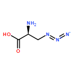
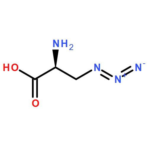
Glutathionylation involves reversible protein cysteine modification that regulates the function of numerous proteins in response to redox stimuli, thereby altering cellular processes. Herein we developed a selective and versatile approach to identifying glutathionylation by using a mutant of glutathione synthetase (GS). GS wild-type catalyzes coupling of γGlu-Cys to Gly to form glutathione. We generated a GS mutant that catalyzes azido-Ala in place of Gly with high catalytic efficiency and selectivity. Transfection of this GS mutant (F152A/S151G) and incubation of azido-Ala in cells efficiently afford the azide-containing glutathione derivative, γGlu-Cys-azido-Ala. Upon H2O2 treatment, clickable glutathione allowed for selective and sensitive detection of glutathionylated proteins by Western blotting or fluorescence after click reaction with biotin-alkyne or rhodamine-alkyne. This approach affords the efficient metabolic tagging of intracellular glutathione with small clickable functionality, providing a versatile handle for characterizing glutathionylation.
Co-reporter:Dilini N. Kekulandara, Kusal T. G. Samarasinghe, Dhanushka N. P. Munkanatta Godage and Young-Hoon Ahn
Organic & Biomolecular Chemistry 2016 - vol. 14(Issue 46) pp:NaN10893-10893
Publication Date(Web):2016/10/28
DOI:10.1039/C6OB02050J
Protein glutathionylation is one of the major cysteine oxidative modifications in response to reactive oxygen species (ROS). We recently developed a clickable glutathione approach for detecting glutathionylation by using a glutathione synthetase mutant (GS M4) that synthesizes azido-glutathione (γGlu-Cys-azido-Ala) in situ in cells. In order to demonstrate the versatility of clickable glutathione and to increase the chemical tools for detecting glutathionylation, we sought to develop clickable glutathione that uses tetrazine-alkene bioorthogonal chemistry. Here we report two alkene-containing glycine surrogates (allyl-Gly and allyl-Ser) for the biosynthesis of clickable glutathione and their use for detection, enrichment, and identification of glutathionylated proteins. Our results provide chemical tools (allyl-Gly and allyl-Ser for GS M4) for versatile characterization of protein glutathionylation. In addition, we show that the active site of GS can be tuned to introduce a small size chemical tag on glutathione for exploring glutathione function in cells.

![1H-Thieno[3,4-d]imidazole-4-pentanoicacid, hexahydro-2-oxo-, 2-[2-(aminooxy)acetyl]hydrazide, (3aS,4S,6aR)-](http://img.cochemist.com/ccimg/139600/139585-03-8.png)
![1H-Thieno[3,4-d]imidazole-4-pentanoicacid, hexahydro-2-oxo-, 2-[2-(aminooxy)acetyl]hydrazide, (3aS,4S,6aR)-](http://img.cochemist.com/ccimg/139600/139585-03-8_b.png)