Co-reporter:Kazuki Miura, Wataru Hakamata, Ayako Tanaka, Takako Hirano, Toshiyuki Nishio
Bioorganic & Medicinal Chemistry 2016 Volume 24(Issue 6) pp:1369-1375
Publication Date(Web):15 March 2016
DOI:10.1016/j.bmc.2016.02.010
Post-translational modifications (PTMs) of proteins play important roles in the physiology of eukaryotes. In the PTMs, non-reversible glycosylations are classified as N-glycosylations and O-glycosylations, and are catalyzed by various glycosidases and glycosyltransferases. However, β-glycosidases are not known to play a role in N- and O-glycan processing, although both glycans provide partial structures as substrates for β-galactosidase and β-N-acetylglucosaminidase in the Golgi apparatus of human cells. We explored human Golgi β-galactosidase using fluorescent substrates based on a quinone methide cleavage (QMC) substrate design platform that was previously developed to image exo-type glycosidases in living cells. As a result, we discovered a novel Golgi β-galactosidase in human cells. It is possible to predict a novel and important function in glycan processing of this β-galactosidase, because various β-galactosyl linkages in N- and O-glycans exist in Golgi apparatus. In addition, these results show that the QMC platform is excellent for imaging exo-type glycosidases.
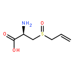
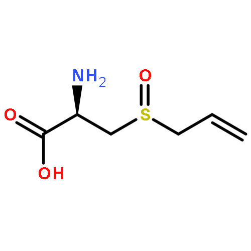
Co-reporter:Wataru Hakamata, Ryosuke Koyama, Mizuki Tanida, Tomomi Haga, Takako Hirano, Makoto Akao, Hitomi Kumagai, and Toshiyuki Nishio
Journal of Agricultural and Food Chemistry 2015 Volume 63(Issue 50) pp:10778-10784
Publication Date(Web):December 5, 2015
DOI:10.1021/acs.jafc.5b05501
A simple method for the isolation of the bioactive compound alliin from garlic, as well as a method for the synthesis of diastereomerically pure alliin and allo-alliin on a preparative laboratory scale, was developed. The absolute configuration of the sulfur atom in alliin and allo-alliin was assigned on the basis of enzyme reactivity, optical rotatory dispersion, and circular dichroism analyses. A comparison of the results from these analyses, in combination with an X-ray diffraction study on a protected allo-alliin derivative, revealed S and R configurations of the sulfur atoms in alliin and allo-alliin, respectively. In addition, the same 1H NMR spectrum was observed for synthetic and natural alliin. The absolute configuration of natural alliin was assigned for the first time on the basis of the NMR spectrum and X-ray coordinates.
Co-reporter:Wataru Hakamata, Kazuki Miura, Takako Hirano, Toshiyuki Nishio
Bioorganic & Medicinal Chemistry 2015 Volume 23(Issue 1) pp:73-79
Publication Date(Web):1 January 2015
DOI:10.1016/j.bmc.2014.11.023
The majority of eukaryotic proteins undergo post-translational modifications (PTMs) involving the attachment of complex glycans, predominantly through N-glycosylation and O-glycosylation. PTMs play important roles in virtually all cellular processes, and aberrant regulation of protein glycosylation and glycan processing has been implicated in various diseases. However, glycan processing on proteins in various cellular contexts has not been visualized. We had previously developed a quinone methide cleavage (QMC) platform for enhanced substrate design. This platform was applied here to screen for novel glycan-processing enzymes. We designed and synthesized fluorescent substrates with β-allopyranoside residues using the QMC platform. When applied in cell-based assays, the fluorescent substrates allowed rapid and clear visualization of β-allosidase activity in the Golgi apparatus of human cultured cells. The QMC platform will likely find broad applications in visualizing the activities of glycan processing enzymes in living cells and in studying PTMs.
Co-reporter:Wataru Hakamata, Saori Tamura, Takako Hirano, and Toshiyuki Nishio
ACS Medicinal Chemistry Letters 2014 Volume 5(Issue 4) pp:321-325
Publication Date(Web):January 16, 2014
DOI:10.1021/ml400398t
The carboxylesterase families of enzymes are key participants in phase I drug metabolism processes. Carboxylesterase families 1 and 2 are of particular clinical relevance. These enzymes produce endoplasmic reticulum localization signals, are primarily localized in the endoplasmic reticulum, and hydrolyze a wide range of ester-containing prodrugs into an activated form. In order to detect enzymes belonging to both families, we developed an optical multicolor imaging technique, which provides a distinct color window for multicolor imaging. This technique required the design and synthesis of three new mechanistic colored probes that fluoresce red, green, or blue and are based on the quinone methide cleavage process. These activity-based probes allow rapid and clear visualization with high specificity against the endoplasmic reticulum in cultured cells based on endoplasmic reticulum localized esterases including both families of carboxylesterase enzymes.Keywords: carboxylesterase; fluorescent probe; Prodrug; quinone methide cleavage;
Co-reporter:Wataru Hakamata, Ryosuke Ishikawa, Yoriko Ushijima, Takumi Tsukagoshi, Saori Tamura, Takako Hirano, Toshiyuki Nishio
Bioorganic & Medicinal Chemistry Letters 2012 Volume 22(Issue 1) pp:62-64
Publication Date(Web):1 January 2012
DOI:10.1016/j.bmcl.2011.11.084
5-Thiazoleacetamide derivatives of AR122 and AR125 were screened as α-glucosidase inhibitors by in silico high-throughput screening from commercial drug-like small compound libraries. Inhibition of α-glucosidase with AR122 and AR125 is time dependent: with no preincubation, AR122 and AR125 are relatively moderate inhibitors, but interestingly, after a 120 min incubation, they were 50-fold more potent (AR122: IC50 = 2.47 μM and AR125: IC50 = 27.1 μM). Plots of ln [residual α-glucosidase activity %] versus preincubation time show a pseudo-first order kinetics for both inhibitors. Through dialysis of enzyme–inhibitor complexes, no activity recovery was shown. These results suggest that AR122 and AR125 constitute a new class of noncarbohydrate mimetic inhibitor with an irreversible mechanism.
Co-reporter:Wataru Hakamata, Aki Machida, Tadatake Oku, Toshiyuki Nishio
Bioorganic & Medicinal Chemistry Letters 2011 Volume 21(Issue 11) pp:3206-3209
Publication Date(Web):1 June 2011
DOI:10.1016/j.bmcl.2011.04.066
CEs are important enzymes that catalyze the hydrolysis of prodrugs. In this Letter, we present a new mechanistic ER-specific fluorescent probe 1 based on CE activity. Permeation of 1 into cells and subsequent hydrolytic activation by CEs causes spontaneously quinone methide cleavage, resulting in bright red fluorescence in ER with high specificity. Probe 1 was developed for CE activity imaging and inhibitor screening at the cellular level.


![Phenol, 4-[[[(1,1-dimethylethyl)dimethylsilyl]oxy]methyl]-](http://img.cochemist.com/ccimg/126100/126070-20-0.png)
![Phenol, 4-[[[(1,1-dimethylethyl)dimethylsilyl]oxy]methyl]-](http://img.cochemist.com/ccimg/126100/126070-20-0_b.png)
![[4-(HYDROXYMETHYL)PHENYL] 2-METHYLPROPANOATE](http://img.cochemist.com/ccimg/70400/70362-64-0.png)
![[4-(HYDROXYMETHYL)PHENYL] 2-METHYLPROPANOATE](http://img.cochemist.com/ccimg/70400/70362-64-0_b.png)
![D-Glucose,2-(acetylamino)-4-O-[2-(acetylamino)-2-deoxy-b-D-glucopyranosyl]-2-deoxy-](http://img.cochemist.com/ccimg/35100/35061-50-8.png)
![D-Glucose,2-(acetylamino)-4-O-[2-(acetylamino)-2-deoxy-b-D-glucopyranosyl]-2-deoxy-](http://img.cochemist.com/ccimg/35100/35061-50-8_b.png)