Co-reporter:Youkun Zheng, Weiwei Liu, Yun Chen, Hui Jiang, Hong Yan, Irina Kosenko, Lubov Chekulaeva, Igor Sivaev, Vladimir Bregadze, and Xuemei Wang
Organometallics September 25, 2017 Volume 36(Issue 18) pp:3484-3484
Publication Date(Web):September 1, 2017
DOI:10.1021/acs.organomet.7b00426
Methicillin-resistant Staphylococcus aureus (MRSA) is a notorious superbug that is potentially life-threatening. Among conventional antibiotics, vancomycin is a “gold standard” agent used to treat serious MRSA infections. Such therapy, however, is often ineffective because of the emergence of less-susceptible strains. Therefore, the exploration of new antimicrobial agents, especially nonantibiotic drugs, to cope with the growing threat of MRSA has become an urgent necessity. Herein, we have investigated the possibility to develop a metallacarborane antimicrobial agent, cobalt bis(1,2-dicarbollide) alkoxy derivative (K121), and we have evaluated the relevant anti-MRSA behaviors. We demonstrated that K121 has a dose-dependent anti-MRSA activity with a low minimal inhibitory concentration of 8 μg/mL and a high selectivity over mammalian cells. In particular, a high bacteria-killing efficiency was observed with eradication of all MRSA cells within 30 min. In addition, K121 showed a high inhibition effect on the formation of bacterial biofilm. More importantly, unlike vancomycin, a repeated use of K121 would not induce drug resistance even after 20 passages of MRSA. The mechanistic study showed that K121 kills MRSA by inducing an increase in the reactive oxygen species (ROS) production and consequentially inducing irreversible damage to the cell wall/membrane, which ultimately leads to the death of MRSA. Our results suggested that K121 may be used as a promising nonantibiotic therapeutic agent against MRSA infections in future clinical practices.
Co-reporter:Lanmei Lai, Xuerui Jiang, Shanying Han, Chunqiu Zhao, Tianyu Du, Fawad Ur Rehman, Youkun Zheng, Xiaoqi Li, Xiaoli Liu, Hui Jiang, and Xuemei Wang
Langmuir September 12, 2017 Volume 33(Issue 36) pp:9018-9018
Publication Date(Web):August 14, 2017
DOI:10.1021/acs.langmuir.7b01516
Alzheimer’s disease is still incurable and neurodegenerative, and there is a lack of detection methods with high sensitivity and specificity. In this study, by taking different month old Alzheimer’s mice as models, we have explored the possibility of the target bioimaging of diseased sites through the initial injection of zinc gluconate solution into Alzheimer’s model mice post-tail vein and then the combination of another injection of ferrous chloride (FeCl2) solution into the same Alzheimer’s model mice post-stomach. Our observations indicate that both zinc gluconate solution and FeCl2 solution could cross the blood–brain barrier (BBB) to biosynthesize the fluorescent zinc oxide nanoclusters and magnetic iron oxide nanoclusters, respectively, in the lesion areas of the AD model mice, thus enabling high spatiotemporal dual-modality bioimaging (i.e., including fluorescence bioimaging (FL) and magnetic resonance imaging (MRI)) of Alzheimer’s disease for the first time. The result presents a novel promising strategy for the rapid and early diagnosis of Alzheimer’s disease.
Co-reporter:Hui Jiang;Liu Liu
Nanoscale (2009-Present) 2017 vol. 9(Issue 28) pp:9792-9796
Publication Date(Web):2017/07/20
DOI:10.1039/C7NR03382F
This study reports the occurrence of a special red-emitted anodic electrochemiluminescence (ECL) emission at +1.4 V (vs. Ag/AgCl) on a glass carbon electrode (GCE) after the addition of thioglycol (TG) to surface-unsaturated glutathione (GSH)-coated Au nanoclusters (NCs), with an emission peak at ∼630 nm. Compared to the ECL at a potential of +1.8 V (vs. Ag/AgCl) and an emission peak at 580 nm (corresponding to fluorescence) for only GSH-coated Au NCs, this ECL emission not only exhibits a lower ECL potential but also shows a significantly red-shifted emission wavelength up to ∼50 nm. We demonstrated that the formation of TG/GSH dual ligand-coated Au NCs is responsible for the red-shifted ECL emission. Other common thiol compounds cannot result in similar effect on GCE, and no ECL is observed on other electrodes such as indium tin oxide and platinum electrodes. This finding offers a great possibility to design novel feasible ECL systems for different complicated applications.
Co-reporter:Jing Ye;Xiawei Dong;Hui Jiang
Journal of Materials Chemistry B 2017 vol. 5(Issue 4) pp:691-696
Publication Date(Web):2017/01/25
DOI:10.1039/C6TB02751B
Temperature variation is related to a series of biological reactions and abnormal medical processes of living cells. Fluorescence-based temperature nanoprobes have great potential for cellular imaging and temperature measurement. In this study, we have established a facile, efficient and green strategy for the preparation of an intracellular temperature nanoprobe specifically by in situ biosynthesized fluorescent copper nanoclusters (CuNCs). Our observations demonstrate that the fluorescent CuNCs could specifically be biosynthesized spontaneously in MDA-MB-231 cancer cells through a particular molecular process, but not in normal cells (i.e., L02 cells). The resultant CuNCs, with an average diameter of 2.4 ± 0.4 nm, were found to exhibit red fluorescence emission (λem = 610 nm) and could further efficiently accumulate for bioimaging in target cancer cells. More importantly, the fluorescence signal of the biosynthesized CuNCs is sensitively thermo-responsive over the physiological temperature range in MDA-MB-231 cells (relative sensitivity: −3.18% per Celsius). This provides an efficient nanothermometer based on the in situ biosynthesized CuNCs for cellular fluorescence imaging and other biomedical applications.
Co-reporter:Shanying Han, Tianyu Du, Hui Jiang, Xuemei Wang
Biosensors and Bioelectronics 2017 Volume 89(Part 1) pp:422-429
Publication Date(Web):15 March 2017
DOI:10.1016/j.bios.2016.04.092
•Graphene-pyrroloquinoline quinone (Gr-PQQ) composite was formed by simple adsorption.•Gr-PQQ film modified electrode was highly sensitive to electrooxidation of NADH.•Gr and PQQ show synergistic effect on enhanced detection performances.•The sensors can successfully determine NADH in human serum samples.A self-assembly composite of graphene-pyrroloquinoline quinone (PQQ) was fabricated and modified on glassy carbon electrode (GCE) for sensitive detection of nicotinamide adenine dinucleotide (NADH). Chitosan (CTS) was applied to disperse graphene to form a stable robust film on GCE. A synergistic effect between PQQ and graphene was observed during the electrocatalytic oxidation of NADH, with about 260 mV reduction in the oxidation potential and 2.5-fold increase in the oxidation current compared with those on the bare GCE. The electrochemical sensors based on the modified electrodes allowed the detection of NADH with a good linear dependence from 0.32 to 220 µM with a high sensitivity of 0.421 µA µM−1 cm−2 and a low detection limit of 0.16 µM (S/N=3). It could also eliminate the interference of electroactive substances like ascorbic acid (AA), uric acid, and dopamine and its derivatives. The outstanding performances of graphene-PQQ/CTS composite capable of improving the electrical conductivity and accelerating the electron transport suggested its promising applications for design of different graphene based composites used in electrochemical sensing and energy fields.
Co-reporter:Youkun Zheng, Lanmei Lai, Weiwei Liu, Hui Jiang, Xuemei Wang
Advances in Colloid and Interface Science 2017 Volume 242(Volume 242) pp:
Publication Date(Web):1 April 2017
DOI:10.1016/j.cis.2017.02.005
•Up-to-date compilation of fluorescent gold nanoclusters (AuNCs)•Biosynthesis and electrochemical reduction strategies can be readily utilized for the preparation of fluorescent AuNCs.•Fluorescent AuNCs have been widely used for biolabeling, biosensing, bioimaging and targeted cancer diagnostics and treatment.Fluorescent gold nanoclusters (AuNCs) are emerging as novel fluorescent materials and have attracted more and more attention in the field of biolabeling, biosensing, bioimaging and targeted cancer treatment because of their unusual physicochemical properties, such as long fluorescence lifetime, ultrasmall size, large Stokes shift, strong photoluminescence, as well as excellent biocompatibility and photostability. Recently, significant efforts have been committed to the preparation, functionalization and biomedical application studies of fluorescent AuNCs. In this review, we have summarized the strategies for preparation and surface functionalization of fluorescent AuNCs in the past several years, and highlighted recent advances in the biomedical applications of the relevant fluorescent AuNCs. Based on these observations, we also give a discussion on the current problems and future developments of the fluorescent AuNCs for biomedical applications.“Eight diagrams” illustrating of the preparation, functionalization and biomedical applications of fluorescent gold nanoclusters.Download high-res image (190KB)Download full-size image
Co-reporter:Hui Jiang, Xiaoqing Su, Yuanyuan Zhang, Junyu Zhou, Danjun Fang, and Xuemei Wang
Analytical Chemistry 2016 Volume 88(Issue 9) pp:4766
Publication Date(Web):April 7, 2016
DOI:10.1021/acs.analchem.6b00112
The photoluminescence (PL) of nonthiolate ligand capped Au nanoclusters (NCs) is usually quenched by thiols due to the tight adsorption of thiols to the Au surface and formation of larger non-PL species. However, we here report an unexpected PL enhancement of cytidine stabilized Au (AuCyt) NCs triggered by thiols, such as reduced glutathione (GSH) at sub-μM level, while such phenomena have not been observed for Au NCs capped with similar adenosine/cytidine nucleotides. The mass spectroscopic results indicate that this enhancement may be caused by the formation of smaller, but highly fluorescent, Au species etched by thiols. This enables the sensitive detection of GSH from 20 nM to 3 μM, with an ultralow detection limit of 2.0 nM. Moreover, the glutathione reductase (GR) activity can be determined by the initial rate of GSH production, i.e., the maximum PL increasing rate, with a linear range of 0.34–17.0 U/L (1 U means reduction of 1.0 μmol of oxidized glutathione per min at pH 7.6 at 25 °C) and a limit of detection of 0.34 U/L. This method allows the accurate assays of GR in clinical serum samples as well as the rapid screening of GR inhibitors, indicating its promising biomedical applications.
Co-reporter:Changhui Li, Xiaoli Liu, Yuanyuan Zhang, Yun Chen, Tianyu Du, Hui Jiang, Xuemei Wang
Analytica Chimica Acta 2016 Volume 933() pp:66-74
Publication Date(Web):24 August 2016
DOI:10.1016/j.aca.2016.05.043
•A novel nonenzymatic H2O2 electrochemical biosensor was constructed based on the CuI/Gr composites.•The biosensor has low detection limit and high sensitivity for H2O2 detection.•SECM imaging study further illustrates the electrochemical catalytic capability for H2O2 reduction.•The H2O2 biosensor is used to detect H2O2 released from living cells.A high-sensitive nonenzymatic hydrogen peroxide (H2O2) biosensor based on cuprous iodide and graphene (CuI/Gr) composites has been explored for the detection of H2O2 released by living cells and monitoring the oxidative stress of cells under excellular stimulation. The biosensor properties were evaluated by cyclic voltammetry (CV), electrochemical impedance spectroscopy (EIS), amperometric i-t curve, and the redox-competition mode of scanning electrochemical microscopy (SECM). Our observations demonstrate that the CuI/Gr nanocomposites modified glassy carbon electrode (GCE) exhibits excellent catalytic activity for H2O2 with relatively low detection limit and a wide linear range from 0.5 μM to 3 mM. Moreover, the redox-competition mode of SECM imaging study further illustrates the improved electrochemical catalytic capability for H2O2 reduction with CuI/Gr nanocomposites deposited on graphite electrode. Hence, the as-prepared nonenzymatic H2O2 biosensor could be used to detect H2O2 release from different kinds of living cells under stimulation while eliminating the interference of ascorbic acid.
Co-reporter:Lanmei Lai, Chunqiu Zhao, Meina Su, Xiaoqi Li, Xiaoli Liu, Hui Jiang, Christian Amatore and Xuemei Wang
Biomaterials Science 2016 vol. 4(Issue 7) pp:1085-1091
Publication Date(Web):27 May 2016
DOI:10.1039/C6BM00233A
Alzheimer's disease (AD) is an irreversible neurodegenerative disease which is difficult to cure. When Alzheimer's disease occurs, the level of zinc ions in the brain changes, and the relevant amount of zinc ions continue decreasing in the cerebrospinal fluid and plasma of Alzheimer's patients with disease exacerbation. In view of these considerations, we have explored a new strategy for the in vivo rapid fluorescence imaging of Alzheimer's disease through target bio-labeling of zinc oxide nanoclusters which were biosynthesized in vivo in the Alzheimer's brain via intravenous injection of zinc gluconate solution. By using three-month-old and six-month-old Alzheimer's model mice as models, our observations demonstrate that biocompatible zinc ions could pass through the blood–brain barrier of the Alzheimer's disease mice and generate fluorescent zinc oxide nanoclusters (ZnO NCs) through biosynthesis, and then the bio-synthesized ZnO NCs could readily accumulate in situ on the hippocampus specific region for the in vivo fluorescent labeling of the affected sites. This study provides a new way for the rapid diagnosis of Alzheimer's disease and may have promising prospects in the effective diagnosis of Alzheimer's disease.
Co-reporter:Jing Ye, Jianling Wang, Qiwei Li, Xiawei Dong, Wei Ge, Yun Chen, Xuerui Jiang, Hongde Liu, Hui Jiang and Xuemei Wang
Biomaterials Science 2016 vol. 4(Issue 4) pp:652-660
Publication Date(Web):26 Jan 2016
DOI:10.1039/C5BM00528K
A new and facile method for rapidly and accurately achieving tumor targeting fluorescent images has been explored using a specifically biosynthesized europium (Eu) complex in vivo and in vitro. It demonstrated that a fluorescent Eu complex could be bio-synthesized through a spontaneous molecular process in cancerous cells and tumors, but not prepared in normal cells and tissues. In addition, the proteomics analyses show that some biological pathways of metabolism, especially for NADPH production and glutamine metabolism, are remarkably affected during the relevant biosynthesis process, where molecular precursors of europium ions are reduced to fluorescent europium complexes inside cancerous cells or tumor tissues. These results proved that the specific self-biosynthesis of a fluorescent Eu complex by cancer cells or tumor tissues can provide a new strategy for accurate diagnosis and treatment strategies in the early stages of cancers and thus is beneficial for realizing precise surgical intervention based on the relevant cheap and readily available agents.
Co-reporter:F. U. Rehman, C. Zhao, H. Jiang and X. Wang
Biomaterials Science 2016 vol. 4(Issue 1) pp:40-54
Publication Date(Web):07 Oct 2015
DOI:10.1039/C5BM00332F
Titanium dioxide (TiO2) is one of the most abundantly used nanomaterials for human life. It is used in sunscreen, photovoltaic devices, biomedical applications and as a food additive and environmental scavenger. Nano-TiO2 in biomedical applications is well documented. It is used in endoprosthetic implants and early theranostics of neoplastic and non-neoplastic maladies as a photodynamic therapeutic agent and as vehicles in nano-drug delivery systems. Herein, we focus on the recent advancements and applications of nano-TiO2 in bio-nanotechnology, nanomedicine and photodynamic therapy (PDT).
Co-reporter:Yun Chen, Qiwei Li, Hui Jiang, Xuemei Wang
Journal of Electroanalytical Chemistry 2016 Volume 781() pp:233-237
Publication Date(Web):15 November 2016
DOI:10.1016/j.jelechem.2016.06.020
In this contribution, a new carbon fiber microelectrode (CFME) for detection of hydrogen peroxide (H2O2) is constructed with a rapid and low-cost method based on the electrochemical etching technique and the electro-deposition of platinum (Pt) nanoparticles, and the potential application in electrochemical bioanalysis has been explored. It is observed that when the CFME modified in 5 mmol/L chloroplatinic acid with 0.5 mol/L H2SO4 used as supporting electrolyte, the as-prepared Pt modified carbon fiber microelectrode (Pt/CFME) demonstrates an excellent electrochemically catalytic activity, which exhibited a wide linear in relevant detection for the reduction of H2O2, ranging from 0.044 to 12.30 mmol/L with the limit of 44 μmol/L. Moreover, our results illustrate that the Pt/CFME electrode can be readily utilized in scanning electrochemical microscope (SECM) studies to detect H2O2 release from human glioblastoma cells U87 when stimulated with ascorbic acid (AA).
Co-reporter:Yuanyuan Zhang, Jincheng Li, Hui Jiang, Chunqiu Zhao and Xuemei Wang
RSC Advances 2016 vol. 6(Issue 68) pp:63331-63337
Publication Date(Web):28 Jun 2016
DOI:10.1039/C6RA10409F
Gold nanoclusters (Au NCs) possess outstanding physical and chemical attributes that make them excellent scaffolds for the construction of novel chemical sensors and biological imaging probes. In this study, a simple one-pot synthesis method, employing L-glutathione as the stabilizer, was presented for the preparation of red fluorescent Au NCs. The prepared Au NCs have no obvious cell cytotoxic effect on cancerous cells (i.e., HeLa, U87, and MCF-7 cells) and non-cancerous cells (i.e., L02 cells) in a wide concentration range. Then the prepared Au NCs were applied for tumor-targeted imaging in vitro and in vivo due to their good photo-stability, strong fluorescence emission, excellent water solubility and bio-compatibility. The observations indicate that the as-prepared Au NCs exhibited a near infrared fluorescence emission at 710 nm for in vivo bioimaging of tumors. Furthermore, Au NCs combining with porphyrin derivatives were applied for photothermal treatment to effectively inhibit the growth of tumors. This raises the possibility of utilizing Au NCs as a fluorescent probe for tumor-targeted rapid imaging and thus realize the facile fluorescence imaging-guided photothermal therapy of tumors.
Co-reporter:Leifeng Chen;Yuanyuan Zhang;Hui Jiang;Chongyang Liu
Chinese Journal of Chemistry 2016 Volume 34( Issue 6) pp:589-593
Publication Date(Web):
DOI:10.1002/cjoc.201500748
Abstract
Noble metal clusters is an emerging class of fluorescent probes, avoiding most of the drawbacks of common fluorescent compounds, and they are simple to prepare and have good water solubility, good biocompatibility and excellent fluorescence properties. In this study, we have explored the synthesis of the cytidine mediated gold-silver nanoclusters (AuAg NCs) and applied it for both in vitro cellular imaging and tumor in vivo detection. Experimental results show that the as-prepared AuAg NCs can be used as a sensitive fluorescent probe for cancer cells/tissue detection. Especially, it is evident that under the relevant light irradiation with the wavelength of 488 nm, obviously bright fluorescence signal could be readily detected from focus location of inoculating tumor mouse, implying its possible application for the effective in vivo tumor bioimaging.
Co-reporter:Chunqiu Zhao;Fawad Ur Rehman;Hui Jiang;Matthias Selke
Science China Chemistry 2016 Volume 59( Issue 5) pp:637-642
Publication Date(Web):2016 May
DOI:10.1007/s11426-016-5568-1
Photodynamic therapy (PDT) is one of the latest biomedical technologies used for treatment of various neoplastic and non-neoplastic diseases. However, there still exist some well-known problems regarding its efficacy, e.g. effective concentration of the drug at the desired sites, the irradiation light dosimetry and biocompatibility of the photosensitizer. The introduction of nanotechnology and nanomaterial like biocompatible nano-titania (i.e., nano-TiO2) may facilitate to solve some of these problems. In this study we have explored the possibility of combining tetra sulphonatophenyl porphyrin (TSPP) with nano-titania (PT) for efficient PDT with least adverse effects. The spectroscopic properties of these nano-composites were characterized by using fluorescence and UV-Vis absorption spectroscopic study. The singlet oxygen quantum yield was determined by using 2,5-diphenyl-3,4-benzofuran (DPBF), while the effect of nano TiO2 with TSPP on the synovial fibroblast cells from human (HSC) and rat models (RSC) were investigated by confocal laser microscopy and 3-(4,5-dimethylthiazol-2-yl)-2,5-diphenyltetrazolium bromide (MTT) assay. Our results suggest that nano TiO2 with TSPP can be readily utilized for effective PDT treatment of Rheumatoid Arthritis (RA).
Co-reporter:Fawad U. Rehman;Chunqiu Zhao;Changyu Wu;Xiaoqi Li;Hui Jiang
Nano Research 2016 Volume 9( Issue 11) pp:3305-3321
Publication Date(Web):2016 November
DOI:10.1007/s12274-016-1208-5
Rheumatoid arthritis (RA) etiology and amelioration remains a challenge in modern therapeutics. Herein, we explored the synergistic effect of allogenic bone marrow stem cell (BMSC) translation and photodynamic treatment of RA with tetra sulfonatophenyl porphyrin (TSPP) and TiO2 nanocomposites as a new strategy for RA theranostics. The translation of BMSCs with miRNAs into infected joints in long bones post-photodynamic therapy is helpful for treating and understanding RA pathophysiology. We observed that allogenic BMSC translation combined with TSPP-TiO2 nanocomposites can significantly (p < 0.01) lower the concentrations of serum biomarkers (tumor necrosis factor-α and interleukin-17) in a collagen induced arthritis (CIA) murine model, both in vitro and in vivo, as well as improve other parameters such as arthritis score, BMSC count, complete blood count, and numbers of platelets, red blood cells, and white blood cells. Moreover, a fluorescent TSPP in the feet or long bones and X-ray bioimaging of RA joints revealed the clinical efficacy of BMSCs combined with TSPP-TiO2 nanocomposites. Microarray data analysis illustrated that rno-mir-375-3p and rno-mir-196b-3p were up-regulated by approximately 100-fold in the BMSCs of ameliorated RA post-photodynamic therapy with TSPP-TiO2 nanocomposites. Our study not only suggests a new approach for RA theranostics, but also helps in understanding RA pathophysiology.
Co-reporter:Changyu Wu, Fawad ur Rehman, Jingyuan Li, Jing Ye, Yuanyuan Zhang, Meina Su, Hui Jiang, and Xuemei Wang
ACS Applied Materials & Interfaces 2015 Volume 7(Issue 44) pp:24848
Publication Date(Web):October 22, 2015
DOI:10.1021/acsami.5b08066
This work presents a new strategy of the combination of surface plasmon resonance (SPR) and electrochemical study for real-time evaluation of live cancer cells treated with daunorubicin (DNR) at the interface of the SPR chip and living cancer cells. The observations demonstrate that the SPR signal changes could be closely related to the morphology and mass changes of adsorbed cancer cells and the variation of the refractive index of the medium solution. The results of light microscopy images and 3-(4,5-dimethyl-2-thiazolyl)-2,5-diphenyl-2H-tetrazolium bromide studies also illustrate the release or desorption of HepG2 cancer cells, which were due to their apoptosis after treatment with DNR. It is evident that the extracellular concentration of DNR residue can be readily determined through electrochemical measurements. The decreases in the magnitudes of SPR signals were linearly related to cell survival rates, and the combination of SPR with electrochemical study could be utilized to evaluate the potential therapeutic efficiency of bioactive agents to cells. Thus, this label-free, real-time SPR–electrochemical detection technique has great promise in bioanalysis or monitoring of relevant treatment processes in clinical applications.Keywords: cancer; electrochemistry; live cells; real-time evaluation; surface plasmon resonance
Co-reporter:Donghua Chen, Chunqiu Zhao, Jing Ye, Qiwei Li, Xiaoli Liu, Meina Su, Hui Jiang, Christian Amatore, Matthias Selke, and Xuemei Wang
ACS Applied Materials & Interfaces 2015 Volume 7(Issue 32) pp:18163
Publication Date(Web):July 31, 2015
DOI:10.1021/acsami.5b05805
Among the noble-metal clusters, very few reports about platinum clusters were used as bioimaging probes of tumors except as a reducing catalyst. It is first established herein that the biocompatible platinum nanoclusters are spontaneously biosynthesized by cancerous cells (i.e., HepG2 (human hepatocarcinoma), A549 (lung cancer), and others) rather than noncancerous cells (i.e., L02 (human embryo liver cells)) when incubated with micromolar chloroplatinic acid solutions. These in situ biosynthesized platinum nanoclusters could be readily realized in a biological environment and emit a bright fluorescence at 460 nm, which could be further utilized to facilitate an excellent cancer-cell-killing efficiency when combined with porphyrin derivatives for photothermal treatment. This raises the possibility of providing a promising and precise bioimaging strategy for specific fluorescent self-biomarking of tumor locations and realizing fluorescence imaging-guided photothermal therapy of tumors.Keywords: bioimaging; cancer; photothermal treatment; platinum nanoclusters; theranostics
Co-reporter:Le Wang, Yuanyuan Zhang, Chuansheng Cheng, Xiaoli Liu, Hui Jiang, and Xuemei Wang
ACS Applied Materials & Interfaces 2015 Volume 7(Issue 33) pp:18441
Publication Date(Web):August 4, 2015
DOI:10.1021/acsami.5b04553
High levels of H2O2 pertain to high oxidative stress and are associated with cancer, autoimmune, and neurodegenerative disease, and other related diseases. In this study, a sensitive H2O2 biosensor for evaluation of oxidative stress was fabricated on the basis of the reduced graphene oxide (RGO) nanocomposites decorated with Au, Fe3O4, and Pt nanoparticles (RGO/AuFe3O4/Pt) modified glassy carbon electrode (GCE) and used to detect the released H2O2 from cancer cells and assess the oxidative stress elicited from H2O2 in living cells. Electrochemical behavior of RGO/AuFe3O4/Pt nanocomposites exhibits excellent catalytic activity toward the relevant reduction with high selection and sensitivity, low overpotential of 0 V, low detection limit of ∼0.1 μM, large linear range from 0.5 μM to 11.5 mM, and outstanding reproducibility. The as-prepared biosensor was applied in the measurement of efflux of H2O2 from living cells including healthy normal cells and tumor cells under the external stimulation. The results display that this new nanocomposites-based biosensor is a promising candidate of nonenzymatic H2O2 sensor which has the possibility of application in clinical diagnostics to assess oxidative stress of different kinds of living cells.Keywords: electrochemical detection; ferroferric oxide nanoparticles; gold nanoparticles; graphene nanosheets; hydrogen peroxide; living cell; Pt nanoparticles
Co-reporter:Xiaoqing Su, Hui Jiang, and Xuemei Wang
Analytical Chemistry 2015 Volume 87(Issue 20) pp:10230
Publication Date(Web):September 14, 2015
DOI:10.1021/acs.analchem.5b02559
The rapid detection and imaging of intracellular thiols is of great importance during the occurrence and development of some chronic diseases. Here we demonstrate the rapid thiols-induced photoluminescence (PL) enhancement of the low luminescent glutathione (GSH) stabilized Au nanoparticles, AuGSH (low). The dynamic PL investigation reveals that the PL enhancement fits a first-order reaction model. The X-ray photoelectron spectroscopic and mass spectroscopic results indicate that AuGSH (low) are mainly comprised of “thiols-insufficient” Au species and the additional thiols can efficiently attach to the “unsaturated” surface of Au nanoparticles, accompanied by significant PL enhancement. The noncytotoxic AuGSH (low) probe can be successfully applied for imaging of intracellular thiols. Generally, this work illustrates the great prospects of facile-prepared AuGSH (low) as a candidate for thiols labeling and imaging.
Co-reporter:Changyu Wu, Afzal Shah, Hongde Ye, Xiao Chen, Jing Ye, Hui Jiang, Baoan Chen, Xuemei Wang, Hong Yan
Analytica Chimica Acta 2015 Volume 857() pp:39-45
Publication Date(Web):1 February 2015
DOI:10.1016/j.aca.2014.12.019
•Electrochemical behaviors of novel ferrocenyl based carboranes (FcCB) were explored with a droplet system.•The shifts of peak potentials with changes of pH values indicated the involvement of proton during electron transfer reaction.•Normal cells and cancer cells could be specifically recognized by using FcCB as probe.•This electrochemical method in a droplet shows great potential application for relevant diagnostics of clinical samples.Novel ferrocenyl based carboranes (FcCBs) and their distinguish behavior for cancer cell recognition have been explored in this contribution. The voltammetric study in a droplet of 10 μL placed on the surface of a glassy carbon electrode demonstrates the excellent electrochemical behavior of FcCBs, which could be further exploited for establishing the promising and sensitive biosensors. The FcCBs’ redox behavior is examined in a wide pH range, and square wave voltammetry revealed the reversible and irreversible nature of first and second anodic peaks. The obvious shifts in peak potentials corresponding with the change of pH values demonstrate the abstraction of electrons to be accompanied with the transfer of protons. By using the droplet electrochemical technique, FcCBs can be employed to distinguish normal and cancer cells with a linear range from 1.0 × 10³ to 3.0 × 10⁴ cells mL−1 and the limit of detection at 800 cells mL−1. The novel carborane derivatives could be utilized as important potential molecular probes for specific recognition of cancer cells like leukemia cells from normal cells.
Co-reporter:Fawad Ur Rehman, Chunqiu Zhao, Changyu Wu, Hui Jiang, Matthias Selke and Xuemei Wang
RSC Advances 2015 vol. 5(Issue 130) pp:107285-107292
Publication Date(Web):01 Dec 2015
DOI:10.1039/C5RA23480H
Photodynamic therapy (PDT) is mostly used to induce apoptosis or necrosis in benign and malignant tumors, along with other microbial infections and suppression of autoimmune diseases including rheumatoid arthritis (RA). The bone marrow stem (BMS) cells are also a focus in translational medicine, tissue engineering and as an autoimmune disease suppressant. In this study we used tetra sulphonatophenyl porphyrin (TSPP) with TiO2 nanowhiskers for RA PDT and evaluated the effect on stress biomarkers (CAT, SOD, GPX, GR, TAO and MDA) in vivo and BMS cell proliferation in vitro. We compared four murine groups, three of which had collagen induced arthritis as TP-L (illuminated), TP-nL (dark) and CIA (control), whereas the other group was normal without disease and treatment. All anti-oxidative enzymes and biomarkers were significantly (p < 0.01) affected by the treatment except TAO (p > 0.05). Moreover, we also evaluated the growth proliferation effect of TSPP–TiO2 (TP) PDT on the in vitro RA infected BMS cells i.e. 25 μl had highest cell count (12.33 × 10⁶ cells per well) and 33% higher growth rate in photoactivated TP when compared with 50 and 100 μl treatment groups. Herein, we report that photoactivated TSPP–TiO2 for RA PDT may be safer than photosensitizers without the titanium nanomaterials in terms of reduced oxidative stress and also promotion of RA BMS cell growth in vitro as a novel finding.
Co-reporter:Meina Su, Jing Ye, Qiwei Li, Wei Ge, Yuanyuan Zhang, Hui Jiang, Christian Amatore and Xuemei Wang
RSC Advances 2015 vol. 5(Issue 91) pp:74844-74849
Publication Date(Web):24 Aug 2015
DOI:10.1039/C5RA14663A
Zinc has important physiological and biochemical functions in the body of human beings, the relevant concentrations of zinc ions were found to considerably decrease when some cancers occurred. Thus, in this contribution, we have explored the possibility of effective imaging or the labeling of cancer cells through in situ biosynthesized zinc nanoclusters. The results demonstrate that we can readily realize the target cellular fluorescence bio-imaging through the in situ biosynthesis of biocompatible zinc nanoclusters from cancerous cells when target cells are cultured with micromolar zinc gluconate solutions. Moreover, in vivo imaging of subcutaneous xenografted tumors in nude mice has also established the validity of this strategy for the rapid and precise target bio-imaging of tumors by subcutaneous injections of zinc gluconate solutions, without significant dissemination to the surrounding normal tissues, resulting from a completely different redox homeostasis of cancer cells from normal cells. This strategy could provide a new ultrasensitive way for rapid & accurate tumor diagnosis/treatment through zinc gluconate, an ideal organic zinc supplement with good biological compatibility and high bioavailability.
Co-reporter:Shengping Gao, Wei Ge, Chunqiu Zhao, Chuansheng Cheng, Hui Jiang and Xuemei Wang
RSC Advances 2015 vol. 5(Issue 33) pp:25870-25876
Publication Date(Web):06 Mar 2015
DOI:10.1039/C5RA01199J
It is well known that nanosilver or silver ions could act as an effective antibacterial agent without the development of bacterial resistance but long term exposure may induce in vivo toxicity. Thus, specific care should be taken before relevant silver-containing materials are used as antibacterial agents. Recently biocompatible polymeric materials are widely used to reduce the toxic effects of nanomaterials, which could be utilized to fabricate antibacterial surface coatings with good biocompatibility. In this study we have developed a simple and green synthesis strategy to prepare Ag@PNIPAM nanocomposites with high purity and good bioactivity for promising bio-applications as highly effective antimicrobial agents. The relevant synthesis takes place in a clean environment without any chemical additives, which ensures ultrahigh active surfaces of the Ag clusters. The as-prepared Ag@PNIPAM nanocomposites exhibit highly effective antimicrobial activities against Staphylococcus aureus (S. aureus) and have a good therapeutic effect for burn wounds.
Co-reporter:Xiaoli Liu, Hui Jiang, Wei Ge, Changyu Wu, Donghua Chen, Qiwei Li, Yun Chen and Xuemei Wang
RSC Advances 2015 vol. 5(Issue 23) pp:17532-17540
Publication Date(Web):03 Feb 2015
DOI:10.1039/C4RA16359A
Developing an efficient nanoparticulate drug-delivery system with a sub-100 nm diameter plays a crucial role in delivering antitumor drugs into cancer cells and improving their therapeutic efficacy. Carbon spheres, due to their large surface areas, unique surface properties and ease of functionalization, can generally deliver a large quantity of therapeutic agents to the target disease sites. In this study, spherical carbon nanoparticles with uniform size (71 nm) and regular shape were synthesized by hydrothermal reaction of bacterial cellulose nanofibers (30–50 nm), which had been achieved by a microorganism synthesis. Then using a simple acidification treatment, we could obtain carbon nanospheres with high drug loading capacity (the drug encapsulation efficiency was found to be about 93.4% and the drug loading efficiency (DL) reached about 52.3%). Meanwhile, the carbon nanospheres also exhibited good pH sensitivity in drug delivery. The results of in vitro experiments demonstrate that the carbon nanospheres prepared played an important part in the increase of the intracellular drug concentration and delayed-efficacy of the drug effect, which make them a promising platform for the delivery of other therapeutic agents beyond DOX.
Co-reporter:Wanjun Zhang, Jing Ye, Yuanyuan Zhang, Qiwei Li, Xiawei Dong, Hui Jiang and Xuemei Wang
RSC Advances 2015 vol. 5(Issue 78) pp:63821-63826
Publication Date(Web):20 Jul 2015
DOI:10.1039/C5RA11321K
Fluorescent bio-imaging has become a major topic of the modern biomedical research field. Fluorescent metal nanoclusters have been proposed as sensitive optical imaging probes aiming for early cancer diagnosis. We have developed a new strategy for the facile synthesis of Au-BSA nanoclusters (NCs) which have stable and bright fluorescence and could be used as a fluorescent probe for bioimaging rapidly and effectively. In this contribution, we have synthesized Au-BSA NCs at 80 °C for 10 minutes with the pH value of 11.5. At the concentration range of 0.1–10 mg mL−1, Au-BSA NCs have no obvious cell cytotoxicity effect on MCF-7, HeLa, L02, U87, and A549 cells. Then the as-prepared Au-BSA NCs were characterized by using TEM and XPS and applied for rapid tumor imaging. The biocompatible BSA stabilized fluorescent gold nanoclusters (NCs) synthesized through one-step hydrothermal reaction possess strong and bright fluorescence that can be readily utilized as a highly sensitive fluorescence probe for tumor-targeted bio-imaging in vitro and in vivo.
Co-reporter:Changyu Wu, Yueli Wu, Xiao Chen, Yuanyuan Zhang, Hui Jiang, Jinglin Zuo, Baoan Chen and Xuemei Wang
Analytical Methods 2015 vol. 7(Issue 16) pp:6479-6482
Publication Date(Web):17 Jul 2015
DOI:10.1039/C5AY01398D
A simple, fast and sensitive droplet electrochemical method for the identification of leukemia cells and leukocytes with a new tetrathiafulvalene (TTF) probe was developed and successfully used in the analysis of clinical samples. This raised the possibility of the utilization of the TTF probe in the identification and diagnosis of leukemia.
Co-reporter:Wei Ge;Xiaoli Liu;Jing Ye;Qiwei Li;Hui Jiang
Science China Chemistry 2015 Volume 58( Issue 4) pp:634-639
Publication Date(Web):2015 April
DOI:10.1007/s11426-014-5254-0
Biocompatible carbon-spheres-based nanocomposites exhibit great potential in biomedical and clinical applications. In this contribution we report the first green photochemical synthesis of carbon spheres through in-situ enwrapping around silver nanoparticles (CS-Ag NPs). Since mesoporous carbon spheres can provide the location for combining Ag NPs and other agents, one-step synthesis of glutathione-stabilized CS-Ag NPs could be readily realized by photoreduction. TEM characterization of CS-Ag NPs nanocomposites illustrates that Ag NPs were superbly wrapped inside the carbon spheres and also adhered to the surfaces of the carbon spheres. These porous CS-Ag NPs show excellent fluorescence and effective antibacterial efficiency, exhibiting ideal lengthened activities against Escherichia coli and Staphylococcus aureus compared with bare Ag NPs. The relevant rationale behind it could be attributed to the fact that CS-Ag NPs nanocomposites can provide some excellent niches for the durable and slow release of silver ions. This raises the possibility of promising applications of CS-Ag NPs nanocomposites as excellent antibacterial agents for the efficient monitoring of some disease-related bacteria.
Co-reporter:Yuanyuan Zhang;Changyu Wu;Hui Jiang;Jinglin Zuo
Science China Chemistry 2015 Volume 58( Issue 7) pp:1193-1199
Publication Date(Web):2015 July
DOI:10.1007/s11426-015-5352-7
Cancer is still one of the important diseases that threatens the health of the people. Multidrug resistance (MDR) is the main factor that leads to the failure of cancer chemotherapy. Thus, MDR diagnosis could facilitate the monitoring of the therapy process and realization of efficient treatment of tumors. In this study, we have tried to use a new tetrathiafulvalene (TTF) derivative (TTF-(COONBu4)2) to sensitively recognize the MDR through the multi-signal responsive strategy. The relevant electrochemical and spectroscopic studies demonstrates the specific binding behavior of TTF-(COONBu4)2 with P-glycoprotein (P-gp) as well as drug-resistant leukemia cells. Especially due to the over-expression of specific components of P-gp on the plasma membranes of drug resistant cells, the electrochemical and hydrophilic/hydrophobic features of drug resistant-leukemia cells are apparently different from those of other kinds of leukemia cells. Meanwhile, Fourier transform infrared spectroscopic study illustrates that the most intense vibration band of TTF moieties in the 1400–1600 cm−1 range is almost smeared out upon binding to P-gp, and the binding of TTF-(COONBu4)2 to P-gp may also lead to changes in protein secondary structure and backbone. This observation may advance the development of the new TTF agent for the promising clinical diagnosis and monitoring of MDR of tumors with the aim of successful chemotherapy for human cancer.
Co-reporter:Lanmei Lai;Chunqiu Zhao;Meina Su;Jing Ye;Hui Jiang
Science Bulletin 2015 Volume 60( Issue 16) pp:1465-1467
Publication Date(Web):2015 August
DOI:10.1007/s11434-015-0851-7
Alzheimer’s disease (AD) is a progressive and age-related irreversible neurodegenerative disease. When AD occurs, the relevant amount of zinc ions in brain considerably changes. In this contribution, we have explored the possibility of in vivo rapid fluorescence imaging of AD through accurate targeting biomarker of zinc gluconate. By using the 3- and 6-month-old Alzheimer’s model mice (AD-1) as the experimental models, our observations demonstrate that zinc gluconate molecules could pass through the blood–brain barrier and then produce hippocampus region-specific accumulation of fluorescent zinc nanoclusters in vivo, thus allowing kinetically controlled selective imaging of AD by fluorescence bio-imaging.阿尔兹海默症是一种与年龄相关的、不可逆转的神经退行性疾病。阿尔兹海默症患者大脑中的锌离子水平会发生明显变化。本文探索了一种基于葡萄糖酸锌的阿尔兹海默症快速荧光标记成像新方法。研究结果表明,葡萄糖酸锌可以通过阿尔兹海默症模型鼠的血脑屏障,富集于病变的海马区并实现对于阿尔兹海默症的精确标记与实时动态靶向荧光成像。
Co-reporter:Yuanyuan Zhang, Hui Jiang, Xuemei Wang
Analytica Chimica Acta 2015 870() pp: 1-7
Publication Date(Web):22 April 2015
DOI:10.1016/j.aca.2015.01.016
•Au NCs and AuAg NCs as fluorescent turn-on and turn-off probes were synthesized.•Au NCs recognize silver ions by fluorescence enhancement to form AuAg NCs.•AuAg NCs can be reused to detect Hg2+ based on fluorescence quenching.•The fluorescent nanoprobes are used to monitor environmental water samples.In this study, we have developed a label-free, dual functional detection strategy for highly selective and sensitive determination of aqueous Ag+ and Hg2+ by using cytidine stabilized Au NCs and AuAg NCs as fluorescent turn-on and turn off probes, respectively. The Au NCs and AuAg NCs showed a remarkably rapid response and high selectivity for Ag+ and Hg2+ over other metal ions, and relevant detection limit of Ag+ and Hg2+ is ca. 10 nM and 30 nM, respectively. Importantly, the fluorescence enhanced Au NCs by doping Ag+ can be conveniently reusable for the detection of Hg2+ based on the corresponding fluorescence quenching. The sensing mechanism was based on the high-affinity metallophilic Hg2+–Ag+ interaction, which effectively quenched the fluorescence of AuAg NCs. Furthermore, these fluorescent nanoprobes could be readily applied to Ag+ and Hg2+ detection in environmental water samples, indicating their possibility to be utilized as a convenient, dual functional, rapid response, and label-free fluorescence sensor for related environmental and health monitoring.Scheme for formation of cytidine-stabilized Au NCs and used as a fluorescent turn-on and turn-off probe for dual functional detection of Ag+ and Hg2+.Figure optionsDownload full-size imageDownload as PowerPoint slide
Co-reporter:Hui Jiang, Yuanyuan Zhang and Xuemei Wang
Nanoscale 2014 vol. 6(Issue 17) pp:10355-10362
Publication Date(Web):03 Jul 2014
DOI:10.1039/C4NR02180K
Ultra-small metallic nanoparticles, or so-called “nanoclusters” (NCs), have attracted considerable interest due to their unique optical properties that are different from both larger nanoparticles and single atoms. To prepare high-quality NCs, the stabilizing agent plays an essential role. In this work, we have revealed and validated that cytidine and its nucleotides (cytidine 5′-monophosphate or cytidine 5′-triphosphate) can act as efficient stabilizers for syntheses of multicolored Au NCs. Interestingly, Au NCs with blue, green and yellow fluorescence emissions are simultaneously obtained using various pH environments or reaction times. The transmission electron microscopy verifies that the size of Au NCs ranges from 1.5 to 3 nm. The X-ray photoelectron spectroscopy confirms that only Au (0) species are present in NCs. Generally, the facile preparation of multicolored Au NCs that are stabilized by cytidine units provides access to promising candidates for multiple biolabeling applications.
Co-reporter:Xiayi Lv, Wei Ge, Qiwei Li, Yueli Wu, Hui Jiang, and Xuemei Wang
ACS Applied Materials & Interfaces 2014 Volume 6(Issue 14) pp:11025
Publication Date(Web):June 20, 2014
DOI:10.1021/am5016099
Rapid and ultrasensitive detection of pathogenic bacteria and their relevant multidrug resistance is particularly important in clinical diagnosis, disease control, and environmental monitoring. In this contribution, we have explored the possibility to rapidly detect some important disease related bacteria based on a nanostructured Au modified indium tin oxide electrode through the antibiotic agents such as doxorubicin. The rapid and real-time electrochemical detection of multidrug resistant bacteria like Escherichia coli and Staphylococcus aureus could be readily realized through the nanostructured Au based biosensor with high sensitivity. The observations of surface-enhanced Raman spectroscopy and laser confocal fluorescence microscopy also demonstrate the effectiveness of the relevant new strategy for the rapid and ultrasensitive electrochemical detection of some disease related bacteria.Keywords: doxorubicin; electrochemical detection; nanostructured Au modified ITO electrode; surface enhanced Raman spectroscopy
Co-reporter:Hui Jiang and Xuemei Wang
Analytical Chemistry 2014 Volume 86(Issue 14) pp:6872
Publication Date(Web):June 16, 2014
DOI:10.1021/ac501734x
Molecular recognition based rapid and simple techniques for identifying subtypes of cancer cells are essential in molecular medicine. In this work, we have designed a molecular recognition mediated electrochemiluminescent (ECL) strategy for label-free and sensitive detection of folate receptor (FR) (+) cells (HeLa cell as a model) on folic acid-functionalized and red emitting CdTe/GSH nanoparticle-modified indium–tin oxide (ITO) electrodes. The ECL emission selectively responses to the rapid binding of FR (+) cells on the modified ITO electrodes due to the block of electron exchange between CdTe nanoparticles and coreacted dissolved oxygen. Microscopic observation verifies that the binding of HeLa cells is more favored than that for HepG2 cells [FR (−) type], resulting in a great difference in ECL intensity. The proposed platform allows the detection of ∼35 cells from 10 μL of cell suspension. This study has laid the foundation for building rapid and low-cost ECL diagnostic devices for specific detection of FR (+) cancer cells, with potential applications in profiling of cancer cell subtypes.
Co-reporter:Yuanyuan Zhang, Xiaoyun Bai, Xuemei Wang, Kwok-Keung Shiu, Yanliang Zhu, and Hui Jiang
Analytical Chemistry 2014 Volume 86(Issue 19) pp:9459
Publication Date(Web):September 5, 2014
DOI:10.1021/ac5009699
A sensitive hydrogen peroxide (H2O2) sensor was constructed based on graphene–Pt (RGO–Pt) nanocomposites and used to measure the release of H2O2 from living cells. The graphene and Pt nanoparticles (Pt NPs) were modified on glassy carbon electrode (GCE) by the physical adsorption and electrodeposition of K2PtCl6 solution, respectively. Through characterization by scanning electron microscopy (SEM) and energy-dispersive X-ray spectroscopy (EDS), it was observed that the electrodeposited Pt NPs were densely covered and well distributed on the entire graphene surface. Electrochemical study demonstrates that the RGO–Pt nanocomposites modified glassy carbon electrode exhibited a high peak current and low overpotential toward the reduction of H2O2. The relevant detection limit of H2O2 is ∼0.2 μM with a wide linear range from 0.5 μM to 3.475 mM, displaying a much higher sensitivity (459 ± 3 mA M–1 cm–2, n = 5) than that of Pt nanoparticles or graphene modified electrode. This novel biosensor can measure the H2O2 release from living cells because of its low detection limit, wide linear range, and higher sensitivity.
Co-reporter:Donghua Chen, Shengping Gao, Wei Ge, Qiwei Li, Hui Jiang and Xuemei Wang
RSC Advances 2014 vol. 4(Issue 76) pp:40141-40145
Publication Date(Web):19 Aug 2014
DOI:10.1039/C4RA07121B
Fluorescent platinum nanoclusters constructed through one-step synthesis from chloroplatinic acid cross swiftly across carcinoma cell membranes for bio-imaging and photothermal treatment.
Co-reporter:Jianling Wang, Jing Ye, Hui Jiang, Shengping Gao, Wei Ge, Yun Chen, Chongyang Liu, Christian Amatore and Xuemei Wang
RSC Advances 2014 vol. 4(Issue 71) pp:37790-37795
Publication Date(Web):13 Aug 2014
DOI:10.1039/C4RA05021E
Simultaneous and multisite tumor rapid-target bioimaging has been realized in this contribution through in vivo biosynthesis of fluorescent gold nanoclusters (GNCs). The selectively biosynthesized fluorescent GNCs in cancer cells or tumor tissues by systemic bio-administration of gold precursors via tail vein injection in tumor bearing mice were found to exhibit a highly efficient tumor targeting effect. Intracellular fluorescence studies demonstrate that in vivo biosynthesized GNCs from cancer cells could efficiently label and image target cells with bright photostable fluorescence, which could be readily exploited for the rapid imaging in vivo of the biodistribution of GNCs in mice and thus efficiently determine the precise target sites of fluorescent GNCs specifically biosynthesized in tumor tissues with high spatiotemporal resolution. Moreover, histopathologic analyses of H&E-stained tissue sections indicate that no side effects for mice treated with gold precursors are found during the process of systemic bio-administration for gold precursors. This raises the possibility of utilizing the in vivo biosynthesized GNCs through intravenous administration of biocompatible gold precursors as promising and effective biomarkers for rapid tumor diagnosis and precise surgical intervention.
Co-reporter:Shengping Gao, Changyu Wu, Hui Jiang, Donghua Chen, Qiwei Li, Xiaoli Liu and Xuemei Wang
RSC Advances 2014 vol. 4(Issue 40) pp:20841-20846
Publication Date(Web):29 Apr 2014
DOI:10.1039/C4RA02082K
Magnetic nanospheres have recently attracted much attention in the biomedical areas due to their good biocompatibility and unique magnetic features. Herein we report the synthesis and characterization of different sized porous superparamagnetic iron oxide nanospheres (SPIONs) (Zn1/3Fe8/3O4) which are based on a new rational method of elevated-temperature hydrolysis of chelate iron alkoxide complexes in solutions of the corresponding alcohol, diethyleneglycol (DEG) and diethanolamine (DEA). The size of the SPIONs is controlled by changing the ratio of the reaction media. It is noted that the highly water dispersible porous SPIONs with narrow size distribution can be tuned from 6.5 to 200 nm, each of which is composed of many single magnetite crystallites of approximately 5.5 nm in size. The SPIONs show superparamagnetic properties at room temperature. The superparamagnetic behavior, high magnetization, and high water dispersibility make these nanospheres ideal candidates for various important applications for drug delivery.
Co-reporter:Yuanyuan Zhang, Hui Jiang, Wei Ge, Qiwei Li, and Xuemei Wang
Langmuir 2014 Volume 30(Issue 36) pp:10910-10917
Publication Date(Web):2017-2-22
DOI:10.1021/la5028702
Fluorescent gold/silver nanoclusters templated by DNA or oligonucleotides have been widely reported since DNA or oligonucleotides could be designed to position a few metal ions at close proximity prior to their reduction, but nucleoside-templated synthesis is more challenging. In this work, a novel type of strategy taking cytidine (C) as template to rapid synthesis of fluorescent, water-soluble gold and silver nanoclusters (C-AuAg NCs) has been developed. The as-prepared C-AuAg NCs have been characterized by UV–vis absorption spectroscopy, fluorescence, transmission electron microscopy (TEM), energy dispersive X-ray spectroscopy (EDS), X-ray photoelectron spectroscopy (XPS), Fourier transform infrared spectroscopy (FT-IR), and inductively coupled plasma mass spectroscopy (ICP-MS). The characterizations demonstrate that C-AuAg NCs with a diameter of 1.50 ± 0.31 nm, a quantum yield ∼9%, and an average lifetime ∼6.07 μs possess prominent fluorescence properties, good dispersibility, and easy water solubility, indicating the promising application in bioanalysis and biomedical diagnosis. Furthermore, this strategy by rapid producing of highly fluorescent nanoclusters could be explored for the possible recognition of some disease-related changes in blood serum. This raises the possibility of their promising application in bioanalysis and biomedical diagnosis.
Co-reporter:DongHua Chen;ShengPing Gao;Fawad Ur Rehman;Hui Jiang
Science China Chemistry 2014 Volume 57( Issue 11) pp:1532-1537
Publication Date(Web):2014 November
DOI:10.1007/s11426-014-5208-6
Recently much attention has been paid to the application of metal hybrid nanoparticles in industrial catalytic fields because of their super-efficient catalytic activity and attractive properties. We explored a novel strategy to prepare GSH-capped Pt-Au-Ag-hybrid nanoclusters through the synergistic effect between ascorbic acid (VC) and glutathione (GSH) with chloroplatinic acid, chloroauric acid, and silver nitrate as precursors. The potential utilization of as-prepared GSH-capped Pt-Au-Ag-hybrid nanoclusters for catalytic applications has been evaluated through the reduction of 4-nitrophenol (4-NP) with NaBH4; we obtained the kinetic data by monitoring with UV-Vis spectroscopy. Our results illustrate that GSH-capped Pt-Au-Ag-hybrid nanoclusters could facilitate the process of reduction of 4-NP in a way that is unprecedented. This approach may offer a novel, non-cytotoxicity, efficient catalyst for industry.
Co-reporter:JingYuan Li;LiXin Shi;YiXiang Shao;Matthias Selke;BaoAn Chen
Science China Chemistry 2014 Volume 57( Issue 6) pp:833-841
Publication Date(Web):2014 June
DOI:10.1007/s11426-014-5092-0
As one of the active compounds derived from Traditional Chinese Medicine, Celastrol (CSL) had cytotoxicity for human leukemia cancer cells K562 and its multidrug-resistant cell line K562/A02. Here, we introduced cysteamine-modified CdTe QDs as the labeling and drug carrier into CSL research and found that the self-assembly and conjugation of anticancer molecular CSL with the Cys-CdTe QDs could significantly increase the drug’s cytotoxicity for K562 cells. More important, these CSL-Cys-CdTe nanocomposites could overcome the multidrug resistance of K562/A02 cells and efficiently inhibit the cancer cell proliferation by realizing the pH-sensitive responsive release of CSL to cancer cells. The enhanced cytotoxicity was caused by the increase of the G2/M phase arrest for K562/A02 cells as well as for K562 cells. Cys-CdTe QDs can readily bind on the cell plasma membranes and be internalized into cancer cells to trace and detect human leukemia cancer cells in real time. In addition, these Cys-CdTe QDs can facilitate the inhibition of the multidrug resistance of K562/A02 cells and readily induce apoptosis. As a good photosensitizer for the therapy, labeling, and tracing of cancer cells, the combination of CSL with Cys-CdTe QDs can optimize the use of and a new potential therapy method for CSL and yield new tools to explore the mechanisms of active compounds from Traditional Chinese Medicine.
Co-reporter:Gen Zhang, Hucheng Chang, Christian Amatore, Yu Chen, Hui Jiang and Xuemei Wang
Journal of Materials Chemistry A 2013 vol. 1(Issue 4) pp:493-499
Publication Date(Web):19 Nov 2012
DOI:10.1039/C2TB00378C
Daunorubicin (DNR) loaded graphene–gold nanocomposites offer a novel strategy for inducing apoptosis in drug resistant leukemia cells (K562/A02; KA). In vitro and in vivo investigations on xenografted tumors in KA nude mice demonstrate that the combination of monoclonal P-glycoprotein (P-gp) antibodies and DNR anticancer drug loaded on graphene–gold nanocomposites (GGN) is an efficient drug delivery vector, with remarkable targeting and binding properties towards drug resistant KA cell lines, and induces apoptosis of KA cells and inhibits tumor growth in KA nude mice. Cellular treatment with DNR-loaded GGN remarkably reduced drug resistant-related P-gp expression and activated apoptosis-related caspase protein expression in KA cells. Cell apoptosis provoked in vitro by such nanocomposites corresponds to a rapid induction of active caspase 8,3 activities and stimulation of poly-(ADP-ribose) polymerase (PARP) proteolytic cleavage. In vivo studies indicate that DNR-loaded GGN nanocomposites effectively overcome the inhibition of drug resistant leukemia cell-induced tumor growth in KA nude mice. This nanocomposite raises the possibility of modulating apoptosis in cancer cells, and of inhibiting tumor growth, showing that nanocomposites of this kind have promising applications in efficient multifunctional therapy.
Co-reporter:Jianling Wang, Guihua Chen, Hui Jiang, Zhiyong Li and Xuemei Wang
Analyst 2013 vol. 138(Issue 16) pp:4427-4435
Publication Date(Web):09 May 2013
DOI:10.1039/C3AN00438D
Recently, a growing amount of attention has been focused on the utility of biosensors for biomedical applications. Combined with nanomaterials and nanostructures, nano-scaled biosensors are installed for biomedical applications, such as pathogenic bacteria monitoring, virus recognition, disease biomarker detection, among others. These nano-biosensors offer a number of advantages and in many respects are ideally suited to biomedical applications, which could be made as extremely flexible devices, allowing biomedical analysis with speediness, excellent selectivity and high sensitivity. This minireview discusses the literature published in the latest years on the advances in biomedical applications of nano-scaled biosensors for disease bio-marking and detection, especially in bio-imaging and the diagnosis of pathological cells and viruses, monitoring pathogenic bacteria, thus providing insight into the future prospects of biosensors in relevant clinical applications.
Co-reporter:Jingyuan Li, Changyu Wu, Peipei Xu, Lixin Shi, Baoan Chen, Matthias Selke, Hui Jiang and Xuemei Wang
RSC Advances 2013 vol. 3(Issue 18) pp:6518-6525
Publication Date(Web):19 Feb 2013
DOI:10.1039/C3RA23424J
We have studied the multifunctional effects of cysteamine-coated cadmium–tellurium quantum dots (Cys–CdTe QDs) conjugated with gambogic acid (GA) for cancer cell labeling and cancer treatment. Our results indicated that the Cys–CdTe QDs (about 3 nm) could readily bind on the cell plasma membranes and then be internalized into cancer cells for real-time tracing and treatment of human leukemia cancers. The positively charged surface of the self-assembled and conjugated GA with the Cys–CdTe QDs could significantly enhance the drug accumulation into leukemia K562 cells and the drug's cytotoxicity, to inhibit the cancer cell proliferation. The GA–Cys–CdTe nanocomposites improved the drug action to overcome the multidrug resistance of K562/A02 cells and facilitated the GA induced G0/G1 phase cancer cell cycle arrest to promote cell apoptosis. Moreover, the sensitive pH-triggered release behavior of the relevant nanocomposites, loaded with GA, greatly reduced the side effects of the anticancer agents to the normal cells/tissues in the blood circulation and facilitated an efficient drug release and accumulation in the target tumor cells. Thus, the combination of an active compound from Traditional Chinese Medicine (TCM), GA, with Cys–CdTe QDs can afford a new strategy for the potential multimode cancer therapy.
Co-reporter:Yue-Li Wu, Qi-Wei Li, Xiao-Lu Zhang, Xiao Chen, Xue-Mei Wang
Chinese Chemical Letters 2013 Volume 24(Issue 12) pp:1087-1090
Publication Date(Web):December 2013
DOI:10.1016/j.cclet.2013.07.008
In this paper, a novel biosensor was prepared by immobilizing glucose oxidase (GOx) on carbon nanotube–gold–titania nanocomposites (CNT/Au/TiO2) modified glassy carbon electrode (GCE). SEM was initially used to investigate the surface morphology of CNT/Au/TiO2 nanocomposites modified GCE, indicating the formation of the nano-porous structure which could readily facilitate the attachment of GOx on the electrode surface. Cyclic voltammogram (CV) and electrochemical impedance spectrum (EIS) were further utilized to explore relevant electrochemical activity on CNT/Au/TiO2 nanocomposites modified GCE. The observations demonstrated that the immobilized GOx could efficiently execute its bioelectrocatalytic activity for the oxidation of glucose. The biosensor exhibited a wider linearity range from 0.1 mmol L−1 to 8 mmol L−1 glucose with a detection limit of 0.077 mmol L−1.A novel biosensor was prepared by immobilizing glucose oxidase on carbon nanotube–gold–titania nanocomposites (CNT/Au/TiO2) modified electrode.
Co-reporter:ShuiHong Li;ChangYu Wu;Xiao Tang;ShengPing Gao;XinQing Zhao
Science China Chemistry 2013 Volume 56( Issue 5) pp:595-603
Publication Date(Web):2013 May
DOI:10.1007/s11426-012-4812-6
Bacterial biofilms are inherently resistant to antimicrobial agents and are difficult to eradicate with conventional antimicrobial agents, resulting in many persistent and chronic bacterial infections. In this contribution, a new strategy for reversing the biofilm-associated antibiotic resistance has been explored by induction of a carborane ruthenium(II)-arene complex (FcRuSB). Our results demonstrate that the FcRuSB could be utilized as an inducer to efficiently reverse the biofilm-associated antibiotic resistance of multidrug-resistant (MDR) clinical isolates of Staphylococcus aureus and Pseudomonas aeruginosa. The induced effect of FcRuSB is correlated with a considerable decrease in the expression of extracellular matrix proteins (EMP) of the two strains. The considerable decrease of the EMP of induced cells, resulting in the reduction of adherence and biofilm formation ability of the two types of MDR pathogens, and then can cause significantly enhanced sensitivity of them to antibiotics.
Co-reporter:Shuihong Li, Zhaojin Wang, Yuanfeng Wei, Changyu Wu, Shengping Gao, Hui Jiang, Xinqing Zhao, Hong Yan, Xuemei Wang
Biomaterials 2013 34(4) pp: 902-911
Publication Date(Web):
DOI:10.1016/j.biomaterials.2012.10.069
Co-reporter:Yinzhu Zhang, Huangping Wang, Hui Jiang and Xuemei Wang
Nanoscale 2012 vol. 4(Issue 11) pp:3530-3535
Publication Date(Web):03 Apr 2012
DOI:10.1039/C2NR30127J
This work designs a new strategy for the direct synthesis of different zinc oxide (ZnO) nanostructures at low temperatures. Micelles of dodecylamine (DDA) assembled in an ethanol–water system have been explored as a template to direct the growth of the ZnO nanostructures. The key species for the formation of the ZnO nanostructures, OH−, can be provided by the water-induced protonation of DDA. The pH of the reaction micro-environment can be regulated by changing the input amount of water and DDA. By controlling the reaction temperature and pH, various ZnO nanostructures, i.e. quantum dots with green or yellow-green emissions, have been prepared. The relationship of the optical properties and the synthetic conditions has been further discussed. This strategy realizes the convenient preparation of ZnO QDs, indicating the potential prospects in the nanotechnology field for their low-cost synthesis. Meanwhile, the cellular toxicity study of ZnO nanoparticles toward cancer cells, including leukemia K562 and K562/A02 cells as well as HepG2 cells, indicates a selective cytotoxic effect of ZnO QDs against a broad range of human cancer cell lines.
Co-reporter:Hui Jiang and Xuemei Wang
Analytical Chemistry 2012 Volume 84(Issue 16) pp:6986
Publication Date(Web):July 22, 2012
DOI:10.1021/ac300983t
Alkaline phosphatase (ALP) catalyzes the hydrolysis and transphosphorylation of a wide variety of phosphoric acid monoesters and plays an important role in clinical diagnosis. In this work, an ALP-responsive anodic electrochemiluminescence (ECL) system based on coreaction of CdSe nanoparticles (NPs) and triethylamine has been designed for facile detection of ALP. The substrate of ALP, i.e., phenyl phosphate salt, shows no effect on the ECL emission whereas its catalytic product of phenol may induce ECL inhibition. For the buffer containing phenyl phosphate, the ECL emission is found to decline in the presence of ALP with different incubation time. The mechanism investigations indicate that the deposition of the electropolymerized phenol products may compete with the electrophoretic-driven adsorption of CdSe NPs on glassy carbon electrode and induce the ECL inhibition, which can be demonstrated by scanning electron microscopy, energy dispersive spectrometry, and anodic stripping voltammetry. Therefore, an inhibition type strategy has been developed to sensitively detect ALP ranging from 0.5 to 6.4 nM (activity ca. 2–25 U/L), with a detection limit of 0.5 nM. The potential interference from the common proteins is negligible. The recovery of ALP in diluted serum samples ranges from 91 to 114%, implicating its potential applications in the complex biological matrixes.
Co-reporter:Hui Jiang and Xuemei Wang
Analyst 2012 vol. 137(Issue 11) pp:2582-2587
Publication Date(Web):28 Mar 2012
DOI:10.1039/C2AN00038E
This work designs an enzyme-stimulated nanogel aggregation system for the naked-eye assays of α-amylase activity. The visible aggregation of the starch-stabilized CdTe nanogels may be accelerated by α-amylase through its efficient cleavage of glycosidic bonds in the starch network, which has been verified by the evidences from transmission electron microscopy and dynamic light scattering spectra. The required aggregation time, as validated by both the theoretical deduction and the experimental results, is inversely proportional to the enzymatic activity. Therefore a facile method has been proposed for the detection of enzyme activity, with an excellent linear range and a low detection limit. This nanogel-based protocol can be successfully applied in the fast and accurate assays of α-amylase activity in saliva samples with a satisfactory correlation with the standard protocol, suggesting its promising applications in the biomedical and clinical fields, especially in point-of-care testing.
Co-reporter:Guihua Chen, Jianling Wang, Changyu Wu, Chen-zhong Li, Hui Jiang, and Xuemei Wang
Langmuir 2012 Volume 28(Issue 33) pp:12393-12399
Publication Date(Web):August 2, 2012
DOI:10.1021/la302355b
The performance of TiO2 nanoparticles is extremely attractive in various areas of chemical and biochemical engineering as they can effectively work by combining the photocatalytic property with various superior properties of the related nanostructure. The relevant photoelectrochemical detection has attracted considerable interest and shown potential applications in a wide range of areas. In this study, we have prepared new nanowhiskers of platinum-doped titanium dioxide (TiO2–Pt), which could be further used to fabricate a novel nanointerface for the sensitive detection of biomolecules including glutathione (GSH). Our observations demonstrate that the sensitive TiO2–Pt nanowhiskers biointerface could be readily fabricated by casting the TiO2–Pt nanowhiskers suspension on a glassy carbon electrode (GCE), which could readily combine the photocatalytic and eletrocatalytic properties of TiO2 nanocomposites to introduce a novel photoelectrocatalytic biosensor for GSH detection in real samples. Compared to other analysis strategies, the TiO2–Pt nanowhiskers-modified GCE showed a considerably high sensitivity for the detection of GSH due to the excellent photoelectrocatalytic ability of the porous TiO2–Pt nanowhiskers. Scanning electron microscopy (SEM), Raman spectroscopy, and electrochemical impedance spectroscopy have shown that Pt can readily blend with porous TiO2 nanowhiskers and facilitate the relevant catalysis property of TiO2, resulting in the enhanced photoelectrocatalytic effect. Thus, through the new strategy of the utilization of the excellent photoelectrocatalytic property of TiO2–Pt nanocomposites, it is possible to realize the rapid electrochemical detection of glutathione with high sensitivity, low cost, and good reproducibility.
Co-reporter:Chunhui Wu, Lixin Shi, Qingning Li, Hui Jiang, Matthias Selke, Hong Yan, Xuemei Wang
Nanomedicine: Nanotechnology, Biology and Medicine 2012 Volume 8(Issue 6) pp:860-869
Publication Date(Web):August 2012
DOI:10.1016/j.nano.2011.10.011
Nanoconjugates composed of drug molecules encapsulated in quantum dots (QDs) attract enormous attention due to their promising bioimaging and biomedical applications. Here, the anticancer efficiency of potential pharmacophore agents (o-carborane (Cb), o-carborane-C-carboxylic acid (Cbac1), and o-carborane-C(1)C(2)-dicarboxylic acid (Cbac2) coupling with cadmium telluride QDs capped with cysteamine (CA-CdTe QDs)) have been explored. Compared with free CA-CdTe QDs, the composites consisting of Cbac1/Cbac2 and safe-dosage QDs can greatly improve the inhibition efficiency toward SMMC-7721 hepatocellular carcinoma cells with the aid of our real-time cell bioelectronic sensing system and the MTT assay. The enhanced cytotoxicity correlates with increased intracellular reactive oxygen species generation and cell apoptosis. Confocal laser scanning fluorescent microscopy shows improved cellular uptake and drug distribution of the Cbac1/Cbac2-CdTe QDs nanoconjugates. This work raises the possibility that the carborane pharmacophore in combination with QDs or other anticancer drugs may be viable for efficient cancer diagnosis and chemotherapy.From the Clinical EditorThis team of investigators demonstrates efficient inhibition of cancer cells by carborane carboxylic acid–Cadmium telluride nanodots in cell cultures of hepatocellular carcinoma. Further research is needed to evaluate long-term safety and potential in vivo applicability.Graphical AbstractIn this contribution, the specific interactions of carborane pharmacophore agents, namely carborane–carboxylic acid derivatives (denoted as Cbac1 and Cbac2), with cadmium telluride quantum dots capped with cysteamine (CA-CdTe QDs) are explored. The corresponding carborane–carboxylic acid derivatives–CdTe nanoconjugates exhibit synergistic inhibition on target cancer cells, raising the possibility of carborane anticancer agents in combination with safe-dosage QDs for cancer chemotherapy.
Co-reporter:ShuiHong Li;ChangYu Wu;XiaYi Lv;Xiao Tang;XinQing Zhao
Science China Chemistry 2012 Volume 55( Issue 11) pp:2388-2395
Publication Date(Web):2012 November
DOI:10.1007/s11426-012-4621-y
Antimicrobial resistance has now become a very serious global public health problem. New drug discovery and development are urgently needed to combat the growing threat of multidrug-resistant (MDR) bacteria. The aim of this study was to explore the potential application of three ferrocene-carborane derivatives as new promising agents to confront the problem of increasing antibiotic resistance. The results of agar diffusion bioassay, minimal inhibitory concentrations (MIC) testing and time-kill assay illustrate their broad-spectrum antimicrobial activities to both American Type Culture Collection (ATCC) control strains and MDR clinical isolates. It is evident that the relevant antimicrobial properties are all in a dose-dependent manner and gradually transform into a bactericidal effect from a bacteriostatic effect with the increasing of the drug concentration. Furthermore, these ferrocene-carborane derivatives have no/little toxic effect on normal cells like HELF cells and lead to little hemolysis at their MICs. This raises the possibility to develop novel antimicrobial drugs using these new ferrocene carborane derivants.
Co-reporter:ChunHui Wu;HongDe Ye;Hui Jiang;Hong Yan
Science China Chemistry 2012 Volume 55( Issue 4) pp:594-603
Publication Date(Web):2012 April
DOI:10.1007/s11426-011-4490-9
The interactions between the new organometallic complexes, ferrocene-substituted dithio-o-carborane conjugates (denoted as FcSB1, FcSB2 and FcSBCO) and hemoglobin (Hb) are investigated by electrochemistry, fluorescence and UV-vis absorption spectroscopy. The results demonstrate that FcSB1, FcSB2 and FcSBCO can bind to the heme iron center through the replacement of the weakly bound H2O/O2 in the distal heme pocket of Hb by their sulfur donor atoms, inducing the allosteric change from the R state (oxygenated conformation, relax) to T state (deoxygenated conformation, tense). The binding affinity is in the order of FcSBCO>FcSB2>FcSB1. Moreover, the fluorescence study illustrates that the three ferrocene-carborane conjugates differently affect the quarterly and tertiary structures as well as the polarity in the surrounding of the Trp and Tyr residues in Hb. Typically, FcSB2 mainly induces alterations of the microenvironment around the β37Trp residue which is located on the α1β2 interface of Hb. Such distinct influences are attributed to the structural features of FcSB1, FcSB2 and FcSBCO containing hydrophobic ferrocenyl and carboranyl units as well as C=O group. Screening the protein-binding behavior can signify the potential bioactivity of such molecules and may be helpful in the future development of promising multifunctional metallodrugs.
Co-reporter:Jingyuan Li, Xuemei Wang, Hui Jiang, Xiaohua Lu, Yudan Zhu and Baoan Chen
Nanoscale 2011 vol. 3(Issue 8) pp:3115-3122
Publication Date(Web):10 Jun 2011
DOI:10.1039/C1NR10185D
As one of the best biocompatible semiconductor nanomaterials, TiO2 nanofibers can act as a good photosensitizer material and show potential application in the field of drug carriers and photodynamic therapy to cure diseases. Celastrol, one of the active components extracted from T. wilfordii Hook F., was widely used in traditional Chinese medicine for many diseases. In this study, the cytotoxicity of celastrol for HepG2 cancer cells was firstly explored. The results showed that celastrol could inhibit cancer cell proliferation in a time-dependent and dose-dependent manner, inducing apoptosis and cell cycle arrest at G2/M phase in HepG2 cells. After the TiO2 nanofibers were introduced into the system of celastrol, the cooperation effect showed that the nanocomposites between TiO2 nanofibers and celastrol could enhance the cytotoxicity of celastrol for HepG2 cells and cut down the drug consumption so as to reduce the side-effect of the related drug. Associated with the photodynamic effect, it is evident that TiO2 nanofibers could readily facilitate the potential application of the active compounds from natural products like celastrol. Turning to the advantages of nanotechnology, the combination of nanomaterials with the related monomer active compounds of promising Chinese medicine could play an important role to explore the relevant mechanism of the drug cellular interaction and promote the potential application of TiO2 nanofibers in the clinical treatment.
Co-reporter:Chunhui Wu, Hongde Ye, Wenjuan Bai, Qingning Li, Dadong Guo, Gang Lv, Hong Yan, and Xuemei Wang
Bioconjugate Chemistry 2011 Volume 22(Issue 1) pp:16
Publication Date(Web):December 16, 2010
DOI:10.1021/bc100158b
The large diversity of structures and unique bonding modes of organometallic complexes make them possible to act as promising candidate therapeutic agents. In this study, the new type of ferrocene-substituted dithio-o-carborane conjugates (FcSB1, FcSB2, and FcSBCO) has been synthesized, and their in vitro antineoplastic activities have been explored by means of the electrochemical study, the real time cell electronic sensing (RT-CES) system, and biological assays. The conjugate−cell interactions were first monitored by electrochemistry, and the results show different cell uptake efficiency for FcSB1, FcSB2, and FcSBCO toward target cells. Both the highly hydrophobic ferrocenyl and carboranyl groups render the conjugates able to rapidly enter cells and exert acute cytotoxicity after 4 h incubation in serum-free media. However, FcSB1, FcSB2, and FcSBCO display different inhibition efficiencies toward SMMC-7721 and HepG2 cancer cells via the G0/G1 arrest mechanism in a physiological environment. The anticancer activity is in the order FcSB2 > FcSB1 > FcSBCO, which is parallel to the order of the redox potentials of the ferrocenyl groups in the three complexes. In particular, FcSB1 and FcSB2 display a potent selective inhibition effect on the proliferation of the cancer cell lines SMMC-7721 and HepG2, but almost no effect on the normal cell line, the human embryonic lung fibroblast (HELF) cells. Thus, these results may provide some clues for use of the ferrocene−carborane conjugates in developing anticancer drugs.
Co-reporter:Hucheng Chang, Xiaojing Wu, Changyu Wu, Yu Chen, Hui Jiang and Xuemei Wang
Analyst 2011 vol. 136(Issue 13) pp:2735-2740
Publication Date(Web):19 May 2011
DOI:10.1039/C1AN15197E
A self-assembly hybrid of gold nanoparticles on graphene modified electrodes for low-potential NADH detection has been achieved. We used the natural polymer chitosan (Chit) to assist the stabilization of graphene in aqueous solution, and immobilize the electronegative Au nanoparticles (NPs) through electrostatic attraction. The synergy of Au NPs with graphene for catalytic oxidation of NADH made the overpotential ca. 220 mV less positive than that on the bare electrode, and remarkably increased the oxidation current. The amperometric sensors based on such modified electrodes for detection of NADH exhibited a good linearity from 1.5 to 320 μM, and showed high sensitivity with a low detection limit of 1.2 μM (S/N = 3). It could also exclude common interfering electroactive compounds like ascorbic acid and possessed good reproducibility and operational stability. Such eminent performance of the Au–RGO/Chit film together with the ability of graphene to significantly enhance the electron transfer between enzymes and the electrode suggested its promise for constructing novel graphene based dehydrogenase biosensors.
Co-reporter:Xiaojing Wu, Hui Jiang, Jiusong Zheng, Xuemei Wang, Zhongze Gu, Chen Chen
Journal of Electroanalytical Chemistry 2011 Volume 656(1–2) pp:174-178
Publication Date(Web):15 June 2011
DOI:10.1016/j.jelechem.2010.11.035
The efficient diagnosis and therapy of cancers have been the hot topics in the clinic and biomedical areas. In this study, we have developed a new strategy for the fast and high sensitive recognition of the drug-sensitive leukemia K562/B.W. cells and drug-resistant leukemia K562/ADM cells by combining the nanocomposites interface of the gold nanoparticles/polylactide nanofibers (Au/PLA) with the electrochemical detection. Our observations demonstrate that the Au/PLA nanocomposites modified electrode could provide a much more hydrophilic interface for the rapid and high selective identification of leukemia cancer cells. It is evident that different types of leukemia cells could be readily distinguished on the Au/PLA nanocomposites modified indium tin oxide (ITO) electrode, which have the promising application to be adopted as a significant way to detect and identify various cancer cells and advance the clinical diagnosis and monitoring of tumors with the aim of successful chemotherapy of human cancers.
Co-reporter:Chensu Wang;Jingyuan Li; Christian Amatore; Yu Chen;Dr. Hui Jiang; Xue-Mei Wang
Angewandte Chemie 2011 Volume 123( Issue 49) pp:11848-11852
Publication Date(Web):
DOI:10.1002/ange.201105573
Co-reporter:Haijun Zhang, Baoan Chen, Hui Jiang, Cailian Wang, Huangping Wang, Xuemei Wang
Biomaterials 2011 32(7) pp: 1906-1914
Publication Date(Web):
DOI:10.1016/j.biomaterials.2010.11.027
Co-reporter:QingNing Li;XiaoJing Wu;Juan Zhao;ChangYu Wu
Science China Chemistry 2011 Volume 54( Issue 5) pp:812-815
Publication Date(Web):2011 May
DOI:10.1007/s11426-010-4123-8
In this study, we have prepared the blending of gold nanoparticles-multiwalled nanotubes (Au-MCNTs) and then applied the new nanocomposites to modify the glassy carbon electrode (GCE) for highly sensitive detection of the interaction between anticancer drug daunorubicin (DNR) and cancer cells. Electrochemical analysis indicates that the Au-MCNT modified GCE shows high sensitivity and could track the real time response of cancer cells under DNR treatments. Therefore, this new nano-interface and Au-MCNT modified electrode could be explored as a rapid, highly sensitive, and convenient real-time detection strategy in cancer related research and would have prospect in other biomedical applications.
Co-reporter:Haijun Zhang, Hui Jiang, Feifei Sun, Huangping Wang, Juan Zhao, Baoan Chen, Xuemei Wang
Biosensors and Bioelectronics 2011 Volume 26(Issue 7) pp:3361-3366
Publication Date(Web):15 March 2011
DOI:10.1016/j.bios.2011.01.020
The multidrug resistance (MDR) in cancer is a major chemotherapy obstacle, rendering many currently available chemotherapeutic drugs ineffective. The aim of this study was to explore the new strategy to early diagnose the MDR by electrochemical sensor based on carbon nanotubes–drug supramolecular interaction. The carbon nanotubes modified glassy carbon electrodes (CNTs/GCE) were directly immersed into the cells suspension of the sensitive leukemia cells K562 and/or its MDR cells K562/A02 to detect the response of the electrochemical probe of daunorubicin (DNR) residues after incubated with cells for 1 h. The fresh evidence from the electrochemical studies based on CNTs/GCE demonstrated that the homogeneous, label-free strategy could directly measure the function of cell membrane transporters in MDR cancer cells, identify the cell phenotype (sensitive or MDR). When the different ratios of the sensitive leukemia cells K562 and its MDR ones K562/A02 were applied as a model of MDR levels to simulate the MDR occurrence in cancer, the cathodic peak current showed good linear response to the fraction of MDR with a correlation coefficient of 0.995. Therefore, the MDR fraction can be easily predicted based on the calibration curve of the cathodic peak current versus the fraction of MDR. These results indicated that the sensing strategy could provide a powerful tool for assessment of MDR in cancer. The new electrochemical sensor based on carbon nanotubes–drug supramolecular nanocomposites could represent promising approach in the rapid diagnosis of MDR in cancer.
Co-reporter:Dr. Hui Jiang ; Xuemei Wang
Chemistry – An Asian Journal 2011 Volume 6( Issue 6) pp:1533-1538
Publication Date(Web):
DOI:10.1002/asia.201000851
Abstract
Herein, we have designed a special strategy for detection of the important neurotransmitter serotonin by coupling the electrodeposition of serotonin oxidative products with the adsorption-controlled anodic electrochemiluminescent dynamics of the CdSe–triethylamine system. The stable electrochemiluminescent emission is found to decline in the presence of serotonin. This uncommon inhibition is attributed to the gradual accumulation of the electropolymerized serotonin products, which decrease the adsorbance of CdSe nanoparticles on the glass carbon electrode, as demonstrated by the scanning electron microscopic images (SEM) and the stripping voltammetric identification. Based on the inhibition effect, a facile strategy has been developed to sensitively detect the serotonin ranging from 0.2–10 μM, with a detection limit of 0.1 μM. The interference from the common species including ascorbic acid and uric acid has been investigated. The recovery rates in the spiked serum samples range from 94–112 %, thereby implicating its potential applications in the complex bioassays.
Co-reporter:Chensu Wang;Jingyuan Li; Christian Amatore; Yu Chen;Dr. Hui Jiang; Xue-Mei Wang
Angewandte Chemie International Edition 2011 Volume 50( Issue 49) pp:11644-11648
Publication Date(Web):
DOI:10.1002/anie.201105573
Co-reporter:Huang-Ping Wang, Hui Jiang and Xue-Mei Wang
Chemical Communications 2010 vol. 46(Issue 37) pp:6900-6902
Publication Date(Web):16 Jun 2010
DOI:10.1039/C0CC00476F
Strong alkaline microcavities were constructed through the water-induced protonation of dodecylamine for the synthesis of fluorescence-tunable ZnO QDs in neutral bulk reaction solution.
Co-reporter:Xiaojing Wu, Hui Jiang, Yanyan Zhou, Jingyuan Li, Chunhui Wu, Changyu Wu, Baoan Chen, Xuemei Wang
Electrochemistry Communications 2010 Volume 12(Issue 7) pp:962-965
Publication Date(Web):July 2010
DOI:10.1016/j.elecom.2010.05.002
A new electrochemical cell sensor, with low cost, simple fabrication, high selectivity and sensitivity was developed in this study. Titanium dioxide nanoparticles (nano-TiO2) were assembled on the disposable indium tin oxide (ITO) electrodes for the immobilization of the drug sensitive leukemia K562/B.W. cells and drug resistant leukemia K562/ADM cells to fabricate the relative cell sensors. The different electrochemical behaviors of the probe allowed us to differentiate one type of leukemia cells from another. Furthermore, the results of electrochemical impedance spectroscopy indicated that the detection limit of the new cell sensor is 1.3 × 10³ cells ml−1 with a linear range of 1.6 × 10⁴ to 1.0 × 10⁷ cells ml−1. These results suggested the promising application of this nano-TiO2 interface to construct the non-labeling potential-discriminative cell biosensors for clinical uses.
Co-reporter:Juan Zhao, Jianshi Jin, Chunhui Wu, Hui Jiang, Yanyan Zhou, Jinglin Zuo and Xuemei Wang
Analyst 2010 vol. 135(Issue 11) pp:2965-2969
Publication Date(Web):13 Sep 2010
DOI:10.1039/C0AN00338G
In this contribution, we have prepared and explored a novel nano-interface based probe for the rapid identification and highly sensitive detection of cancer cells by means of an electrochemical study. The new probe tetrathiafulvalene (TTF) carboxylate salt (TTF-(COONBu4)2, ditetrabutylammonium salt for propylenedithio-4′,5′-tetrathiafulvalene-4,5-dicarboxylate), which has specific spectral and electrochemical properties, has been synthesized and assembled with carbon nanotubes to form a new type of nanocomposite. A simple method of fabricating the β-CD/MWCNT modified electrodes based on functionalized multi-walled carbon nanotubes (MWCNTs) and β-cyclodextrin (β-CD) has been explored by using glassy carbon electrodes (GCEs), which could remarkably enhance the sensitivity of the biomolecular detection. Our results demonstrate that the combination of the new probe TTF-(COONBu4)2 with β-CD/MWCNT modified electrodes could be readily utilized to sensitively detect cancer cells such as liver cancer cells SMMC-7721 and HepG2, drug sensitive leukemia K562/B.W cells and drug resistant leukemia K562/ADM cells, with a detection limit of ∼103 cells mL−1. This may provide a novel strategy for the potential and promising application of the new TTF molecular probe in the development of multi-signal responsive biosensors for the early diagnosis of cancers.
Co-reporter:Min Song, Xuemei Wang, Wei Liu, Jinglin Zuo
Journal of Colloid and Interface Science 2010 Volume 343(Issue 1) pp:48-51
Publication Date(Web):1 March 2010
DOI:10.1016/j.jcis.2009.10.084
In this study, a novel anthracene–tetrathiafulvalene derivative has been synthesized and immobilized on single-walled carbon nanotubes through non-covalent sidewall functionalization. The new anthracene–tetrathiafulvalene (TTF) derivative-encapsulated SWNT nanocomposites were characterized using SEM, TEM, and Raman spectra and were utilized for biomolecular recognition. Our observations demonstrate that the new anthracene–TTF derivative-encapsulated SWNT nanocomposites can readily facilitate the biosensing and sensitive detection of DNA, which could be further explored for promising applications in bioelectronics and biosensors.A novel anthracene–tetrathiafulvalene derivative has been synthesized and immobilized on single-walled carbon nanotubes through non-covalent sidewall functionalization.
Co-reporter:Chunhui Wu, Lixin Shi, Qingning Li, Hui Jiang, Matthias Selke, Long Ba and Xuemei Wang
Chemical Research in Toxicology 2010 Volume 23(Issue 1) pp:82
Publication Date(Web):December 4, 2009
DOI:10.1021/tx900291c
The application of quantum dots (QDs) in various biomedical areas requires detailed studies of their toxicity. We report a new strategy for probing the biocompatibility of these nanocrystals, namely, a dynamic investigation of cellular uptake images, cell growth curves, metabolic activity changes, and apoptosis aspects of cadmium telluride QDs capped with cysteamine (Cys-CdTe QDs) on human hepatocellular carcinoma SMMC-7721 cells. We used a real-time cell electronic sensing (RT-CES) system in combination with fluorescence microscopy, 3-(4,5-dimethyl-thiazol-zyl)-2,5-diphenyltetrazolium bromide assay, and flow cytometry (FCM) analysis. As observed from fluorescence images and RT-CES system results, Cys-CdTe QDs can readily bind on the cell plasma membrane and then enter into the cancer cell, causing decreased adherence of cancer cells during the initial 6−12 h, while the metabolic activity apparently decreased. After 24 h, the metabolic activity of the cancer cells was significantly reduced, with continued reduction in metabolic activity observed at even longer incubation times. Moreover, FCM observation and DNA fragmentation analysis clearly indicate apoptosis-related phenomena when SMMC-7721 cells were treated with the Cys-CdTe QDs. Thus, our study reveals details of the cellular aging and death process induced by Cys-CdTe QDs.
Co-reporter:Min Song;Hui Jiang;Liqin Ge
Journal of Applied Polymer Science 2010 Volume 117( Issue 3) pp:1613-1617
Publication Date(Web):
DOI:10.1002/app.32052
Abstract
To form the PS/PAH fibers, positively charged poly(allylamine hydrochloride) (PAH) was coated on the surface of polystyrene (PS) fibers. Then, the ordered Hb-PS/PAH film was prepared by the self-assembly of hemoglobin (Hb) on the surface of fibers and characterized by electrochemical and spectroscopic methods. Cyclic voltammetry of the self-assembled Hb-PS/PAH films modified on glassy carbon electrode (GCE) displayed a quasi-reversible electrochemical response in pH 5.0 buffers. Moreover, the Hb-PS/PAH films also exhibited the electrocatalytic activity to the reduction of H2O2. Consequently, the Hb-PS/PAH films are favorable for the direct electrochemistry of heme containing proteins, suggesting their potential application as the promising sensitive biosensor. © 2010 Wiley Periodicals, Inc. J Appl Polym Sci, 2010
Co-reporter:Gang Lv, Zhaoxu Chen, Jie Zheng, Fadong Wei, Hui Jiang, Renyun Zhang, Xuemei Wang
Journal of Molecular Structure: THEOCHEM 2010 Volume 939(1–3) pp:44-52
Publication Date(Web):15 January 2010
DOI:10.1016/j.theochem.2009.09.039
The interacting patterns and mechanism of the binding affinity between the local anaesthetic procaine and four DNA bases (adenine, cytosine, guanine and thymine) in neutral form have been investigated in gas phase using the Austin Model l and density functional methods. The results show that the complexes are mainly stabilized by the H-bonding interactions. The bond critical point properties of the optimized complexes were analyzed by using the atoms in molecules theory with DFT method and the results show that the presence of the C−H···O or C−H···N hydrogen bonding. The natural bond orbital analysis was performed to quantitatively evaluate the hydrogen bonding interaction. The interacting energy shows that the binding of procaine with guanine is the most strong, whereas its binding to cytosine exhibits relatively weaker stability. The strength order of the relevant transferred charge between procaine and DNA base with natural population analysis are consist with the HOMO–LUMO gap results for each complex. And the order is accord with the relevant electrochemical experimental results.
Co-reporter:Jingyuan Li;Dadong Guo;Huangping Wang
Nanoscale Research Letters 2010 Volume 5( Issue 6) pp:
Publication Date(Web):2010 June
DOI:10.1007/s11671-010-9603-4
Nanomaterials have widely been used in the field of biological and biomedicine, such as tissue imaging, diagnosis and cancer therapy. In this study, we explored the cytotoxicity and photodynamic effect of different-sized ZnO nanoparticles to target cells. Our observations demonstrated that ZnO nanoparticles exerted dose-dependent and time-dependent cytotoxicity for cancer cells like hepatocellular carcinoma SMMC-7721 cells in vitro. Meanwhile, it was observed that UV irradiation could enhance the suppression ability of ZnO nanoparticles on cancer cells proliferation, and these effects were in the size-dependent manner. Furthermore, when ZnO nanoparticles combined with daunorubicin, the related cytotoxicity of anticancer agents on cancer cells was evidently enhanced, suggesting that ZnO nanoparticles could play an important role in drug delivery. This may offer the possibility of the great potential and promising applications of the ZnO nanoparticles in clinical and biomedical areas like photodynamic cancer therapy and others.
Co-reporter:Yanyan Zhou, Lixin Shi, Qingning Li, Hui Jiang, Gang Lv, Juan Zhao, Chunhui Wu, Matthias Selke, Xuemei Wang
Biomaterials 2010 Volume 31(Issue 18) pp:4958-4963
Publication Date(Web):June 2010
DOI:10.1016/j.biomaterials.2010.02.053
Photoluminescent semiconductor quantum dots (QDs) have received significant attention in biological and biomedical fields because of their attractive properties. In this contribution, we have explored and evaluated the utilization of water-soluble nanocrystal CdTe quantum dots (QDs) capped with negatively charged 3-mercapitalpropionic acid (MPA)-QDs to enhance the drug uptake into the target cancer cells and the efficiency of the biomarker and cancer treatments, by using the cytotoxicity evaluation, total internal reflection fluorescence microscopy, electrochemistry and UV–Vis absorption spectroscopy. Our results illustrate that the MPA-CdTe QDs could effectively facilitate the interaction of anticancer agent daunorubicin (DNR) with leukemia cells and the efficiency of biolabeling in cancer cells. Therefore, the present study affords a new potential method for simultaneous cellular inhibition and imaging of cancer cells.
Co-reporter:Chun-Hui Wu, De-Hong Wu, Xuan Liu, Gulnisa Guoyiqibayi, Da-Dong Guo, Gang Lv, Xue-Mei Wang, Hong Yan, Hui Jiang and Zu-Hong Lu
Inorganic Chemistry 2009 Volume 48(Issue 6) pp:2352-2354
Publication Date(Web):February 16, 2009
DOI:10.1021/ic900009j
Two new ruthenium(II) arene complexes, 2a (C24H34B10FeRuS2) and 2b (C15H26B10O2RuS2), bearing a carborane unit and other different functional groups were synthesized, and their cytostatic effects on cancerous cells were evaluated. Our observations illustrate that a structural change from a ferrocene unit to a carboxyl group could lead to high selectivity toward cancer cells and facilitate the efficient inhibition of the proliferation of target cells, indicating that the tuning of the overall properties of the ruthenium(II) arene complex by appropriate ligand tagging is critical to creating a selective antineoplastic agent.
Co-reporter:De-Hong Wu, Chun-Hui Wu, Yi-Zhi Li, Da-Dong Guo, Xue-Mei Wang and Hong Yan
Dalton Transactions 2009 (Issue 2) pp:285-290
Publication Date(Web):10 Nov 2008
DOI:10.1039/B810831E
The addition reactions of the 16e half-sandwich complexes (p-cymene)M(S2C2B10H10) (1S, M = Ru; 2S, M = Os) and Cp*Ir(E2C2B10H10) (3S, E = S; 3Se, E = Se) with ethynylferrocene lead selectively to the 18e complexes (p-cymene)Ru(S2C2B10H9)(H2CCFc) (Fc = ferrocenyl) (4S), (p-cymene)Os(S2C2B10H9)(H2CCFc) (5S), Cp*Ir(S2C2B10H9)(H2CCFc) (6S) and Cp*Ir(Se2C2B10H9)(H2CCFc) (6Se), in which the alkyne is regio- and stereoselectively inserted into one of the M–E bonds that may further lead to metal-induced B–H activation, hydrogen atom transfer from the carboranevia the metal center to the inserted alkyne, and the generation of a M–B bond. In all complexes the S-η2-(Fc)C–C and C–B(M) moieties occupy a cisoid position. The four new complexes are characterized by IR, MS, NMR spectroscopy and microanalysis, and the X-ray structural analysis of 4S is performed. 4S was observed to promote the uptake of anticancer drug daunorubicin in drug-resistant leukemia K562 cells.
Co-reporter:Qin Shen, Xuemei Wang
Journal of Electroanalytical Chemistry 2009 Volume 632(1–2) pp:149-153
Publication Date(Web):1 July 2009
DOI:10.1016/j.jelechem.2009.04.009
In the electroanalytical fields, carbon nanotubes (CNTs) have been usually utilized to modify the electrodes to enhance the relevant detection sensitivity because of their unique physical and chemical properties. In this research, novel nanocomposites of β-cyclodextrin and multi-wall CNT (MWCNTs) have been prepared and deposited on the glassy carbon electrodes to form the β-CyDex/MWNT modified electrodes. These novel modified electrodes offer a new strategy for the simultaneous determination of guanine (G), adenine (A) and thymine (T). Well separated voltammetric peaks are obtained between G and A (330 mV), A and T (170 mV) present in the analyte mixture. The detection limits of A, T and G for individual analysis can be calculated to be 0.75, 6.768 and 33.67 ± 7.8% nmol L−1, respectively. The functional properties of the composite films from differential pulse voltammetry (DPV) show that the β-CyDex/MWNT nanocomposites modified electrodes are quite sensitive and could be readily applied in biosensor devices.
Co-reporter:Gang Lv, Fadong Wei, Hui Jiang, Yanyan Zhou, Xuemei Wang
Journal of Molecular Structure: THEOCHEM 2009 Volume 915(1–3) pp:98-104
Publication Date(Web):15 December 2009
DOI:10.1016/j.theochem.2009.08.023
The interaction between small gold clusters and thymine was studied using density functional method (DFT). Geometries of neutral thymine with Aun (n = 2–4) clusters were optimized using the B3LYP method. The 6-31G+(d,p) basis set was used for thymine and the Stuttgart/Dresden effective core potential (ECP) basis set SDD was employed for the gold atom. Structural parameters and energy properties were discussed for the optimized complexes stabilized with the AuO anchoring bond and the nonconventional Au···HN hydrogen bond. The calculated interacting energy results show that the binding of Aun is the most strong at thymine (H1,O2) site, whereas its binding at (H3,O2) displays relatively weaker stability. Furthermore, analysis of the charge distributions of the optimized complexes by using the natural bond orbital analysis indicates charge is transferred from the lone pair of oxygen in thymine to the n* and σ* orbitals of Au during the interaction. The calculated second-order perturbation stabilization energies show that the AuO anchoring bond plays an important role in forming the Aun·Thymine complexes.
Co-reporter:Fang He, Qin Shen, Hui Jiang, Jian Zhou, Jian Cheng, Dadong Guo, Qingning Li, Xuemei Wang, Degang Fu, Baoan Chen
Talanta 2009 Volume 77(Issue 3) pp:1009-1014
Publication Date(Web):15 January 2009
DOI:10.1016/j.talanta.2008.07.063
Co-reporter:Renyun Zhang, Chunhui Wu, Xuemei Wang, Qian Sun, Baoan Chen, Xiaomao Li, Sebastian Gutmann, Gang Lv
Materials Science and Engineering: C 2009 29(5) pp: 1697-1701
Publication Date(Web):
DOI:10.1016/j.msec.2009.01.021
Co-reporter:Hong-Li Hu, Hui Jiang, Xue-Mei Wang, Bao-An Chen
Electrochemistry Communications 2008 Volume 10(Issue 8) pp:1121-1124
Publication Date(Web):August 2008
DOI:10.1016/j.elecom.2008.05.030
The Au nanoparticles (Au NPs) modified interface has been fabricated by multi-potential step electrodeposition in this study. Based on the nano-Au interface, we have proposed an electrochemical approach to detect the cancer cell numbers sensitively with a detection limit of about 500 cells. More interestingly, the drug sensitive leukemia K562 cells and drug resistant leukemia K562/adriamycin could be electrochemically distinguished on the interface by the oxidation potential, which did not show any evident differences on the bare electrode. These results indicate the promising application of this nano-interface for constructing the unlabeled potential-discriminative cell biosensors.
Co-reporter:Min Song;Jingyuan Li;Renyun Zhang;Baoan Chen;Degang Fu
Journal of Biomedical Materials Research Part A 2008 Volume 86A( Issue 4) pp:942-946
Publication Date(Web):
DOI:10.1002/jbm.a.31692
Abstract
In this report, we have explored the possibility to facilitate the drug delivery efficiency for cancer cells through the surface chemistry modification of gold nanoparticles (AuNPs), where the functional AuNPs were synthesized by the ligand exchange reaction between triphenyl phosphide-stabilized precursor nanoparticles and mercaptopropionic acid. Our observations demonstrate that the combination of the anticancer drug daunorubicin with relevant AuNPs could be used as an efficient way to mark the cancer cells, which may afford the potential application for the early diagnosis of the respective cancers. Besides, the synergistic enhanced effect of the relevant gold nanoparticles on the drug uptake of target cancer cells could provide a new strategy to inhibit the multidrug resistance of the respective cancers. © 2007 Wiley Periodicals, Inc. J Biomed Mater Res 86A: 942–946, 2008
Co-reporter:Renyun Zhang, Sebastian Gutmann, Jian Zhou, Xueyan Zhao, Hongguang Li, Liqiang Zheng, Xuemei Wang
Applied Surface Science 2008 Volume 255(Issue 2) pp:585-588
Publication Date(Web):15 November 2008
DOI:10.1016/j.apsusc.2008.06.172
Abstract
In this study, the specific DNA binding behavior of a water soluble C60 derivative (C60DF) and redox-controlled hydrophilic-to-hydrophobic transformations of the relative C60DF monolayer on surfaces of glassy carbon electrode (GCE) have been demonstrated by using in situ electrochemical static contact angle analysis. The results illustrate that in situ electrochemical contact angle detection based on C60DF functional film can be utilized to sensitively probe some specific bio-molecular recognition like DNA interaction, which will provide the new strategy to develop the promising multi-signal responsive biosensors for relevant biological process.
Co-reporter:Jian ZHOU;Ren-Yun ZHANG;Chun-Hui WU;Xue-Yan ZHAO;Li-Qiang ZHENG;De-Gang FU;Bao-An CHEN;Xue-Mei WANG
Chinese Journal of Chemistry 2008 Volume 26( Issue 1) pp:116-120
Publication Date(Web):
DOI:10.1002/cjoc.200890003
Abstract
Dendrofullerene (C60DF) is a novel fullerene derivative with potential and promising biomedical applications. In this work, electrochemical/contact angle behavior of C60DF in the cellular system has been explored by in-situ electrochemical contact angle measurement. This measuring system is a newly developed technique which can provide electrochemical and contact angle detection simultaneously. The electrochemical results indicate that dendrofullerene may effectively bind and permeate the tumor cell membrane and then distribute into the cancer cells. Our observations of in-situ electrochemical contact angle measurement also illustrate that the permeation and interaction of C60DF with target cancer cells may lead to some variation of the configurational structure of the relative cell membrane and thus result in the change of hydrophilic/hydrophobic properties of target cellular system. Furthermore, through confocus fluorescence microscopy study we found that, upon application of C60DF, the intracellular accumulation of anticancer drug daunorubicin in leukemia K562 cells could be remarkably enhanced by C60DF. Therefore fullerene derivatives were demonstrated to be a good candidate that can play an important role in improving the intracellular drug uptake in the target cancer cells.
Co-reporter:Da Dong Guo, Chun Hui Wu, Xue Mei Wang, Bao An Chen
Chinese Chemical Letters 2008 Volume 19(Issue 5) pp:577-580
Publication Date(Web):May 2008
DOI:10.1016/j.cclet.2008.01.038
In this study, we have fabricated the functionalized nickel nanoparticles and investigated their effects on cellular uptake of quercetin in leukemia K562 cancer cells by using electrochemical assay. The results indicate that nickel nanoparticles could efficiently enhance the quercetin uptake and increase the intracellular accumulation in cancer cells, implying the great potential of functionalized nickel nanoparticles in target cancer therapy.
Co-reporter:Min Song, Chao Pan, Jingyuan Li, Renyun Zhang, Xuemei Wang, Zhongze Gu
Talanta 2008 Volume 75(Issue 4) pp:1035-1040
Publication Date(Web):30 May 2008
DOI:10.1016/j.talanta.2008.01.005
The poly (N-isopropylacrylamide)-co-polystyrene (PNIPAM-co-PS) nanofibers have been fabricated by electrospinning, and the blends of PNIPAM-co-PS nanofibers with titanium dioxide (TiO2) nanoparticles have been characterized and utilized as the new nanocomposites to enhance the relevant detection sensitivity of biomolecular recognition of an anticancer drug daunorubicin. Our observations demonstrate that upon application of the nanoTiO2–PNIPAM-co-PS polymer nanocomposites, the drug molecules could be readily deposited on the surface of the relevant blends so that the remarkable enhancement effect of the new nanocomposites on the respective biorecognition of daunorubicin could be observed, suggesting the potential valuable application of the blending of the nanoTiO2 and PNIPAM-co-PS polymer nanocomposites in high sensitive bioanalysis.
Co-reporter:Dadong Guo, Chunhui Wu, Hui Jiang, Qingning Li, Xuemei Wang, Baoan Chen
Journal of Photochemistry and Photobiology B: Biology 2008 Volume 93(Issue 3) pp:119-126
Publication Date(Web):11 December 2008
DOI:10.1016/j.jphotobiol.2008.07.009
Failure of chemotherapy to the malignant tumor is usually induced by multidrug resistance (MDR). The development of anti-MDR agents for efficient drug delivery is of great importance in cancer therapy. Recent reports have demonstrated that some anticancer drugs could be readily self-assembled on some biocompatible nanomaterials covalently or non-covalently, which could effectively afford the sustained drug delivery for the target cancer cells and reduce the relevant toxicity towards normal cells and tissues. Thus these biocompatible nanomaterials may play an important role in the relevant biological and biomedical system. In this paper, we have explored the cytotoxic effect of anticancer drug daunorubicin on leukemia cancer cells in the absence and presence of different sized ZnO nanoparticles via fluorescence microscopy, UV–Vis absorption spectroscopy, electrochemical analysis as well as MTT assay. Meanwhile, the cytotoxicity suppression of daunorubicin together with different sized ZnO nanoparticles in the absence and presence of UV irradiation on leukemia cancer cells were also investigated using MTT assay. The results indicate that the combination of the different sized ZnO nanoparticles and daunorubicin under UV irradiation could have synergistic cytotoxic effect on leukemia cancer cells, indicating the great potential of ZnO nanoparticles in relevant clinical and biomedical applications.
Co-reporter:Renyun Zhang, Xuemei Wang, Kwok-Keung Shiu
Journal of Colloid and Interface Science 2007 Volume 316(Issue 2) pp:517-522
Publication Date(Web):15 December 2007
DOI:10.1016/j.jcis.2007.08.016
In this study, we have demonstrated that hemoglobin can be coupled to acid-treated multiwall carbon nanotubes in the presence of 1-ethyl-3-(3-dimethylaminopropyl) carbodiimide (EDC) and assembled as hemoglobin–carbon nanotube (Hb–CNT) composites. Our observations of the electrochemical studies demonstrate that the electrochemical response of Hb–CNT assembled in the presence of EDC is much higher than that in the absence of EDC. It is evident that the direct electron transfer of hemoglobin could be effectively accelerated in the Hb–CNT assembly by using EDC on a glassy carbon electrode (GCE), and the relative electron transfer rate constant KsKs is found to be 1.02±0.05 s−11.02±0.05 s−1. The results of our studies illustrate that the assembly of hemoglobin–multiwall carbon nanotubes using EDC could provide a novel strategy to effectively facilitate the direct electrochemistry of heme-containing proteins, which could be further utilized as a promising biosensor for some specific biological substrate and related biological process.A new strategy to effectively facilitate facilitating the direct electrochemistry of hemoglobin is based on the covalently associated Hb–CNT assembly by using EDC.
Co-reporter:Xuemei Wang;Renyun Zhang;Chunhui Wu;Yongyuan Dai;Min Song;Sebastian Gutmann;Feng Gao;Gang Lv;Jingyuan Li;Xiaomao Li;Zhiqun Guan;Degang Fu;Baoan Chen
Journal of Biomedical Materials Research Part A 2007 Volume 80A(Issue 4) pp:852-860
Publication Date(Web):27 OCT 2006
DOI:10.1002/jbm.a.30901
Although much effort has been extended to the efficient cancer therapies, the drug resistance is still a major obstacle in cancer chemotherapeutic treatments. Almost 90% of the cancer therapy failure is caused by the relative problems. Recently, the application of drug coated polymer nanospheres and nanoparticles to inhibit the related drug resistance has attracted much attention. In this report, we have explored a novel strategy to inhibit the multidrug resistance of the targeted tumor cells by combining the unique properties of tetraheptylammonium capped Fe3O4 magnetic nanoparticles with the drug accumulation of anticancer drug daunorubicin. Our results of confocal fluorescence and atomic force microscopy (AFM) as well as electrochemical studies demonstrate the remarkable synergistic effect of Fe3O4 nanoparticles on drug uptake of daunorubicin in leukemia K562 cells. These observations indicate that the interaction between the magnetic nanoparticles Fe3O4 and biologically active molecules on the membrane of leukemia cell lines may contribute to their beneficial effect on cellular uptake so that the synergistic enhanced effect of magnetic nanoparticles Fe3O4 on drug uptake of drug resistance leukemia K562 cells could be observed upon application of the Fe3O4 nanoparticles. © 2006 Wiley Periodicals, Inc. J Biomed Mater Res, 2006
Co-reporter:Jingyuan Li ;Chunxia Wang;Baoan Chen ;Yongyuan Dai;Renyun Zhang Dr.;Min Song Dr.;Gang Lv Dr.;Degang Fu
ChemMedChem 2007 Volume 2(Issue 3) pp:
Publication Date(Web):8 JAN 2007
DOI:10.1002/cmdc.200600264
The enhancement effect of 3-mercaptopropionic acid capped gold nanoparticles (NPs) in drug delivery and as biomarkers of drug-resistant cancer cells has been demonstrated through fluorescence microscopy and electrochemical studies. The results of cell viability experiments and confocal fluorescence microscopy studies illustrate that these functionalized Au NPs could play an important role in efficient drug delivery and biomarking of drug-resistant leukemia K562/ADM cells. This could be explored as a novel strategy to inhibit multidrug resistance in targeted tumor cells and as a sensitive method for the early diagnosis of certain cancers. Our observations also indicate that the interaction between the functionalized Au NPs and biologically active molecules on the surface of leukemia cells may contribute the observed enhancement in cellular drug uptake.
Co-reporter:Jingyuan Li, Chunhui Wu, Feng Gao, Renyun Zhang, Gang Lv, Degang Fu, Baoan Chen, Xuemei Wang
Bioorganic & Medicinal Chemistry Letters 2006 Volume 16(Issue 18) pp:4808-4812
Publication Date(Web):15 September 2006
DOI:10.1016/j.bmcl.2006.06.069
We report a novel approach to enhance the efficient accumulation and utilization of anticancer drug daunorubicin on cancer cells through the combination with CdS nanoparticles. Our observations using confocal fluorescence scanning microscopy as well as electrochemical analysis methods demonstrate that CdS nanoparticles can readily bind with daunorubicin on the external membrane of the targeted cells and facilitate the uptake of drug molecules in the human leukemia K562 cells. Besides, our results also indicate that the competitive binding of CdS nanoparticles with accompanying anticancer drug to the membrane of leukemia K562 cells could efficiently prevent the drug release by the drug-sensitive and drug-resistant leukemia cells and thus inhibit the possible multidrug resistance of cancer cells, which could be further utilized to improve the future drug efficiency in respective tumor chemotherapies.A novel approach is reported to enhance the efficient accumulation of daunorubicin on cancer cells through the combination with CdS nanoparticles.
Co-reporter:Min Song, Renyun Zhang, Yongyuan Dai, Feng Gao, Huimei Chi, Gang Lv, Baoan Chen, Xuemei Wang
Biomaterials 2006 Volume 27(Issue 23) pp:4230-4238
Publication Date(Web):August 2006
DOI:10.1016/j.biomaterials.2006.03.021
The appearance of drug-resistant (especially, multidrug-resistant (MDR)) tumor cells is a major obstacle to the success of chemotherapy; thus, the development of effective anti-MDR agents plays an important role in the tumor therapy. In this report, the considerable effect of nano-TiO2 and UV illumination on the drug resistance of target cancer cells has been explored, and the fresh evidence from the fluorescence spectroscopy and microscopy as well as electrochemical studies demonstrates the significant enhancement effect of nano-TiO2 to the drug uptake by drug-resistant leukemia cells. Besides, it is also observed that the combination of the nano-TiO2 and UV irradiation with the accompanying anticancer drug daunorubicin could provoke some considerable changes of the cell membrane of the target leukemia cells, which indicates that nano-TiO2 could not only increase the drug accumulation in target cancer cells, but also act as an effective anti-MDR agent to inhibit the relative drug resistance.
Co-reporter:Renyun Zhang;Haihua Chen;Chunhui Wu;Zhongze Gu
Macromolecular Rapid Communications 2005 Volume 26(Issue 18) pp:1469-1472
Publication Date(Web):5 SEP 2005
DOI:10.1002/marc.200500401
Summary: Monodisperse carboxy polystyrene (PS) spheres were synthesized and utilized as an interface to increase the detection sensitivity of some biomolecules. The binding behavior of dacarbazine (DTIC), an anticancer drug, with DNA bases in the absence and presence of the PS spheres was investigated. The results indicated a remarkable effect of PS on the binding behavior of DTIC to DNA bases and significantly enhanced detection sensitivity for the relative biomolecular recognition.
Co-reporter:Gen Zhang, Bin Bin Lai, Yan Yan Zhou, Bao An Chen, Xue Mei Wang, Qun Lu, Yan-Hua Chen
Nanomedicine: Nanotechnology, Biology and Medicine (October 2011) Volume 7(Issue 5) pp:595-603
Publication Date(Web):1 October 2011
DOI:10.1016/j.nano.2011.01.013
Nanomaterials can enhance the delivery and treatment efficiency of anticancer drugs, but the mechanisms of the tumor-reducing activity of ferrous-ferric oxide (Fe3O4) nanoparticles (NPs) with daunorubicin (DNR) have not been established. Here we investigate the synergistic effects of Fe3O4 NPs with DNR on the induction of apoptosis using K562 leukemia cells. Fe3O4 NPs increased the ability of DNR to induce apoptosis in both adriamycin-sensitive and adriamycin-resistant K562 cells through the caspase 8-poly(ADP-ribose) polymerase pathway. Fe3O4 NPs combined with DNR also effectively inhibited the tumor growth induced by the inoculation of K562 cells into nude mice. The increased cell apoptotic rate was closely correlated with the enhanced inhibition of tumor growth. Biodistribution studies in xenograft tumors indicated that Fe3O4 NPs could be potentially excreted from the body via the gastrointestinal system. In conclusion, our study suggests that Fe3O4 NPs combined with anticancer drugs could serve as a better alternative for targeted therapeutic approaches to cancer treatments.From the Clinical EditorIn this paper, the synergistic effects on tumor growth of ferrous-ferric oxide nanoparticles with daunorubicin are investigated. The combined treatment was demonstrated to be superior in a leukemia cell line murine model in vivo.
Co-reporter:Fawad Ur Rehman, Chunqiu Zhao, Hui Jiang, Matthias Selke, Xuemei Wang
Photodiagnosis and Photodynamic Therapy (March 2016) Volume 13() pp:267-275
Publication Date(Web):1 March 2016
DOI:10.1016/j.pdpdt.2015.08.005
•TiO2 nanowhiskers lower the adverse effects of Tetra Sulphonatophenyl Porphyrin (TSPP),•Combined TiO2 with TSPP protect the circulatory and excretory system in vivo.•TSPP 0.1 mM combined with TiO2 0.6 mM nanowhiskers are safer than any other combination either dark or illuminated.BackgroundTetra Sulphonatophenyl Porphyrin (TSPP) is well known photosensitizer for photodynamic therapy; nevertheless, its well-known adverse effects hamper its potential use. Recently, nano TiO2’s potential role in biomedical has been defined for various disease theranostics, including cancer and other infections. Thus, in this contribution we have explored the possibility of utilizing TiO2 nanowhiskers as novel strategy to lower TSPP adverse effects both in vitro, and in vivo.MethodsVarious concentrations of TSPP, TiO2-TSPP, and TiO2 were injected to three different rat groups, while fourth group was kept as control. Toxic effects were evaluated on excretory and circulatory system by using histopathology, fluorescent microscopy, complete blood cells count (CBC) and serum enzymes.ResultsIn complete blood cells count, all cells were significantly (p < 0.01) affected by the various concentration and treatment groups. The various dose concentrations and treatment also significantly (p < 0.01) affected the serum enzyme parameters including AST, ALT, LDH, Creatinine and BUN level. The low concentration of TSPP-TiO2 was found to be the safest, on the bases of serum enzyme parameters, CBC, histopathology, and fluorescent microscopic analysis. The MTT assay was used to evaluate in vitro cytotoxicity, and the results demonstrated maximum viability in illuminated TSPP-TiO2 nanowhiskers group when compared with TSPP treated group.ConclusionsIt was evident that increase in concentration of TSPP increased the toxic effects; however, the TiO2 nanowhiskers combination with TSPP decreased these adverse effects. Moreover, TSPP (0.1 mM) combined with TiO2 nanowhiskers (0.6 mM) was safer than TSPP (0.1 mM) alone.Download high-res image (149KB)Download full-size image
Co-reporter:Jing Ye, Xiawei Dong, Hui Jiang and Xuemei Wang
Journal of Materials Chemistry A 2017 - vol. 5(Issue 4) pp:NaN696-696
Publication Date(Web):2016/12/06
DOI:10.1039/C6TB02751B
Temperature variation is related to a series of biological reactions and abnormal medical processes of living cells. Fluorescence-based temperature nanoprobes have great potential for cellular imaging and temperature measurement. In this study, we have established a facile, efficient and green strategy for the preparation of an intracellular temperature nanoprobe specifically by in situ biosynthesized fluorescent copper nanoclusters (CuNCs). Our observations demonstrate that the fluorescent CuNCs could specifically be biosynthesized spontaneously in MDA-MB-231 cancer cells through a particular molecular process, but not in normal cells (i.e., L02 cells). The resultant CuNCs, with an average diameter of 2.4 ± 0.4 nm, were found to exhibit red fluorescence emission (λem = 610 nm) and could further efficiently accumulate for bioimaging in target cancer cells. More importantly, the fluorescence signal of the biosynthesized CuNCs is sensitively thermo-responsive over the physiological temperature range in MDA-MB-231 cells (relative sensitivity: −3.18% per Celsius). This provides an efficient nanothermometer based on the in situ biosynthesized CuNCs for cellular fluorescence imaging and other biomedical applications.
Co-reporter:
Analytical Methods (2009-Present) 2015 - vol. 7(Issue 16) pp:NaN6482-6482
Publication Date(Web):2015/07/17
DOI:10.1039/C5AY01398D
A simple, fast and sensitive droplet electrochemical method for the identification of leukemia cells and leukocytes with a new tetrathiafulvalene (TTF) probe was developed and successfully used in the analysis of clinical samples. This raised the possibility of the utilization of the TTF probe in the identification and diagnosis of leukemia.
Co-reporter:F. U. Rehman, C. Zhao, H. Jiang and X. Wang
Biomaterials Science (2013-Present) 2016 - vol. 4(Issue 1) pp:
Publication Date(Web):
DOI:10.1039/C5BM00332F
Co-reporter:Shengping Gao, Xiaoli Liu, Chunqiu Zhao, Meina Su, Hui Jiang and Xuemei Wang
Journal of Materials Chemistry A 2016 - vol. 4(Issue 40) pp:NaN6515-6515
Publication Date(Web):2016/09/12
DOI:10.1039/C6TB01943A
New multifunctional nanospheres have been designed and synthesized through a green and facile strategy, which could be readily used in multi-modality tumor imaging through near-infrared fluorescence (NIRF) imaging, magnetic resonance imaging (MRI) and computed tomography (CT) imaging. Such nanospheres are made of porous superparamagnetic Zn1/3Fe8/3O4 nanosphere cores covered by a thin layer shell of Ag clusters. While the shell of Ag clusters provides efficient NIRF imaging, the Zn1/3Fe8/3O4 nanosphere cores allow external magnetic manipulation and readily facilitates their utilization for MRI and CT imaging.
Co-reporter:Gen Zhang, Hucheng Chang, Christian Amatore, Yu Chen, Hui Jiang and Xuemei Wang
Journal of Materials Chemistry A 2013 - vol. 1(Issue 4) pp:NaN499-499
Publication Date(Web):2012/11/19
DOI:10.1039/C2TB00378C
Daunorubicin (DNR) loaded graphene–gold nanocomposites offer a novel strategy for inducing apoptosis in drug resistant leukemia cells (K562/A02; KA). In vitro and in vivo investigations on xenografted tumors in KA nude mice demonstrate that the combination of monoclonal P-glycoprotein (P-gp) antibodies and DNR anticancer drug loaded on graphene–gold nanocomposites (GGN) is an efficient drug delivery vector, with remarkable targeting and binding properties towards drug resistant KA cell lines, and induces apoptosis of KA cells and inhibits tumor growth in KA nude mice. Cellular treatment with DNR-loaded GGN remarkably reduced drug resistant-related P-gp expression and activated apoptosis-related caspase protein expression in KA cells. Cell apoptosis provoked in vitro by such nanocomposites corresponds to a rapid induction of active caspase 8,3 activities and stimulation of poly-(ADP-ribose) polymerase (PARP) proteolytic cleavage. In vivo studies indicate that DNR-loaded GGN nanocomposites effectively overcome the inhibition of drug resistant leukemia cell-induced tumor growth in KA nude mice. This nanocomposite raises the possibility of modulating apoptosis in cancer cells, and of inhibiting tumor growth, showing that nanocomposites of this kind have promising applications in efficient multifunctional therapy.
Co-reporter:De-Hong Wu, Chun-Hui Wu, Yi-Zhi Li, Da-Dong Guo, Xue-Mei Wang and Hong Yan
Dalton Transactions 2009(Issue 2) pp:NaN290-290
Publication Date(Web):2008/11/10
DOI:10.1039/B810831E
The addition reactions of the 16e half-sandwich complexes (p-cymene)M(S2C2B10H10) (1S, M = Ru; 2S, M = Os) and Cp*Ir(E2C2B10H10) (3S, E = S; 3Se, E = Se) with ethynylferrocene lead selectively to the 18e complexes (p-cymene)Ru(S2C2B10H9)(H2CCFc) (Fc = ferrocenyl) (4S), (p-cymene)Os(S2C2B10H9)(H2CCFc) (5S), Cp*Ir(S2C2B10H9)(H2CCFc) (6S) and Cp*Ir(Se2C2B10H9)(H2CCFc) (6Se), in which the alkyne is regio- and stereoselectively inserted into one of the M–E bonds that may further lead to metal-induced B–H activation, hydrogen atom transfer from the carboranevia the metal center to the inserted alkyne, and the generation of a M–B bond. In all complexes the S-η2-(Fc)C–C and C–B(M) moieties occupy a cisoid position. The four new complexes are characterized by IR, MS, NMR spectroscopy and microanalysis, and the X-ray structural analysis of 4S is performed. 4S was observed to promote the uptake of anticancer drug daunorubicin in drug-resistant leukemia K562 cells.
Co-reporter:Jing Ye, Jianling Wang, Qiwei Li, Xiawei Dong, Wei Ge, Yun Chen, Xuerui Jiang, Hongde Liu, Hui Jiang and Xuemei Wang
Biomaterials Science (2013-Present) 2016 - vol. 4(Issue 4) pp:NaN660-660
Publication Date(Web):2016/01/26
DOI:10.1039/C5BM00528K
A new and facile method for rapidly and accurately achieving tumor targeting fluorescent images has been explored using a specifically biosynthesized europium (Eu) complex in vivo and in vitro. It demonstrated that a fluorescent Eu complex could be bio-synthesized through a spontaneous molecular process in cancerous cells and tumors, but not prepared in normal cells and tissues. In addition, the proteomics analyses show that some biological pathways of metabolism, especially for NADPH production and glutamine metabolism, are remarkably affected during the relevant biosynthesis process, where molecular precursors of europium ions are reduced to fluorescent europium complexes inside cancerous cells or tumor tissues. These results proved that the specific self-biosynthesis of a fluorescent Eu complex by cancer cells or tumor tissues can provide a new strategy for accurate diagnosis and treatment strategies in the early stages of cancers and thus is beneficial for realizing precise surgical intervention based on the relevant cheap and readily available agents.
Co-reporter:Lanmei Lai, Chunqiu Zhao, Meina Su, Xiaoqi Li, Xiaoli Liu, Hui Jiang, Christian Amatore and Xuemei Wang
Biomaterials Science (2013-Present) 2016 - vol. 4(Issue 7) pp:NaN1091-1091
Publication Date(Web):2016/05/27
DOI:10.1039/C6BM00233A
Alzheimer's disease (AD) is an irreversible neurodegenerative disease which is difficult to cure. When Alzheimer's disease occurs, the level of zinc ions in the brain changes, and the relevant amount of zinc ions continue decreasing in the cerebrospinal fluid and plasma of Alzheimer's patients with disease exacerbation. In view of these considerations, we have explored a new strategy for the in vivo rapid fluorescence imaging of Alzheimer's disease through target bio-labeling of zinc oxide nanoclusters which were biosynthesized in vivo in the Alzheimer's brain via intravenous injection of zinc gluconate solution. By using three-month-old and six-month-old Alzheimer's model mice as models, our observations demonstrate that biocompatible zinc ions could pass through the blood–brain barrier of the Alzheimer's disease mice and generate fluorescent zinc oxide nanoclusters (ZnO NCs) through biosynthesis, and then the bio-synthesized ZnO NCs could readily accumulate in situ on the hippocampus specific region for the in vivo fluorescent labeling of the affected sites. This study provides a new way for the rapid diagnosis of Alzheimer's disease and may have promising prospects in the effective diagnosis of Alzheimer's disease.
Co-reporter:Huang-Ping Wang, Hui Jiang and Xue-Mei Wang
Chemical Communications 2010 - vol. 46(Issue 37) pp:NaN6902-6902
Publication Date(Web):2010/06/16
DOI:10.1039/C0CC00476F
Strong alkaline microcavities were constructed through the water-induced protonation of dodecylamine for the synthesis of fluorescence-tunable ZnO QDs in neutral bulk reaction solution.

![N-[4-[[(2-amino-3,4-dihydro-4-oxo-6-pteridinyl)methyl]amino]benzoyl]-L-Glutamic acid 5-(2,5-dioxo-1-pyrrolidinyl) ester](http://img.cochemist.com/ccimg/153500/153445-05-7.png)
![N-[4-[[(2-amino-3,4-dihydro-4-oxo-6-pteridinyl)methyl]amino]benzoyl]-L-Glutamic acid 5-(2,5-dioxo-1-pyrrolidinyl) ester](http://img.cochemist.com/ccimg/153500/153445-05-7_b.png)


![4,5-Dioxo-4,5-dihydro-1H-pyrrolo[2,3-f]quinoline-2,7,9-tricarboxylic acid](/data/chemimg/654800/72909-34-3.png)
![4,5-Dioxo-4,5-dihydro-1H-pyrrolo[2,3-f]quinoline-2,7,9-tricarboxylic acid](/data/chemimg/654800/72909-34-3_b.png)


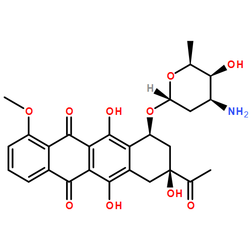

